Preprint
Review

This version is not peer-reviewed.

From Diet to Drugs: Integrative Perspectives on Iodine, Thyroid Dysfunction, and Microbiota in the Elderly

Submitted:

26 September 2025

Posted:

30 September 2025

Read the latest preprint version here

Abstract

Background/Objectives: Iodine intake demonstrates a U-shaped relationship with thyroid dysfunction, with both deficiency and excess contributing to adverse outcomes. Older adults are especially susceptible due to age-related alterations in thyroid physiology, reduced functional reserve, impaired adaptation to iodine excess, comorbidities, and polypharmacy. This review aims to synthesize evidence on how ageing influences iodine–thyroid interactions and to identify factors that complicate clinical management in older populations. Methods: A narrative review of the literature was conducted, focusing on studies addressing iodine metabolism, thyroid function, and the modifying roles of gut microbiota, nutrient cofactors, pharmacological exposures, renal function, and metabolic ageing in older adults. Results: Ageing affects iodine handling and thyroid function through multiple mechanisms. Dysbiosis may contribute to thyroid autoimmunity and hormone metabolism via immune modulation, micronutrient utilization, and enterohepatic recycling. Declining renal clearance prolongs iodide retention, while the frequent use of amiodarone, iodinated contrast, lithium, and medications interfering with levothyroxine absorption increases iatrogenic risk. Concurrent metabolic changes—such as adiposity, insulin resistance, and chronic low-grade inflammation—further impair iodine utilization and thyroid hormone action. Conclusions: Recognition of the complex interactions between ageing, iodine metabolism, and thyroid function is essential for accurate diagnosis and individualized management in older adults. Strategies should incorporate age-adjusted reference ranges, systematic medication review, micronutrient optimization, and iodine prophylaxis policies compatible with salt-reduction initiatives. Emerging microbiome-targeted interventions warrant further investigation as potential modulators of iodine–thyroid dynamics in ageing populations.

Keywords: 
;  ;  ;  ;  ;  ;  ;  ;  

1. Introduction

Iodine is an essential micronutrient required for thyroid hormone synthesis, and its intake determines much of the epidemiology of thyroid disease [40]. The relationship between iodine and thyroid dysfunction follows a special form/curve: deficiency predisposes to goiter and hypothyroidism, while chronic excess may trigger hypothyroidism and autoimmunity [41]. By 2050, over 21% of the global population will be aged 60 years or older. This demographic is particularly vulnerable due to altered thyroid physiology [42,43], multimorbidity, and frequent drug exposures.
Over recent decades, widespread adoption of mandatory or voluntary salt iodization (USI), together with improvements in animal feed supplementation, food-processing standards, and agricultural practices, has markedly reduced endemic goiter and severe developmental consequences of iodine deficiency in many regions [5,6]. USI has dramatically reduced endemic goiter and iodine deficiency disorders, particularly in historically deficient regions [44,45]. Nevertheless, global iodine nutrition has become increasingly heterogeneous: while many populations now maintain adequate iodine status, significant pockets of persistent deficiency remain, especially among marginalized rural and remote communities, and paradoxically, iodine excess (either episodic or chronic) has emerged in certain settings due to high environmental iodine exposure, supplements, seaweed consumption, or iatrogenic sources [45,46,47].
These trends underscore the need for public health strategies that move beyond a binary 'deficiency vs adequacy' framework toward dynamic, context-sensitive monitoring and fine-tuned interventions tailored to population and subpopulation needs [48].
Sources of iodine and determinants of intake are multiple and variable. The principal population-level intervention remains iodized salt, but dietary sources (seafood and seaweed, dairy and eggs, iodophor-treated foodstuffs), fortification of animal feeds (which raises iodine content of milk and meat) [49], multivitamin/mineral supplements, and iatrogenic sources (iodinated contrast media, amiodarone, topical iodophors, etc.) all contribute to individual exposure. Geochemical factors (soil iodine content) [50], inland versus coastal ecology [50], and food-system changes (increasing consumption of processed foods often made with non-iodized industrial salt) modify the effectiveness of USI and create regional variability. Public-health successes therefore reflect not only legislation and salt-supply chain management but also the broader food environment and clinical practices that deliver intermittent high iodine loads.
Monitoring and assessment practices underpin programmatic decision-making. Population iodine status is most commonly assessed by the median urinary iodine concentration (UIC) in spot urine samples [51]. The World Health Organisation (WHO) benchmarks define adequacy in non-pregnant population groups by a median UIC of 100–199 µg/L (with 150–249 µg/L indicating adequacy in pregnancy) [52]. School-age children have classically served as sentinel populations for surveillance because of logistical convenience, but their status does not reliably reflect the iodine sufficiency of pregnant and lactating women—groups with higher physiologic requirements and the greatest vulnerability to deficiency-related neurodevelopmental harm. Consequently, contemporary surveillance frameworks increasingly emphasize targeted monitoring of pregnant women, women of reproductive age, and neonates (e.g., neonatal thyroid-stimulating hormone -TSH- screening), combined with repeated population-level surveys to capture temporal trends.
Despite these monitoring tools, programmatic challenges persist. First, UIC reflects recent intake and is highly variable for each individual [46]; single spot measurements are meaningful only when aggregated as population medians, and interpreting borderline values therefore requires careful contextualisation. Second, the global push toward dietary salt reduction for cardiovascular prevention complicates iodine prophylaxis: lower consumption of household salt reduces a major vehicle for iodine delivery unless iodization levels and fortification strategies are adapted and industrial food producers use iodized salt. Third, the decentralization of food production and the rise in consumption of processed and restaurant foods—often produced with non-iodized industrial salt—can erode the coverage and impact of USI unless regulatory frameworks extend to industrial salt. Emerging risks of iodine excess are increasingly recognized and multifactorial. In some populations, chronic excess arises from high habitual intake of iodine-rich foods (notably certain seaweeds and supplements) [16,17], over-fortification of salt or foodstuffs [53] or environmental/clinical exposures that acutely raise intrathyroidal iodide (like iodinated contrast agents, amiodarone therapy, repeated topical povidone-iodine application) [19,20].
Epidemiologic observations during and after salt-iodization programs in previously deficient regions have documented transient rises in autoimmune thyroiditis and subclinical hypothyroidism—phenomena attributed to increased iodination of thyroglobulin, greater oxidative stress within thyrocytes, and the unmasking of underlying autoimmune susceptibility [21,22,23,24]. At individual level, the elderly may be relatively more susceptible to iodine-induced hypothyroidism because of diminished glandular reserve, altered renal clearance prolonging iodide exposure, and a higher prevalence of thyroid autoantibodies; conversely, certain coastal populations with traditional seaweed consumption maintain high habitual iodine intakes without overt disease, illustrating marked interpopulation heterogeneity in biological response and genetic/environmental modifiers. Also, thyroid malfunction is frequently linked in older adults with dislypemia, atherosclerosis, cognitive decline, neuromuscular dysfunction, sarcopenia, osteoporosis, and frailty [25,26]. This narrative review synthesizes evidence on iodine nutrition and hypothyroidism in older adults. We emphasize intersecting modifiers, mainly age-related changes in thyroid physiology, including impaired escape from the acute Wolff–Chaikoff effect; comorbidities and pharmacologic exposures that alter iodine handling or thyroid hormone metabolism; and changes in nutrition, gastrointestinal physiology and microbiota with ageing that may modulate iodine absorption and autoimmunity. We highlight knowledge gaps, epidemiological and mechanistic data, and clinical implications for surveillance, prevention, and management.

2. Materials and Methods

This work was conceived as a narrative review synthesizing current evidence on the relationship between iodine intake, thyroid dysfunction, and ageing-related modifiers, with a particular emphasis on the role of gut microbiota, nutrient status, pharmacologic exposures, and metabolic ageing. Though this is a narrative review, we used a structured search strategy to ensure broad coverage of the subject (Figure 1).

2.1. A Comprehensive Literature Search

was conducted between January and June 2025 using PubMed/MEDLINE, Scopus, Web of Science, and Embase databases. Search terms combined controlled vocabulary [MeSH/Emtree] and free-text keywords including: “iodine,” “thyroid,” “hypothyroidism,” “hyperthyroidism,” “thyroid autoimmunity,” “ageing,” “elderly,” “microbiota,” “dysbiosis,” “polypharmacy,” “amiodarone,” “iodinated contrast,” “levothyroxine absorption,” “micronutrients” and “Romania”. Boolean operators [AND/OR] and truncation were applied to broaden sensitivity. Reference lists of retrieved papers and relevant reviews were also manually screened to identify additional eligible studies.

2.2. Eligibility Criteria

We included peer-reviewed publications in English from 1980 to 2025 that investigated:
  • The impact of iodine deficiency or excess on thyroid function in adults ≥60 years,
  • Age-specific physiological changes in thyroid hormone regulation,
  • Gut microbiota interactions with thyroid autoimmunity or hormone metabolism,
  • Nutrient cofactors relevant to thyroid function (selenium, zinc, iron),
  • Pharmacological exposures (iodine-rich or thyroid-disrupting drugs, absorption modifiers),
  • Epidemiological or clinical data relevant to iodine status in Romania.

2.3. Exclusion Criteria

Were non-human studies unless mechanistically informative, pediatric or pregnancy-focused studies, and conference abstracts without peer-reviewed full text.

2.4. Data Extraction and Synthesis

Data from eligible studies were extracted into evidence matrices and organized according to predefined themes: [i] iodine intake and thyroid physiology, [ii] ageing-related thyroid adaptations, [iii] Wolff–Chaikoff effect and autoregulatory failure, [iv] microbiota–thyroid axis, [v] nutritional and gastrointestinal changes, [vi] pharmacological and iatrogenic factors, and [vii] metabolic ageing. Findings were narratively synthesized and critically appraised, highlighting convergences, inconsistencies, and gaps in knowledge (Table 1)

2.5. Quality Assessment

Given the heterogeneity of study designs (epidemiological surveys, clinical cohorts, mechanistic studies, and reviews), formal meta-analysis was not feasible. Instead, methodological rigor was evaluated qualitatively, emphasizing sample size, adjustment for confounders, and reproducibility. Where appropriate, higher-level evidence (systematic reviews, meta-analyses, large cohort studies) was prioritized in interpretation.

3. Results and Discussions

3.1. Romania as Case Example

Romania provides a practical example of USI efficacy. Historically, Romania was characterized by widespread endemic goiter, with two-thirds of the country affected, particularly in mountainous Carpathian regions, where severe iodine deficiency and cretinism were documented before the mid-20th century [54,55]. In response, voluntary salt iodization and distribution of potassium iodide tablets to pregnant women and children began in 1962 [55], which significantly reduced the incidence of endemic cretinism; however, mild to moderate deficiency persisted through the 1970s and 1980s, as evidenced by goiter prevalence and low urinary iodine in both children and adults. A landmark shift occurred in 2002 with the adoption of mandatory universal salt iodization for household and bakery use, leading to normalization of median urinary iodine in schoolchildren, though pregnant women remained at risk in some counties [56]. However, despite a longstanding mandatory universal salt iodization policy since 2002, which includes household and bakery salt, iodine intake in Romania demonstrates a mixed picture. Nationally representative data indicate that school-aged children generally achieve adequate iodine status, with median urinary iodine concentration (UIC) around 141 µg/L and consistent levels across historic endemic and non-endemic regions [54]. Pregnant women remain at risk: multiple studies have documented persistent mild iodine deficiency, with median UIC values approximately 116 µg/L and up to nearly half of pregnant cohorts below sufficiency thresholds [57]. Neonatal screening adds a concerning signal: elevated neonatal TSH (> 5 mIU/L) exceeds the 3% adequacy threshold in most counties, indirectly reflecting insufficient maternal iodine intake [54]. While children have generally attained adequacy, these findings suggest that vulnerable groups remain at risk. Importantly, iodine status among older adults in Romania has not been systematically assessed, representing a significant knowledge gap with potential implications for hypothyroidism and autoimmunity in an ageing population. Given Romania’s rapidly ageing population, the absence of iodine data in older adults represents a major blind spot in public health surveillance.

3.2. Why Focus on Older Adults

Hypothyroidism is disproportionately prevalent in older adults, affecting approximately 8–12% of women and 3–6% of men over 65 years, much of it undiagnosed [58,59,60]. In the Atherosclerosis Risk in Communities (ARIC) study, populations over 65 had a higher prevalence of undiagnosed hypothyroidism (6.88%, from which 0.82% clinically manifest and 6.06% subclinical) (34). Risk is amplified by age-related physiological and clinical factors. Thyroid gland reserve declines with age, TSH set-points shift upward, and the capacity to escape from the Wolff–Chaikoff effect is attenuated [61]. Some studies show that 7%-14% of older individuals have serum TSH levels above the upper limit of reference ranges [62,63,64,65]. The rise in TSH is independent of the presence of antithyroid antibodies [66]. However, there are also studies that found decreased serum TSH in elderly [67,68]. Frequently, an opposite relation between TSH and age was noticed in iodine-deficient populations where the main thyroid pathology consists of the presence of nodules and higher thyroid autoimmunity instances as people age [69].
Older adults also have higher prevalence of multimorbidity and are more frequently exposed to drugs that interfere with thyroid function or iodine metabolism. These include amiodarone, lithium, interferon-α, tyrosine kinase inhibitors, as well as common agents such as proton-pump inhibitors, calcium, and iron supplements that reduce levothyroxine absorption [70]. Beyond pharmacology, gastrointestinal changes (atrophic gastritis, reduced gastric acid, altered microbiota) may impair iodine absorption and modulate thyroid autoimmunity [71]. Moreover, renal function decline with ageing prolongs plasma iodide half-life, potentially enhancing the risk from high-iodine medical exposures. Epidemiological evidence from iodine-replete countries shows that both deficiency- and excess-induced hypothyroidism disproportionately affect the elderly, reinforcing the need for targeted iodine surveillance in this population [72]. In the context of rapid global ageing, ensuring adequate—but not excessive—iodine intake in older adults is becoming a critical component of endocrine public health. Another factor to consider is the reduced salt intake in older adults. In many settings, discretionary household salt is the dominant iodine vehicle; thus, sodium restriction—common in older adults with hypertension or chronic kidney disease—can reduce iodine intake if iodized table salt is simply removed without replacement. U.S. NHANES data showed that adults reporting low-salt diets had lower urinary iodine and higher odds of iodine deficiency, underscoring this linkage at the population level [73]. Prospective data in hypertensive outpatients (mean age ~64 years) indicate that sodium reduction does not jeopardize iodine status when the small amount of discretionary salt that remains is iodized; importantly, most processed/restaurant foods in that setting used non-iodized industrial salt, so table salt remained the key iodine source. These findings align with World Health Organization and the Iodine Global Network (IGN) guidance: salt-reduction and salt-iodization agendas are compatible if iodization extends to all edible salt [including industrial/food-service and any salt substitutes] and if programs monitor iodine status in at-risk groups (pregnant women and older adults) [74,75].
For older patients with chronic kidney disease (CKD) the picture is doubly nuanced: they are often counseled to restrict sodium, which may lower iodine intake, yet reduced renal clearance alters iodine kinetics and can heighten susceptibility to iodine-induced dysfunction after large iatrogenic loads—arguing for individualized monitoring rather than blanket assumptions about adequacy.
Together, these features make older adults a high-risk group for both deficiency- and excess-related hypothyroidism, even in countries with established iodine prophylaxis. This intersection of epidemiology, physiology, and pharmacology justifies targeted monitoring and tailored clinical management strategies in ageing populations (Figure 2).

3.3. Age-Related Thyroid Physiology and Iodine-Related Pathophysiology

3.3.1. Iodine’s Role in Thyroid Hormone Synthesis

Iodine is an essential trace element required for the biosynthesis of thyroxine and triiodothyronine Inorganic iodide is actively transported from the circulation into thyroid follicular cells via the sodium–iodide symporter (NIS) on the basolateral membrane, then translocated into the follicular lumen through pendrin and other apical transporters. At the colloid–cell interface, thyroid peroxidase (TPO) catalyzes the oxidation of iodide to iodine and its covalent binding to tyrosyl residues of thyroglobulin (iodination), producing monoiodotyrosine (MIT) and diiodotyrosine (DIT). Coupling of one DIT and one MIT yields triiodothyronine (T3), while coupling of two DIT residues yields T4 (thyroxine). These iodothyronines are stored in the colloid until proteolytic release into the circulation under TSH stimulation [76]. Adequate and stable iodine availability is therefore critical to maintain normal rates of hormone synthesis, while both deficiency and excess perturb these enzymatic and transport processes, predisposing to thyroid dysfunction.

3.3.2. Age-Specific Thyroid Physiology

Less investigated, age specific changes are noted in thyroid physiology. Thus, TSH shifts with ageing. Large epidemiological datasets (NHANES, EPIC, HIMS, HUNT) show that serum TSH concentrations tend to increase modestly with chronological age, even in individuals without overt thyroid disease [51,52,53,54]. This rightward shift of the reference distribution appears most pronounced after the seventh decade, with median TSH values rising by ~0.5–1.0 mIU/L compared with middle-aged adults.
Some explanatory mechanisms might include:
  • Altered hypothalamic–pituitary set-point — possible reduced sensitivity of hypothalamic thyrotropin -releasing hormon (TRH) neurons and/or pituitary thyrotrophs to circulating free T4 [77]
  • Reduced TSH bioactivity — some studies suggest changes in glycosylation patterns that may make TSH less biologically potent, thus requiring higher serum levels to achieve the same thyroidal stimulation [78]
  • Adaptive or benign physiological change — higher TSH in elderly may reflect a homeostatic adaptation to slow metabolism and reduced tissue demand for thyroid hormones, not necessarily pathological hypothyroidism
This age-related shift underlies the clinical debate about whether higher “normal” TSH ranges should be used in older adults, to avoid overdiagnosis and overtreatment of subclinical hypothyroidism [79]. Also with ageing, the thyroid gland undergoes structural and functional changes. They range from histological changes (increased fibrosis, decreased follicular cell mass, accumulation of colloid, and variable atrophy) and vascular and stromal alterations (microvascular rarefaction and reduced capillary density can impair hormone export) to autoimmunity accumulation (lifetime exposure to immune triggers increases the prevalence of anti-TPO (thyroid peroxidase) and anti-Tg antibodies, which can further reduce functional reserve) [80]. These changes lower the gland’s capacity to maintain euthyroidism under stress (e.g., illness, abrupt iodine load, medication interference). As a result, older adults may be less able to compensate for environmental or pharmacologic disruptions in hormone synthesis.
Collectively, these alterations render older adults more susceptible to both overt hypothyroidism and subtle thyroid dysfunction, even in the absence of frank disease.

3.3.3. The Wolff–Chaikoff Effect

This effect is an acute autoregulatory response of the thyroid gland to excess intrathyroidal iodide, first described in 1948 [81]. When intrathyroidal inorganic iodide concentrations exceed a critical threshold (aprox 10⁻³–10⁻² M), organification via thyroid peroxidase (TPO) is transiently inhibited, leading to a sharp reduction in the synthesis of monoiodotyrosine (MIT) and diiodotyrosine (DIT), and thus T4 and T3 production. This block is thought to be mediated by increased intrathyroidal iodolactone and iodoaldehyde formation, which interfere with TPO activity and hydrogen peroxide generation, although the precise molecular mediators remain incompletely defined. In euthyroid individuals, escape from the Wolff–Chaikoff effect typically occurs within 24–48 hours despite continued iodine excess. This is achieved primarily by downregulation of the sodium–iodide symporter (NIS) at the basolateral membrane of thyrocytes, reducing iodide influx and lowering intrathyroidal iodide below the inhibitory threshold, thereby allowing TPO activity and hormone synthesis to resume.
Failure to escape — due to impaired NIS regulation, reduced functional follicular cell mass, or autoimmune/inflammatory injury — can lead to sustained hypothyroidism. This is more common in neonates, individuals with chronic autoimmune thyroiditis, and older adults
In older individuals, there are several factors that may delay or blunt the escape response. One of them is the reduced NIS downregulation efficiency, possibly due to age-related changes in gene expression and cellular signaling in thyrocytes. Another might be the lower gland reserve, with fewer functioning follicular cells to re-initiate organification once NIS expression changes. Coexisting autoimmunity adds inflammatory injury, that can slow recovery of hormone synthesis after iodine excess. Frequent comorbidities (e.g., CKD) that prolong iodine clearance, extend the period of exposure. In the mean time, high iodine exposure might also be frequent due to iatrogenic interventions.
Clinically, this means that older adults are more likely to develop iodine-induced hypothyroidism after exposures such as amiodarone initiation [82] high-dose iodine supplements [83], or iodinated radiologic contrast [84].
All these physiological shifts have clinical relevance. They mean that the “buffer capacity” of the ageing thyroid is reduced. In practice, modest deviations in iodine intake, acute iodine exposures, or drug interactions can tip an older patient into subclinical or overt hypothyroidism, even if younger adults would remain unaffected. This justifies age-aware interpretation of thyroid function tests and heightened vigilance in high-risk clinical scenarios [77].

3.4. The U-Shaped Curve: Deficiency and Excess as Dual Hazards

Population studies consistently demonstrate a U-shaped association between habitual iodine intake and the risk of thyroid dysfunction [85]. Chronic iodine deficiency (<100 µg/day in adults) impairs T4 and T3 synthesis, leading to elevated TSH and compensatory thyroid hypertrophy [goiter]. Prolonged deficiency increases the prevalence of hypothyroidism and nodular disease, often with coexisting functional autonomy. Conversely, chronic iodine excess (>300–500 µg/day) can trigger sustained inhibition of organification (failure to escape the Wolff–Chaikoff effect) and promote thyroid autoimmunity, particularly in genetically predisposed individuals] [64,65]. This risk is mediated through increased iodination of thyroglobulin, enhanced oxidative stress within thyrocytes, and unmasking of latent autoimmune susceptibility [86,87]. Epidemiologic observations during and after salt-iodization programs in formerly deficient regions have documented transient rises in autoimmune thyroiditis and subclinical hypothyroidism, particularly in women [88]. While these trends stabilize over time, they underscore the narrow margin between deficiency and excess, and the importance of gradual, carefully monitored iodization strategies (150–250 µg/day for adults). In older adults, who already carry a higher background prevalence of thyroid autoantibodies, even modest excess exposure may accelerate progression to clinical hypothyroidism.

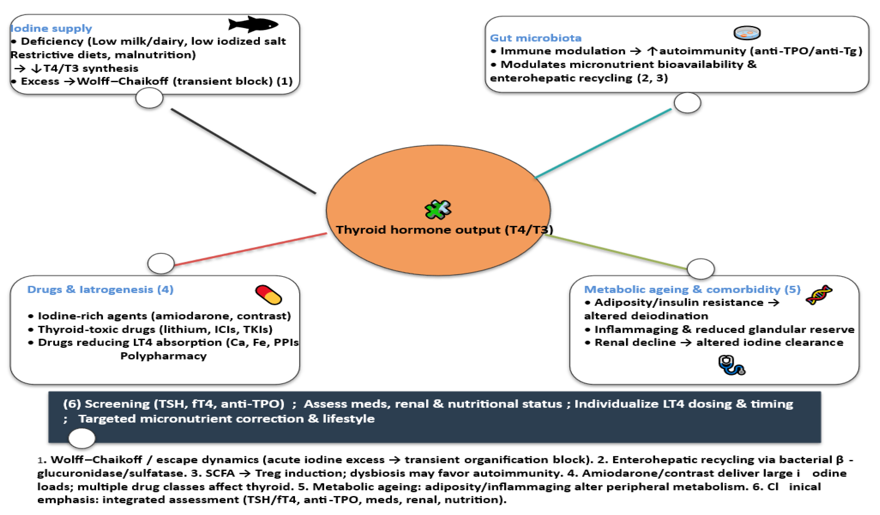
3.5. Modifiers of Iodine Status and Thyroid Function in Ageing

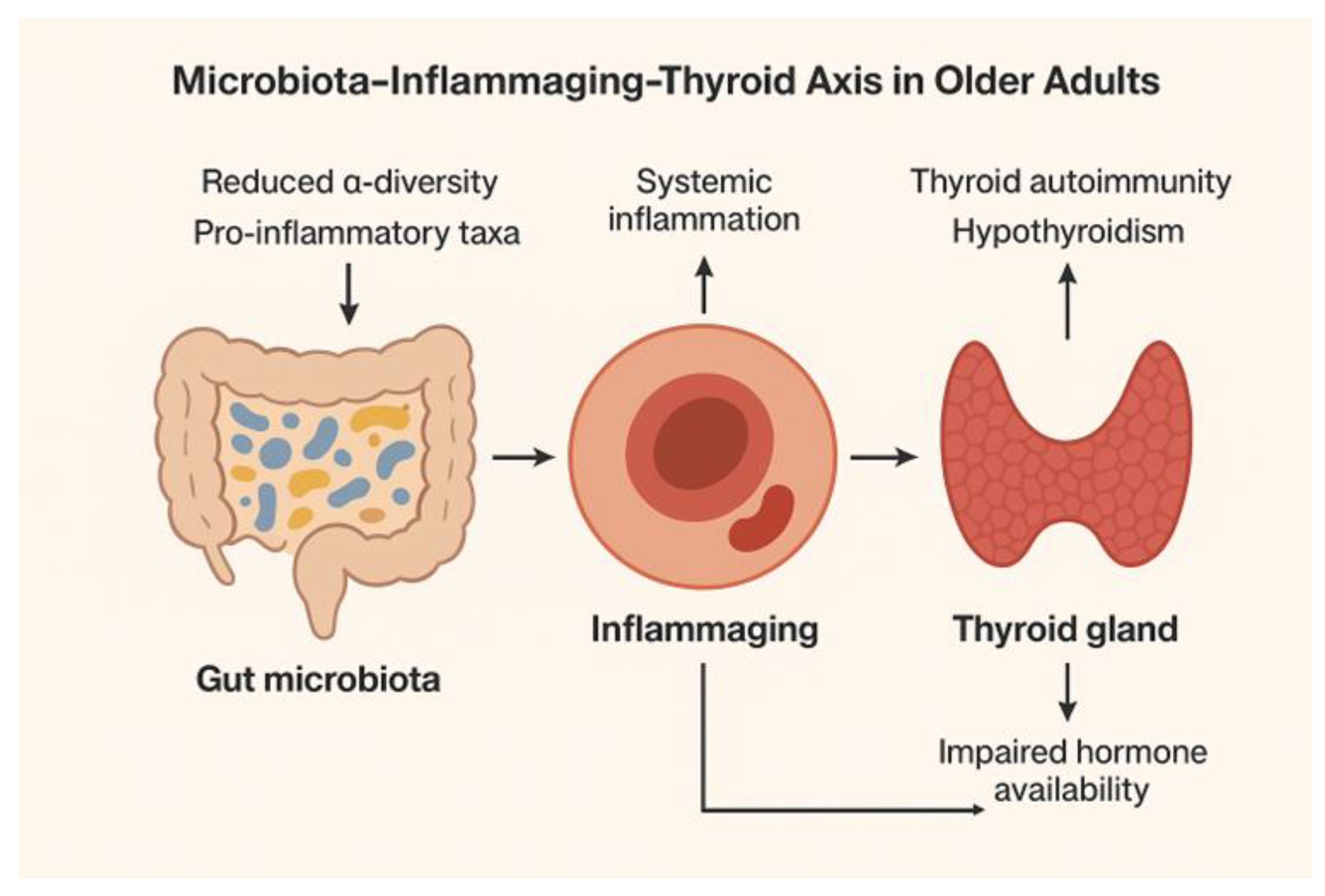
3.5.1. Gut Physiology and Microbiota

In older adults, gut microbiota exerts multidimensional effects on thyroid function through immune modulation, nutrient metabolism, and hormone recirculation. Age-related dysbiosis may contribute to increased susceptibility to hypothyroidism and autoimmunity, while targeted interventions with probiotics, prebiotics, and diet hold potential but require rigorous clinical evaluation. Emerging evidence indicates that gut microbiota plays a pivotal role in modulating thyroid function, particularly in older adults, in whom age-related changes in microbiota composition may lead to an increased risk of thyroid malfunction [89]. The interactions are multifactorial, encompassing immune modulation, micronutrient metabolism, enterohepatic circulation, and peripheral thyroid hormone metabolism.
  • Immune Modulation and Thyroid Autoimmunity
The gut microbiota exerts profound effects on systemic immune responses, shaping both innate and adaptive immunity. Dysbiosis—characterized by reduced microbial diversity and expansion of pathobionts—can promote pro-inflammatory signaling and the breakdown of immune tolerance. In older adults, this contributes to the increased prevalence of autoimmune thyroid diseases (AITD), including Hashimoto’s thyroiditis [90]. Microbial metabolites, such as short-chain fatty acids (SCFAs), regulate regulatory T cells (Tregs) and influence the production of cytokines that modulate thyroid autoantibody formation [anti-thyroid peroxidase (anti-TPO) and anti-thyroglobulin (anti-Tg) [91] These metabolites act through G-protein–coupled receptors (e.g., FFAR2/3), modulate gene expression epigenetically, and thereby regulate immune cell behavior [92] Experimental models suggest that certain bacterial taxa, including Bacteroides and Clostridia clusters IV and XIVa, enhance Treg differentiation, whereas reduced abundance may promote autoimmunity [73,74].
  • Microbial Effects on Micronutrient Metabolism
Gut bacteria influence the absorption and bioavailability of micronutrients [93] including those critical for thyroid function, including iodine, selenium, and iron [94]. Selenium, in particular, is essential for selenoproteins involved in thyroid hormone synthesis and antioxidant defense. Dysbiosis can impair selenium absorption and alter microbial metabolism, indirectly affecting thyroid hormone production and oxidative stress in the gland [95]. Similarly, microbial deiodinase-like activity might influence local iodine utilization and thyroid hormone synthesis, suggesting that gut composition may impact endocrine homeostasis [7877].
  • Influence on Enterohepatic Circulation and Peripheral Thyroid Hormone Metabolism
Thyroid hormones (T4, T3) are conjugated in the liver [glucuronidation/sulfation] and excreted into bile; a portion is deconjugated in the intestine by microbial β-glucuronidases and sulfatases, allowing enterohepatic recirculation and reabsorption of free iodothyronines. This enterohepatic loop has been shown in classic animal and human work [biliary secretion of iodothyronines; deconjugation by intestinal bacteria; interruption by bile-acid sequestrants], and contemporary reviews explicitly recognize the microbiota’s role via these deconjugating enzymes. Direct modulation of T4→T3 conversion by the microbiota has not been demonstrated; T4→T3 activation is mediated by host deiodinases, though microbiota can indirectly influence peripheral hormone availability by altering deconjugation and bile-acid metabolism [96,97].
Dysbiosis in older adults, characterized by reduced microbial diversity and altered enzymatic activity, may impair β-glucuronidase–mediated deconjugation of thyroid hormone metabolites, thereby modifying the efficiency of enterohepatic recirculation. This could reduce peripheral hormone availability and subtly influence systemic thyroid status [98]. Although microbiota do not catalyze the T4→T3 conversion directly, changes in microbial composition may shape hormone reabsorption and metabolic outcomes, contributing to the variability observed in age-related subclinical hypothyroidism.
  • Age-Related Microbiota Changes (Figure 3)
Aging is accompanied by profound alterations in gut microbial ecology. Multiple cohort studies demonstrate that older adults typically exhibit reduced α-diversity and a decline in beneficial commensals, particularly butyrate-producing Firmicutes (e.g., Faecalibacterium prausnitzii, Roseburia) alongside enrichment of potentially pro-inflammatory taxa such as Enterobacteriaceae and other Proteobacteria [99].
These compositional changes are not merely incidental but are closely linked with systemic inflammatory tone. Reduced short-chain fatty acid (SCFA) production and increased lipopolysaccharide (LPS) exposure contribute to gut barrier dysfunction, endotoxemia, and the phenomenon termed “inflammaging”—a state of chronic low-grade inflammation associated with impaired immune regulation [100]. This pro-inflammatory environment has downstream relevance to the thyroid. Chronic inflammation can promote loss of immune tolerance, favoring thyroid autoimmunity (e.g., anti-TPO and anti-Tg antibodies). Moreover, dysbiosis-driven alterations in bile acid metabolism and β-glucuronidase activity may influence enterohepatic recirculation of thyroid hormones, subtly modulating peripheral hormone availability [96]. While direct clinical links between aging microbiota and hypothyroidism are not fully established, the biological plausibility is strengthened by these converging mechanisms. Epidemiologic work has highlighted the variability within older populations. For example, frail elderly often present with pronounced loss of diversity and expansion of Proteobacteria, correlating with elevated systemic inflammatory markers and poorer metabolic outcomes [101]. In contrast, centenarians—despite advanced chronological age—frequently retain greater microbial richness, with enrichment of taxa such as Akkermansia and certain SCFA producers. These individuals tend to exhibit lower levels of systemic inflammation and autoantibody prevalence, suggesting a protective role of preserved microbial diversity in buffering against immune dysregulation and possibly thyroid dysfunction [85,86]. These findings are consistent with a protective phenotype and the survivor bias has to be taken into consideration. Thus, age-related microbiota changes can be conceptualized not as a uniform decline but as a spectrum ranging from resilience to dysbiosis. Individuals maintaining balanced microbial ecosystems may experience less inflammaging and lower risk of thyroid autoimmunity, whereas those with pronounced dysbiosis could face compounding risks from chronic inflammation, micronutrient malabsorption (iodine, selenium, zinc), and impaired enterohepatic recirculation of thyroid hormones. These findings underscore the need for longitudinal studies that integrate microbiome, endocrine, and immune parameters in older adults to clarify causal pathways and intervention opportunities.
  • Evidence linking microbiota composition to hypothyroidism and thyroid autoimmunity
Human studies generally show that patients with Hashimoto’s thyroiditis [HT] harbor distinct gut microbial profiles versus euthyroid controls [102], often with lower relative abundance of SCFA-producing taxa [e.g., Faecalibacterium, Roseburia] and enrichment of pro-inflammatory or pathobiont lineages, though results vary by geography, diet, and methods [87,88]. Examples include cohort reports of altered diversity and shifts in Firmicutes/Bacteroidetes with hyperthyroidism [103], and taxa differences involving Akkermansia, Bifidobacterium, Prevotella. Subclinical hypothyroidism (SCH) has likewise been associated with gut dysbiosis, including reduced SCFA producers and links to SIBO and autoantibody positivity in some cohorts [104]. Animal models support biological plausibility; fecal microbiota transfer from hypothyroid or dysbiotic donors might induce or exacerbate hypothyroidism in mice, and GF/antibiotic models show microbiota-dependent modulation of thyroid function [90,91]. However, human causal inference remains limited/indirect, microbiota composition being influenced by many other factors (diet, BMI, proton pump inhibitors, levothyroxine use, geographic diet, etc). Recent syntheses and mechanistic reviews converge on a model whereby dysbiosis reduces SCFAs, increases permeability/LPS exposure, and perturbs bile-acid/β-glucuronidase networks relevant to enterohepatic thyroid hormone handling—changes that could facilitate loss of tolerance (anti-TPO/anti-Tg) and subtle peripheral hormone shifts. A 2024 Mendelian-randomization analysis further suggests a potential causal pathway between specific gut taxa, immune cell mediation, and HT risk, though such inference still requires careful validation [105]. Human data are largely observational and subject to confounding — causal inference remains limited and requires longitudinal/intervention studies.
  • Potential of Probiotics, Prebiotics, and Symbiotics in Modulating Thyroid Outcomes
Interventional studies using probiotics, prebiotics and synbiotics in thyroid disorders remain limited but encouraging [106]. Meta-analyses indicate that probiotics can reduce systemic inflammatory markers [107], and randomized trials suggest improvements in intestinal barrier function [108,109] (e.g., lower zonulin). In Graves’ disease, in a small trial, adjunctive Bifidobacterium longum with methimazole improved thyroid function and autoantibodies, supporting a gut–thyroid axis mechanism [110]. While Lactobacillus rhamnosus strains consistently modulate inflammation and barrier integrity [111], direct effects on thyroid hormone metabolism are better documented in animal studies with Lactobacillus reuteri [112] rather than Lactobacillus rhamnosus; thus, strain-specific claims should be made cautiously pending larger human trials. Clinical trials in older adults with thyroid disorders are scarce, and evidence remains preliminary [113]. In recent research [101,102,103] there have been reported some mixed effects of prescribing routinely prebiotic, probiotic, and synbiotic supplementation on clinical and laboratory outcomes in hypothyroidism [114]. Critical appraisal indicates that while modulation of gut microbiota is a potential therapeutic avenue, standardized protocols, strain-specific efficacy data, and long-term outcomes are urgently needed before routine clinical recommendations can be made.

3.5.2. Changes in Dietary Habits, Nutrition and Gastrointestinal Physiology

Aging is accompanied by profound alterations in dietary intake, gastrointestinal structure and function, and nutrient absorption [115] all of which bear relevance for thyroid health and iodine metabolism.
  • Dietary Transitions in Aging
Nutritional patterns in older populations diverge from those of younger adults due to multiple interdependent factors. Physiological changes in appetite regulation (e.g., blunted ghrelin signaling, increased cholecystokinin) combine with psychosocial determinants (loneliness, depression, economic hardship), leading to lower caloric and nutrient intake [106,107]. Lower caloric intake is often accompanied by reduced protein and micronutrient density, increasing the risk of borderline deficiencies even in high-income settings. Older adults often consume less dairy and seafood, which are major iodine contributors in many countries, partly due to lactose intolerance, dental problems, cost, and personal preferences. At the same time, increased reliance on processed foods in institutionalized or socially vulnerable elderly may uncouple iodine intake from natural sources, depending on fortification policies. Another factor that may play a role is salt use variability: Concerns about hypertension and cardiovascular disease often drive reduced discretionary salt use in seniors. When iodized salt is the main prophylaxis vehicle, this may inadvertently compromise iodine status unless compensated by fortified foods or supplements [116].
Supplement intake is inconsistent in older populations: some seniors adopt multivitamins or kelp-based iodine supplements, while others avoid supplements altogether. This produces wide inter-individual variability, with risks of both mild chronic deficiency and iatrogenic excess. Importantly, seniors are more likely to consume over-the-counter supplements without medical guidance, which can destabilize thyroid homeostasis.
  • Gastrointestinal Structural and Functional Alterations
The GI tract undergoes age-related modifications that influence nutrient assimilation.
Gastric function: Hypochlorhydria and atrophic gastritis are prevalent in aging, compounded by frequent proton pump inhibitor (PPI) use. Low gastric acidity reduces iodine liberation from organic food matrices, impairs solubilization of iron and zinc, and alters microbiota composition [117]. Impaired gastric intrinsic factor secretion also predisposes to vitamin B12 deficiency, which may indirectly influence thyroid symptoms through overlapping neurocognitive manifestations.
Pancreatic and biliary function: Exocrine pancreatic insufficiency, though mild in many older adults, impairs lipid digestion and absorption of fat-soluble vitamins (A, D, E, K) [118] as well as selenium, a key cofactor for glutathione peroxidases and iodothyronine deiodinases. Changes in bile acid composition and reduced pool size alter enterohepatic circulation of hormones, with potential implications for thyroid hormone metabolism.
Intestinal absorption: While gross small-intestinal morphology is often preserved with aging, subtle functional changes occur: reduced expression of certain nutrient transporters, increased mucosal permeability, and higher prevalence of microscopic enteropathy [119]. These changes may contribute to reduced efficiency of iodine, selenium, and zinc absorption
Colonic function and microbiota: Slowed colonic transit and constipation alter the gut microbial milieu, influencing bile acid metabolism and short-chain fatty acid production. Dysbiosis, as discussed in previous sections, can secondarily affect enterohepatic recirculation of thyroid hormones and systemic inflammation, thus linking gastro-intestinal physiology to thyroid outcomes in the elderly.
Consequences on nutrient absorption: Normally absorbed rapidly in the stomach and duodenum, iodine uptake may be compromised in hypochlorhydria and reduced gastric surface area. Even mild impairments can become clinically relevant when combined with lower intake. Selenium deficiency diminishes activity of deiodinases (responsible for T4-to-T3 conversion) and glutathione peroxidases, exacerbating oxidative stress within thyrocytes. Zinc deficiency, more common in institutionalized elderly, alters thyroid hormone receptor activity and impairs TSH signaling [120]. These micronutrient deficiencies often coexist with impaired gastro-intestinal absorption, creating a cumulative burden on thyroid function. In frail elderly, low protein intake reduces hepatic synthesis of thyroxine-binding globulin [121]. This alters the distribution and availability of thyroid hormones, complicating the interpretation of thyroid function tests and sometimes mimicking hypothyroid biochemistry.

3.5.3. Pharmacologic and Iatrogenic Factors Affecting Thyroid Function in Older Adults

Thyroid function in older adults is particularly vulnerable to pharmacologic and iatrogenic influences due to age-related physiological changes, polypharmacy, and comorbidities. A systematic approach involving regular monitoring, judicious drug selection, and attention to drug–nutrient interactions is critical for preserving thyroid health in the aging population Several drugs can directly or indirectly alter thyroid hormone synthesis, metabolism, or bioavailability, thereby affecting clinical outcomes.
  • Iodine-Rich Drugs
Excess iodine can disrupt thyroid function through the Wolff–Chaikoff effect, a rapid autoregulatory block of hormone synthesis; failure to escape this inhibition may lead to prolonged hypothyroidism in susceptible older adults or those with autoimmune disease [81]. Ingesting high iodine loads—such as via iodinated contrast media used in imaging—can precipitate iodine-induced hyperthyroidism (Jod–Basedow phenomenon) or hypothyroidism, especially in elderly patients with nodular thyroid disease or impaired autoregulation [122]. Amiodarone, a potent I¬-containing antiarrhythmic (~37% by weight), interferes with peripheral deiodination (inhibiting type I 5′-deiodinase), reduces T4→T3 conversion, and increases reverse T3, leading to various thyroid dysfunctions. It may induce hypothyroidism (AIH) via the Wolff–Chaikoff effect—particularly in iodine-replete settings—or hyperthyroidism (AIT) in patients with multinodular or latent thyroid disease, risks that increase with age and underlying glandular autonomy [115,116,117].
  • Thyroid-Toxic Drugs
Certain drugs can directly injure thyroid tissue or trigger autoimmune thyroiditis. Used in psychiatric disorders, lithium inhibits thyroid hormone release, leading to hypothyroidism in up to 20% of patients, with higher susceptibility in older adults. Lithium can also cause goiter via increased TSH stimulation [118,119], Immune Checkpoint Inhibitors (ICIs) (Anti-PD-1, anti-PD-L1, and anti-CTLA-4) therapies used in oncology can trigger thyroiditis through immune activation, resulting in transient thyrotoxicosis followed by hypothyroidism. Incidence rises with combination regimens, and older patients may present with atypical or subclinical manifestations [123]. Tyrosine Kinase Inhibitors (TKIs), such as sunitinib or sorafenib, may reduce thyroid hormone synthesis, increase hormone clearance, or cause destructive thyroiditis. Older patients may be particularly sensitive due to reduced thyroid reserve [124].
  • Drugs Interfering with Levothyroxine Absorption
Optimal thyroid hormone replacement can be impaired by numerous agents through direct chelation, pH alteration, or bile acid interactions [125]. Calcium and Iron supplements form insoluble complexes with levothyroxine, reducing absorption. Proton Pump Inhibitors (PPIs) rise gastric pH, thus decreasing levothyroxine dissolution and absorption. Bile-acid binders like cholestyramine and colestipol sequester levothyroxine in the gut. High fiber, soy products, or certain proteins can slow gastrointestinal transit or bind thyroid hormone, impairing bioavailability [126]. Careful timing of levothyroxine administration relative to these agents is essential, typically spacing doses by 2–4 hours.
  • Polypharmacy Patterns and Cumulative Impact in Older Adults
Older adults frequently take multiple medications for cardiovascular, metabolic, and psychiatric conditions. Polypharmacy can amplify the risk of thyroid dysfunction [127] through additive effects of iodine-rich or thyroid-toxic drugs. Also, there might be multiple agents impairing levothyroxine absorption [128] or drug–drug interactions affecting hepatic metabolism of thyroid hormones (e.g., CYP450 inducers such as rifampicin). We also have to consider the whole framework of metabolic changes, with increased susceptibility to subclinical hypothyroidism or thyrotoxicosis due to reduced thyroid reserve and altered homeostatic responses with aging. While polypharmacy is highly prevalent in older adults and creates multiple paths for pharmacologic and absorption interference with thyroid hormone management, epidemiological evidence directly linking polypharmacy to abnormal TSH or free T4 levels in community populations is lacking. Nevertheless, clinical guidelines recommend cautious, individualized monitoring of thyroid function in elderly patients due to heightened risk of drug interactions and altered hormone metabolism.
  • Clinical Implications and Management
As a consequence, regular thyroid function testing is recommended in older adults on amiodarone, lithium, ICIs, TKIs, or high-dose iodine exposure. Minimizing unnecessary polypharmacy and carefully timing levothyroxine relative to interfering agents. Awareness of age-specific pharmacokinetics and comorbidities can guide drug selection and dosing to reduce thyroid-related complications.

3.5.4. Metabolic Ageing and Thyroid–Iodine Interaction

Ageing is accompanied by profound alterations in metabolic homeostasis that influence thyroid physiology and the utilization of iodine. Beyond changes in intake and renal excretion, intrinsic metabolic shifts modulate hormone synthesis, conversion, and tissue responsiveness. Metabolic ageing creates a permissive environment where insulin resistance, adipokine imbalance, inflammaging, and micronutrient insufficiencies converge to affect thyroid hormone production, peripheral metabolism, and iodine utilization. This multifactorial landscape underscores the need for individualized nutritional and metabolic assessment in geriatric thyroid care.
  • Metabolic Syndrome, Obesity, and Insulin Resistance
Insulin resistance and central obesity, common in older adults, affect deiodinase activity, particularly type 2 deiodinase in peripheral tissues. This can lead to altered T4 to T3 conversion and higher circulating reverse T3, a biologically inactive metabolite [129].
Increased adiposity elevates leptin and proinflammatory adipokines, which can stimulate hypothalamic TRH secretion, may also promote low-grade tissue resistance to thyroid hormones, complicating metabolic adaptation. In vitro and in vivo rodent studies show that leptin directly stimulates pro-TRH gene expression and TRH release in hypothalamic neurons, acting via STAT3-related mechanisms [127,128]. Reviews confirm that leptin is a proinflammatory adipokine regulating the HPT axis; adipokines such as resistin and adiponectin modulate deiodinase activity and peripheral thyroid hormone metabolism [130]. Epidemiological studies indicate that older adults with metabolic syndrome often present with slightly elevated TSH and lower free T3, reflecting a complex interplay between insulin signaling, hepatic metabolism, and adipose-driven cytokine activity. The Health, Aging and Body Composition (Health ABC) study found that each one-unit increase in TSH was associated with a 3% increase in odds of prevalent metabolic syndrome; the association was stronger for TSH values in the upper normal range [131].
Reviews and cross-sectional analyses report positive correlations of TSH and BMI/waist circumference in euthyroid adults, potentially mediated by leptin and insulin resistance pathways [132].
  • Chronic Low-Grade Inflammation (“Inflammaging”)
Immune modulation and thyroid autoimmunity are important topics in older people. Age-related chronic low-grade inflammation—characterized by elevated IL-6, TNF-α, and CRP—can influence thyroid autoimmunity by promoting antigen presentation and autoreactive lymphocyte activation, subtly increasing anti-TPO or anti-Tg prevalence [99]. Inflammaging may impair thyroidal iodine uptake through oxidative stress and altered sodium-iodide symporter expression, potentially reducing hormone synthesis efficiency, particularly under borderline iodine intake.
  • Influence of renal function decline on iodine excretion.
After midlife, the glomerular filtration rate (GFR) declines by ~0.75–1 mL/min/1.73 m² per year, even in the absence of overt kidney disease [133]. Because >90% of dietary iodine is excreted renally, this progressive decline leads to longer systemic retention of iodine in older adults, amplifying susceptibility to iodine-induced hypothyroidism [failure to escape the Wolff–Chaikoff effect] or hyperthyroidism [especially in nodular thyroids]. Older adults often present with reduced thyroidal functional reserve [atrophy, fibrosis, nodular autonomy], meaning that modest delays in iodine clearance can have disproportionate endocrine effects. Clinical studies show that elderly populations exposed to iodinated contrast media or amiodarone exhibit higher rates of thyroid dysfunction than younger cohorts with similar exposures [134]. This indicates a synergism between age-related renal and thyroidal changes. Even without chronic kidney disease, common geriatric factors—such as reduced protein and dairy intake, variable use of iodized salt, and lower fluid intake—alter iodine intake and excretion dynamics [135]. In parallel, polypharmacy (e.g., diuretics, ACE inhibitors, NSAIDs) can subtly reduce renal clearance. Thus, in older adults, the margin between iodine sufficiency and excess narrows considerably, making precise monitoring of iodine exposure particularly relevant [136].
  • Micronutrient Co-factors in Metabolic Ageing.
Selenium is essential.for deiodinase enzymes and antioxidant selenoproteins, which mitigate oxidative stress in the thyroid. Selenium deficiency reduces activity of selenoproteins (e.g., glutathione peroxidases and deiodinases), thereby increasing oxidative stress in thyrocytes and potentiating iodine-induced damage [137]. Older adults often exhibit borderline selenium deficiency due to dietary limitations and reduced absorption, exacerbating thyroid vulnerability under inflammatory conditions. Zinc is required for both TRH gene expression and the activity of type 1 deiodinase, which converts T4 into active T3. Animal models of zinc deficiency demonstrate reduced serum T3 and impaired hypothalamic–pituitary–thyroid signaling [138]. In humans, zinc deficiency has been associated with low T3 and elevated reverse T3, and supplementation restored thyroid function in small clinical studies [139]. Older adults are particularly vulnerable to zinc insufficiency due to reduced intake and absorption, making this an important factor in age-related thyroid dysregulation. Iron is a cofactor for thyroid peroxidase (TPO), the enzyme responsible for iodide oxidation and thyroglobulin iodination. Iron deficiency reduces TPO activity and thyroid hormone synthesis [140]. In both animals and humans, iron deficiency leads to lower T4 and T3 concentrations; repletion restores normal thyroid function [141]. Subclinical iron deficiency, common in older adults due to dietary changes, comorbidities and polypharmacy, may thus contribute to subtle hypothyroid states despite adequate iodine intake.

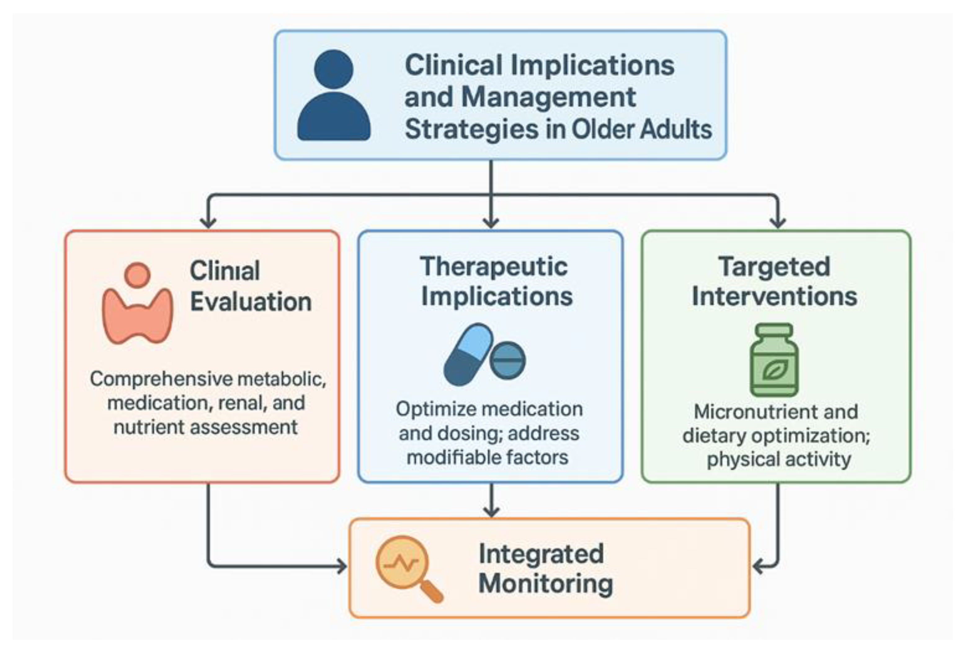
3.6. Clinical Implications and Management Strategies in Older Adults

Our study emphasizes the complex interplay of ageing, metabolic shifts, nutrient status, pharmacologic exposures, and gut–thyroid interactions that have tangible implications for clinical care. Older adults are exposed to polypharmacy, iatrogenic thyroid disruptors, altered dietary patterns, declining renal function, and metabolic dysregulation. Some of these factors can independently and mostly synergistically influence thyroid hormone synthesis, metabolism, and action. Recognition of these factors is essential for early detection, accurate diagnosis, and individualized management (Figure 4). Older adults frequently exhibit higher TSH and lower free T3 even in the absence of overt disease [40,141]. Clinicians must carefully review each patient and avoid overtreatment. Comprehensive evaluation of older patients should include metabolic profile, medication history, renal function, micronutrient status, and autoimmune markers to contextualize thyroid test abnormalities. Also thyroid function can fluctuate with acute illness, polypharmacy, or nutritional changes. Testing has to be dinamically applied before initiating therapy is advised to avoid overtreatment. At the same time, there are important therapeutic implications. If the patient uses levothyroxine, its dosing has to be carried out taking into account reduced lean body mass, altered absorption, and administration of other drugs. Dividing doses or adjusting timing relative to calcium, iron, or fiber supplements can optimize absorption [142]. One has also to address the correction modifiable factors—micronutrient insufficiencies, gut dysbiosis, and metabolic derangements— thus improving thyroid function without pharmacologic escalation [143]. In patients with elevated anti-TPO or anti-Tg, periodic surveillance is warranted to detect transition from subclinical to overt hypothyroidism, particularly in the context of metabolic syndrome or inflammaging. Micronutrient intake has to be optimized. Adequate intake of iodine, selenium, zinc, and iron should be ensured, preferably through diet, with supplements considered only when deficiencies are confirmed [144,145]. Regarding dietary patterns, emphasis on protein sufficiency, moderate caloric intake, and fiber-rich foods supports both gut microbiota diversity and metabolic health. Physical activity must also be included in the lifestyle advise. Resistance training and aerobic exercise improve insulin sensitivity, mitigate inflammaging, and may indirectly support thyroid hormone efficacy at the tissue level. Effective management requires a precision-medicine approach—considering individual metabolic status, nutrient sufficiency, polypharmacy, and gut health—rather than relying solely on biochemical thresholds. Optimizing diet, micronutrient intake, and lifestyle, while judiciously using pharmacologic therapy, can improve outcomes and quality of life. Regular medication review is critical to minimize iatrogenic thyroid dysfunction, especially in those taking multiple agents that interfere with hormone metabolism or absorption. Microbiome-targeted interventions can be added. Emerging evidence suggests probiotics, prebiotics, or dietary modulation could complement conventional therapy by stabilizing gut–thyroid crosstalk, though robust clinical trials in older adults are still limited [146,147].
Integrated monitoring is very important in older patients. Subclinical disease prevalence can be high. The interplay of inflammaging [86,148], insulin resistance, and micronutrient insufficiencies may amplify susceptibility to autoimmune thyroid disease and subtle thyroid hormone deficiencies, often presenting as nonspecific clinical features or biochemical abnormalities.
Future perspectives: Interventions targeting the gut microbiota, metabolic optimization, and careful modulation of drug exposures represent promising areas for research. Longitudinal studies in older adults are needed to define evidence-based strategies for prevention, early detection, and management of thyroid dysfunction in this population. In essence, thyroid care in older adults must be holistic, individualized, and integrative, bridging endocrinology, geriatrics, nutrition, immunology, and pharmacology. Recognizing the complexity of thyroid–ageing interactions allows clinicians to mitigate risk factors, personalize therapy, and promote healthy endocrine ageing.

3.7. Limitations

This review has several limitations that should be acknowledged. First, as a narrative review, the selection of studies was not conducted according to a systematic protocol, and the possibility of publication bias and incomplete retrieval of relevant data cannot be excluded. Second, the evidence base itself is heterogeneous, encompassing epidemiological studies, mechanistic animal models, and clinical observations with variable quality and generalizability. This precludes quantitative synthesis or meta-analysis. Third, many of the studies addressing iodine status, thyroid dysfunction, and ageing derive from younger or mixed-age populations, with relatively few investigations specifically targeting older adults. Consequently, some of the extrapolations presented here rely on indirect inference. Fourth, data regarding the gut microbiota–thyroid axis remain preliminary, with most findings observational or from small interventional studies; causality and clinical applicability in elderly populations remain uncertain. Fifth, regional insights such as those from Romania highlight important public health aspects but are limited by scarce nationally representative data on iodine status in older adults. Finally, while pharmacologic and metabolic influences were discussed, the interactions between polypharmacy, nutrient status, and thyroid function in ageing populations are complex and incompletely characterized, limiting firm conclusions. Despite these constraints, the review synthesizes available knowledge across disciplines and identifies critical gaps to inform future research.

4. Conclusions

Ageing modifies iodine metabolism and thyroid function through a complex interplay of dietary intake, physiological changes, comorbidities, and pharmacological exposures. Population studies indicate that older adults remain at risk of both iodine deficiency and excess, depending on regional fortification, dietary habits, and the prevalence of chronic conditions Mechanistic insights highlight that impaired thyroid autoregulation, oxidative stress, and inflammatory pathways amplify susceptibility to iodine-induced dysfunction
Emerging evidence underscores the role of the gut microbiota, polypharmacy, and micronutrient status as critical modifiers of thyroid homeostasis in older adults. Age-related microbiota shifts, combined with deficiencies in selenium or iron, can exacerbate oxidative stress and thyroid autoimmunity, whereas common medications (e.g., amiodarone, lithium, iodine-containing contrast agents) can precipitate acute thyroid malfunctioning. Metabolic comorbidities such as type 2 diabetes and chronic kidney disease further complicate iodine handling, highlighting the need for individualized assessment of thyroid risk in geriatric populations. From a public health and clinical perspective, these findings argue for a nuanced approach to iodine monitoring in older adults. Recommendations should integrate dietary surveys, medication review, and evaluation of coexisting micronutrient deficiencies. Interventions, including targeted fortification, supplementation, and careful use of iodine-rich medications, must be tailored to the elderly to prevent both deficiency and excess. Future research should aim to clarify the microbiome-thyroid axis, explore drug–nutrient interactions in ageing, and define optimal iodine thresholds in populations with diverse comorbidities.

Author Contributions

Conceptualization, C.Z.; methodology, C.C. and C.Z.; resources, C.C., C.Z., M.P.; writing—review and editing, C.Z., M.P.; supervision, C.C.; All authors have read and agreed to the published version of the manuscript.

Funding

This research received no external funding.

Transparency statement

The authors declare that Figure 2, Figure 3 and Figure 4 were created with the assistance of an AI image-generation tool (Chat GPT 4.) The authors have reviewed and edited the output and take full responsibility for the content of this publication.

Conflicts of Interest

The authors declare no conflicts of interest.

References

  1. Smyth, P.P.A. Iodine, Seaweed, and the Thyroid. European Thyroid Journal 2021, 10, 101–108. [Google Scholar] [CrossRef]
  2. Luo, Y.; Kawashima, A.; Ishido, Y.; Yoshihara, A.; Oda, K.; Hiroi, N.; Ito, T.; Ishii, N.; Katayama, S.; Tsuboi, K.; Endo, T. Iodine Excess as an Environmental Risk Factor for Autoimmune Thyroid Disease. International Journal of Molecular Sciences 2014, 15, 12895–12912. [Google Scholar] [CrossRef]
  3. Sohn, S.Y.; Inoue, K.; Rhee, C.M.; Leung, A.M. Risks of Iodine Excess. Endocrine Reviews 2024, 45, 858–879. [Google Scholar] [CrossRef] [PubMed]
  4. Bath, S.C.; Steer, C.D.; Golding, J.; Emmett, P.; Rayman, M.P. Effect of Inadequate Iodine Status in UK Pregnant Women on Cognitive Outcomes in Their Children: Results from the Avon Longitudinal Study of Parents and Children [ALSPAC]. The Lancet 2013, 382, 331–337. [Google Scholar] [CrossRef] [PubMed]
  5. Zimmermann, M.B.; Boelaert, K. Iodine Deficiency and Thyroid Disorders. The Lancet Diabetes & Endocrinology 2015, 3, 286–295. [Google Scholar] [CrossRef] [PubMed]
  6. Lazarus, J.H. Iodine Status in Europe in 2014. European Thyroid Journal 2014, 3, 3–6. [Google Scholar] [CrossRef]
  7. Zimmermann, M.B.; Andersson, M. Global Perspectives in Endocrinology: Coverage of Iodized Salt Programs and Iodine Status in 2020. The Journal of Clinical Endocrinology & Metabolism 2021, 106, 3597–3606. [Google Scholar] [CrossRef]
  8. Diab, N.; Daya, N.R.; Juraschek, S.P.; et al. Prevalence and Risk Factors of Thyroid Dysfunction in Older Adults in the Community. Sci Rep. 2019, 9, 13156. [Google Scholar] [CrossRef]
  9. Hollowell, J.G.; Staehling, N.W.; Flanders, W.D.; et al. Serum TSH, T[43], and thyroid antibodies in the United States population [1988 to 1994]: National Health and Nutrition Examination Survey [NHANES III]. J. Clin. Endocrinol. Metab. 2002, 87, 489–499. [Google Scholar] [CrossRef]
  10. Boekholdt, S.M.; Titan, S.M.; Wiersinga, W.M.; et al. Initial thyroid status and cardiovascular risk factors: The EPIC-Norfolk prospective population study. Clin. Endocrinol. 2010, 72, 404–410. [Google Scholar] [CrossRef]
  11. Yeap, B.B.; Manning, L.; Chubb, S.A.; Hankey, G.J.; Golledge, J.; Almeida, O.P.; Flicker, L. Reference ranges for thyroid-stimulating hormone and free thyroxine in older men: Results from the Health In Men Study. J. Gerontol. A Biol. Sci. Med. Sci. 2017, 72, 444–449. [Google Scholar] [CrossRef] [PubMed]
  12. Åsberg, A.; Berg, M.; Hov, G.G.; Lian, I.A.; Løfblad, L.; Mikkelsen, G. The HUNT study: Long-term within-subject variation of thyroid-stimulating hormone [TSH]. Scand. J. Clin. Lab. Invest. 2025, 85, 143–147. [Google Scholar] [CrossRef] [PubMed]
  13. Kahaly, G.J.; Dienes, H.P.; Beyer, J.; Hommel, G. Iodide induces thyroid autoimmunity in patients with endemic goiter: a randomized, double-blind, placebo-controlled trial. Eur. J. Endocrinol. 1998, 139, 290–297. [Google Scholar] [CrossRef] [PubMed]
  14. Kalarani, I.B.; Veerabathiran, R. Impact of iodine intake on the pathogenesis of autoimmune thyroid disease in children and adults. Ann. Pediatr. Endocrinol. Metab. 2022, 27, 256–264. [Google Scholar] [CrossRef]
  15. Alexander, K.L.; Targan, S.R.; Elson, C.O. 3rd. Microbiota activation and regulation of innate and adaptive immunity. Immunol. Rev. 2014, 260, 206–220. [Google Scholar] [CrossRef]
  16. Sai, A.; Shetty, G.B.; Shetty, P.; Nanjeshgowda, H.L. Influence of gut microbiota on autoimmunity: a narrative review. Brain Behav. Immun. Integr. 2024, 5, 100046. [Google Scholar] [CrossRef]
  17. Abubakar, D.; Abdullahi, H.; Ibrahim, I. Bridging microbiomes: Exploring oral and gut microbiomes in autoimmune thyroid diseases—New insights and therapeutic frontiers. Gut Microbes Rep. 2025, 2, 2452471. [Google Scholar] [CrossRef]
  18. Sawicka-Gutaj, N.; Gruszczyński, D.; Zawalna, N.; Nijakowski, K.; Muller, I.; Karpiński, T.; Salvi, M.; Ruchała, M. Microbiota alterations in patients with autoimmune thyroid diseases: A systematic review. Int. J. Mol. Sci. 2022, 23, 13450. [Google Scholar] [CrossRef]
  19. Rinninella, E.; Raoul, P.; Cintoni, M.; Franceschi, F.; Miggiano, G.A.D.; Gasbarrini, A.; Mele, M.C. What is the healthy gut microbiota composition? A changing ecosystem across age, environment, diet, and diseases. Microorganisms 2019, 7, 14. [Google Scholar] [CrossRef]
  20. Chang, S.-C.; Lin, S.-F.; Chen, S.-T.; Chang, P.-Y.; Yeh, Y.-M.; Lo, F.-S.; Lu, J.-J. Alterations of gut microbiota in patients with Graves’ disease. Front. Cell. Infect. Microbiol. 2021, 11, 663131. [Google Scholar] [CrossRef]
  21. Jiang, W.; Lu, G.; Gao, D.; Lv, Z.; Li, D. The relationships between the gut microbiota and its metabolites with thyroid diseases. Front. Endocrinol. [Lausanne] 2022, 13, 943408. [Google Scholar] [CrossRef]
  22. Hao, Y.; Xu, Y.; Ban; Y, Li, J; Wu, B; Ouyang, Q, et al. Efficacy evaluation of probiotics combined with prebiotics in patients with clinical hypothyroidism complicated with small intestinal bacterial overgrowth during the second trimester of pregnancy. Front Cell Infect. Microbiol. 2022; 12:983027.
  23. Ouyang, Q.; Xu, Y.; Ban, Y.; Li, J.; Cai, Y.; Wu, B., et al. Probiotics and prebiotics in subclinical hypothyroidism of pregnancy with small intestinal bacterial overgrowth. Probiotics Antimicrob. Proteins 2024; 16, 579–588.
  24. Zawadzka, K.; Kałuzińska, K.; Świerz, M.J.; Sawiec, Z.; Antonowicz, E.; Leończyk-Spórna, M.; et al. Are probiotics, prebiotics, and synbiotics beneficial in primary thyroid diseases? A systematic review with meta-analysis. Ann. Agric. Environ. Med. 2023; 30, 217–23.
  25. Grassi, M.; Petraccia, L.; Mennuni, G.; et al. Changes, functional disorders, and diseases in the gastrointestinal tract of elderly. Nutr. Hosp. 2011, 26, 659–668. [Google Scholar] [PubMed]
  26. Morley, J.E. Anorexia of ageing: A key component in the pathogenesis of both sarcopenia and cachexia. J. Cachexia Sarcopenia Muscle 2017, 8, 523–526. [Google Scholar] [CrossRef] [PubMed]
  27. Dunne, P.; Kaimal, N.; MacDonald, J.; Syed, A.A. Iodinated contrast-induced thyrotoxicosis. CMAJ 2013, 185, 144–147. [Google Scholar] [CrossRef] [PubMed]
  28. Narayana, S.K.; Woods, D.R.; Boos, C.J. Management of amiodarone-related thyroid problems. Ther. Adv. Endocrinol. Metab. 2011, 2, 115–126. [Google Scholar] [CrossRef]
  29. Medić, F.; Bakula, M.; Alfirević, M.; Bakula, M.; Mucić, K.; Marić, N. Amiodarone and thyroid dysfunction. Acta Clin. Croat. 2022, 61, 327–341. [Google Scholar] [CrossRef]
  30. Martino, E.; Bartalena, L.; Bogazzi, F.; Braverman, L.E. The effects of amiodarone on the thyroid. Endocr. Rev. 2001, 22, 240–254. [Google Scholar] [CrossRef]
  31. Ozsoy, S.; Mavili, E.; Aydin, M.; Turan, T.; Esel, E. Ultrasonically determined thyroid volume and thyroid functions in lithium-naïve and lithium-treated patients with bipolar disorder: A cross-sectional and longitudinal study. Hum. Psychopharmacol. 2010, 25, 174–178. [Google Scholar] [CrossRef]
  32. Biondi, B.; Kahaly, G.J.; Robertson, R.P. Thyroid dysfunction and diabetes mellitus: Two closely associated disorders. Endocr. Rev. 2019, 40, 789–824. [Google Scholar] [CrossRef]
  33. Nillni, E.A.; Vaslet, C.; Harris, M.; Hollenberg, A.; Bjørbak, C.; Flier, J.S. Leptin regulates prothyrotropin-releasing hormone biosynthesis. Evidence for direct and indirect pathways. J. Biol. Chem. 2000, 275, 36124–36133. [Google Scholar] [CrossRef]
  34. Garofalo, V.; Condorelli, R.A.; Cannarella, R.; Aversa, A.; Calogero, A.E.; La Vignera, S. Relationship between iron deficiency and thyroid function: A systematic review and meta-analysis. Nutrients 2023, 15. [Google Scholar] [CrossRef]
  35. Somwaru, L.L.; Arnold, A.M.; Joshi, N.; Fried, L.P.; Cappola, A.R. High frequency of and factors associated with thyroid hormone over-replacement and under-replacement in men and women aged 65 and over. J. Clin. Endocrinol. Metab. 2009, 94, 1342–1345. [Google Scholar] [CrossRef]
  36. Zimmermann, M.B.; Kohrle, J. The impact of iron and selenium deficiencies on iodine and thyroid metabolism: Biochemistry and relevance to public health. Thyroid 2002, 12, 867–878. [Google Scholar] [CrossRef]
  37. Skalnaya, M.G.; Skalny, A.V. Essential trace elements in human health: A physician’s view. Tomsk Publishing House of Tomsk State University: Tomsk, Russia, 2018.
  38. Virili, C.; Centanni, M. Does microbiota composition affect thyroid homeostasis? Endocrine 2015, 49, 583–587. [Google Scholar] [CrossRef]
  39. Zhao, F.; Feng, J.; Li, J.; Zhao, L.; Liu, Y. Alterations of the gut microbiota in Hashimoto’s thyroiditis patients. Thyroid 2018, 28, 175–186. [Google Scholar] [CrossRef] [PubMed]
  40. Laurberg, P.; Cerqueira, C.; Ovesen, L.; Rasmussen, L.B.; Perrild, H.; Andersen, S.; Pedersen, I.B.; Carlé, A. Iodine Intake as a Determinant of Thyroid Disorders in Populations. Best Practice & Research Clinical Endocrinology & Metabolism 2010, 24, 13–27. [Google Scholar] [CrossRef] [PubMed]
  41. Wang, B.; He, W.; Li, Q.; Jia, X.; Yao, Q.; Song, R.; Qin, Q.; Zhang, J.A. U-Shaped Relationship between Iodine Status and Thyroid Autoimmunity Risk in Adults. European Journal of Endocrinology 2019, 181, 255–266. [Google Scholar] [CrossRef] [PubMed]
  42. World Health Organization. Ageing and Health. Available online: https://www.who.int/news-room/fact-sheets/detail/ageing-and-health [accessed on 26 August 2025].
  43. Harper, S.; Howse, K.; Chan, A. Editorial: Integrating Age Structural Change into Global Policy. Frontiers in Public Health 2022, 10, 1005907. [Google Scholar] [CrossRef]
  44. Andersson, M.; de Benoist, B.; Rogers, L. Epidemiology of Iodine Deficiency: Salt Iodisation and Iodine Status. Best Practice & Research Clinical Endocrinology & Metabolism 2010, 24, 1–11. [Google Scholar] [CrossRef]
  45. Bertinato, J. Iodine Nutrition: Disorders, Monitoring and Policies. Advances in Food and Nutrition Research 2021, 96, 365–415. [Google Scholar] [CrossRef]
  46. Wainwright, P.; Cook, P. The Assessment of Iodine Status—Populations, Individuals and Limitations. Annals of Clinical Biochemistry 2019, 56, 7–14. [Google Scholar] [CrossRef] [PubMed]
  47. Hatch-McChesney, A.; Lieberman, H.R. Iodine and Iodine Deficiency: A Comprehensive Review of a Re-Emerging Issue. Nutrients 2022, 14, 3474. [Google Scholar] [CrossRef] [PubMed]
  48. World Health Organization. Iodine Deficiency in Europe: A Continuing Public Health Problem. Available online: https://www.who.int/publications/i/item/9789241593960 [accessed on 26 August 2025].
  49. Niero, G.; Visentin, G.; Censi, S.; Righi, F.; Manuelian, C.L.; Formigoni, A.; Tenti, S.; Masoero, F.; Capelletti, M.; Gottardo, F.; Mason, F.; Masoero, G.; Bittante, G.; Schiavon, S. Invited Review: Iodine Level in Dairy Products—A Feed-to-Fork Overview. Journal of Dairy Science 2023, 106, 2213–2229. [Google Scholar] [CrossRef] [PubMed]
  50. Ahmad, S.; Bailey, E.H.; Arshad, M.; et al. Multiple Geochemical Factors May Cause Iodine and Selenium Deficiency in Gilgit-Baltistan, Pakistan. Environmental Geochemistry and Health 2021, 43, 4493–4513. [Google Scholar] [CrossRef]
  51. King’s College London. Urinary Iodine Concentration. Available online: https://www.kcl.ac.uk/open-global/biomarkers/mineral/iodine/urinary-iodine-concentration [accessed on 26 August 2025].
  52. World Health Organization. Iodine Deficiency. Available online: https://www.who.int/data/nutrition/nlis/info/iodine-deficiency [accessed on 26 August 2025].
  53. World Health Organization. Guideline: Fortification of Food-Grade Salt with Iodine for the Prevention and Control of Iodine Deficiency Disorders. Available online: https://www.ncbi.nlm.nih.gov/books/NBK254244/ [accessed on 26 August 2025].
  54. Bath, S.C.; Walter, A.; Taylor, A.; Wright, J.; Rayman, M.P. Iodine Deficiency in the UK: An Analysis of the UK National Diet and Nutrition Survey Rolling Programme. British Journal of Nutrition 2014, 112, 2066–2073. [Google Scholar] [CrossRef]
  55. Pearce, E.N.; Lazarus, J.H.; Moreno-Reyes, R.; Zimmermann, M.B. Consequences of Iodine Deficiency and Excess in Pregnant Women: An Overview of Current Known and Unknowns. American Journal of Clinical Nutrition 2016, 104, 918S–923S. [Google Scholar] [CrossRef]
  56. Bath, S.C.; Rayman, M.P. A Review of Iodine Status of UK Pregnant Women and Its Impact on Neonatal Development. British Journal of Nutrition 2015, 114, 1623–1631. [Google Scholar] [CrossRef]
  57. Ittermann, T.; Albrecht, D.; Arohonka, P.; et al. Standardized Map of Iodine Status in Europe. Thyroid 2020, 30, 1346–1354. [Google Scholar] [CrossRef]
  58. Kim, M.I. Hypothyroidism in Older Adults. In Feingold, K.R., Ahmed, S.F., Anawalt, B., et al., Eds.; Endotext: South Dartmouth, MA, USA, 2000; Available online: https://www.ncbi.nlm.nih.gov/books/NBK279005/ (accessed on 14 July 2020).
  59. Vanderpump, M.P. The epidemiology of thyroid disease. Br. Med. Bull. 2011, 99, 39–51. [Google Scholar] [CrossRef]
  60. Madariaga, A.G.; Palacios, S.S.; Guillén-Grima, F.; Galofré, J.C. The incidence and prevalence of thyroid dysfunction in Europe: A meta-analysis. J. Clin. Endocrinol. Metab. 2014, 99, 923–931. [Google Scholar] [CrossRef]
  61. Carlé, A.; Andersen, S.L.; Boelaert, K.; Laurberg, P. Management of endocrine disease: Subclinical thyrotoxicosis: Prevalence, causes and choice of therapy. Eur. J. Endocrinol. 2017, 176, R325–R337. [Google Scholar] [CrossRef] [PubMed]
  62. Sawin, C.T.; Castelli, W.P.; Hershman, J.M.; McNamara, P.; Bacharach, P. The aging thyroid. Thyroid deficiency in the Framingham Study. Arch. Intern. Med. 1985, 145, 1386–1388. [Google Scholar] [CrossRef] [PubMed]
  63. Brochmann, H.; Bjøro, T.; Gaarder, P.I.; Hanson, F.; Frey, H.M. Prevalence of thyroid dysfunction in elderly subjects. A randomized study in a Norwegian rural community [Naerøy]. Acta Endocrinol. 1988, 117, 7–12. [Google Scholar]
  64. Bagchi, N.; Brown, T.R.; Parish, R.F. Thyroid dysfunction in adults over age 55 years. A study in an urban US community. Arch. Intern. Med. 1990, 150, 785–787. [Google Scholar] [CrossRef]
  65. Luboshitzky, R.; Oberman, A.S.; Kaufman, N.; Reichman, N.; Flatau, E. Prevalence of cognitive dysfunction and hypothyroidism in an elderly community population. Isr. J. Med. Sci. 1996, 32, 60–65. [Google Scholar]
  66. Surks, M.I.; Hollowell, J.G. Age-specific distribution of serum thyrotropin and antithyroid antibodies in the US population: Implications for the prevalence of subclinical hypothyroidism. J. Clin. Endocrinol. Metab. 2007, 92, 4575–4582. [Google Scholar] [CrossRef]
  67. Mariotti, S.; Franceschi, C.; Cossarizza, A.; et al. The aging thyroid. Endocr. Rev. 1995, 16, 686–715. [Google Scholar] [CrossRef]
  68. Mariotti, S.; Barbesino, G.; Caturegli, P.; et al. Complex alteration of thyroid function in healthy centenarians. J. Clin. Endocrinol. Metab. 1993, 77, 1130–1134. [Google Scholar]
  69. Volzke, H.; Alte, D.; Kohlmann, T.; et al. Reference intervals of serum thyroid function tests in a previously iodine-deficient area. Thyroid 2005, 15, 279–285. [Google Scholar] [CrossRef]
  70. Benvenga, S.; Di Bari, F.; Vita, R. Undertreated hypothyroidism due to calcium or iron supplementation corrected by oral liquid levothyroxine. Endocrine 2017, 56, 138–145. [Google Scholar] [CrossRef]
  71. Franceschi, C.; Ostan, R.; Mariotti, S.; Monti, D.; Vitale, G. The aging thyroid: A reappraisal within the geroscience integrated perspective. Endocr. Rev. 2019, 40, 1250–1270. [Google Scholar] [CrossRef] [PubMed]
  72. Zimmermann, M.B.; Boelaert, K. Iodine deficiency and thyroid disorders. Lancet Diabetes Endocrinol. 2015, 3, 286–295. [Google Scholar] [CrossRef] [PubMed]
  73. Tayie, F.A.K.; Jourdan, K. Hypertension, dietary salt restriction, and iodine deficiency among adults. Am. J. Hypertens. 2010, 23, 1095–1102. [Google Scholar] [CrossRef] [PubMed]
  74. Zhang, Y.; Wang, J.; Li, X.; Ma, W.; Wang, J.; Wang, H.; Xu, J. A cross-sectional survey of iodized salt usage in dining establishments—13 PLADs, China, 2021–2022. China CDC Wkly. 2023, 5, 751–755. [Google Scholar] [CrossRef]
  75. World Health Organization. Iodization of salt for the prevention and control of iodine deficiency disorders. Available online: https://www.who.int/tools/elena/interventions/salt-iodization [accessed on 9 August 2025].
  76. Sorrenti, S.; Baldini, E.; Pironi, D.; Lauro, A.; D'Orazi, V.; Tartaglia, F.; Tripodi, D.; Lori, E.; Gagliardi, F.; Praticò, M.; et al. Iodine: Its role in thyroid hormone biosynthesis and beyond. Nutrients 2021, 13, 4469. [Google Scholar] [CrossRef]
  77. Taylor, P.N.; Lansdown, A.; Witczak, J.; Khan, R.; Rees, A.; Dayan, C.M.; Okosieme, O. Age-related variation in thyroid function—A narrative review highlighting important implications for research and clinical practice. Thyroid Res. 2023, 16, 7. [Google Scholar] [CrossRef]
  78. Mooradian, A.D. Age-related thyroid hormone resistance: A friend or foe. Endocr. Metab. Sci. 2023, 11, 100132. [Google Scholar] [CrossRef]
  79. Leng, O.; Razvi, S. Hypothyroidism in the older population. Thyroid Res. 2019, 12, 2. [Google Scholar] [CrossRef]
  80. Lee, J.; Yi, S.; Kang, Y.E.; Kim, H.W.; Joung, K.H.; Sul, H.J.; Kim, K.S.; Shong, M. Morphological and functional changes in the thyroid follicles of the aged murine and humans. J. Pathol. Transl. Med. 2016, 50, 426–435. [Google Scholar] [CrossRef]
  81. Markou, K.; Georgopoulos, N.; Kyriazopoulou, V.; Vagenakis, A.G. Iodine-induced hypothyroidism. Thyroid 2001, 11, 501–510. [Google Scholar] [CrossRef]
  82. Medić, F.; Bakula, M.; Alfirević, M.; Bakula, M.; Mucić, K.; Marić, N. Amiodarone and thyroid dysfunction. Acta Clin. Croat. 2022, 61, 327–341. [Google Scholar] [CrossRef]
  83. Sun, X.; Shan, Z.; Teng, W. Effects of increased iodine intake on thyroid disorders. Endocrinol. Metab. [Seoul] 2014, 29, 240–247. [Google Scholar] [CrossRef]
  84. Hu, Y.; Zhong, X.; Peng, D.; Zhao, L. Iodinated contrast media [ICM]-induced thyroid dysfunction: a review of potential mechanisms and clinical management. Clin. Exp. Med. 2025, 25. [Google Scholar] [CrossRef] [PubMed]
  85. Jin, M.; Zhang, Z.; Li, Y.; Teng, D.; Shi, X.; Ba, J.; Chen, B.; Du, J.; He, L.; Lai, X.; Teng, X.; Li, Y.; Chi, H.; Liao, E.; Liu, C.; Liu, L.; Qin, G.; Qin, Y.; Quan, H.; Shi, B.; Sun, H.; Tang, X.; Tong, N.; Wang, G.; Zhang, J.A.; Wang, Y.; Xue, Y.; Yan, L.; Yang, J.; Yang, L.; Yao, Y.; Ye, Z.; Zhang, Q.; Zhang, L.; Zhu, J.; Zhu, M.; Ning, G.; Mu, Y.; Zhao, J.; Teng, W.; Shan, Z. U-shaped associations between urinary iodine concentration and the prevalence of metabolic disorders: a cross-sectional study. Thyroid 2020, 30, 1053–1065. [Google Scholar] [CrossRef] [PubMed]
  86. Leung, A.M.; Braverman, L.E. Iodine-induced thyroid dysfunction. Curr. Opin. Endocrinol. Diabetes Obes. 2012, 19, 414–419. [Google Scholar] [CrossRef] [PubMed]
  87. Leung, A.M.; Braverman, L.E. Consequences of excess iodine. Nat. Rev. Endocrinol. 2014, 10, 136–142. [Google Scholar] [CrossRef]
  88. Pedersen, I.B.; Knudsen, N.; Carlé, A.; Vejbjerg, P.; Jørgensen, T.; Perrild, H.; Ovesen, L.; Rasmussen, L.B.; Laurberg, P. A cautious iodization programme bringing iodine intake to a low recommended level is associated with an increase in the prevalence of thyroid autoantibodies in the population. Clin. Endocrinol. [Oxf.] 2011, 75, 120–126. [Google Scholar] [CrossRef]
  89. Fang, L.; Ning, J. Recent advances in gut microbiota and thyroid disease: pathogenesis and therapeutics in autoimmune, neoplastic, and nodular conditions. Front. Cell. Infect. Microbiol. 2024, 14, 1465928. [Google Scholar] [CrossRef]
  90. Liu, J.; Qin, X.; Lin, B.; et al. Analysis of gut microbiota diversity in Hashimoto’s thyroiditis patients. BMC Microbiol. 2022, 22, 318. [Google Scholar] [CrossRef]
  91. Mendoza-León, M.J.; Mangalam, A.K.; Regaldiz, A.; González-Madrid, E.; Rangel-Ramírez, M.A.; Álvarez-Mardonez, O.; Vallejos, O.P.; Méndez, C.; Bueno, S.M.; Melo-González, F.; Duarte, Y.; Opazo, M.C.; Kalergis, A.M.; Riedel, C.A. Gut microbiota short-chain fatty acids and their impact on the host thyroid function and diseases. Front. Endocrinol. [Lausanne] 2023, 14, 1192216. [Google Scholar] [CrossRef]
  92. Zhu, X.; Zhang, C.; Feng, S.; et al. Intestinal microbiota regulates the gut-thyroid axis: the new dawn of improving Hashimoto thyroiditis. Clin. Exp. Med. 2024, 24, 39. [Google Scholar] [CrossRef]
  93. Abulizi, N.; Quin, C.; Brown, K.; Chan, Y.K.; Gill, S.K.; Gibson, D.L. Gut mucosal proteins and bacteriome are shaped by the saturation index of dietary lipids. Nutrients 2019, 11, 418. [Google Scholar] [CrossRef]
  94. Shulhai, A.M.; Rotondo, R.; Petraroli, M.; Patianna, V.; Predieri, B.; Iughetti, L.; et al. The role of nutrition on thyroid function. Nutrients 2024, 16, 2496. [Google Scholar] [CrossRef]
  95. Ventura, M.; Melo, M.; Carrilho, F. Selenium and thyroid disease: From pathophysiology to treatment. Int. J. Endocrinol. 2017, 2017, 1297658. [Google Scholar] [CrossRef] [PubMed]
  96. Fenneman, A.C.; Bruinstroop, E.; Nieuwdorp, M.; van der Spek, A.H.; Boelen, A. A comprehensive review of thyroid hormone metabolism in the gut and its clinical implications. Thyroid 2023, 33, 32–44. [Google Scholar] [CrossRef] [PubMed]
  97. Pellock, S.J.; Redinbo, M.R. Glucuronides in the gut: Sugar-driven symbioses between microbe and host. J. Biol. Chem. 2017, 292, 8569–8576. [Google Scholar] [CrossRef] [PubMed]
  98. Fröhlich, E.; Wahl, R. Microbiota and thyroid interaction in health and disease. Trends Endocrinol. Metab. 2019, 30, 479–490. [Google Scholar] [CrossRef]
  99. Simpson, S.J.; Le Couteur, D.G.; Raubenheimer, D. Putting the balance back in diet. Cell 2015, 161, 18–23. [Google Scholar] [CrossRef]
  100. Odamaki, T.; Kato, K.; Sugahara, H.; et al. Age-related changes in gut microbiota composition from newborn to centenarian: A cross-sectional study. BMC Microbiol. 2016, 16, 90. [Google Scholar] [CrossRef]
  101. Franceschi, C.; Garagnani, P.; Parini, P.; Giuliani, C.; Santoro, A. Inflammaging: A new immune-metabolic viewpoint for age-related diseases. Nat. Rev. Endocrinol. 2018, 14, 576–590. [Google Scholar] [CrossRef]
  102. Claesson, M.J.; Jeffery, I.B.; Conde, S.; Power, S.E.; O'Connor, E.M.; Cusack, S.; Harris, H.M.; Coakley, M.; Lakshminarayanan, B.; O'Sullivan, O.; et al. Gut microbiota composition correlates with diet and health in the elderly. Nature 2012, 488, 178–184. [Google Scholar] [CrossRef]
  103. Campaniello, D.; Corbo, M.R.; Sinigaglia, M.; Speranza, B.; Racioppo, A.; Altieri, C.; Bevilacqua, A. How diet and physical activity modulate gut microbiota: Evidence, and perspectives. Nutrients 2022, 14, 2456. [Google Scholar] [CrossRef] [PubMed]
  104. Biagi, E.; Franceschi, C.; Rampelli, S.; Severgnini, M.; Ostan, R.; Turroni, S.; Consolandi, C.; Quercia, S.; Scurti, M.; Monti, D.; et al. Gut microbiota and extreme longevity. Curr. Biol. 2016, 26, 1480–1485. [Google Scholar] [CrossRef] [PubMed]
  105. Shi, C.; Chen, J.; Chen, S.; He, S.; Zhang, Y.; Zhang, Y.; Zhang, Y.; Zhang, Y.; Yu, L.; Yu, L. Cross-talk between the gut microbiota and hypothyroidism: A bidirectional two-sample Mendelian randomization study. Front. Nutr. 2024, 11, 1286593. [Google Scholar] [CrossRef] [PubMed]
  106. Pei, X.Q.; Wang, W.H.; Gao, Y.H.; Zhang, T.X.; Liu, J.Y.; Zhao, Z.D.; Zhang, H.W. Role of immune cells in mediating the effect of gut microbiota on Hashimoto's thyroiditis: A 2-sample Mendelian randomization study. Front. Microbiol. 2024, 15, 1463394. [Google Scholar] [CrossRef]
  107. Knezevic, J.; Starchl, C.; Tmava Berisha, A.; Amrein, K. Thyroid-gut-axis: How does the microbiota influence thyroid function? Nutrients 2020, 12, 1769. [Google Scholar] [CrossRef]
  108. Mazidi, M.; Rezaie, P.; Ferns, G.A.; Vatanparast, H. Impact of probiotic administration on serum C-reactive protein concentrations: Systematic review and meta-analysis of randomized control trials. Nutrients 2017, 9, 20. [Google Scholar] [CrossRef]
  109. Zheng, Y.; Zhang, Z.; Tang, P.; Wu, Y.; Zhang, A.; Li, D.; Wang, C.Z.; Wan, J.Y.; Yao, H.; Yuan, C.S. Probiotics fortify intestinal barrier function: A systematic review and meta-analysis of randomized trials. Front. Immunol. 2023, 14, 1143548. [Google Scholar] [CrossRef]
  110. Lamprecht, M.; Bogner, S.; Schippinger, G.; et al. Probiotic supplementation affects markers of intestinal barrier, oxidation, and inflammation in trained men; A randomized, double-blinded, placebo-controlled trial. J. Int. Soc. Sports Nutr. 2012, 9, 45. [Google Scholar] [CrossRef]
  111. Huo, D.; Cen, C.; Chang, H.; et al. Probiotic Bifidobacterium longum supplied with methimazole improved the thyroid function of Graves’ disease patients through the gut-thyroid axis. Commun. Biol. 2021, 4, 1046. [Google Scholar] [CrossRef]
  112. Zhou, J.S.; Gill, H.S. Immunostimulatory probiotic Lactobacillus rhamnosus HN001 and Bifidobacterium lactis HN019 do not induce pathological inflammation in mouse model of experimental autoimmune thyroiditis. Int. J. Food Microbiol. 2005, 103, 97–104. [Google Scholar] [CrossRef] [PubMed]
  113. Voicu, S.N.; Scărlătescu, A.I.A.; Apetroaei, M.M.; Nedea, M.I.I.; Blejan, I.E.; Udeanu, D.I.; Velescu, B.Ș.; Ghica, M.; Nedea, O.A.; Cobelschi, C.P.; Arsene, A.L. Evaluation of neuro-hormonal dynamics after the administration of probiotic microbial strains in a murine model of hyperthyroidism. Nutrients 2024, 16, 1077. [Google Scholar] [CrossRef] [PubMed]
  114. Parveen, N.; Chittawar, S.; Khandelwal, D. Gut-thyroid axis and emerging role of probiotics in thyroid disorders. Thyroid Research and Practice. 2025, 21, 4–12. [Google Scholar] [CrossRef]
  115. Shu, Q.; Kang, C.; Li, J.; Hou, Z.; Xiong, M.; Wang, X.; Peng, H. Effect of probiotics or prebiotics on thyroid function: A meta-analysis of eight randomized controlled trials. PLoS One 2024, 19, e0296733. [Google Scholar] [CrossRef]
  116. Zempleni, S.; Christensen, S. The risk for malnutrition increases with high age. Pressbooks Nebraska n.d.. Available online: https://pressbooks.nebraska.edu/nutr251/chapter/the-risk-for-malnutrition-increases-with-high-age/.
  117. Zimmermann, M.B.; Andersson, M. Global endocrinology: Global perspectives in endocrinology: Coverage of iodized salt programs and iodine status in 2020. Eur. J. Endocrinol. 2021, 185, R13–R21. [Google Scholar] [CrossRef]
  118. Heidelbaugh, J.J.; Metz, D.C.; Yang, Y.-X. Proton pump inhibitors: Are they overutilized in clinical practice and do they pose significant risk? Int. J. Clin. Pract. 2012, 66, 145. [Google Scholar] [CrossRef]
  119. Löhr, J.-M.; Panic, N.; Vujasinovic, M.; Verbeke, C.S. The ageing pancreas: A systematic review of the evidence and analysis of the consequences. Pancreatology 2018, 18, 446–460. [Google Scholar]
  120. Drozdowski, L.; Thomson, A.B. Aging and the intestine. World J. Gastroenterol. 2006, 12, 7578–7584. [Google Scholar] [CrossRef]
  121. Vural, Z.; Avery, A.; Kalogiros, D.I.; Coneyworth, L.J.; Welham, S.J.M. Trace mineral intake and deficiencies in older adults living in the community and institutions: A systematic review. Nutrients 2020, 12, 1072. [Google Scholar] [CrossRef]
  122. Pałkowska-Goździk, E.; Lachowicz, K.; Rosołowska-Huszcz, D. Effects of dietary protein on thyroid axis activity. Nutrients 2017, 10, 5. [Google Scholar] [CrossRef]
  123. Zhou, Q.; Xue, S.; Zhang, L.; Chen, G. Trace elements and the thyroid. Front. Endocrinol. [Lausanne] 2022, 13, 904889. [Google Scholar] [CrossRef]
  124. Zhan, L.; Feng, H.F.; Liu, H.Q.; Guo, L.T.; Chen, C.; Yao, X.L.; Sun, S.R. Immune checkpoint inhibitors-related thyroid dysfunction: Epidemiology, clinical presentation, possible pathogenesis, and management. Front. Endocrinol. [Lausanne] 2021, 12, 649863. [Google Scholar] [CrossRef] [PubMed]
  125. Clemons, J.; Gao, D.; Naam, M.; Breaker, K.; Garfield, D.; Flaig, T.W. Thyroid dysfunction in patients treated with sunitinib or sorafenib. Clin. Genitourin. Cancer 2012, 10, 225–231. [Google Scholar] [CrossRef] [PubMed]
  126. Liwanpo, L.; Hershman, J.M. Conditions and drugs interfering with thyroxine absorption. Best Pract. Res. Clin. Endocrinol. Metab. 2009, 23, 781–792. [Google Scholar] [CrossRef]
  127. Eghtedari, B.; Correa, R. Levothyroxine. In StatPearls [Internet]; StatPearls Publishing: Treasure Island, FL, USA, 2025. [Google Scholar]
  128. Livecchi, R.; Coe, A.B.; Reyes-Gastelum, D.; Banerjee, M.; Haymart, M.R.; Papaleontiou, M. Concurrent use of thyroid hormone therapy and interfering medications in older US veterans. J. Clin. Endocrinol. Metab. 2022, 107, e2738–e2742. [Google Scholar] [CrossRef] [PubMed]
  129. Liu, H.; Lu, M.; Hu, J.; Fu, G.; Feng, Q.; Sun, S.; Chen, C. Medications and food interfering with the bioavailability of levothyroxine: A systematic review. Ther. Clin. Risk Manag. 2023, 19, 503–523. [Google Scholar] [CrossRef]
  130. Guo, F.; Bakal, K.; Minokoshi, Y.; Hollenberg, A.N. Leptin signaling targets the thyrotropin-releasing hormone gene promoter in vivo. Endocrinology 2004, 145, 2221–2227. [Google Scholar] [CrossRef]
  131. Aydogan, B.İ.; Sahin, M. Adipocytokines in thyroid dysfunction. ISRN Inflamm. 2013, 2013, 646271. [Google Scholar] [CrossRef]
  132. Waring, A.C.; Rodondi, N.; Harrison, S.; Kanaya, A.M.; Simonsick, E.M.; Miljkovic, I.; Satterfield, S.; Newman, A.B.; Bauer, D.C.; Health, Ageing, and Body Composition [Health ABC] Study. Thyroid function and prevalent and incident metabolic syndrome in older adults: The Health, Ageing and Body Composition Study. Clin. Endocrinol. [Oxf.] 2012, 76, 911–918. [Google Scholar] [CrossRef]
  133. Delitala, A.P.; Scuteri, A.; Fiorillo, E.; Lakatta, E.G.; Schlessinger, D.; Cucca, F. Role of adipokines in the association between thyroid hormone and components of the metabolic syndrome. J. Clin. Med. 2019, 8, 764. [Google Scholar] [CrossRef]
  134. Glassock, R.J.; Rule, A.D. The implications of anatomical and functional changes of the aging kidney: With an emphasis on the glomeruli. Kidney Int. 2016, 91, 561–572. [Google Scholar] [CrossRef] [PubMed]
  135. Nishiyama, S.; Mikeda, T.; Okada, T.; et al. Chronic kidney disease and thyroid disorders in adult Japanese patients. Endocr. J. 2018, 65, 1069–1076. [Google Scholar] [CrossRef]
  136. Katagiri, R.; Yuan, X.; Kobayashi, S.; et al. Effect of dietary sodium intake on iodine status in Japanese adults: Cross-sectional analysis of the NHNS. Eur. J. Clin. Nutr. 2017, 71, 123–129. [Google Scholar] [CrossRef]
  137. Chehade, J.M.; Belal, H.F. Cross-section of thyroidology and nephrology: Literature review and key points for clinicians. J. Clin. Transl. Endocrinol. 2024, 37, 100359. [Google Scholar] [CrossRef]
  138. Schomburg, L. Selenium, selenoproteins and the thyroid gland: Interactions in health and disease. Nat. Rev. Endocrinol. 2011, 8, 160–171. [Google Scholar] [CrossRef]
  139. Beckett, G.J.; Arthur, J.R. Thyroid hormone and trace elements. J. Nutr. 2005, 135, 591–595. [Google Scholar]
  140. Baltaci, A.K.; Mogulkoc, R. Zinc and thyroid function. Biol. Trace Elem. Res. 2018, 183, 31–38. [Google Scholar]
  141. Zimmermann, M.B. The influence of iron status on iodine utilization and thyroid function. Annu. Rev. Nutr. 2006, 26, 367–389. [Google Scholar] [CrossRef]
  142. Biondi, B.; Cappola, A.R.; Cooper, D.S. Subclinical hypothyroidism: A review. JAMA 2019, 322, 153–160. [Google Scholar] [CrossRef]
  143. Centanni, M.; et al. Thyroxine in goiter, Helicobacter pylori infection, and chronic gastritis. N. Engl. J. Med. 2006, 354, 1787–1795. [Google Scholar]
  144. Aakre, I.; Solli, D.D.; Markhus, M.W.; Mæhre, H.K.; Dahl, L.; Henjum, S.; Alexander, J.; Meltzer, H.M. Commercially Available Kelp and Seaweed Products: Valuable Iodine Source or Risk of Excess Intake? Food & Nutrition Research 2021, 65, 7584. [Google Scholar] [CrossRef]
  145. Dold, S.; Zimmermann, M.B.; Jukic, T.; Kusic, Z.; Jia, Q.; Sang, Z.; Quirino, A.; San Luis, T.O.L.; Fingerhut, R.; Kupka, R.; Timmer, A.; Garrett, G.S.; Andersson, M. Universal Salt Iodization Provides Sufficient Dietary Iodine to Achieve Adequate Iodine Nutrition during the First 1000 Days: A Cross-Sectional Multicenter Study. The Journal of Nutrition 2018, 148, 587–598. [Google Scholar] [CrossRef]
  146. Gorstein, J.L.; Bagriansky, J.; Pearce, E.N.; Kupka, R.; Zimmermann, M.B. Estimating the Health and Economic Benefits of Universal Salt Iodization Programs to Correct Iodine Deficiency Disorders. Thyroid 2020, 30, 1802–1809. [Google Scholar] [CrossRef]
  147. Leung, A.M.; Avram, A.M.; Brenner, A.V.; et al. Potential Risks of Excess Iodine Intake and Exposure: Statement by the American Thyroid Association Public Health Committee. Thyroid 2022, 32, 254–258. [Google Scholar] [CrossRef]
  148. Haldimann, M.; Alt, A.; Blanc, A.; Blondeau, K. Iodine Content of Food Groups. Journal of Food Composition and Analysis 2005, 18, 461–471. [Google Scholar] [CrossRef]
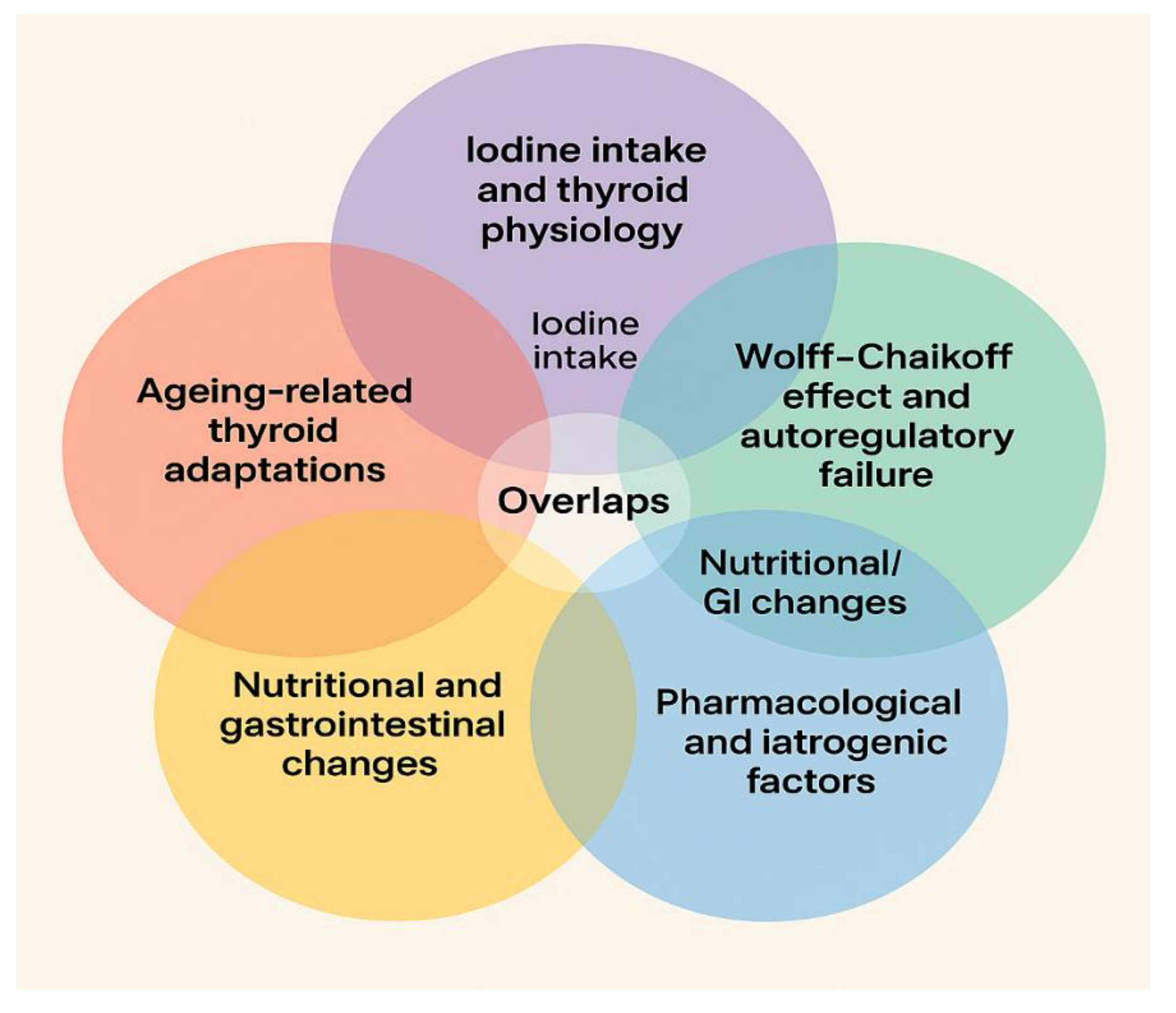
Figure 1. Overlaping subjects in articles taken into consideration in the review.
Preprints 178368 g001
Figure 2. Factors influencing thyroid hormone secretion and priorities for older patients.
Preprints 178368 g002
Figure 3. Microbiota, inflamation and thyroid functioning.
Preprints 178368 g003
Figure 4. Older adults require a special integrated clinical management in order to ensure an adequate functioning of thyroid.
Preprints 178368 g004
Table 1. Studies included in the narrative review.
Topic References Notes / Focus / Overlaps
  • Iodine intake and thyroid physiology
1–30, 58, 60, 62–67 Deficiency/excess, hormone synthesis, population surveys; overlaps with Wolff–Chaikoff effect
  • Ageing-related thyroid adaptations
3, 4, 31–42, 44, 54–57 Elderly thyroid function, structural/hormonal changes; overlaps with metabolic ageing & nutritional changes
  • Wolff–Chaikoff effect and autoregulatory failure
19–21, 58, 60–66, 110–113 Iodine-induced hypothyroidism, autoregulation mechanisms; overlaps with iodine intake
  • Microbiota–thyroid axis
68–75, 77–100, 142–143 Gut–thyroid interactions, autoimmunity, probiotics; overlaps with nutritional/GI changes
  • Nutritional and gastrointestinal changes
101–103, 105–109, 115, 138, 140 Malnutrition, protein/mineral intake, GI aging; overlaps with microbiota axis & ageing adaptations
  • Pharmacological and iatrogenic factors
110–114, 116–121 Drugs affecting thyroid, contrast agents, interactions; overlaps with Wolff–Chaikoff effect in some cases
  • Metabolic ageing
122–137, 139, 141 Thyroid–metabolism interplay, diabetes, adipokines, kidney; overlaps with ageing adaptations
Disclaimer/Publisher’s Note: The statements, opinions and data contained in all publications are solely those of the individual author(s) and contributor(s) and not of MDPI and/or the editor(s). MDPI and/or the editor(s) disclaim responsibility for any injury to people or property resulting from any ideas, methods, instructions or products referred to in the content.
Copyright: This open access article is published under a Creative Commons CC BY 4.0 license, which permit the free download, distribution, and reuse, provided that the author and preprint are cited in any reuse.
Prerpints.org logo

Preprints.org is a free preprint server supported by MDPI in Basel, Switzerland.

Subscribe

Disclaimer

Terms of Use

Privacy Policy

Privacy Settings

© 2026 MDPI (Basel, Switzerland) unless otherwise stated