

Submitted:
27 May 2026
Posted:
28 May 2026
You are already at the latest version
Abstract
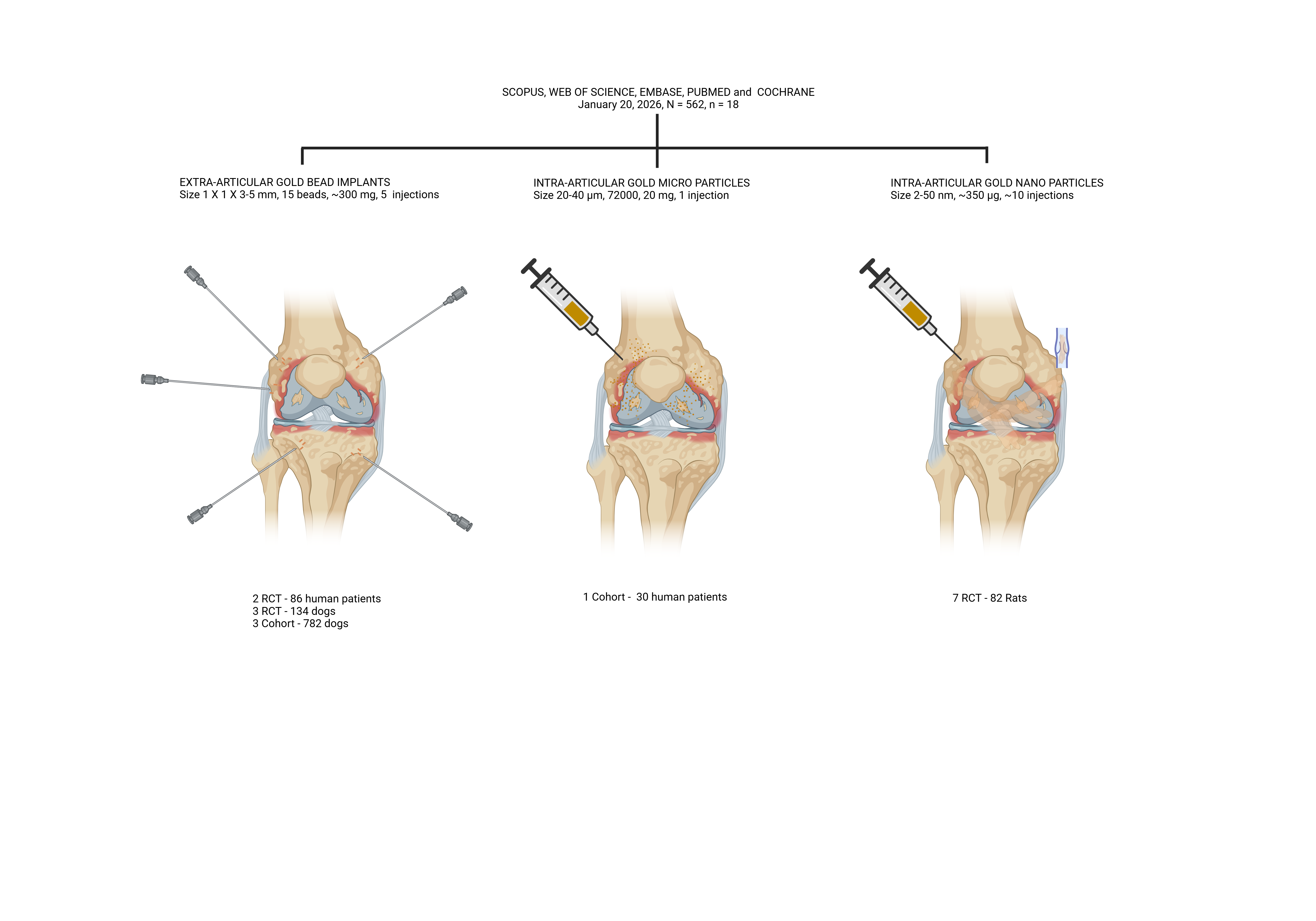
Background and objectives: Gold-based therapies are increasingly being studied for their potential to alleviate osteoarthritic (OA) symptoms, although their overall evidence remains inconclusive. Methods: We conducted a systematic search (January 20, 2026) across five databases. We included both human and animal cohort studies and randomized controlled trials that reported on the effects of metallic gold nanoparticles (AuNP), microparticles (µGold), and bead implants (GBI) on osteoarthritic symptoms. The risk of bias was assessed using the Cochrane, CAMARADES, and ROBINS-I tools. Results: A total of 18 studies met the inclusion criteria. We analyzed 7 studies on AuNP, 1 on µGold, and 10 on GBI, grouping them by study type. In the analysis of five randomized studies on BMI in human and animal OA, a significant difference between the intervention and the control was found in one human and one animal study. The seven randomized studies examining AuNP for animal OA reported significant differences between the intervention and control groups. The cohort studies assessing the success of BMI in animals demonstrate different 95% CI’s, with a total success of 668/782. One human cohort study of µGold demonstrates a wide 95% CI of success. Conclusions: Although the evidence is of low quality and shows moderate heterogeneity, the results support the use of AuNP, µGold, and GBI for OA treatment. Notably, µGold may provide superior localized delivery via a single intra-articular administration and potentially enable greater gold-ion release than GBI and AuNP do.
Keywords:
osteoarthritis
; gold microparticles
; gold nanoparticles
; gold bead implants
; human studies
; animal model
; systemic review
; narrative synthesis
1. Introduction
Gold-based therapies have been extensively studied for their potential to treat inflammatory and degenerative diseases, especially joint disorders like osteoarthritis (OA) and rheumatoid arthritis [1]. Among these approaches, gold bead implantation (GBI) [2,3], gold microparticles (µGold) [4,5], and gold nanoparticles (AuNP) [6] have emerged as prominent modalities, each with distinct properties influencing their clinical use. Both µGold and GBI interventions seek to harness gold’s anti-inflammatory properties and biocompatibility; however, their differences in size, delivery method, and therapeutic efficacy define their clinical applications [2,3,4,5]. Although AuNP are utilized in diagnostics, imaging, photothermal therapy, radiotherapy, and drug delivery [6], AuNP have not been reported to be used in clinical treatment on their own.
GBI, traditionally used in permanent implantation procedures, has been employed in veterinary and human medicine, particularly in chronic pain management and degenerative joint conditions [2,3,7,8,9]. Their localized presence provides long-term therapeutic pain relief and anti-inflammatory effects, but potential concerns remain regarding implant migration, localized fibrosis, and surgical risks associated with implantation [10,11]. Traditionally, GBI involves the surgical placement of small gold beads into specific anatomical locations, such as around artificial joints or acupuncture points, to provide long-term pain relief and anti-inflammatory effects [7,8,9].
In contrast, µGold offers a minimally invasive alternative, administered via intra-articular injection, potentially reducing procedural risks while maintaining effective immune modulation and anti-inflammatory effects [4,5,12,13]. Recent exploratory human studies show promising results using intra-articular injections of gold microparticles in patients with knee and hip osteoarthritis, leading to significant pain reduction and improved joint function, with no reported adverse effects [4,12,13]. These outcomes suggest that µGold may offer a safer and more effective alternative to traditional GBI.
The effectiveness of gold-based local anti-inflammatory treatments is closely linked to the distribution of gold ions within the inflamed tissue [14,15]. Gold ions are slowly released from the implanted gold by macrophages through a process called dissolucytosis. The distribution of gold ions into the interstitial cellular fluid and cells from the gold surfaces is within a few mm [14,15]. This may explain why a multitude of gold particles, 72.000 gold micro particles (µGold) [4,12,13] may offer superior therapeutic outcomes compared to larger GBI implants [2,3] or repeated injection of AuNPs [6,16,17]. The micron-sized particles composed of 99.99% pure gold further expand the available gold surface drastically compared to gold beads and substantially increase the amount of released gold ions. The tiny particles can be injected directly into joints without any side effects [4,12,13]. Another advantage is that a thin 21G needle can be used to apply gold microparticles, making the treatment easier and almost painless compared to using a 14G needle and stylet to inject millimeter-sized gold beads [2,3,7].
This study aims to investigate whether there is a spatial advantage among µGold, GBI, and AuNP. It is hypothesized that µGold therapy, which involves a single, minimally invasive injection, is more effective than multiple GBI and repeated AuNP injections in treating joint diseases. The rationale behind this hypothesis is that having more gold implant particles present and stable in proximity to the inflamed tissue is expected to enhance the dissolucytotic and immunosuppressive effects, ultimately improving outcomes and reducing side effects.
2. Materials and Methods
2.1. Reporting
This report follows the PRISMA guidelines [18]. The completed PRISMA checklist is provided in the Supplementary Materials (S1). The protocol was not registered because it is a narrative analysis of a systematic review, and it was not possible to register it given the inclusion of both animal and human studies.
2.2. Information Sources and Search Strategy
This study identified human and animal studies of local injections of gold microparticles (µGold), gold bead implantation (GBI), and gold nanoparticles (AuNP) for osteoarthritis and other inflammatory joint disorders. Five databases were searched: Medline (PubMed), Embase, Cochrane, Scopus, and Web of Science, with no language or publication date restrictions. The categories used were ("gold microparticles"[All Fields] OR "gold implantation"[All Fields] OR "gold bead implantation"[All Fields] OR "gold nanoparticles"[All Fields]) AND ("osteoarthritis"[All Fields] OR "joint disease"[All Fields] OR "rheumatoid arthritis"[All Fields] OR "joint inflammation"[All Fields] OR "cartilage repair"[All Fields] OR "synovial inflammation"[All Fields] OR "degenerative joint disease"[All Fields]). The search was conducted by SR and SK from Aalborg University, Denmark, on January 20, 2026, to investigate the effects of µGold, GBI, and AuNPs on osteoarthritis and other inflammatory joint disorders. Complete details of the electronic search strategies are provided in Appendix 1.
2.3. Study Selection
Duplicate entries were manually identified and removed by the authors, and the authors independently screened eligible studies in two steps in accordance with the PRISMA Guidelines [18]. In the first step, we screened all titles and abstracts according to the predefined inclusion and exclusion criteria. The inclusion criteria comprised human and animal studies that investigate the effects of µGold, GBI, and AuNP on osteoarthritis and other inflammatory joint diseases, with quantifiable data (N, n1, n2, number improved, ratio, mean, SD, or SEM) reported for each group. In cases of conflicts, the authors re-evaluated the title and abstract and reached a mutual consensus after discussion.
In the second step, the two reviewers independently read all full-text articles from the previous step. In addition to the previously mentioned inclusion criteria, the final inclusion criteria were human and animal studies investigating the effects of µGold, GBI, and AuNP when locally injected periarticularly or intraarticularly, specifically measuring the number of subjects improved in randomized and cohort studies, or the mean and SD of inflammatory markers in randomized studies. Additionally, for each study, the number of injections used is specified. Only studies reporting outcomes after a minimum of 8 weeks for humans and large animals, and 1 week for small animals, were included to ensure that the initial intention-to-treat, local, and general anesthesia had subsided, allowing for a clearer evaluation of early- to late-stage treatment outcomes. This criterion was set to ensure that the effects of initial treatment, local anesthesia, and general anesthesia had subsided, allowing for a clearer evaluation of treatment outcomes from early through late stages. Reasons for exclusion were pre-specified as follows: 1) no in-vivo experiment, 2) no extractable or available data, 3) wrong route of administration, and 4) not µGold, GBI, or AuNP.
2.4. Quality Assessment
The quality of the included randomized human studies was assessed for bias by the Cochrane Collaboration’s tool for assessing risk of bias [19], which assess various types of bias, including random sequence generation, allocation concealment, blinding of participants and personnel, blinding of outcome assessment, incomplete outcome data, selective reporting, and other biases. The authors conducted the quality assessment, and any disagreements that arose were resolved through discussion.
The quality of the included randomized animal studies was assessed by the Collaborative Approach to Meta-Analysis and Review of Animal Data from Experimental Studies (CAMARADES) checklist [20,21]. The tool includes 14 questions designed to identify potential biases in the study design. Scores range from 0 to 14, with a higher score reflecting a greater methodological quality of the study [20,22].
The quality of the included non-randomized studies was assessed for bias by the Cochrane Collaboration’s tool for assessing risk of bias [23] version 2 of the ROBINS-I tool, launched on 22 November 2024 [24], which includes an assessment of bias in seven domains: (1) bias due to confounding (control for confounders (different size of animals included, different joints treated, different causes of osteoarthritis) and control for postintervention variables (time varying, other treatment); (2) bias in classification of interventions (clearly defined intervention groups); (3) bias in selection of participants into the study (or into the analysis) (selection of participants into the study (or into the analysis) based on participant characteristics observed after the start of intervention); (4) bias due to deviations from intended interventions (deviations from the intended intervention beyond what would be expected in usual practice, and adheres to the assigned intervention regimen); (5) bias due to missing data (outcome data available for all, or nearly all, participants, and participants excluded due to missing data on other variables needed for the analysis); (6) bias arising from measurement of the outcome (outcome measure influenced by knowledge of the intervention received, outcome assessors aware of the intervention received by study participants, and systematic errors in measurement of the outcome related to the intervention received); (7) and bias in selection of the reported results (multiple outcome measurements, multiple analyses of the intervention-outcome relationship, and different subgroups). Each bias domain is evaluated as low, moderate, serious, or critical according to the ROBINS-I framework. The quality assessment was carried out by the authors, and any disagreements were resolved by discussion.
2.5. Data Extraction
For each eligible study, the following characteristics were extracted: year of publication, number of participants, type and formulation of gold used (e.g., µGold, GBI, and AuNP), target condition (e.g., osteoarthritis or other inflammatory joint diseases), and route of administration (i.e., extra-articular or intra-articular injection). For randomized and cohort studies, we extracted data on the number of subjects who improved. For randomized studies with quantifiable outcomes, we extracted group-level data (N, n1, n2, number improved, proportion, mean, SD, or SEM) on inflammatory markers and osteoarthritic scores.
2.6. Statistical Analysis
To conduct the analysis, the statistical software Stata MP 18.0 was utilized. For randomized studies, we calculated the log odds-ratio or Cohen’s d with a 95% confidence interval (CI). For cohort studies, we calculated the 95% CI of the proportions of successes.
Generative artificial intelligence (GenAI) has not been used in this paper (e.g., to generate text, data, or graphics, or to assist in study design, data collection, analysis, or interpretation).
3. Results
3.1. Study Selection
The study selection process is illustrated in the PRISMA flow diagram (Figure 1).
On January 20, 2026, the search yielded 562 studies. Of these, 177 were duplicates, and 337 were excluded based on irrelevance from the title and abstract. After assessing the 48 full-text articles, 25 studies were deemed eligible. Five animal studies using protein-conjugated AuNP [25,26,27,28,29], and two human studies using hyaluronic acid as a carrier [12,13] were excluded. Included in the final analysis were 18 studies [2,3,4,7,8,9,30,31,32,33,34,35,36,37,38,39,40,41].
3.2. Study Characteristics
The studies were published between 1992 and 2024. Table 1, Table 2 and Table 3 presents the characteristics of the included studies.
The study characteristics display a heterogeneous group of studies that can be divided into twelve randomized trials [2,3,7,8,9,30,31,32,33,34,40,41], and six cohorts [4,35,36,37,38,39], in fifteen animal [7,8,9,30,31,32,33,34,35,36,37,38,39,40,41], and three human studies [2,3,4], in ten studies using GBI [2,3,7,8,9,35,36,37,38,39], seven studies using AuNP [30,31,32,33,34,40,41], and one study using µGold [4]. In seven studies [7,8,9,35,36,37,38], no metrics support the claim that the number of subjects improved. In two studies [2,3] metrics supported the claim that the number of subjects increased, but the metrics were not extractable. In one study, metrics support that the number of subjects improved.
The GBI studies [2,3,7,8,9,35,36,37,38,39] (Table 1, Table 2 and Table 3) were dog and human trials using 3-mm GBI (diameter 1 mm, length 2-4 mm) at three or more sites around the osteoarthritic joint. The follow-up period is between one month and one year. No adverse events were reported in three studies [2,3,37], and four studies do not report events [7,8,36,38]. Bleeding is reported in one study [39], and one case of infection in another study [9]. Durkes [35], who reported the first study on BMI and included the largest number of individuals, observed several possible adverse events. Intra-articularly placed BMI may cause pain and must be removed. The most common problem is superficial infection due to improper sterile technique, and another concern is the possibility of injuring the sciatic nerve. The number of injections for three beads ranged from 3 to 16. The gold dosage ranged from 91 to 728 mg, and the total surface area ranged from 0.71 to 1.78 cm2.
The AuNP studies [31,32,33,34,40,41] were all conducted in small-animal trials and employed a wide range of formulations, delivery strategies, and follow-up periods (Table 3). The studies were performed on rat ankles and knees. The number of injections ranged from 1 to 12, and follow-up ranged from 9 to 60 days. The AuNP size ranged between 20 and 50 nm. The total dosage ranges from 27 to 687.5 µg, and the total surface area ranges from 0.81 to 106.8 cm2.
The µGold study [4] was performed on human knee OA using a single injection. The patient’s synovial fluid [4] was used as a carrier of the µGold. The dosage of µGold was 20 mg gold microparticles (72,000 50particles, 20–40 μm in diameter) with a total surface of 2.05 cm2. No adverse effects were observed during the two years following the treatment.
3.3. Study Quality and Publication Bias
The Cochrane Collaboration's tool for assessing risk of bias [19,42] was applied to two studies [2,3], resulting in a low risk of bias (Table 4).
The CAMARADES checklist for assessing risk of bias [20,21] was used for the randomized animal trials [7,8,9,30,31,32,33,34,40,41] (Table 5). All studies (n = 12) were published in peer-reviewed journals using rats, or dogs as animal models. The risk-of-bias scores ranged from 2 to 12, with a median of 8.
The Cochrane Collaboration’s tool for assessing risk of bias [23], version 2 of the ROBINS-I tool, launched on 22 November 2024 [24], was used for the human [4] and animal cohort studies [35,36,37,38,39] (Table 6). Table 6 tabulates 5 serious and 20 critical risks of bias across 35 assessments of the animal studies.
3.4. Statistical Analysis Results
The 18 included randomized and cohort studies examining the effects of BMI, AuNP, and µGold were analyzed using 95% confidence intervals for log-odds ratios for binomial outcomes in randomized studies, Cohen’s d for continuous outcomes in randomized studies, and proportions of successes in cohort studies. In the analysis of five randomized studies on BMI for human and animal OA, two studies found a significant difference between the intervention and control groups [2,3,7,8,9]. A significant difference between the intervention and the control was found in one human [3] and one animal study [8] (Table 1 and Table 2). The seven randomized studies examining AuNP for animal OA [31,32,33,34,40,41] reported significant differences between the intervention and control groups (Table 2). The cohort studies assessing the success of BMI in animals [35,36,37,38,39] demonstrate different 95% CI’s, with a total success of 668/782 (0.854 (0.829-0.879)) (Table 3). One human cohort study of µGold demonstrates a wide 95% CI of success [4] (Table 3)
4. Discussion
This narrative review is the first to evaluate the effects of extra-articular and intra-articular gold treatments for osteoarthritis (OA) in both human and animal trials. Significant effects of GBI were found in one human randomized controlled trial [3] and one animal randomized controlled trial [8] when comparing extra-articular GBI to a placebo. However, no significant effects were found in one human randomized trial [2] and two animal randomized trials [7,9].
In five animal cohort studies, extra-articular GBI was associated with reported improvement [35,36,37,38,39]. The analysis indicated a significant effect of intra-articular gold nanoparticles (AuNP) on OA in seven randomized animal trials compared with placebo [30,31,32,33,34,40,41]. One human cohort study of intra-articular µGold reported improvement in patients with knee OA [4].
The analysis of the study characteristics revealed that the GBI and AuNP trials involved multiple injections, whereas the µGold trials utilized a single injection.
When calculating the total gold surface area that facilitates the diffusion of gold ions into the interstitial cellular fluid and cells from the gold surfaces [14,15], the area for this process, known as dissolucytosis [4,15,43] was greater in the AuNP studies than in the BMI studies and the µGold study. This may explain the more significant effect of the intervention in these studies, especially when considering the size of small animals.
The data analysis and examination of study characteristics support the hypothesis that µGold may offer a spatial advantage over GBI and AuNP. The findings indicate that a single injection of µGold, with its large surface area conducive to dissolucytosis, bolsters this hypothesis. However, several weaknesses include the limited number of studies in each category and the fact that the µGold studies are uncontrolled cohorts.
The effectiveness of intra-articular AuNP may be reduced by the macrophages' ability to remove and scavenge nanoparticles [31,43,44], which may explain the need for multiple injections. This is clinically relevant in a human setup, as multiple injections will require several outpatient visits and increase the risk of infection. Intravenous [45] and intraperitoneal [46] injections and oral administration [47] of AuNP can relieve OA symptoms in animal studies and thereby demonstrate the macrophages’ ability to remove and transport the AuNP to all other tissues [48] and indicate that there is a systemic effect of AuNP. There is an accumulation of injected AuNP in the liver, spleen, and lungs [48,49] and granular degeneration is observed in these tissues [48], which may pose a risk. There is an intracellular release of gold ions from the AuNP, which initiates the anti-inflammatory and other responses [14,50]. Even though AuNPs are considered highly biocompatible nano constructs, potential toxicity, primarily related to internalization pathways, has been demonstrated by the release of free gold ions in various cells and tissues [51]. In five AuNP studies excluded from this analysis, different proteins were used as carriers. The proteins can enhance the AuNP’s therapeutic effects by acting as carriers, improving stability, targeting diseased cells, and facilitating controlled release of gold ions, or may have a direct therapeutic effect.
The macrophages cannot remove or transport µGold and GBI, and the gold microparticles and beads stay in place. Only cells close to the µGold and GBI become loaded with gold ions, implying that no gold is transported to other organs, and may exclude any toxic effect [14,15,43]. The dose-response relationship for gold in the treatment of arthritis depends strongly on the delivery form (nanoparticles, microparticles, or salts). Oral micro-dosing of AuNP at a daily dose of 0.34 mg for 8 weeks (19 mg) is safe and effective for patients with rheumatoid arthritis and osteoarthritis [52]. Continuous injection of gold clusters (15 mg Au/kg) has been observed in experimental models to cause abnormal blood values and effects on the kidneys and adrenal glands [53]. Lower doses (e.g., 10 mg Au/kg) are not associated with obvious toxicity in these models [53]. The clinical literature describes that the risk of serious and potentially irreversible side effects increases significantly when the total (cumulative) dose exceeds 1.0 to 1.5 grams of gold [54].
Gold is generally inert (non-reactive) in its metallic form (Au0), making it biocompatible and indigestible. Its clinical and biological effects occur primarily when gold is exposed to an extracellular environment. Facilitated by M1 macrophages, it is converted into gold compounds (gold ions, Au+/Au3+) that can bind to proteins and enzymes, or when used as nanoparticles, can enter cells and interact physically [54,55].
Disulfide bonds are essential determinants of protein stability and biological function in medically relevant proteins, including enzymes, hormones, antibodies, and cell-surface receptors, where they stabilize the native three-dimensional structure and limit conformational flexibility [56]. Gold exhibits an unusually strong affinity for sulfur, forming stable S-Au–S interactions with cysteine thiols and disulfide-containing motifs, a property extensively characterized in chemical and nano-biological systems [57,58]. Experimental evidence demonstrates that gold(I) compounds and gold-based nanomaterials can directly interact with and disrupt protein disulfide bonds via thiol–disulfide exchange or formation of S–Au-S interactions, as shown for model proteins such as insulin and for clinically relevant agents including auranofin [59]. These interactions can destabilize native protein folding, leading to partial unfolding or functional impairment, effects that are mechanistically linked both to the therapeutic activity of gold-containing drugs and to their potential toxicity [60,61]. Disruption of these stabilizing elements, therefore, has immediate consequences for protein conformation and biological activity [62,63,64]. Closely related to this process is gold-mediated disruption of zinc-binding motifs. Structural zinc ions stabilize numerous protein domains by coordinating cysteine- and histidine-rich motifs in tetrahedral geometries essential for proper folding. These zinc sites are intrinsically labile, as they rely on soft sulfur ligands that are highly susceptible to chemical attack. Gold compounds do not substitute Zn iso-structurally; instead, they expel Zn from these coordination environments and subsequently bind cysteine residues in linear S–Au–S interactions. Conceptually, this transformation can be represented as Zn–Cys₄ → 2×Au–Cys₂, resulting in the collapse of the zinc-binding architecture and unfolding of the associated domains [62,63,64]. Importantly, S-Au–S binding and Zn displacement are mechanistically linked by their shared dependence on sulfur coordination chemistry and frequently act in concert within the same protein. Many proteins contain both disulfide bonds and zinc-binding sites, such that exposure to gold can propagate structural destabilization across multiple stabilizing elements. This convergence explains why gold compounds often induce broad conformational perturbations rather than narrowly targeted inhibition. Taken together, these observations highlight that gold compounds function as modulators of protein structural integrity rather than simple enzyme inhibitors. Recognition of S-Au–S binding and Zn displacement as interconnected pathways emphasizes the importance of structural protein chemistry (Figure 2).
This review has several limitations. The 18 included studies are divided into human and animal studies and use three different gold sizes. Furthermore, substantial variation in the number of individuals included in each study and high heterogeneity did not support a meta-analysis. In addition, there is a high variation in study quality and bias. There is a risk of overemphasizing statistical significance when confidence intervals are wide or near zero. Conjugation of protein to AuNP may influence the results of the treatment, and five animal studies using protein-conjugated AuNP [25,26,27,28,29] were excluded, which reduced the number of animal individuals that could have been included in the analysis. Hyaluronic acid has a clinical effect, and two human studies [12,13] using hyaluronic acid as a carrier for gold microparticles was excluded, reducing the number of patients who could have been included in the analysis by 182.
The use of gold to treat inflammatory diseases such as rheumatoid arthritis became significant in the early 20th century and is well-documented [4,65]. However, the gold thiocompounds (i.e., sulfur-containing gold-based drugs such as auranofin) must be administered with caution, as they are toxic to the liver, kidneys, and skin, and have been replaced by more modern disease-modifying antirheumatic drugs [66]. Later, it became evident that gold ions alter antigen processing and reduce cytokine expression in macrophages [67]. With the introduction of GBI for the treatment of animal OA using acupuncture points for insertion in the 70-ties [35,68], it became interesting to investigate the effect of different gold therapies for OA [4,6,16,17,30], based on the limited benefit of the intra-articular injections that are a major part of the treatment program for osteoarthritis, which includes exercise, analgesics, and dietary weight management [69,70]. The intra-articular treatment with corticosteroids and hyaluronic acid provides limited benefit for 3-4 weeks [69,70]. Systematic reviews do not support viscosupplementation [71], and there is no evidence for platelet-rich plasma [72], stem cells [73], or micro-fragmented adipose tissue [74].
There is limited evidence for µGold compared with the previously mentioned evidence for other intra-articular treatments for osteoarthritis. The actual human clinical studies [4,12,13] of µGold for osteoarthritis are cohort studies without a control group. Another issue is that pure metallic gold is a basic element that cannot be patented and is not FDA-approved as a drug or implant. There is a need to demonstrate significant and sustained clinical effectiveness beyond placebo, as well as favorable risk-benefit profiles, particularly with respect to adverse events. Future studies of µGold need to address the challenges posed by the heterogeneity of OA patients and symptoms. In addition, the difficulty in establishing measurable metrics of functional improvements and the long-term safety of delivery further complicates regulatory approval. OA pain relief and functional improvement are not linear, making it difficult to demonstrate a consistent, clinically meaningful benefit that outweighs the treatment's risks [75]. A planned randomized, double-blind, controlled trial of µGold for knee OA is registered with ClinicalTrials.gov (NCT07202390).
5. Conclusions
GBI, µGold, and AuNP have shown potential to relieve OA symptoms in animal and human studies, supporting their potential as therapeutic agents. µGold may have a spatial advantage in using a single intra-articular administration and a higher possibility of release of gold ions compared to GBI and AuNP. The evidence is low to moderate with a moderate degree of heterogeneity. Large randomized, double-blind human studies comparing intra-articular µGold and AuNP with a placebo are necessary to draw proper conclusions.
Author Contributions
Conceptualization: Sten Rasmussen, Stephen Gunaratnam Klavsen; Methodology: Sten Rasmussen, Stephen Gunaratnam Klavsen; Formal analysis: Sten Rasmussen, Stephen Gunaratnam Klavsen; Investigation: Sten Rasmussen, Stephen Gunaratnam Klavsen; Writing – original draft: Sten Rasmussen, Stephen Gunaratnam Klavsen; Writing – review & editing: Sten Rasmussen.
Funding
This study received no external funding.
Data Availability Statement
The protocol and the data supporting the findings of this study are available from the corresponding author upon reasonable request.
Conflicts of Interest
The authors declare no conflict of interest.
Abbreviations
The following abbreviations are used in this manuscript:
| OA | Osteoarthritis |
| µGold | Gold microparticles |
| GBI | Gold bead particles |
| AuNPs | Gold nanoparticles |
| e.a. | Extra-articular |
| i.a. | Intra-articular |
Appendix A
Search strategy, January 20, 2026:
Scopus
(“gold microparticles” OR “gold implantation” OR “gold bead implantation” OR “gold nanoparticles”) AND ("osteoarthritis" OR "joint disease" OR "rheumatoid arthritis" OR "joint inflammation" OR "cartilage repair" OR "synovial inflammation" OR "degenerative joint disease") AND ("clinical trial" OR "human study" OR "animal study" OR "preclinical study" OR "in vivo") NOT ("gold dental implant" OR "gold stent" OR "gold alloy").
N = 305
Web of Science
TS=("gold microparticles" OR "gold implantation" OR "gold bead implantation" OR “gold nanoparticles”) AND TS=("osteoarthritis" OR "joint disease" OR "rheumatoid arthritis" OR "joint inflammation" OR "cartilage repair" OR "synovial inflammation" OR "degenerative joint disease")
225
Embase
('gold microparticles'/exp OR 'gold implantation'/exp OR 'gold bead implantation'/exp OR 'gold nanoparticles'/exp) AND ('osteoarthritis'/exp OR 'joint disease'/exp OR 'rheumatoid arthritis'/exp OR 'joint inflammation'/exp OR 'cartilage repair'/exp OR 'synovial inflammation'/exp OR 'degenerative joint disease'/exp) AND ('clinical trial'/exp OR 'human study'/exp OR 'animal study'/exp OR 'preclinical study'/exp OR 'in vivo study'/exp)
N=206
PubMed:
(“gold microparticles” OR “gold implantation” OR “gold bead implantation” OR “gold nanoparticles”) AND ("osteoarthritis" OR "joint disease" OR "rheumatoid arthritis" OR "joint inflammation" OR "cartilage repair" OR "synovial inflammation" OR "degenerative joint disease")
N = 79
Cochrane
("gold microparticles" OR "gold nanoparticles" OR "gold implantation" OR "gold bead implantation") AND ("osteoarthritis" OR "joint disease" OR "rheumatoid arthritis" OR "joint inflammation" OR "cartilage repair" OR "synovial inflammation" OR "degenerative joint disease")
N = 4
References
- Rasmussen, S.; Frederickson, C.; Danscher, G. Inhibition of Local Inflammation by Implanted Gold: A Narrative Review of the History and Use of Gold. J. Rheumatol. 2023, 50. [Google Scholar] [CrossRef]
- Nejrup, K.; de Fine Olivarius, N.; Jacobsen, J.L.; Siersma, V. Randomised Controlled Trial of Extraarticular Gold Bead Implantation for Treatment of Knee Osteoarthritis: A Pilot Study. Clin. Rheumatol. 2008, 27, 1363–1369. [Google Scholar] [CrossRef] [PubMed]
- Kjerkegaard, H.K.; Kirkeby, R.; Christensen, T.B.; Schlünzen, L. Double-Blinded, Placebo-Controlled Trial of the Pain-Relieving Effect of Gold Bead Implantation on Cervical Osteoarthritis. Med. Acupunct. 2011, 23, 87–91. [Google Scholar] [CrossRef]
- Rasmussen, S.; Kjær Petersen, K.; Kristiansen, M.K.; Skallerup, J.; Aboo, C.; Thomsen, M.E.; Skjoldemose, E.; Jørgensen, N.K.; Stensballe, A.; Arendt-Nielsen, L. Gold Micro-Particles for Knee Osteoarthritis. Eur. J. Pain (United Kingdom) 2022, 26. [Google Scholar] [CrossRef] [PubMed]
- Märki, N.; Witte, S.; Kuchen, S.; Reichenbach, S.; Ramseyer, A.; Gerber, V.; Spadavecchia, C. Safety of Intra-Articular Gold Microimplants in Horses–A Randomized, Blinded, Controlled Experimental Study. J. Equine Vet. Sci. 2018, 60, 59–66.e2. [Google Scholar] [CrossRef]
- Huang, H.; Liu, R.; Yang, J.; Dai, J.; Fan, S.; Pi, J.; Wei, Y.; Guo, X. Gold Nanoparticles: Construction for Drug Delivery and Application in Cancer Immunotherapy. Pharmaceutics 2023, 15, 1868. [Google Scholar] [CrossRef]
- Hielm-Bjorkman, A.; Raekallio, M.; Kuusela, E.; Saarto, E.; Markkola, A.; Tulamo, R. Double-blind Evaluation of Implants of Gold Wire at Acupuncture Points in the Dog as a Treatment for Osteoarthritis Induced by Hip Dysplasia. Vet. Rec. 2001, 149, 452–456. [Google Scholar] [CrossRef]
- Jaeger, G.T.; Larsen, S.; Søli, N.; Moe, L. Double-blind, Placebo-controlled Trial of the Pain-relieving Effects of the Implantation of Gold Beads into Dogs with Hip Dysplasia. Vet. Rec. 2006, 158, 722–726. [Google Scholar] [CrossRef]
- Bolliger, C.; DeCamp, C.E.; Stajich, M.; Flo, G.; Martinez, S.; Bennett, R.; Bebchuk, T. Gait Analysis of Dogs with Hip Dysplasia Treated with Gold Bead Implantation Acupuncture. J. Eur. Soc. Vet. Orthop. Traumatol. 2002, 15, 116–122. [Google Scholar]
- Lie, K.-I.; Jæger, G.; Nordstoga, K.; Moe, L. Inflammatory Response to Therapeutic Gold Bead Implantation in Canine Hip Joint Osteoarthritis. Vet. Pathol. 2011, 48, 1118–1124. [Google Scholar] [CrossRef]
- Jæger, G.T.; Stigen, Ø.; Devor, M.; Moe, L. Gold Bead Implantation in Acupoints for Coxofemoral Arthrosis in Dogs: Method Description and Adverse Effects. Animals 2012, 2, 426–436. [Google Scholar] [CrossRef] [PubMed]
- Rasmussen, S.; Petersen, K.K.; Aboo, C.; Andersen, J.S.; Skjoldemose, E.; Jørgensen, N.K.; Stensballe, A.; Arendt-Nielsen, L. Intra-Articular Injection of Gold Micro-Particles with Hyaluronic Acid for Painful Knee Osteoarthritis. BMC Musculoskelet. Disord. 2024, 25, 211. [Google Scholar] [CrossRef] [PubMed]
- Rasmussen, S.; Skjoldemose, E.; Jørgensen, N.K. Intraarticular Gold Microparticles Using Hyaluronic Acid as the Carrier for Hip Osteoarthritis. A 2-Year Follow-up Pilot Study. Sci. Rep. 2024, 14, 26249. [Google Scholar] [CrossRef] [PubMed]
- Danscher, G. In Vivo Liberation of Gold Ions from Gold Implants. Autometallographic Tracing of Gold in Cells Adjacent to Metallic Gold. Histochem. Cell Biol. 2002, 117, 447–452. [Google Scholar] [CrossRef] [PubMed]
- Larsen, A.; Stoltenberg, M.; Danscher, G. In Vitro Liberation of Charged Gold Atoms: Autometallographic Tracing of Gold Ions Released by Macrophages Grown on Metallic Gold Surfaces. Histochem. Cell Biol. 2007, 128, 1–6. [Google Scholar] [CrossRef]
- Filho, M.C.B.; dos Santos Haupenthal, D.P.; Zaccaron, R.P.; de Bem Silveira, G.; de Roch Casagrande, L.; Lupselo, F.S.; Alves, N.; de Sousa Mariano, S.; do Bomfim, F.R.C.; de Andrade, T.A.M.; et al. Intra-articular Treatment with Hyaluronic Acid Associated with Gold Nanoparticles in a Mechanical Osteoarthritis Model in Wistar Rats. J. Orthop. Res. 2021, 39, 2546–2555. [Google Scholar] [CrossRef]
- Sarkar, A.; Carvalho, E.; D’souza, A.A.; Banerjee, R. Liposome-Encapsulated Fish Oil Protein-Tagged Gold Nanoparticles for Intra-Articular Therapy in Osteoarthritis. Nanomedicine 2019, 14, 871–887. [Google Scholar] [CrossRef]
- Page, M.J.; McKenzie, J.E.; Bossuyt, P.M.; Boutron, I.; Hoffmann, T.C.; Mulrow, C.D.; Shamseer, L.; Tetzlaff, J.M.; Akl, E.A.; Brennan, S.E.; et al. The PRISMA 2020 Statement: An Updated Guideline for Reporting Systematic Reviews. J. Clin. Epidemiol. 2021, 134, 178–189. [Google Scholar] [CrossRef]
- Higgins, J.P.T.; Altman, D.G.; Gotzsche, P.C.; Juni, P.; Moher, D.; Oxman, A.D.; Savovic, J.; Schulz, K.F.; Weeks, L.; Sterne, J.A.C. The Cochrane Collaboration’s Tool for Assessing Risk of Bias in Randomised Trials. BMJ 2011, 343, d5928–d5928. [Google Scholar] [CrossRef]
- Macleod, M.R.; O’Collins, T.; Howells, D.W.; Donnan, G.A. Pooling of Animal Experimental Data Reveals Influence of Study Design and Publication Bias. Stroke 2004, 35, 1203–1208. [Google Scholar] [CrossRef]
- Ritskes-Hoitinga, M.; Leenaars, M.; Avey, M.; Rovers, M.; Scholten, R. Systematic Reviews of Preclinical Animal Studies Can Make Significant Contributions to Health Care and More Transparent Translational Medicine. In Cochrane Database of Systematic Reviews; Tovey, D., Ed.; John Wiley & Sons, Ltd: Chichester, UK, 2014. [Google Scholar]
- Wilson, E.; Ramage, F.J.; Wever, K.E.; Sena, E.S.; Macleod, M.R.; Currie, G.L. Designing, Conducting, and Reporting Reproducible Animal Experiments. J. Endocrinol. 2023, 258. [Google Scholar] [CrossRef]
- Sterne, J.A.; Hernán, M.A.; Reeves, B.C.; Savović, J.; Berkman, N.D.; Viswanathan, M.; Henry, D.; Altman, D.G.; Ansari, M.T.; Boutron, I.; et al. ROBINS-I: A Tool for Assessing Risk of Bias in Non-Randomised Studies of Interventions. BMJ 2016, i4919. [Google Scholar] [CrossRef]
- Sterne, J.; Higgins, J. ROBINS-1 V2 Tool.
- Campos, W.N. da S.; Leite, A.E.T.; Sonego, D.A.; de Andrade, M.A.; Pizzinatto, F.D.; Marangoni, V.S.; Zucolotto, V.; Nakazato, L.; Colodel, E.M.; Souza, R.L. de Síntese e Caracterização de Nanopartículas de Ouro Conjugadas Com Curcumina e Seus Efeitos Na Osteoartrite Experimental Induzida. Ciência Rural 2017, 47. [Google Scholar] [CrossRef]
- Sarkar, A.; Carvalho, E.; D’souza, A.A.; Banerjee, R. Liposome-Encapsulated Fish Oil Protein-Tagged Gold Nanoparticles for Intra-Articular Therapy in Osteoarthritis. Nanomedicine 2019, 14, 871–887. [Google Scholar] [CrossRef]
- Shahen, S.M.; Mohamed, M.R.; Ali, M.R.K.; Samaka, R.M.; Hamdy, G.M.; Talaat, R.M. Therapeutic Potential of Targeted-gold Nanospheres on Collagen-induced Arthritis in Rats. Clin. Exp. Pharmacol. Physiol. 2021, 48, 1346–1357. [Google Scholar] [CrossRef] [PubMed]
- Wei, Y.; Nie, Y.; Han, Z.; Huang, H.; Liao, X.; Wang, X.; Fan, Z.; Zheng, Y. Au@polydopamine Nanoparticles/Tocilizumab Composite as Efficient Scavengers of Oxygen Free Radicals for Improving the Treatment of Rheumatoid Arthritis. Mater. Sci. Eng. C 2021, 118, 111434. [Google Scholar] [CrossRef] [PubMed]
- Wang, C.; Hua, S.; Feng, T.; Zheng, Y.; Hu, Y.; Zhao, C.; Wang, S.; Hu, Y.; Zhou, M.; Liang, F. Starlike Au Nanoparticle Unleashing SiDDIT3 and Photothermal Power to Combat Ferroptosis - Driven Osteoarthritis. J. Nanobiotechnology 2025, 23, 487. [Google Scholar] [CrossRef]
- Tsai, C.; Shiau, A.; Chen, S.; Chen, Y.; Cheng, P.; Chang, M.; Chen, D.; Chou, C.; Wang, C.; Wu, C. Amelioration of Collagen-induced Arthritis in Rats by Nanogold. Arthritis Rheum. 2007, 56, 544–554. [Google Scholar] [CrossRef]
- Leonavičienė, L.; Kirdaitė, G.; Bradūnaitė, R.; Vaitkienė, D.; Vasiliauskas, A.; Zabulytė, D.; Ramanavičienė, A.; Ramanavičius, A.; Ašmenavičius, T.; Mackiewicz, Z. Effect of Gold Nanoparticles in the Treatment of Established Collagen Arthritis in Rats. Medicina 2012, 48, 91–101. [Google Scholar] [CrossRef]
- Kirdaite, G.; Leonaviciene, L.; Bradunaite, R.; Vasiliauskas, A.; Rudys, R.; Ramanaviciene, A.; Mackiewicz, Z. Antioxidant Effects of Gold Nanoparticles on Early Stage of Collagen-Induced Arthritis in Rats. Res. Vet. Sci. 2019, 124, 32–37. [Google Scholar] [CrossRef] [PubMed]
- Abdel-Hakem, N.; Abo-El-Atta, A.; Samaka, R.; El-Shahat, M.; Bassyouni, I.; Talaat, R. Improving the Anti-Inflammatory/Anti-Angiogenic Properties of Gold Nanoparticles in the Treatment of Experimental Rheumatoid Arthritis. Curr. Trends Immunol. 2022, 23, 33–42. [Google Scholar]
- dos Santos Haupenthal, D.P.; Resmini, M.B.; Da Silva, L.A.; Colares, M.C.; de Roch Casagrande, L.; Milanez Venturini, L.; de Andrade, T.A.M.; do Bomfim, F.R.C.; Thirupathi, A.; Emilio Feuser, P.; et al. Intra-Articular Treatment with Triamcinolone Hexacetonide Associated with Gold Nanoparticles Reduces Cartilage Degeneration in an Animal Model of Osteoarthritis. Curr. Drug Targets 2023, 24, 287–296. [Google Scholar] [CrossRef]
- Durkes, T. Gold Bead Implants. Probl. Vet. Med. 1992, 4, 207–211. [Google Scholar] [PubMed]
- Klitsgaard, J. Gold Implants - Practical Experiences with 400 Hip Dysplasia Cases in the Dog. In Proceedings of the International Veterinary Acupuncture Society Proceedings 22nd International Congress, Spiez, Schweiz, September 5 1996; pp. 1–5. [Google Scholar]
- Thoresen, S. A “New” Method of Placing Gold Implants to Treat Hip Dysplasia in the Dog. Description and Evaluation of 50 Cases 1990-1996. In Proceedings of the International Veterinary Acupuncture Society Proceedings 22nd Annual International Congress, Spiez, Schweiz, September 5 1996; pp. 1–7. [Google Scholar]
- Kothbauer, O. Über Die Implantation von Goldpartikeln Zur Therapeutischen Beeinflussung von Schmerzhaften Prozessen Im Hüftgelenksbereich von Hunden – Dargestellt an Drei Fallbeispielen. Wien. Tierarztl. Monatsschr. 1997, 84, 47–52. [Google Scholar]
- Bartholomé, M.; Schulze, E. Efficacy of Gold Bead Acupuncture Treatment of Chronic Hip, Elbow and Spine Pain in 79 Dogs Following Unsuccessful Conventional Treatments. Am. J. Tradit. Chin. Vet. Med. 2020, 15, 17–30. [Google Scholar]
- Chen, C.; Tang, C.; Chen, K.; Deng, M.; Pu, X.; Hu, T.; Chen, Z.; Zhong, L.; Jiang, Y.; Huang, Y. Hyaluronanized Gold Nanoparticles Functionalized with CTX-II Antibody for Early Diagnosis and Treatment of Knee Osteoarthritis. Chem. Eng. J. 2025, 506, 160078. [Google Scholar] [CrossRef]
- Choi, S.; Lee, S.-S.; Choi, J.; Yeo, H.; Kim, H.; Yun, H.; Lee, S.; An, H.-J. Polydopamine-Coated Gold Nanoparticles Promote Cartilage Regeneration and Alleviate Osteoarthritis in Rats. Biomed. Pharmacother. 2025, 193, 118828. [Google Scholar] [CrossRef]
- Ryan, R.; Hill, S.; Prictor, M.; McKenzie, J. Study Quality Guide; Cochrane Consumers and Communication Review Group: Melbourne, Australia, May 2013. [Google Scholar]
- Danscher, G.; Rasmussen, S. NanoGold and ΜGold Inhibit Autoimmune Inflammation: A Review. Histochem. Cell Biol. 2023, 159. [Google Scholar] [CrossRef]
- Logozzi, M.; Mizzoni, D.; Bocca, B.; Di Raimo, R.; Petrucci, F.; Caimi, S.; Alimonti, A.; Falchi, M.; Cappello, F.; Campanella, C.; et al. Human Primary Macrophages Scavenge AuNPs and Eliminate It through Exosomes. A Natural Shuttling for Nanomaterials. Eur. J. Pharm. Biopharm. 2019, 137, 23–36. [Google Scholar] [CrossRef]
- Li, X.; Wang, H.; Zou, X.; Su, H.; Li, C. Methotrexate-Loaded Folic Acid of Solid-Phase Synthesis Conjugated Gold Nanoparticles Targeted Treatment for Rheumatoid Arthritis. Eur. J. Pharm. Sci. 2022, 170, 106101. [Google Scholar] [CrossRef]
- Ruvalcaba-Ontiveros, R.I.; González-Chávez, S.A.; Carrasco-Hernández, A.R.; López-Loeza, S.M.; Castellanos-Ponce, I.; Vázquez-Olvera, G.; Neri-Flores, M.Á.; Espino-Solís, G.P.; Duarte-Moller, J.A.; Pacheco-Tena, C.; et al. Treatment with Silica–Gold Nanostructures Decreases Inflammation-Related Gene Expression in Collagen-Induced Arthritis. Biomater. Sci. 2022, 10, 5216–5229. [Google Scholar] [CrossRef]
- Koliyote, S.; Shaji, J. The in Vivo Antiarthritic Activity of Guggulosomes Prepared Using Gold Nanoparticles Generated from Stem Extract of Tinospora Cardifolia (Thunb.) Miers. Ann. Phytomed. An. Int. J. 2022, 11. [Google Scholar] [CrossRef]
- Terentyuk, G.S.; Maslyakova, G.N.; Suleymanova, L.V.; Khlebtsov, B.N.; Kogan, B.Ya.; Akchurin, G.G.; Shantrocha, A.V.; Maksimova, I.L.; Khlebtsov, N.G.; Tuchin, V.V. Circulation and Distribution of Gold Nanoparticles and Induced Alterations of Tissue Morphology at Intravenous Particle Delivery. J. Biophotonics 2009, 2, 292–302. [Google Scholar] [CrossRef] [PubMed]
- Sadauskas, E.; Jacobsen, N.R.; Danscher, G.; Stoltenberg, M.; Vogel, U.; Larsen, A.; Kreyling, W.; Wallin, H. Biodistribution of Gold Nanoparticles in Mouse Lung Following Intratracheal Instillation. Chem. Cent. J. 2009, 3, 16. [Google Scholar] [CrossRef] [PubMed]
- Pinho, R.A.; Haupenthal, D.P.S.; Fauser, P.E.; Thirupathi, A.; Silveira, P.C.L. Gold Nanoparticle-Based Therapy for Muscle Inflammation and Oxidative Stress. J. Inflamm. Res. 2022, 15, 3219–3234. [Google Scholar] [CrossRef]
- Sabella, S.; Carney, R.P.; Brunetti, V.; Malvindi, M.A.; Al-Juffali, N.; Vecchio, G.; Janes, S.M.; Bakr, O.M.; Cingolani, R.; Stellacci, F.; et al. A General Mechanism for Intracellular Toxicity of Metal-Containing Nanoparticles. Nanoscale 2014, 6, 7052. [Google Scholar] [CrossRef]
- Han, X.; Avelar, E.; Mathai, A.; Vollmer, D.; Lehman, R. A Clinical Study to Evaluate the Safety and Efficacy of Oral Administration of Microscopic Dose Gold Nanoparticle (AuNP) on Knee Joint Health and Function in Arthritis Patients. J. Funct. Morphol. Kinesiol. 2022, 7, 52. [Google Scholar] [CrossRef]
- Zhang Toxicologic Effects of Gold Nanoparticles in Vivo by Different Administration Routes. Int. J. Nanomed. 2010, 771. [CrossRef]
- Balfourier, A.; Kolosnjaj-Tabi, J.; Luciani, N.; Carn, F.; Gazeau, F. Gold-Based Therapy: From Past to Present. Proc. Natl. Acad. Sci. 2020, 117, 22639–22648. [Google Scholar] [CrossRef]
- Danscher, G.; Rasmussen, S. NanoGold and ΜGold Inhibit Autoimmune Inflammation: A Review. Histochem. Cell Biol. 2023, 159, 225–232. [Google Scholar] [CrossRef]
- Manteca, A.; Alonso-Caballero, Á.; Fertin, M.; Poly, S.; De Sancho, D.; Perez-Jimenez, R. The Influence of Disulfide Bonds on the Mechanical Stability of Proteins Is Context Dependent. J. Biol. Chem. 2017, 292, 13374–13380. [Google Scholar] [CrossRef]
- Häkkinen, H. The Gold–Sulfur Interface at the Nanoscale. Nat. Chem. 2012, 4, 443–455. [Google Scholar] [CrossRef]
- Love, J.C.; Estroff, L.A.; Kriebel, J.K.; Nuzzo, R.G.; Whitesides, G.M. Self-Assembled Monolayers of Thiolates on Metals as a Form of Nanotechnology. Chem. Rev. 2005, 105, 1103–1170. [Google Scholar] [CrossRef]
- Quadros Barsé, L.; Düchting, P.; Lupilov, N.; Bandow, J.E.; Krämer, U.; Leichert, L.I. Auranofin Induces Disulfide Bond–Mimicking S-Au Adducts in Protein Thiol Pairs. J. Biol. Chem. 2025, 301, 108159. [Google Scholar] [CrossRef]
- Awotunde, O.; Okyem, S.; Chikoti, R.; Driskell, J.D. Role of Free Thiol on Protein Adsorption to Gold Nanoparticles. Langmuir 2020, 36, 9241–9249. [Google Scholar] [CrossRef]
- Wang, P.; Wang, X.; Wang, L.; Hou, X.; Liu, W.; Chen, C. Interaction of Gold Nanoparticles with Proteins and Cells. Sci. Technol. Adv. Mater. 2015, 16, 034610. [Google Scholar] [CrossRef]
- Chang, S.; Jiao, X.; Hu, J.-P.; Chen, Y.; Tian, X.-H. Stability and Folding Behavior Analysis of Zinc-Finger Using Simple Models. Int. J. Mol. Sci. 2010, 11, 4014–4034. [Google Scholar] [CrossRef]
- Cassandri, M.; Smirnov, A.; Novelli, F.; Pitolli, C.; Agostini, M.; Malewicz, M.; Melino, G.; Raschellà, G. Zinc-Finger Proteins in Health and Disease. Cell Death Discov. 2017, 3, 17071. [Google Scholar] [CrossRef]
- Gil-Moles, M.; Basu, U.; Büssing, R.; Hoffmeister, H.; Türck, S.; Varchmin, A.; Ott, I. Gold Metallodrugs to Target Coronavirus Proteins: Inhibitory Effects on the Spike-ACE2 Interaction and on PLpro Protease Activity by Auranofin and Gold Organometallics**. Chem. – A Eur. J. 2020, 26, 15140–15144. [Google Scholar] [CrossRef]
- Berners-Price, S.J.; Filipovska, A. Gold Compounds as Therapeutic Agents for Human Diseases. Metallomics 2011, 3, 863. [Google Scholar] [CrossRef]
- Clark, P.; Tugwell, P.; Bennett, K.J.; Bombardier, C.; Shea, B.; Wells, G.A.; Suarez-Almazor, M.E. Injectable Gold for Rheumatoid Arthritis. In Cochrane Database of Systematic Reviews; 1997. [Google Scholar] [CrossRef]
- Burmester, G.R.; Barthel, H.R. [Mechanism of Action of Gold in Treatment of Rheumatoid Arthritis]. Z. Rheumatol. 1996, 55, 299–306. [Google Scholar]
- Yoo, H.-G.; Yoo, W.-H. Acupuncture with Gold Thread for Osteoarthritis of the Knee. N. Engl. J. Med. 2013, 369, e37. [Google Scholar] [CrossRef]
- Bannuru, R.R.; Osani, M.C.; Vaysbrot, E.E.; Arden, N.K.; Bennell, K.; Bierma-Zeinstra, S.M.A.; Kraus, V.B.; Lohmander, L.S.; Abbott, J.H.; Bhandari, M.; et al. OARSI Guidelines for the Non-Surgical Management of Knee, Hip, and Polyarticular Osteoarthritis. Osteoarthr. Cartil. 2019, 27, 1578–1589. [Google Scholar] [CrossRef]
- Richard, M.J.; Driban, J.B.; McAlindon, T.E. Pharmaceutical Treatment of Osteoarthritis. Osteoarthr. Cartil. 2023, 31, 458–466. [Google Scholar] [CrossRef]
- Pereira, T.V.; Jüni, P.; Saadat, P.; Xing, D.; Yao, L.; Bobos, P.; Agarwal, A.; Hincapié, C.A.; da Costa, B.R. Viscosupplementation for Knee Osteoarthritis: Systematic Review and Meta-Analysis. BMJ 2022, e069722. [Google Scholar] [CrossRef]
- Peck, J.; Slovek, A.; Miro, P.; Vij, N.; Traube, B.; Lee, C.; Berger, A.A.; Kassem, H.; Kaye, A.D.; Sherman, W.F.; et al. A Comprehensive Review of Viscosupplementation in Osteoarthritis of the Knee. Orthop. Rev. . 2021, 13. [Google Scholar] [CrossRef]
- Kim, K.-I.; Kim, M.-S.; Kim, J.-H. Intra-Articular Injection of Autologous Adipose-Derived Stem Cells or Stromal Vascular Fractions: Are They Effective for Patients With Knee Osteoarthritis? A Systematic Review With Meta-Analysis of Randomized Controlled Trials. Am. J. Sports Med. 2023, 51, 837–848. [Google Scholar] [CrossRef]
- Barfod, K.W.; Blønd, L.; Mikkelsen, R.K.; Bagge, J.; Hölmich, L.R.; Kallemose, T.; Troelsen, A.; Hölmich, P. Treatment of Knee Osteoarthritis with a Single Injection of Autologous Micro-Fragmented Adipose Tissue Is Not Superior to a Placebo Saline Injection: A Blinded Randomised Controlled Trial with 2-Year Follow-Up. Br. J. Sports Med. 2025, bjsports-2024-108732. [Google Scholar] [CrossRef]
- Felson, D.T.; Neogi, T.; 75. Emerging Treatment Models in Rheumatology: Challenges for Osteoarthritis Trials. Arthritis Rheumatol. 2018, 70, 1175–1181. [Google Scholar] [CrossRef]
Figure 1.
PRISMA flow diagram.
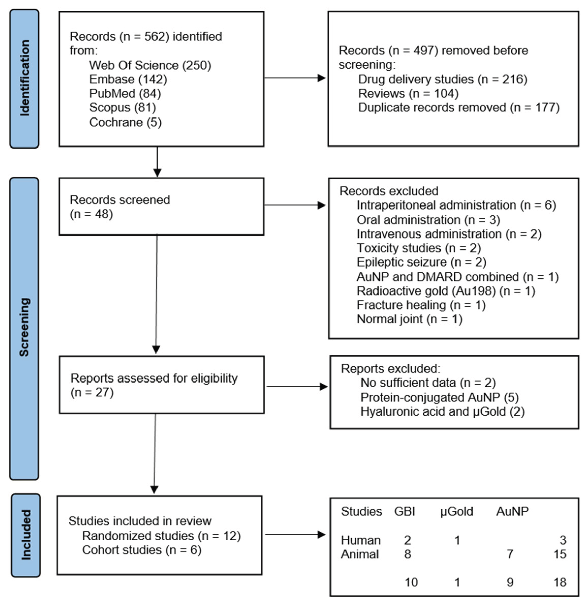
Figure 2.
The schematic illustrates the structural and biochemical consequences of gold ions released from metallic gold particles on gold–sulfur interactions on disulfide-bonded proteins.
Figure 2.
The schematic illustrates the structural and biochemical consequences of gold ions released from metallic gold particles on gold–sulfur interactions on disulfide-bonded proteins.
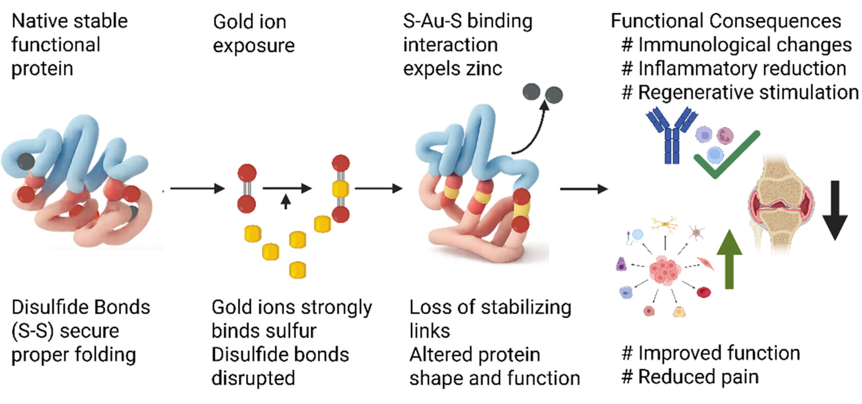

Table 1.
Study characteristics of two human randomized controlled trials of GBI for knee and cervical osteoarthritis. N = number of individuals; I = intervention group; C = control group; F-u = follow-up. Results in numbers improved. Log odds-ratio with 95% CI.
Table 1.
Study characteristics of two human randomized controlled trials of GBI for knee and cervical osteoarthritis. N = number of individuals; I = intervention group; C = control group; F-u = follow-up. Results in numbers improved. Log odds-ratio with 95% CI.
| Study | N-I | N-C | Intervention | Size/Weight/Area | Control | F-u | Measure | I | C | 95% CI |
|---|---|---|---|---|---|---|---|---|---|---|
| Nejrup 2008 [2] | 20 | 20 | Knee OA; 3 GBI at 5 points | 1x2.5mm/454mg/1.18cm2 | Needle | 1 y | Improved | 5 | 4 | 0.29 (-1.20, 1.78) |
| Kjerkegaard 2011 [3] | 24 | 22 | Cervical OA; 3 GBI at 2-3 lamina | 1x2.5mm/>364mg/>0.942cm2 | Needle | 1 y | Improved | 16 | 2 | 3.00 (1.31, 4.68) |
Table 2.
Study characteristics of ten animal randomized controlled studies evaluating gold bead particles (GBI) and gold nanoparticles (AuNP) for osteoarthritis. N = number of individuals; I = intervention group; C = control group; F-u = follow-up; e.a. = extra articular; i.a. = intra articular. Results are presented in numbers, improved, and log odds-Ratio, or in mean, SD, and Cohen’s d.
Table 2.
Study characteristics of ten animal randomized controlled studies evaluating gold bead particles (GBI) and gold nanoparticles (AuNP) for osteoarthritis. N = number of individuals; I = intervention group; C = control group; F-u = follow-up; e.a. = extra articular; i.a. = intra articular. Results are presented in numbers, improved, and log odds-Ratio, or in mean, SD, and Cohen’s d.
| Study | Diagnosis | N-I | N-C | Intervention | Size/Weight/Area | Control | F-u | Measure | I | C | 95% CI |
|---|---|---|---|---|---|---|---|---|---|---|---|
| Hielm-Björkman 2001 [7] | Dog hip OA | 19 | 19 | 3 GBI x 3 e.a. | 1x2mm/273mg/0.71cm2 | Needle | 24w | Improved | 12 | 10 | 0.43(-0.86, 1.73) |
| Bolliger 2002 [9] | Dog hip OA | 9 | 9 | 3 GBI x ≥ 3 e.a. | 1mm/>91mg/>0.282 cm2 | Needle | 3 m | Improved | 3 | 3 | 0(-1.96, 1.96) |
| Jæger 2006 [8] | Dog hip OA | 36 | 42 | 3 GBI x 5 e.a. | 1x2mm/455mg/1.78cm2 | Needle | 6 m | Improved | 25 | 16 | 1.51(.52, 2.49) |
| Tsai 2007 [30] | Rat ankle OA | 6 | 5 | AuNP x 1, i.a. | 20nm/27µg/6.45cm2 | Buffer | 9 d | OA score 0-5 | 3(1.2) | 5.5(.6) | -2.55(-4.15, -0.96) |
| Leonavičienė 2012 [31] | Rat ankle OA | 8 | 8 | AuNP x 12 i.a. | 50nm/225.6µg/14.01cm2 | Saline | 28 d | OA score 0-3 | .19(.3) | 1.8(.4) | -4.55(-6.41, -2.7) |
| Kirdaite 2019 [32] | Rat ankle OA | 8 | 8 | AuNP x 7 i.a. | 50nm/131.6µg/8.18cm2 | Saline | 10 d | OA score 0-3 | 1 (.9) | 2.(.9) | -1.11(-2.16, 0.06) |
| Abdel-Hakem 2022 [33] | Rat ankle OA | 10 | 10 | AuNP x1 i.a. | 25nm/6.5µg/0.81cm2 | None | 35 d | TNF alfa | 45(13) | 85(25) | -2.0(-3.08, -0.93) |
| Haupenthal 2023 [34] | Rat knee OA | 6 | 6 | AuNP x 2 i.a. | 20nm/687.5µg/106.8cm2 | None | 60 d | TNF alfa | 23(8) | 26(10) | -0.33(-1.47, 0.81) |
| Chen 2025 [40] | Rat knee OA | 3 | 3 | AuNP x 3 i.a. | 20nm/45µg/10.8cm2 | None | 4 w | OA score 0-5 | 1.7(.4) | 2.4(.4) | -2.76(-6.29, 0.77) |
| Choi 2025 [41] | Rat knee OA | 6 | 6 | AuNP x 1 i.a. | 50nm/100µg/6.2cm2 | None | 6 w | OA score 0-10 | 2.8(1) | 3.8(1) | -1(-2.38, 0.38) |
Table 3.
Study characteristics of six animal and one human non-controlled studies of GBI and µGold for osteoarthritis. F-u. = follow-up; e.a. = extra articular; i.a. = intra articular; n.a. = not available. Proportion with 95% confidence interval.
Table 3.
Study characteristics of six animal and one human non-controlled studies of GBI and µGold for osteoarthritis. F-u. = follow-up; e.a. = extra articular; i.a. = intra articular; n.a. = not available. Proportion with 95% confidence interval.
| Study | Individuals and diagnosis | Intervention | Size/Weight/Area | F-u | Measure | Results | 95% CI |
|---|---|---|---|---|---|---|---|
| Durkes 1992 [35] | 250 dogs, hip OA, dysplasia | 3 GBI at 3-16 point e.a. | 1x3mm/364mg/0.879cm2 | >3 m | Improved | 200 | 0.80(0.75, 0.85) |
| Klitsgaard 1996 [36] | 400 dogs, hip OA or dysplasia | 3 GBI at 3 points e.a. | 1x2.5mm/341mg/0.848cm2 | 2 m | Improved | 360 | 0.90(0.87, 0.93) |
| Thoresen 1996 [37] | 50 dogs, hip OA and dysplasia | 1 GBI at 1 point e.a. | n.a. | 1 m | Improved | 48 | 0.96(0.88, 1.00) |
| Kothbauer 1997 [38] | 3 dogs, hip OA and dysplasia | 3 GBI at 3 points e.a. | 1x2mm/272mg0.707cm2 | >1 y | Improved | 3 | 1.00(0.50, 1.00) |
| Bartholomé 2020 [39] | 79 dogs, hip, elbow, spine OA | 3 GBI at 3-5 points e.a. | 1x3-5mm/728mg/1.70cm2 | 2 m | Improved | 57 | 0.72(0.62, 0.82) |
| Rasmussen 2022 [4] | 30 patients with knee OA | µGold 20 mg i.a. | 20-40nm/20mg/2.05cm2 | 2 y | Improved | 24 | 0.80(0.64, 0.93) |
Table 4.
Risk of bias in two human randomized controlled studies investigating extraarticular gold bead implant (GBI) for osteoarthritis using the Cochrane Collaboration’s tool for assessing risk of bias. Random sequence generation (randomization), allocation concealment, blinding of participants and personnel, blinding of outcome assessment, incomplete outcome data, selective reporting, and other bias.
Table 4.
Risk of bias in two human randomized controlled studies investigating extraarticular gold bead implant (GBI) for osteoarthritis using the Cochrane Collaboration’s tool for assessing risk of bias. Random sequence generation (randomization), allocation concealment, blinding of participants and personnel, blinding of outcome assessment, incomplete outcome data, selective reporting, and other bias.
| Id | Randomization | Allocation | Blinding | Assessment | Data | Reporting | Other |
|---|---|---|---|---|---|---|---|
| Nejrup 2008 [2] | Low | Low | Low | Low | Low | Low | Low |
| Kjerkegaard 2011 [3] | Low | Low | Low | Low | Low | Low | Unclear |
Table 5.
Risk of bias in 10 animal RCT’s investigating gold for osteoarthritis, arthritic joint disease, or hip dysplasia using the CAMARADES checklist. Studies fulfilling the criteria of: (1) peer reviewed publication; (2) control of temperature; (3) random allocation to treatment or control; (4) allocation concealment; (5) blinded assessment of outcome; (6) use of anesthetic without significant intrinsic neuroprotective activity; (7) animal model (osteoarthritis, arthritis and/or hip dysplasia); (8) sample size calculation; (9) compliance with animal welfare regulations; (10) statement of potential conflict of interests; (11) physiological monitoring; (12) prespecified in- and exclusion criteria; (13) reporting animals excluded from analysis; (14) and reporting of study funding.
Table 5.
Risk of bias in 10 animal RCT’s investigating gold for osteoarthritis, arthritic joint disease, or hip dysplasia using the CAMARADES checklist. Studies fulfilling the criteria of: (1) peer reviewed publication; (2) control of temperature; (3) random allocation to treatment or control; (4) allocation concealment; (5) blinded assessment of outcome; (6) use of anesthetic without significant intrinsic neuroprotective activity; (7) animal model (osteoarthritis, arthritis and/or hip dysplasia); (8) sample size calculation; (9) compliance with animal welfare regulations; (10) statement of potential conflict of interests; (11) physiological monitoring; (12) prespecified in- and exclusion criteria; (13) reporting animals excluded from analysis; (14) and reporting of study funding.
| Id | 1 | 2 | 3 | 4 | 5 | 6 | 7 | 8 | 9 | 10 | 11 | 12 | 13 | 14 | Score | |
|---|---|---|---|---|---|---|---|---|---|---|---|---|---|---|---|---|
| Hielm-Björkman 2001 [7] | Dog, GBI | x | x | x | x | x | x | x | x | x | x | x | x | 12 | ||
| Bolliger 2002 [9] | Dog, GBI | x | x | x | x | x | x | x | 7 | |||||||
| Jæger 2006 [8] | Dog, GBI | x | x | x | x | x | x | x | x | x | x | x | x | 12 | ||
| Tsai 2007 [30] | Rat, AuNP | x | x | 2 | ||||||||||||
| Leonavičienė 2012 [31] | Rat, AuNP | x | x | x | x | x | x | x | x | 8 | ||||||
| Kirdaite 2019 [32] | Rat, AuNP | x | x | x | x | x | x | 6 | ||||||||
| Abdel-Hakem 2022 [33] | Rat, AuNP | x | x | x | x | x | x | 6 | ||||||||
| Haupenthal 2023 [34] | Rat, AuNP | x | x | x | x | x | x | x | x | x | x | 10 | ||||
| Chen 2025 [40] | Rat, AuNP | x | x | x | x | x | x | x | x | 8 | ||||||
| Choi 2025 [41] | Rat, AuNP | x | x | x | x | x | x | x | x | 8 |
Table 6.
Risk of bias in 6 non-randomized cohort studies investigating gold for osteoarthritis, arthritic joint disease, or hip dysplasia using the ROBINS-I V2 tool. Studies fulfilling the criteria of: (1) bias due to confounding; (2) bias in classification of interventions; (3) bias in selection of participants into the study (or into the analysis); (4) bias due to deviations from intended interventions; (5) bias due to missing data; (6) bias arising from measurement of the outcome; (7) and bias in selection of the reported results. Each bias domain is evaluated as low (L), moderate (M), serious (S), or critical (C).
Table 6.
Risk of bias in 6 non-randomized cohort studies investigating gold for osteoarthritis, arthritic joint disease, or hip dysplasia using the ROBINS-I V2 tool. Studies fulfilling the criteria of: (1) bias due to confounding; (2) bias in classification of interventions; (3) bias in selection of participants into the study (or into the analysis); (4) bias due to deviations from intended interventions; (5) bias due to missing data; (6) bias arising from measurement of the outcome; (7) and bias in selection of the reported results. Each bias domain is evaluated as low (L), moderate (M), serious (S), or critical (C).
| Id | 1 | 2 | 3 | 4 | 5 | 6 | 7 | |
|---|---|---|---|---|---|---|---|---|
| Durkes 1992 [35] | Dog, GBI | S | L | C | L | C | C | C |
| Klitsgaard 1996 [36] | Dog, GBI | S | L | C | L | C | C | C |
| Thoresen 1996 [37] | Dog, GBI | C | L | C | C | C | C | C |
| Kothbauer 1997 [38] | Dog, GBI | C | L | C | C | C | C | C |
| Bartholomé 2020 [39] | Dog, GBI | S | L | M | L | L | S | S |
| Rasmussen 2022 [4] | Human, µGold | L | L | L | L | L | M | M |
Disclaimer/Publisher’s Note: The statements, opinions and data contained in all publications are solely those of the individual author(s) and contributor(s) and not of MDPI and/or the editor(s). MDPI and/or the editor(s) disclaim responsibility for any injury to people or property resulting from any ideas, methods, instructions or products referred to in the content. |
© 2026 by the authors. Licensee MDPI, Basel, Switzerland. This article is an open access article distributed under the terms and conditions of the Creative Commons Attribution (CC BY) license (http://creativecommons.org/licenses/by/4.0/).
Copyright: This open access article is published under a Creative Commons CC BY 4.0 license, which permit the free download, distribution, and reuse, provided that the author and preprint are cited in any reuse.


