Submitted:
22 August 2025
Posted:
26 August 2025
Read the latest preprint version here
Abstract
The incidence of kidney diseases has been increasing in the last decade due to extended lifespan, which is often related to polymorbidity. Many chronic conditions slowly and progressively impair the kidney function and lead to chronic kidney disease (CKD). This condition is associated with high morbidity and mortality, elevated costs for renal replacement therapy (RRT) and heavy psychosomatic burden. At the same time, therapeutic options are limited to prophylactic and renoprotective medications and measurements, which can slow progression but not to restore the impaired function of the organ. With the development of cellular therapies new perspectives arise on the horizon with promising potential - mesenchymal stem cells (MSCs) and induced pluripotent cells (iPSCs). Here we review the current possibility of both cell types slowing and reversing the CKD course and assess their cost implication.
Keywords:
cellular therapy
; regenerative medicine
; kidney diseases
; nephrology
1. Introduction
The rising incidence of kidney diseases is an alarming trend which lead to a critical a health and financial consequences. It is esimtated that almost one billion people in low income and low-middle income countries have kidney diseases, which are undiagnosed due to lack of prophylaxis. This is a concerning phenomenon, snice the current therapy of many nephrological conditions is limited to prevention and delaying the progression to CKD [1]. Almost all illnesses that affect the kidney led to reorganization of its parenchyma to fibrotic tissue and impaired organ function [2]. CKD, dialysis and transplantation are related to high morbidity and mortality rates and at the same time represent a high economic burden for the population [1]. Acute kidney injury (AKI) is a common complication in critically ill patients, which as well as CKD is related with high mortality and costs. Additionally, in the most of the cases blood pressure control, fluid management, exclusion of nephrotoxic agents and in sevear cases RRT are among the few options that clinicians have to treat this condition [19]. That is to say that the two most common syndroms in the field of the field of nephrology are with high incidence, can lead to fatal complications and at the same time menegment opportunities are limited and sometimes very expensive. New etiological drugs are needed that can decrease morbidity and mortality and improve the quality of life of nephrological patients.
Luckily, the development of cellular therapies and more precisely MSCs and iPSCs gives us new therapeutic options and possibilities for in vitro experiments that improve our understanding of kidney diseases. The results of many preclinical and clinical trials give indications of their effectiveness, but still there are no available cellular therapies in routine clinical practice [3]. Here we review the applications of cellular therapies in the field of nephrology and assess their economic potential.
2. Mesenchymal Stem Cells and Kidney Diseases
A promising therapeutic perspective are MSCs, which have been extensively investigated in the last two decades and many ongoing clinical trials have shown multiple benefits of their usage. The International Society of Cell & Gene Therapy (ISCT) defines MSCs as plastic-adherent cells that express CD73, CD90 and CD105 and are lacking the expression of hematopoietic and endothelial markers CD11b, CD14, CD19, CD34, CD45, CD79a and HLA-DR. Mesenchymal stem can differentiate into various tissues in vitro and in vivo and are capable of self-renewal [4,5]. They can be isolated from different tissue: adipose, bone marrow, umbilical cord, dental pulp, muscle and many others. Although MSCs of different origins share similar fibroblast-like structure and surface markers, they vary in their differentiation capacity and protein expression pattern and ISCT recommends annotating the tissue they are derived from. For research and clinical purposes adipose(AD), bone marrow(BM) and umbilical cord(UC) tissues are most often used since they are accessible and relatively safe [4,6,7,8].
The MSCs own regenerative, anti-inflammatory, immunomodulating, antifibrotic, proangiogenic and antiapoptotic effects, which make them a promising perspective for treatment of many diseases [9]. Even though in the last years MSCs have been extensively investigated, the intimate mechanism by which they affect the damaged tissues is not fully understood. Some researchers point out their homing ability to be of great importance, since it allows them to migrate to injured tissues, where they induce regeneration by cell to cell contact and secretion of different active molecules [10,11,12]. Others reported that most of the MSCs are trapped in lungs vessels after intravenous application and their effect is mediated by extracellular vesicles with different size and origin - exosomes (30–150 nm in diameter), microvesicles (150–500 nm in diameter), and apoptotic bodies (800–500 nm in diameter). Thus chemokines, interleukins and growth factors that are contained in the vesicles affect the targeted organ by paracrine mechanism [13,14]. Important soluble factors, secreted by MSCs, are reported by different groups: interferon γ (IFN-γ), tumor necrosis factor α(TNF-α), transforming growth factor β (TGF-β), Fibroblast growth factor (FGF), Insulin growth factor 1 (IGF1), hepatocyte growth factor (HGF), vascular endothelial growth factor (VEGF), Interleukin 6 (IL-6) and microRNAs [15,16].
In the field of nephrology there is a vast need for treatment options, since acute kidney injury (AKI) and chronic kidney disease (CDK) are with rising incidence and at the same time therapeutic options are limited to rather preventative and renoprotective measurements and renal replacement therapies (RRT). Often kidneys are affected by different conditions e.g., diabetes and hypertension, that slowly damage the cells and structures of the organ and lead to its function deterioration. Common management strategies are changes in diet, weight loss, avoidance of nephrotoxins, use of RAAS and SGLT inhibitors, aldosterone antagonists and control of underlying diseases [17,18,19]. Often despite the standard care diverse pathological processes progress with different pace and end up replacing the nephrons with non-functional connective tissue: interstitial fibrosis and glomerulosclerosis and thus lead to CKD. Immune-mediated inflammation, epithelial-to-mesenchymal transition (EMT), extracellular matrix (ECM) reorganization by metalloproteases and ischemia are the usual pathophysiological mechanisms behind the developing nephrosclerosis, and they can possibly be targeted by MSCs therapies [2,20,21].
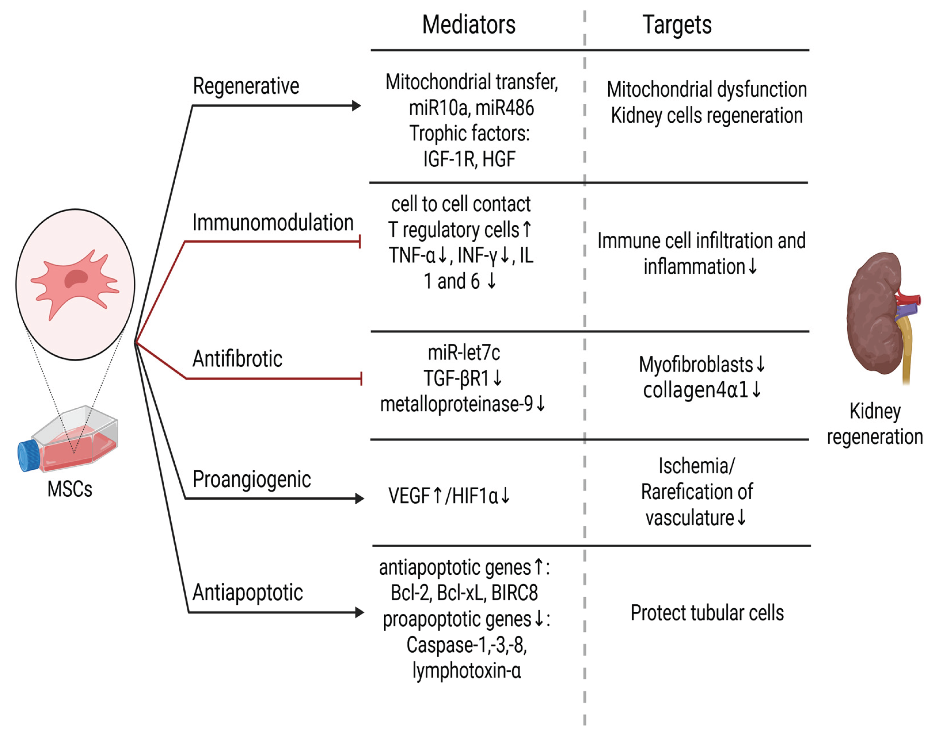
On Figure 1 are summarized beneficial effects that MSCs can have on damaged kidney and their possible mechanism. It is shown in vivo and iv vitro that MSCs therapy reduce scarification of the kidney by suppressing a key regulator in this process - TGF-β1/TGF-βR1 and thus decrease collagen 4α1 accumulation. miR-let7c was identified as a responsible mediator, which can additionally downregulate metalloprotease 9 – an enzyme known to contribute to renal ECM reorganization by fibrosis [2,22]. TGF-β is known to promote fibrosis by enhancing production of extracellular proteins and simultaneously to downregulate the expression of matrix degrading plasminogen-activator inhibitor [23]. This is an important possibility for treating interstitial fibrosis and glomerulosclerosis, which, as it was already mentioned, are common pathological processes that are developing by many nephrological diseases.
The immunomodulatory effect of MSCs is crucial for the treatment of different condition s: nephrosclerosis, glomerulonephritis and kidney transplantation, since innate and acquired immunity are a general regulator of these processes [24,25]. It was shown that injured kidneys are infiltrated by immune cells: macrophages, T and B -lymphocytes and NK cells and they promote interstitial fibrosis and glomerular sclerosis [20,23]. MSCs inhibit the production of proinflammatory cytokines like TNF-α, INF-γ, IL-1, IL-6, while increasing the level of their antagonistic IL-10[15]. Furthermore, these cells can induce the regulatory T cells and thus promote kidney regeneration and simultaneously suppress inflammation [26,27].
Additionally, glomerulonephritis is a heterogeneous group of kidney diseases, and their etiology is mainly related to immune system dysregulation, which makes them also a proper target for MSCs therapy [28]. Rampino et al. reported that in an experimental animal model treatment with MSCs intravenously (via tail vein) can significantly decrease the proteinuria, serum levels of creatinine, IL-6 and TGF-β and sparing glomeruli from monocyte infiltration and injury. Some of the stem cells were found in the kidney, which supports the hypothesis of their homming ability [29]. These findings were confirmed by Kunter et al. with similar demonstration with the difference that only application in renal artery gave significant results while MSCs infusion in tail vein failed to reproduce the regenerative outcomes [30]. Interestingly, another group that compared the effect of Everolimus to BM-MSCs intravenous infusion, found out that despite the reduced expression of renal T cells, IL-17 and activated macrophages they were unable to inhibit glomerular injury and apoptosis [31]. Although these articles have at first glance controversial results a deeper review can find an explanation. First, Rampino et al. and Kunter et al. used higher cell number respectively 2x10⁶ and 3x10⁶, while Zedan et al. injected 1x10⁵ – 1x10⁶ cells. Second, the last group did not characterize the cell suspension according to ISCT prescriptions and they probably injected a heterogeneous group of cells and not only MSCs.
These differences provoke us to consider that in comparison to other chemical drugs, MSCs are a “living medicine” and the number of isolated cells, their fitness and condition, distinct origin as well as their multipotent effects makes cellular therapies very challenging and well-regulated reproducible protocols are needed [14].
The immunomodulatory effects of this cellular therapy were further proved in a clinical case of a patient with vasculitis and kidney involvement, where standard care fails to induce remission, but allogenic MSCs application leads to recovery with no adverse event [32].
Besides their effect on immune system, MSCs release trophic factors: HDF, IGF-1 and microRNAs: miR10a and miR486[33]. They are capable of mitochondrial transfer towards renal cells with dysfunctional mitochondria and all these factors promote regeneration [34]. In addition, by increasing level of VEGF and decreasing HIF-1α mesenchymal cells are reversing rarefication of the kidney vasculature and thus declining ischemia. Antiapoptotic genes (Bcl-2, Bcl-xL, BIRC8) upregulation with simultaneously proapoptotic genes suppression also contribute to MSCs protective effect on tubular cells [15,35].
Given all benefits of this type of therapy, combined with its reliable safety profile, it is very tempting and there are more than 50 clinical trials on clincialtrials.gov, which concern different nephrological diseases. Many papers, from the recent past confirmed the good tolerability of MSCs in patients but are controversial about their efficacy in CKD, AKI, immune-mediated nephritis and kidney transplantation. These differences are related to the many variable factors like derivation from distinct tissues: BM, AD, UC or other, the individual condition of the donor, the concrete protocol and many others. An opportunity for minimization of this deviation is the use of suspension of allogenic stem cells from a few to dozen healthy donors [36]. Application methods can differ as well e.g.,: intravenously [37] or direct injection in renal parenchyma [38]. Despite the challenges in this involving field, regenerative medicines shows promising results. MSCs are able to induced remission in many autoimmune diseases, to reverse CKD progression and extend of kidney graft lifespan. This indicates that mesenchymal cells can be effective in treatment of not all, but maybe in the majority of nephrological conditions [36].
3. Induced Pluripotent Stem Cells and Kidney Diseases
Although MSCs may be considered as one of the first choices for cell-based therapies, there have been studies suggesting that uremia may affect MSCs’ functionality in patients with CKD (e.g., can decrease numbers and differentiation capacity of endothelia progenitor cells [39]. Therefore, Tajiri et al. sought to investigate if induced pluripotent stem cells (iPSCs) from CKD patients would retain their potential to differentiate into nephron progenitors. Indeed, iPSCs derived from patients on haemodialysis (HD-iPSCs) had comparable differentiation abilities in vitro and could form vascularised glomeruli upon implantation in mice which demonstrated their therapeutic potential as an autologous source for CKD treatment [40]
Of note, iPSCs offer a number of advantages over other types of SCs, but a key one is that they can be obtained in a much less invasive and traumatic manner and in much higher numbers than, for example, bone marrow derived MSCs. Importantly, iPSCs can also be directly injected in attempt to slow the progression of CKD too. Caldas et al. conducted an in vivo study in a rat model based on a 5/6 renal mass reduction mimicking CKD, where they aimed to compare the benefits of BM-MSCs to iPSCs. The authors showed that injection of 0.5 million SCs in the renal parenchyma immediately after nephrectomy could effectively slow down CKD progression as measured on day 60 after the intervention by creatinine clearance and other parameters, reduction of glomerulosclerosis, and lower macrophage infiltration (lower score in the iPSCs group vs the BM-MSCs one). Nevertheless, 5 out of 8 kidneys injected with iPSCs developed Willms’ tumors (rather than benign teratomas) originating from the injected cells (as assessed by SRY gene expression) highlighting the danger of this type of therapeutic approach. No tumors were observed within 60 days in the BM-MSC group [41]. Shankar et al. in 2022 also showed the tumorigenic potential of both iPSCs (forming teratomas) and, more interestingly, iPSC-derived kidney organoids in a study where 5 out of 103 mice developed Wilms’ tumor upon subcutaneous injection of such pre-differentiated 3D structures [42] .
While iPSCs can be injected directly, another approach is to differentiate them into renal progenitors prior to therapy. The way to this strategy was paved in 2014 first by Taguchi et al. who demonstrated through elaborate in vitro protocols that iPSC can differentiate into intermediate mesoderm and subsequently into nephron progenitors (metanephric mesenchyme cells) [43] .
It has been 10 years since the first reports of promising results with mouse models of AKI. Imberti et al. and Tayohara et al. showed respectively that iPSC-derived renal progenitors injected through the tail vein can engraft in cispplatin-induced AKI tubuli and reduce renal tubular damage and improve tubular function, and that implantation of in vitro generated 3D proximal renal tubule-like structures can ameliorate renal injury in mice [44,45] . Of note, Tayohara et al. also identified 3 renoprotective factors secreted by these differentiated iPSCs – angiopoietin, VEGF-A and hepatocyte growth factor [45] .
A similar strategy using progenitors rather than iPSCs was more recently adopted by Araoka et al. The research group developed and validated an expansion media for iPSC-derived nephron progenitor cells (NPCs) and used these in mouse models of aristolochic acid-induced CKD and cisplatin-induced AKI. In both conditions, iPSC-NPCs successfully improved kidney function, delayed disease progression (as judged by reduced fibrosis and senescence). Importantly, conditioned medium from these cells was also demonstrated to hold therapeutic potential and one key mediator of the improved survival in CKD and AKI was found to be VEGF-A secreted by the iPSC-NPCs [46] Of note, in attempt to avoid teratoma or Wilm’s tumor formation following injection of iPSCs, Ribeiro et al. validated a strategy of pretreating the cells with mitomycin C (an antimitotic drug aiming to reduce the proliferation of cells) prior implantation in nephrectomised mouse kidneys. The effect on CKD attenuation was compared to implantation of iPSC-derived renal progenitors and the authors found comparable results both in terms of efficacy and safety, re-opening the door to using directly iPSCs for cell therapy in CKD [47] .
Importantly, more intricate co-culture protocols of separately generated nephron and ureteric duct progenitors can recapitulate kidney organoids that contain simultaneously cell differentiated into glomeruli, renal tubules, and collecting ducts. Implantation of these complex iPSCs-derived organoids under the kidney capsule of mice resulted in functional vascularization of the glomerulus-like structures, which suggest the great therapeutic potential that iPSCs have [48].
Lastly, although iPSCs’ ultimate goal would be in cell-based therapies, intermediate mesoderm to mesentheric mesenchyme or ureteric bud progenitor differentiation protocols are in fact more readily available and more widely used for in vitro disease modeling in the context of the generation of different types of organoids (nephrons, collecting ducts or containing both). Such experimental systems have been available for more than 5 years and include brunching ureteric bud (Wolfian duct), collecting duct that can recapitulate aspects of multicystic dysplastic kidney (MCDK) , and others [49,50] including of genetic kidney diseases using either patient-derived or gene edited iPSCs [51] . Models of AKI have also been established through cisplatin-mediated toxic damage of such organoids mimicking the proximal tubule [52]. Several studies have suggested that drug screening can also be carried out in such 3D systems, for example showing that imatinib may have a reno-protective effect upon cisplatin-induced kidney [53,54].
In summary, while iPSCs have the potential to lead to malignancies in vivo, iPSC-derived progenitor cells have not been linked to adverse effects in animal models and several studies demonstrate that lab-engineered renal structures are capable of ameliorating renal function. Therefore, even though there are no current clinical trials with iPSCs in CKD, there are several with iPSC-derived cardiomyocytes for the treatment of heart failure (e.g., NCT04945018, NCT04982081, NCT05647213), which shows that iPSC cell-based therapies may hold potential in CKD, AKI and other kidney pathologies as well [55] .
4. Assessment of Economic Potential of MSCs and iPSCs in Kidney Diseases
The global prevalence of CKD is steadily increasing, driven by population ageing and the growing incidence of comorbid conditions such as type 2 diabetes and hypertension. This epidemiological trend translates into a considerable economic burden on healthcare systems. In the United States, combined Medicare expenditures for CKD and end-stage renal disease (ESRD) have surpassed US$114 billion, while the National Health Service (NHS) in England allocated approximately US$1.95 billion to CKD-related care during the 2009–2010 fiscal year. As the number of affected individuals continues to rise, there is an urgent need to prioritize healthcare delivery and optimize resource allocation to meet the escalating demand for CKD management [56].
The increasing incidence of acute kidney injury (AKI) and CKD, coupled with their severe complications, underscores the necessity for more effective therapeutic strategies capable of mitigating renal damage and delaying progression to ESRD [57]. Patients with CKD are also at elevated risk of cardiovascular complications, which further amplifies the economic impact of the disease on healthcare systems [58]. Given that most acute and chronic kidney conditions are incurable and associated with significant morbidity, current therapeutic approaches are not only costly but frequently lead to adverse outcomes [59].
In evaluating the cost-effectiveness of mesenchymal stem cell (MSC) therapy for chronic kidney disease (CKD), it is essential to contextualize its potential benefits against the established economic and health outcome benchmarks of conventional treatment modalities.
A scoping review, synthesizing evidence from 78 studies conducted in high-income countries (Europe, North America, and Australia), outlines the substantial economic and patient-centered burden associated with CKD. The review demonstrates a clear escalation in direct healthcare costs as the disease progresses: annual per-patient costs for early-stage CKD (stages 1–3) range from $1,600 to $25,037, increasing to $5,367–$53,186 for stages 4–5. For individuals with ESRD, expenditures rise markedly, ranging between $20,110 and $100,593 per year. From a societal perspective, total costs for stages 4–5 in Europe are estimated between $10,750 and $28,428 annually, with productivity losses contributing up to two-thirds of the total in certain contexts.
Treatment modality strongly influences cost profiles. Hemodialysis (HD) is the most expensive renal replacement therapy, followed by peritoneal dialysis (PD). Home-based dialysis incurs lower costs than in-center modalities. Although kidney transplantation (KTx) involves higher initial costs ($14,067–$80,876 in the first year), it is associated with reduced long-term expenditures and superior outcomes, including improved health-related quality of life (HRQoL; EQ-5D: 0.82–0.83) and increased life expectancy. In contrast, dialysis patients report lower HRQoL (EQ-5D: 0.58–0.68) and reduced survival rates [60].
Findings from the Inside CKD global burden study further emphasize the escalating economic impact of chronic kidney disease across 31 countries and regions. Average annual per-patient medical costs were shown to increase markedly with disease progression, rising from US$3,060 at stage G3a to US$8,736 at stage G5. The initiation of kidney replacement therapy (KRT) is associated with substantially higher expenditures, with mean annual costs of US$57,334 for haemodialysis, US$49,490 for peritoneal dialysis, and US$75,326 during the first year following kidney transplantation. Although post-transplant maintenance costs decrease to approximately US$16,672 per year, they remain significantly elevated compared to earlier CKD stages.
Furthermore, the management of CKD-related complications contributes additional financial strain, with estimated costs of US$18,294 for myocardial infarction and US$10,168 for stroke. The study also identified significant cross-country variability and persistent data gaps, particularly concerning early-stage CKD and long-term transplant maintenance. These findings underscore the pressing need for improved health economic reporting to inform evidence-based policy-making and strategic resource allocation [58].
Although kidney transplantation represents the most cost-effective treatment option, its broader implementation is limited by significant barriers, including the increasing number of elderly or frail patients with end-stage renal disease and the persistent shortage of suitable donor organs and potential organ rejection. Given the clinical experience and evidence suggesting that dialysis may offer limited survival benefit for very elderly individuals or those with significant comorbidities and reduced physical function, the decision to opt for conservative management (active supportive care without dialysis) in such cases is understandable. This calls for a shift in the traditional treatment paradigm, whereby conservative care is considered a first-line approach for frail, elderly patients. Such a strategy should include measures to slow the progression of kidney disease, prevent complications, and emphasize advance care planning and comprehensive supportive interventions [61,62].
Clinical trials (both ongoing and completed) have explored the use of mesenchymal stem cells (MSCs) in the treatment of various renal disorders. Early findings indicate that MSCs are safe, well-tolerated, and demonstrate promising efficacy in improving renal pathology [57,59,63].
MSCs have demonstrated significant therapeutic potential in improving renal function in CKD through several regenerative and anti-inflammatory mechanisms. Furthermore, MSCs exert pronounced anti-fibrotic effects. These mechanisms not only improve renal structural and functional outcomes but also have the potential to delay the progression of CKD and postpone the onset ESRD the most resource-intensive phase of kidney failure requiring dialysis or transplantation [64]. Thus, MSC therapy represents a promising strategy for not only restoring renal function but also extending the time before irreversible renal failure occurs.
A cost-effectiveness analysis evaluated MSC therapy for diabetic kidney disease (DKD) in comparison to usual care (UC) and SGLT2 inhibitors (SGLT2i) using a Markov model. The base-case scenario (age 71, eGFR 35 mL/min/1.73m²) found that MSC therapy generated more QALYs (6.39) and was less costly ($158,770) than UC (6.28 QALYs at $159,978). However, SGLT2i therapy dominated both, yielding 6.46 QALYs at a lower cost ($158,131). Despite this, threshold and probabilistic sensitivity analyses demonstrated that MSC therapy may become cost-effective under certain conditions - particularly among younger patients or when MSC therapy achieves a stronger effect in delaying progression to ESRD. For instance, in a scenario modelling a 40-year-old cohort (reflecting lower-bound eligibility in ongoing MSC trials), MSC therapy was found to have a 29% probability of being cost-effective compared to 0.1% for UC. The authors emphasize that despite SGLT2i dominance, MSC therapy may still be valuable under specific clinical conditions -particularly in younger patients, or when SGLT2i use is contraindicated. Importantly, potential adverse events associated with SGLT2i, such as diabetic ketoacidosis or genitourinary infections, may shift clinical preference toward MSC therapy, particularly for patients at elevated risk or those with a GFR <60 mL/min/1.73m². Moreover, jurisdictional variation in SGLT2i pricing suggests scenarios in which MSC may represent a more favourable option. Notably, the analysis acknowledged that the model did not account for all potential benefits of MSC or SGLT2i therapy, including effects on cardiovascular risk and body weight. Furthermore, the estimated benefit of MSC therapy in delaying ESRD was based on surrogate data, and future evidence confirming its regenerative effects on kidney tissue may further improve its cost-effectiveness by reducing the hazard ratio of disease progression [65].
Furthermore, cell-based and cell-free therapeutic approaches are increasingly recognized for their transformative potential in regenerative medicine. Analyses indicate that the global stem cell market, initially valued at USD 9.4 billion in 2020, is on track to grow steadily, reaching an estimated USD 16 billion by 2028, with a projected annual growth rate of 8.8%. Within this broader market, mesenchymal stromal cells (MSCs) are emerging as a key segment, anticipated to attain a market value of USD 6.1 billion by the same year. With a higher growth rate of 12.6%[66]. The expanding market capitalization, combined with emerging evidence from studies proposing cost-reduction strategies [66,67,68], suggests that MSC therapies are likely to become more economically accessible in the near future.
iPSCs have also demonstrated regenerative potential for kidney tissue in experimental studies [69]
While both MSCs and iPSCs are emerging as candidate regenerative therapies for CKD, their clinical readiness and economic viability differ substantially. Comparative analyses underscore that MSCs possess several translational advantages: they exhibit low tumorigenicity, have an immunoprivileged status allowing for allogeneic use, benefit from established isolation protocols from various adult tissues, and offer scalability that facilitates production for widespread clinical application.
In contrast, iPSCs provide the theoretical advantage of patient-specific autologous therapies and broader pluripotent differentiation capacity but remain constrained by significant challenges, including oncogenic risks and the necessity for rigorous quality assurance. MSCs have been evaluated in more clinical trials than iPSCs, largely due to the higher tumorigenic potential associated with iPSCs.
Although significant progress has been made in the development of iPSC technology, further research is essential to deepen understanding of the biology of pluripotency and cellular differentiation and to overcome the remaining barriers to safe and effective clinical application. Between 2018 and 2021, the number of registered clinical studies investigating MSCs substantially exceeded those focused on iPSCs, reflecting the more established status of MSCs in therapeutic practice. In contrast, iPSCs remain an emerging technology that requires further refinement to achieve reliable, clinical-grade production for safe human use [70].
Furthermore, clinical applications of iPSCs necessitate the production of large cell quantities (approximately 70 million cells per treatment), which must be manufactured efficiently, with consistent reproducibility and high quality [71].
Substantial cost challenges remain for iPSC-based therapies. Recent analyses of pluripotent stem cell banking highlight that labor, facility maintenance, and quality control contribute significantly to manufacturing costs, with examples such as the Korea National Institute of Health reporting annual facility maintenance costs of approximately $2.2 million, and labor accounting for up to 50% of total expenses at other centers such as CiRA in Japan. Professional stem cell banks currently price iPSC vials at approximately $1,000–$1,500[72].
Advances in scalable manufacturing, such as bioreactor-based production, process optimization, and automation, are anticipated to reduce costs and improve the economic viability of pluripotent stem cell therapies in the future [73].
Techno-economic analyses suggest that automated iPSC production may overcome key limitations of traditional manual methods, which are labor-intensive, costly, and subject to operator variability. For example, the StemCellFactory (SCF), a fully automated iPSC production platform, has demonstrated that automation can achieve 42% lower total costs over eight years compared to manual production, while improving reproducibility, throughput, and standardization. These manufacturing innovations are anticipated to enhance the future economic viability of iPSC-based regenerative therapies for chronic kidney disease [71].
While this analysis synthesizes available evidence on the economic burden of CKD and the potential role of regenerative therapies such as MSCs and induced pluripotent stem cells iPSCs, important limitations should be noted. Most data informing cost estimates for both MSC and iPSC therapies are at an early stage, reflecting projections or surrogate outcomes rather than results from large-scale, long-term clinical trials. As such, current evaluations of cost-effectiveness are preliminary and must be interpreted with caution.
In particular, the clinical application of MSCs for CKD remains experimental, with most trials focused on short-term safety and efficacy endpoints, not long-term health and economic outcomes. For iPSCs, their therapeutic use is even less mature, with no approved indications and substantial technical, regulatory, and safety barriers that must be overcome before reliable cost-effectiveness assessment is feasible.
While early findings suggest that MSCs and iPSCs hold promise as regenerative therapies for CKD, robust evidence supporting their comparative clinical effectiveness and economic value remains limited. Future research should address these gaps through comprehensive clinical and health-economic evaluations before definitive conclusions on their cost-effectiveness can be drawn.
Conclusions
Cellular therapies are promising strategies to overcome morbidity, mortality and improve quality of life to patients suffering from kidney disease. MSCs are already in many ongoing clinical trials due to their safe profile and good tolerability and their multipotent effect. Despite of that their intimate mechanism remain not fully understand. Additionally, sometimes there are contraversial results due to many factors that their potency depends on like concrete protocol, differences between donors and others.
On the other hand, iPSCs remain in preclinical settings considering their tumorigenic potential. Despite that fact the pluripotent cells have a great potential to differentiate and replace injured tissues and cells but further investigation will be needed untill they can be widly used in clinical trials.
Although the economical analysis of the cellular therapies is based mainly on hypothetical information of future perspectives of MSCs and iPSCs costs and CKD expences, it appears that once this remedies are routinely integrated in clinical practice they can improve life expectancy and quality on a fair price.
Author Contributions
Draft preparation— B.V., Y.S., K.D. and V.Z. Writing, reviewing, editing and visualisation— B.V., Y.S., K.D. All authors have read and agreed to the published version of the manuscript..
References
- Francis et al., “Chronic kidney disease and the global public health agenda: an international consensus,” Nature Reviews Nephrology 2024 20:7, vol. 20, no. 7, pp. 473–485, Apr. 2024. [CrossRef]
- Hewitson, T. D. “Renal tubulointerstitial fibrosis: Common but never simple,” Am J Physiol Renal Physiol, vol. 296, no. 6, pp. 1239–1244, 2009. [CrossRef]
- Salybekov, A.; Kinzhebay, A.A.; Kobayashi, S. Cell therapy in kidney diseases: advancing treatments for renal regeneration. Front Cell Dev Biol, vol. 12, p. 1505601, 2024. [CrossRef]
- Viswanathan, S. et al., “Mesenchymal stem versus stromal cells: International Society for Cell & Gene Therapy (ISCT®) Mesenchymal Stromal Cell committee position statement on nomenclature,” Cytotherapy, vol. 21, no. 10, pp. 1019–1024, Oct. 2019. [CrossRef]
- Horwitz, E. M. et al. “Clarification of the nomenclature for MSC: The International Society for Cellular Therapy position statement,” Cytotherapy, vol. 7, no. 5, pp. 393–395, 2005. [CrossRef]
- Y. J. Jeon, J. Kim, J. H. Cho, H. M. Chung, and J. Il Chae, “Comparative Analysis of Human Mesenchymal Stem Cells Derived From Bone Marrow, Placenta, and Adipose Tissue as Sources of Cell Therapy,” J Cell Biochem, vol. 117, no. 5, pp. 1112–1125, May 2016. [CrossRef]
- R. Kunimatsu et al., “Comparative characterization of stem cells from human exfoliated deciduous teeth, dental pulp, and bone marrow-derived mesenchymal stem cells,” Biochem Biophys Res Commun, vol. 501, no. 1, pp. 193–198, Jun. 2018. [CrossRef]
- Y. Li et al., “Comparative analysis of human mesenchymal stem cells from bone marrow and adipose tissue under xeno-free conditions for cell therapy,” Stem Cell Res Ther, vol. 6, no. 1, Apr. 2015. [CrossRef]
- R. Margiana et al., “Clinical application of mesenchymal stem cell in regenerative medicine: a narrative review,” Stem Cell Research & Therapy 2022 13:1, vol. 13, no. 1, pp. 1–22, Jul. 2022. [CrossRef]
- T. L. Popielarczyk, W. R. Huckle, and J. G. Barrett, “Human Bone Marrow-Derived Mesenchymal Stem Cells Home via the PI3K-Akt, MAPK, and Jak/Stat Signaling Pathways in Response to Platelet-Derived Growth Factor,” Stem Cells Dev, vol. 28, no. 17, pp. 1191–1202, Sep. 2019. [CrossRef]
- R. A. Shahror, A. A. A. Ali, C. C. Wu, Y. H. Chiang, and K. Y. Chen, “Enhanced Homing of Mesenchymal Stem Cells Overexpressing Fibroblast Growth Factor 21 to Injury Site in a Mouse Model of Traumatic Brain Injury,” Int J Mol Sci, vol. 20, no. 11, Jun. 2019. [CrossRef]
- M. S. Chen, C. Y. Lin, Y. H. Chiu, C. P. Chen, P. J. Tsai, and H. S. Wang, “IL-1 β-Induced Matrix Metalloprotease-1 Promotes Mesenchymal Stem Cell Migration via PAR1 and G-Protein-Coupled Signaling Pathway,” Stem Cells Int, vol. 2018, 2018. [CrossRef]
- A. Lotfy, N. M. AboQuella, and H. Wang, “Mesenchymal stromal/stem cell (MSC)-derived exosomes in clinical trials,” Stem Cell Res Ther, vol. 14, no. 1, pp. 1–18, Dec. 2023. [CrossRef]
- J. Galipeau and L. Sensébé, “Mesenchymal stromal cells: clinical challenges and therapeutic opportunities,” Cell Stem Cell, vol. 22, no. 6, p. 824, Jun. 2018. [CrossRef]
- L. Birtwistle, X. M. Chen, and C. Pollock, “Mesenchymal Stem Cell-Derived Extracellular Vesicles to the Rescue of Renal Injury,” Int J Mol Sci, vol. 22, no. 12, p. 6596, Jun. 2021. [CrossRef]
- N. Song, M. Scholtemeijer, and K. Shah, “Mesenchymal Stem Cell Immunomodulation: Mechanisms and Therapeutic Potential,” Trends Pharmacol Sci, vol. 41, no. 9, pp. 653–664, Sep. 2020. [CrossRef]
- A. Khwaja, “KDIGO Clinical Practice Guidelines for Acute Kidney Injury,” Nephron Clin Pract, vol. 120, no. 4, pp. c179–c184, Oct. 2012. [CrossRef]
- P. E. Stevens et al., “KDIGO 2024 Clinical Practice Guideline for the Evaluation and Management of Chronic Kidney Disease,” Kidney Int, vol. 105, no. 4, pp. S117–S314, Apr. 2024. [CrossRef]
- Tamargo, M. Hanouneh, and C. E. Cervantes, “Treatment of Acute Kidney Injury: A Review of Current Approaches and Emerging Innovations,” Journal of Clinical Medicine 2024, Vol. 13, Page 2455, vol. 13, no. 9, p. 2455, Apr. 2024. [CrossRef]
- M. Zeisberg and E. G. Neilson, “Mechanisms of tubulointerstitial fibrosis,” Journal of the American Society of Nephrology, vol. 21, no. 11, pp. 1819–1834, 2010. [CrossRef]
- P. Boor, T. Ostendorf, and J. Floege, “Renal fibrosis: novel insights into mechanisms and therapeutic targets,” Nature Reviews Nephrology 2010 6:11, vol. 6, no. 11, pp. 643–656, Sep. 2010. [CrossRef]
- B. Wang et al., “Mesenchymal stem cells deliver exogenous MicroRNA-let7c via exosomes to attenuate renal fibrosis,” Molecular Therapy, vol. 24, no. 7, pp. 1290–1301, Aug. 2016. [CrossRef]
- K. Sean Eardley and P. Cockwell, “Macrophages and progressive tubulointerstitial disease,” Kidney Int, vol. 68, no. 2, pp. 437–455, Aug. 2005. [CrossRef]
- N. Li and J. Hua, “Interactions between mesenchymal stem cells and the immune system,” Cell Mol Life Sci, vol. 74, no. 13, p. 2345, Jul. 2017. [CrossRef]
- Mudrabettu et al., “Safety and efficacy of autologous mesenchymal stromal cells transplantation in patients undergoing living donor kidney transplantation: a pilot study,” Nephrology (Carlton), vol. 20, no. 1, pp. 25–33, Jan. 2015. [CrossRef]
- K. English, J. M. Ryan, L. Tobin, M. J. Murphy, F. P. Barry, and B. P. Mahon, “Cell contact, prostaglandin E2 and transforming growth factor beta 1 play non-redundant roles in human mesenchymal stem cell induction of CD4+CD25Highforkhead box P3+ regulatory T cells,” Clin Exp Immunol, vol. 156, no. 1, p. 149, Apr. 2009. [CrossRef]
- M. T. Gandolfo et al., “Foxp3+ regulatory T cells participate in repair of ischemic acute kidney injury,” Kidney Int, vol. 76, no. 7, pp. 717–729, 2009. [CrossRef]
- M. Jin, Y. Xie, Q. Li, and X. Chen, “Stem Cell-Based Cell Therapy for Glomerulonephritis,” Biomed Res Int, vol. 2014, p. 124730, 2014. [CrossRef]
- T. Rampino et al., “Mesenchymal stromal cells improve renal injury in anti-Thy 1 nephritis by modulating inflammatory cytokines and scatter factors,” Clin Sci, vol. 120, no. 1, pp. 25–36, Jan. 2011. [CrossRef]
- U. Kunter et al., “Transplanted mesenchymal stem cells accelerate glomerular healing in experimental glomerulonephritis,” Journal of the American Society of Nephrology, vol. 17, no. 8, pp. 2202–2212, 2006. [CrossRef]
- M. M. Zedan et al., “Effect of Everolimus versus Bone Marrow-Derived Stem Cells on Glomerular Injury in a Rat Model of Glomerulonephritis: A Preventive, Predictive and Personalized Implication,” Int J Mol Sci, vol. 23, no. 1, Jan. 2021. [CrossRef]
- M. Gregorini et al., “Antineutrophil cytoplasmic antibody- associated renal vasculitis treated with autologous mesenchymal stromal cells: Evaluation of the contribution of immune-mediated mechanisms,” Mayo Clin Proc, vol. 88, no. 10, pp. 1174–1179, Oct. 2013. [CrossRef]
- M. Tapparo et al., “Renal Regenerative Potential of Extracellular Vesicles Derived from miRNA-Engineered Mesenchymal Stromal Cells,” Int J Mol Sci, vol. 20, no. 10, p. 2381, May 2019. [CrossRef]
- Y. Plotnikov, T. G. Khryapenkova, S. I. Galkina, G. T. Sukhikh, and D. B. Zorov, “Cytoplasm and organelle transfer between mesenchymal multipotent stromal cells and renal tubular cells in co-culture,” Exp Cell Res, vol. 316, no. 15, pp. 2447–2455, Sep. 2010. [CrossRef]
- Y. Zhou et al., “Exosomes released by human umbilical cord mesenchymal stem cells protect against cisplatin-induced renal oxidative stress and apoptosis in vivo and in vitro,” Stem Cell Res Ther, vol. 4, no. 2, pp. 1–13, Apr. 2013. [CrossRef]
- N. Perico, F. Casiraghi, and G. Remuzzi, “Clinical Translation of Mesenchymal Stromal Cell Therapies in Nephrology,” J Am Soc Nephrol, vol. 29, no. 2, p. 362, Feb. 2017. [CrossRef]
- N. Perico et al., “Safety and Preliminary Efficacy of Mesenchymal Stromal Cell (ORBCEL-M) Therapy in Diabetic Kidney Disease: A Randomized Clinical Trial (NEPHSTROM),” J Am Soc Nephrol, vol. 34, no. 10, pp. 1733–1751, Oct. 2023. [CrossRef]
- J. Stavas et al., “Rilparencel (Renal Autologous Cell Therapy-REACT®) for Chronic Kidney Disease and Type 1 and Type 2 Diabetes: Phase 2 Trial Design Evaluating Bilateral Kidney Dosing and Redosing Triggers,” Am J Nephrol, vol. 55, no. 3, p. 389, Feb. 2024. [CrossRef]
- K. De Groot et al., “Uremia causes endothelial progenitor cell deficiency,” Kidney Int, vol. 66, no. 2, pp. 641–646, Aug. 2004. [CrossRef]
- S. Tajiri et al., “Regenerative potential of induced pluripotent stem cells derived from patients undergoing haemodialysis in kidney regeneration,” Scientific Reports 2018 8:1, vol. 8, no. 1, pp. 1–12, Oct. 2018. [CrossRef]
- H. C. Caldas et al., “Induced Pluripotent Stem Cells Reduce Progression of Experimental Chronic Kidney Disease but Develop Wilms’ Tumors,” Stem Cells Int, vol. 2017, no. 1, p. 7428316, Jan. 2017. [CrossRef]
- A. S. Shankar et al., “Kidney Organoids Are Capable of Forming Tumors, but Not Teratomas,” Stem Cells, vol. 40, no. 6, p. 577, Jun. 2022. [CrossRef]
- Taguchi et al., “Redefining the in vivo origin of metanephric nephron progenitors enables generation of complex kidney structures from pluripotent stem cells,” Cell Stem Cell, vol. 14, no. 1, pp. 53–67, Jan. 2014. [CrossRef]
- Imberti et al., “Renal progenitors derived from human iPSCs engraft and restore function in a mouse model of acute kidney injury,” Sci Rep, vol. 5, p. 8826, 2015. [CrossRef]
- T. Toyohara et al., “Cell Therapy Using Human Induced Pluripotent Stem Cell-Derived Renal Progenitors Ameliorates Acute Kidney Injury in Mice,” Stem Cells Transl Med, vol. 4, no. 9, pp. 980–992, Sep. 2015. [CrossRef]
- T. Araoka et al., “Human iPSC–derived nephron progenitor cells treat acute kidney injury and chronic kidney disease in mouse models,” Sci Transl Med, vol. 17, no. 792, Apr. 2025. [CrossRef]
- P. de C. Ribeiro et al., “Therapeutic potential of human induced pluripotent stem cells and renal progenitor cells in experimental chronic kidney disease,” Stem Cell Res Ther, vol. 11, no. 1, pp. 1–10, Dec. 2020. [CrossRef]
- H. Tsujimoto et al., “A Modular Differentiation System Maps Multiple Human Kidney Lineages from Pluripotent Stem Cells,” Cell Rep, vol. 31, no. 1, p. 107476, Apr. 2020. [CrossRef]
- S. I. Mae et al., “Expansion of Human iPSC-Derived Ureteric Bud Organoids with Repeated Branching Potential,” Cell Rep, vol. 32, no. 4, Jul. 2020. [CrossRef]
- S. I. Mae et al., “Generation of branching ureteric bud tissues from human pluripotent stem cells,” Biochem Biophys Res Commun, vol. 495, no. 1, pp. 954–961. [CrossRef]
- T. Traitteur, C. Zhang, and R. Morizane, “The application of iPSC-derived kidney organoids and genome editing in kidney disease modeling,” iPSCs - State of the Science, pp. 111–136, Jan. 2022. [CrossRef]
- S. Kishi et al., “Proximal tubule ATR regulates DNA repair to prevent maladaptive renal injury responses,” J Clin Invest, vol. 129, no. 11, pp. 4797–4816, Nov. 2019. [CrossRef]
- N. Ning, Z. Liu, X. Li, Y. Liu, and W. Song, “Progress of Induced Pluripotent Stem Cell-Derived Renal Organoids in Clinical Application,” Kidney Diseases, vol. 11, no. 1, pp. 1–10, Jan. 2025. [CrossRef]
- H. Oishi, N. Tabibzadeh, and R. Morizane, “Advancing preclinical drug evaluation through automated 3D imaging for high-throughput screening with kidney organoids,” Biofabrication, vol. 16, no. 3, Jul. 2024. [CrossRef]
- K. Osafune, “iPSC technology-based regenerative medicine for kidney diseases,” Clin Exp Nephrol, vol. 25, no. 6, p. 574, Jun. 2021. [CrossRef]
- Darlington et al., “Costs and Healthcare Resource Use Associated with Risk of Cardiovascular Morbidity in Patients with Chronic Kidney Disease: Evidence from a Systematic Literature Review,” Adv Ther, vol. 38, no. 2, pp. 994–1010, Feb. 2021. [CrossRef]
- Aghajani Nargesi, L. O. Lerman, and A. Eirin, “Mesenchymal stem cell-derived extracellular vesicles for kidney repair: current status and looming challenges,” Stem Cell Res Ther, vol. 8, no. 1, Dec. 2017. [CrossRef]
- V. Jha et al., “Global Economic Burden Associated with Chronic Kidney Disease: A Pragmatic Review of Medical Costs for the Inside CKD Research Programme,” Adv Ther, vol. 40, no. 10, pp. 4405–4420, Oct. 2023. [CrossRef]
- L. T. J. Hickson, S. M. Herrmann, B. A. McNicholas, and M. D. Griffin, “Progress toward the Clinical Application of Mesenchymal Stromal Cells and Other Disease-Modulating Regenerative Therapies: Examples from the Field of Nephrology,” Kidney360, vol. 2, no. 3, pp. 542–557, Mar. 2021. [CrossRef]
- S. Elshahat, P. Cockwell, A. P. Maxwell, M. Griffin, T. O’Brien, and C. O’Neill, “The impact of chronic kidney disease on developed countries from a health economics perspective: A systematic scoping review,” PLoS One, vol. 15, no. 3, 2020. [CrossRef]
- Liu, F. Cheng, S. Pan, and Z. Liu, “Stem cells: a potential treatment option for kidney diseases,” Stem Cell Research & Therapy 2020 11:1, vol. 11, no. 1, pp. 1–20, Jun. 2020. [CrossRef]
- R. Vanholder et al., “Reducing the costs of chronic kidney disease while delivering quality health care: a call to action,” Nat Rev Nephrol, vol. 13, no. 7, pp. 393–409, Jul. 2017. [CrossRef]
- N. Perico, F. Casiraghi, and G. Remuzzi, “Clinical Translation of Mesenchymal Stromal Cell Therapies in Nephrology,” J Am Soc Nephrol, vol. 29, no. 2, pp. 362–375, Feb. 2018. [CrossRef]
- Chen et al., “Mesenchymal Stem Cell Therapy in Kidney Diseases: Potential and Challenges,” Cell Transplant, vol. 32, Jan. 2023. [CrossRef]
- L. E. Barry et al., “Mesenchymal stromal cell therapy compared to SGLT2-inhibitors and usual care in treating diabetic kidney disease: A cost-effectiveness analysis,” PLoS One, vol. 17, no. 11, Nov. 2022. [CrossRef]
- R. M. Silva, S. S. Rosa, J. A. L. Santos, A. M. Azevedo, and A. Fernandes-Platzgummer, “Enabling Mesenchymal Stromal Cells and Their Extracellular Vesicles Clinical Availability-A Technological and Economical Evaluation,” Journal of extracellular biology, vol. 4, no. 3, Mar. 2025. [CrossRef]
- P. G. Childs, S. Reid, M. Salmeron-Sanchez, and M. J. Dalby, “Hurdles to uptake of mesenchymal stem cells and their progenitors in therapeutic products,” Biochemical Journal, vol. 477, no. 17, p. 3349, Sep. 2020. [CrossRef]
- A. L. Russell, R. C. Lefavor, and A. C. Zubair, “Characterization and cost-benefit analysis of automated bioreactor-expanded mesenchymal stem cells for clinical applications,” Transfusion (Paris), vol. 58, no. 10, pp. 2374–2382, Oct. 2018. [CrossRef]
- P. De Carvalho Ribeiro, L. F. Oliveira, M. A. Filho, and H. C. Caldas, “Differentiating Induced Pluripotent Stem Cells into Renal Cells: A New Approach to Treat Kidney Diseases,” Stem Cells Int, vol. 2020, p. 8894590, 2020. [CrossRef]
- K. Thanaskody, A. S. Jusop, G. J. Tye, W. S. Wan Kamarul Zaman, S. A. Dass, and F. Nordin, “MSCs vs. iPSCs: Potential in therapeutic applications,” Front Cell Dev Biol, vol. 10, p. 1005926, Nov. 2022. [CrossRef]
- Nießing, R. Kiesel, L. Herbst, and R. H. Schmitt, “Techno-Economic Analysis of Automated iPSC Production,” Processes 2021, Vol. 9, Page 240, vol. 9, no. 2, p. 240, Jan. 2021. [CrossRef]
- J. H. Kim, E. Kawase, K. Bharti, O. Karnieli, Y. Arakawa, and G. Stacey, “Perspectives on the cost of goods for hPSC banks for manufacture of cell therapies,” npj Regenerative Medicine 2022 7:1, vol. 7, no. 1, pp. 1–8, Sep. 2022. [CrossRef]
- J. A. Mills, T. Donnelly, M. Brennan, and B. Marques, “Transitioning to Scalable Bioreactors for Allogeneic iPSC-Derived Cell Therapies: Cost Savings and Global Patient Access,” Cytotherapy, vol. 27, no. 5, p. S148, May 2025. [CrossRef]
Figure 1.
Effects of MSCs on kidney regeneration and their targets.
Disclaimer/Publisher’s Note: The statements, opinions and data contained in all publications are solely those of the individual author(s) and contributor(s) and not of MDPI and/or the editor(s). MDPI and/or the editor(s) disclaim responsibility for any injury to people or property resulting from any ideas, methods, instructions or products referred to in the content. |
© 2025 by the authors. Licensee MDPI, Basel, Switzerland. This article is an open access article distributed under the terms and conditions of the Creative Commons Attribution (CC BY) license (http://creativecommons.org/licenses/by/4.0/).
Copyright: This open access article is published under a Creative Commons CC BY 4.0 license, which permit the free download, distribution, and reuse, provided that the author and preprint are cited in any reuse.


