Submitted:
14 August 2025
Posted:
15 August 2025
You are already at the latest version
Abstract
Background: Silver fluoride medicaments effectively arrest caries progression but cause permanent staining. Metal nanoparticles are increasingly utilised in paediatric dentistry due to their antimicrobial properties. Aim: This study reports the synthesis and char-acterisation of silver and selenium nanoparticles stabilised with natural agents to en-hance their antimicrobial potential. Methods: Six silver and selenium nanoparticles were chemically synthesised and stabilised using biocompatible eco-friendly capping agents, including casein, bovine serum albumin, chitosan, citrate, and tannic acid. Characterisation was performed using Tyndall scattering, UV-visible spectroscopy, transmission electron microscopy, and ICP-OES. Results: The synthesised particles were spherical in shape, ranging in size from 2.5 to 24 nm for silver, and 35 to 43 nm for selenium. Elemental analysis confirmed the absence of heavy metals. Conclusion: These findings highlight the potential of stabilised metal nanoparticles for antimicrobial ap-plications in paediatric dentistry.
Keywords:
nanotechnology
; metal nanoparticles
; silver nanoparticles
; selenium nanopaticles
; nanoparticles synthesis
; biocompatible capping agents
; paediatric dentistry
A/ Prof. Sobia Zafar. and Amjad Almuqrin. 288 Herston Rd, Herston QLD 4006
1. Introduction
Early Childhood Caries (ECC) is a leading childhood illness as well as a significant cause of avoidable hospital admissions [1]. Managing ECC can be complex due to the young age of affected patients and the rapid, aggressive nature of the disease. Conventional treatments typically involve operative interventions such as tooth preparation, restorations, and pulp therapy, or surgical procedures such as extractions. These interventions often necessitate a high level of child cooperation and may require general anaesthesia when performed on uncooperative patients.
Silver Fluoride (SF) topical solutions effectively arrest caries progression in young children who may not yet comply with standard treatments, which minimises the need for invasive procedures [2]. However, the ionic silver component in these solutions causes permanent tooth staining, raising aesthetic concerns among parents and dental practitioners limiting their acceptance as a first line treatment option [3].
In recent years, metal-based nanomaterials have attracted attention in dentistry due to their potent antimicrobial properties, tuneable physicochemical characteristics, and ability to disrupt oral biofilms [4]. Nanoparticles (NPs), with their ultrafine size (1–100 nm) and high surface-to-volume ratio, exhibit superior reactivity, facilitating strong interactions with oral microbes [5]. This enables the effective eradication of oral pathogens, and penetration into biofilms [6,7]. Among the various nanomaterials explored, silver NPs (AgNPs) are particularly notable for their broad-spectrum antibiofilm efficacy, supporting their integration into dental composites [8], adhesives [9], and implants [10]. Selenium NPs (SeNPs), although less extensively studied in dentistry, are emerging as promising biocompatible antimicrobial agents [11], with additional functions in antioxidant defence and immune modulation [12,13]. The growing interest in AgNPs and SeNPs highlights the broader potential of nanotechnology to enhance antimicrobial efficacy in the oral environment while maintaining compatibility with host tissues.
The stability of NPs remains a significant challenge as they tend to agglomerate or aggregate post-synthesis, which reduces bioavailability and antimicrobial efficacy. Agglomeration, driven by weak van der Waals forces, forms loosely bound clusters that retain surface area but hinder biofilm penetration and ion release [14]. In contrast, aggregation involves strong chemical bonding, forming fused structures with significantly reduced surface area and reactivity [15]. Both processes impair NP performance, necessitating effective stabilisation strategies to preserve their antimicrobial and bioactive potential in clinical applications.
Capping plays a key role by stabilising NPs and enhancing their biological actions. For instance, casein, bovine serum albumin (BSA), chitosan, citrate, and tannic acid have been shown to be effective capping agents due to their biocompatibility, cost-effectiveness, and functional benefits, and scalability for clinical applications. All of the foregoing capping agents are natural compounds with low toxicity. Casein and BSA provide a protein-based stabilisation matrix, which improves NP dispersion and cellular uptake [16]. Chitosan exerts antimicrobial actions, and the charge modifications it causes enhance cellular interactions [17]. Citrate stabilises NPs, to maintain colloidal stability [18]. Tannic acid, a polyphenol with antioxidant properties, further enhances NP stability and biological performance [19]. Therefore, the present study incorporated casein, BSA, chitosan, citrate, and tannic acid as capping agents.
The aim of the study was to synthesise and characterise capped silver and selenium NPs for potential topical antimicrobial applications in paediatric dentistry. Specifically, we assessed their physiochemical characteristics and elemental composition. The graphical illustration is shown in (Figure 1).
2. Materials and Methods
2.1. Preparation of NPs
Based on published methods in the literature, six silver and selenium NPs were synthesised chemically, i.e. four AgNPs and two SeNPs (Table 1). The synthesis process involved a chemical reduction approach, where a precursor solution was treated with a reducing agent and a capping agent to facilitate nanoparticle formation, followed by centrifugation, washing, and storage at 4°C. An overview of the synthesis stages and the final appearance of the NPs is illustrated in (Figure 2). Notably, the distinct colour changes are attributable to the surface plasmon resonance (SPR) phenomenon. The SPR is highly sensitive to particle size, shape, and dielectric environment, confirming successful nanoparticle formation and morphological changes.
Ag1: The Ag1 synthesis method was adapted from previous work 20, employed a chemical reduction approach. In brief, 3.0 mL of 0.6% w/v casein (Sigma-Aldrich, St. Louis, MO, USA) and 0.1 mL of 1.0 M NaOH (Sigma-Aldrich, St. Louis, MO, USA) were combined in a 25 mL glass flask. After 10 minutes of gentle magnetic stirring at room temperature, the 14.9 mL of deionised (DI) water was added, and the mixture heated to 60°C, and stirred for 5 minutes. Lastly, 2 mL of 20 mM silver nitrate (AgNO3) (Sigma-Aldrich, St. Louis, MO, USA) was gradually added dropwise to the mixture and the solution magnetically stirred for 60 minutes at 60°C. During this time, the solution colour changed from transparent to golden yellow. Subsequently, the solution was centrifuged at 5000 rpm at 4°C and washed three times with DI water, with the pellet retained and the supernatant discarded at each washing step. The Ag1 NP were then resuspended in DI water and transferred to light-proof sterile vials, and kept at 4°C. The same approach was used for Ag2 synthesis, substituting casein with BSA (0.6% w/v) (Sigma-Aldrich, St. Louis, MO, USA). The final solution was again a vivid, yellow colour.
AgL1: The synthesis method was modified from past work [21], using 70°C rather than 100°C. In brief, 500 mL of DI water was heated to 70°C in a 2 L beaker on a magnetic stirrer. To this was added 0.1 g of AgNO₃ (Sigma-Aldrich, St. Louis, MO, USA). After mixing for 5 minutes, 0.1 g of tri-sodium citrate dihydrate (Sigma-Aldrich, St. Louis, MO, USA) dissolved in 10 mL of DI water and added dropwise with magnetic stirring. The solution was heated to 94°C and mixing was undertaken in the dark for 2.5 hours, with the beaker covered to minimise evaporative water loss. By the end of this period, the solution was a faint yellow colour. Stirring continued for a further 1 hour while solution cooled to room temperature. The final solution was decanted into a brown glass bottle and stored at room temperature.
AgL2: The AgL2 was synthesised following a modification of the method of Bastus et al. (2014) [22]. In brief, 100 mL of DI water was heated to 80°C and divided into equal portions of 50 mL. To the first was added 0.147 g trisodium citrate (Sigma-Aldrich, St. Louis, MO, USA) to give a concentration of 5 mM followed by 4.25 mg of tannic acid (Sigma-Aldrich, St. Louis, MO, USA) giving a concentration of 0.025 mM. A second solution of 0.0425 g of AgNO3 (Sigma-Aldrich, St. Louis, MO, USA) in 10 mL of DI water was prepared (25 mM). This was then preheated in a microwave to 80°C and added in one step to the first solution. The mixture kept at 80°C under vigorous stirring. The solution turned bright yellow immediately, indicating nanoparticle formation. Stirring continued for a further 1 hour while solution cooled to room temperature. The final solution was decanted into a brown glass bottle and stored at room temperature.
Se1 and Se2: These NPs were synthesised using a previously described method [23] with minor modifications. Initially, a stock solution of 1% wt/v chitosan (Sigma-Aldrich, St. Louis, MO, USA) was prepared in 1% (v/v) acetic acid (Millipore Corporation, Summit Drive, MA, USA). Then, 2 mL of the chitosan solution was mixed with 8 mL of 10 mM Na2SeO3 (Sigma-Aldrich, St. Louis, MO, USA), and the mixture magnetically stirred for 10 minutes at room temperature. Subsequently, 4 mL of ice-cold 0.1 M sodium borohydride (NaBH4) (Sigma-Aldrich, St. Louis, MO, USA) was added gradually under constant stirring for 60 minutes. The solution became dark red in colour. The NPs were washed three times with DI water by centrifugation at 14000 rpm. Finally, the NPs were stored in light-proof sterile tubes at 4°C.
A similar protocol was used for Se2, using BSA in water instead of chitosan in acetic acid. A 2 mL volume of 1% w/v BSA solution was mixed with 8 mL of 10 mM Na2SeO3 solution (Sigma-Aldrich, St. Louis, MO, USA). Then, 4 mL of ice-cold 0.1M NaBH4 was added dropwise. An orange colour developed as the SeNPs were formed. As before, the SeNPs were centrifuged, washed twice with ultra-pure water and stored at 4°C.
2.2. Characterisation
Tyndall scattering: The Tyndall effect is a light-scattering phenomenon utilised to promptly confirm NP formation in colloidal solutions. When an intense light source, such as a laser beam, passes through the suspension, the NPs scatter the light, producing a visible path (Figure 3). The extent of scattering varies with particle size and the wavelength of the incident light, with smaller NPs scattering more effectively at shorter wavelengths.
In this study, three handheld laser pointers of different wavelengths were used: violet (405 ± 10 nm), green (532 ± 10 nm) and red (670 ± 10 nm), each with a maximum power output of < 1000 mW. A 1 mL aliquot of each NP suspension was dispersed in 3.5 mL glass cuvette, and the laser beams were directed horizontally through the dispersion at a consistent distance of 5 cm from cuvette’s surface to maintain a uniform light path. Deionised (DI) water was used as a negative control to compare scattering effects in the absence of NPs. Afterward, three images of each NPs suspension were captured using a smartphone camera (model: iPhone 15 Pro; Apple Inc., Cupertino, CA, USA) positioned perpendicular to the laser path. The experiment was performed in a light-controlled dark setting to minimise external light reflections. The observed scattering patterns were qualitatively analysed, comparing the spread and the intensity of scattered light across different wavelengths to determine the presence of NPs and estimate their relative size.
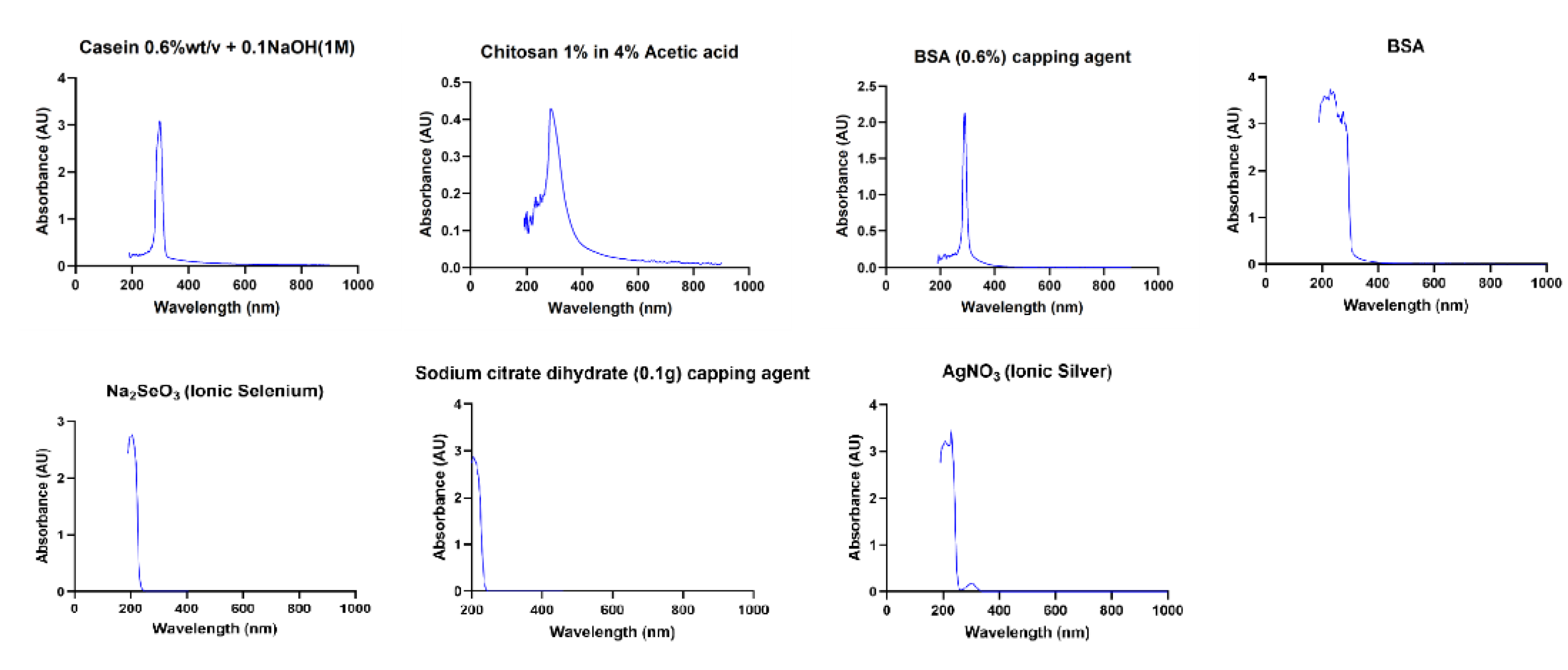
UV-Visible spectroscopy: The ultraviolet-visible (UV-Vis) absorbance spectra of NP suspensions were measured using a spectrophotometer (GENESYS™ 10S UV-Vis spectrophotometer, Thermo Fisher Scientific, Waltham, MA, USA) immediately after NP synthesis. Prior to measurement, samples were vortex mixed and ultrasonicated for 5 minutes to ensure NP dispersion, before being transferred into 3.5 mL quartz cuvettes for analysis. Absorbance spectra were collected within the UV-visible range, with deionised (DI) water used as the baseline. Aqueous solutions of the capping agents were also analysed separately under the same conditions for reference.
Transmission Electron Microscopy (TEM): Six NP samples were transferred to the Central Analytical Research Facility (CARF) of the Queensland University of Technology (QUT). The samples were vortex mixed and ultrasonicated for at least 5 minutes to ensure a homogeneous dispersion. Subsequently, the samples were drop mounted onto 300 mesh lacey formvar/carbon-coated copper grids. The imaging process utilised a JEOL JEM-2100 TEM (JEOL Ltd), operated at 200 kV, with a 4K Fx416 TVIPS CCD camera.
Inductively Coupled Plasma Optical Emission Spectroscopy (ICP-OES): Elemental concentrations of NPs were determined using a PerkinElmer 8300DV ICP-OES fitted with an ESI SC-4DX autosampler. ICP-OES was used to determine the overall concentration of silver and selenium NPs, as conventional drying and weighing methods were impractical due to the presence of organic capping agents. Drying the samples could alter their chemical properties, as the organic capping agents might degrade or undergo structural changes, affecting NP stability. This analysis allowed experiments to be designed based on Ag or Se concentrations, independent of capping agents. ICP-OES was also used to screen for trace contaminants of heavy metals, such as lead and cadmium.
Four aqueous nanomaterial samples were diluted in 2% ultra-pure nitric acid at a 1:10 dilution factor. Multi-element quantitation was performed using the PerkinElmer 8300DV ICP-OES, equipped with an ESI SC-4DX autosampler and a PrepFAST 2 sample handling unit for online internal standardization and auto-dilution of samples and calibration standards. Sub-boiling distilled nitric acid was used to prepare all standards and blank solutions. Instrument calibration was conducted using external standardisation, with multi-element standards prepared in-house from ICP-MS grade single-element stock solutions (High Purity Standards, Charleston, USA). A combination of axial and radial viewing modes was used, with radial mode specifically employed for alkali metals (Li, Na, K). Method robustness, accuracy, and precision were verified through the continuous analysis of multiple Certified Reference Materials (CRMs) covering a range of common matrices and analyte concentrations (National Institute of Standards and Technology, Gaithersburg, MD, USA; United States Geological Survey, Reston, VA, USA).
3. Results
Tyndall scatter: All NPs suspensions exhibited visible light Tyndall scattering when exposed to laser beams at 405 nm, 532 nm, and 670 nm, confirming the presence of suspended NPs, while the DI water negative control showed no scattering, as shown in (Figure 4). Scattering was generally more pronounced at longer wavelengths (532 nm and 670 nm), which aligns with expected light interactions in colloidal systems. The intensity of scattering varied between samples, with Ag2 and Se2 showing stronger scattering compared to Ag1 and Se1, suggesting differences in particle concentration or size. The similarities in scattering intensity between AgL1 and AgL2 indicate comparable NP composition and dispersion characteristics.
UV-Visible spectroscopy: As illustrated in (Figure 5), absorbance spectra confirmed the formation of silver and selenium NPs. Ag1, AgL1, and AgL2 exhibited distinct extinction bands with λmax at 420 nm, while Se2 showed an extinction band with λmax at 410 nm. The absorbance spectrum of Ag2 showed a broader extinction band, with no well-defined λmax, and Se1 exhibited a similarly broad absorbance curve without a sharp peak.
The well-defined λmax at 420 nm in Ag1, AgL1, and AgL2 corresponds to the known SPR of silver NPs, while the broader extinction bands in Ag2 and Se1 suggest the influence of BSA and chitosan capping agents, potentially broadening the absorbance curves and affecting NPs dispersion and size distribution. The λmax at around 400 nm in Se2 indicates a more uniform NPs size, whereas the broader extinction band in Se1 suggests greater size variation.
TEM: TEM images are shown in (Figure 6). These revealed that the synthesized NPs were predominantly spherical in shape, with the following average diameters (means + standard deviations): Ag1 3.6 nm + 3.4; Ag2 2.5 nm + 0.8, AgL1 3.92 nm + 0.71, AgL2 24.17 nm + 5.42; Se1 34.9 nm + 8.5; and Se2 43.2 nm + 13.2.
ICP-OES: Elemental analysis confirmed successful NP synthesis (Table 2 and Figure 7). Ag concentrations were 57.44 mg/L for Ag1, 265.8 mg/L for Ag2, 121.6 mg/L for AgL1, and 119.2 mg/L for AgL2. Similarly, Se concentrations were 683.1 mg/L for Se1 and 642.8 mg/L for Se2. These measured values align with expected synthesis conditions. No detectable contamination with heavy metals such as lead (Pb) or cadmium (Cd) was observed, confirming the purity of the synthesised NPs.
4. Discussion
Aiming at exploring plausible alternatives or augmentative options for ionic silver in SF formulations, six silver and selenium NPs were prepared utilising naturally sourced stabilising agents. The fabrication process was verified using rapid, onsite methods (Tyndall scattering and UV-Vis spectroscopy) and further characterised in terms of particle size, morphology, and elemental composition via TEM and ICP-OES respectively. AgNPs ranged from 2.5 to 24 nm, while SeNPs ranged from 35 to 43 nm. UV-Vis and TEM analyses validated successful NPs preparation and spherical morphology. ICP-OES quantified the elemental composition and concentration. Furthermore, it confirmed the absence of toxic heavy metals. Of particular interest, protein-capped AgNPs, particularly the BSA-stabilised Ag2, exhibited the smallest and most uniform size distribution. In combination, these findings suggest that capped metal NPs could provide improved long-term stability and represent a practical topical antimicrobial choice for caries arrest in paediatric dentistry.
The selection of AgNPs and SeNPs as potential antimicrobial and cariostatic agents was guided by their high antimicrobial potency [24], biocompatibility when used at appropriate concentrations [25], extended shelf life if properly stabilised [26], and low sensitivity to light [27]. While ionic silver is well established in antimicrobial applications, the transition to nanoparticulate forms offers enhanced bioactivity; however, introduces new challenges; specifically, agglomeration and instability under physiological conditions. Such instability can compromise their effectiveness and safety, ultimately shorten their shelf life and limit their clinical utility [28]. To address these issues, the application of suitable stabilisers to metal NPs has become a widely adopted strategy. By preventing uncontrolled NPs growth, it can enhance NP reactivity and stability and maintain bioactivity, thereby supporting antimicrobial performance comparable to that of SF formulations.
Building upon this rationale and recognising that SF formulations currently used in dentistry are water-based, a similar aqueous medium was adopted for AgNPs synthesis. Additionally, using an aqueous formulation should improve biocompatibility by reducing the need for organic solvents. Residues of such solvents may introduce toxicity concerns in clinical settings. Chemical synthesis provides a straightforward and scalable approach for NP fabrication and allows for the incorporation of capping agents [29]. As a controlled, reproducible approach it provides consistency and batch-to-batch uniformity, provided there is precise control of the reaction conditions, including the temperature and time, over prolonged periods. The cost of reagents must also be considered in terms of large-scale production.
Following the successful synthesis, morphological analysis was carried out to assess the physical properties of the generated NPs. The TEM analysis revealed variations in NP sizes, which could influence their antimicrobial activity. Smaller NPs generally exhibit greater bioactivity due to their higher surface area-to-volume ratio, and greater physical penetration into complex microbial membranes. Among the AgNPs, Ag1, Ag2, and AgL1 had small sizes (<5 nm), and Ag2 (BSA-capped) had the narrowest size distribution. In contrast, nanoparticles of AgL2 (citrate and tannic acid-capped) were significantly larger (24.17 ± 5.42 nm), which may influence their interactions with bacterial biofilms. There is a well-established correlation between NPs size and antimicrobial behaviour, where reduced dimensions enhance diffusion, membrane disruption, and intracellular interactions thereby boost antimicrobial actions [30].
The SeNPs were notably larger than the AgNPs and exhibited size variations according to the choice of capping agent, with Se1 (chitosan-capped) at 34.9 ± 8.5 nm and Se2 (BSA-capped) at 43.2 ± 13.2 nm. The larger size of SeNPs versus AgNPs may lower their effectiveness as antimicrobial agents, and this aspect remains to be explored. Given that size directly affects the extent of microbial contact and reactive surface exposure, optimising SeNPs dimensions through refined synthesis methods could enhance their therapeutic applicability. Several strategies can optimise the fabrication protocol to achieve smaller SeNPs, which include elevating the reaction temperature, adjusting the pH of the reaction medium, altering the capping agent or utilising stronger reducing agent.
In addition to particle size, nanoparticle shape is central to modulating biological interactions including biodistribution, cellular uptake and cytotoxicity. In general, spherical NPs exhibit lower cytotoxicity compared to anisotropic forms such as rods [31], sheets [32], prisms [33], and needles [34]. Accordingly, the synthesis strategy adopted in this study prioritised the formation of spherical NPs, with the goal being to minimise toxicity while preserving antimicrobial potency. The TEM images confirmed that the NPs exhibited a predominantly spherical morphology.
Spherical shape NPs probably reflects the thermodynamic for minimal surface energy in the aqueous synthesis medium. Moderate to high temperatures, as followed here, promotes NPs isotropic growth where nucleation rate and surface diffusion are rapid. Thus, NPs form equally in all directions in symmetric and uniform sphere shape [35]. Another shape controlling factor is employing the optimal capping agent which binds evenly to the NPs surface, prevents anisotropy and maintains the sphericity. Further studies are needed to explore the toxicity of the various NPs, to select those with optimal biological performance and low toxicity.
Spectroscopy data validated the formation of both silver and selenium nanoparticles through characteristic SPR absorbance patterns. Three of the four AgNPs showed a sharp peak between 400-450 nm, which aligns with literature on the 420 nm peak [20,21,22,36]. These peaks are indicators to collective oscillations of conduction electrons on the silver NPs surface when excited by incident light, which is a phenomenon highly sensitive to particle size, shape, dielectric environment, and interparticle spacing. The Ag2 sample which was capped with BSA was blue shifted and had a peak absorbance at a lower wavelength, which might indicate some absorbance by the BSA in the visible violet or ultraviolet range, or particle size variations [37]. A similar strong absorbance by the capping agent, which dominates over the light absorbance by the nanoparticles, likely explains the observed broad absorbance of the chitosan capped samples of Se1. There is also the possibility that Se1 NPs have a broad size range due to the polymeric nature of chitosan as a stabiliser. In addition, the oxidation state of selenium could influence λmax positioning. Se2 peaked sharply at approximately 400 nm a spectral signature characteristic of elemental selenium nanoparticles, indicating a relatively uniform particle population.
Taken together, these optical characterisations not only confirmed NPs formation but also highlighted the influence of capping agents on their spectral behaviour as further supported by compositional analysis.
The elemental composition analysis confirmed successful NPs synthesis and a lack of heavy metal contaminants (especially Cr, Cu, Mn, Ni and Pb). Minor carryover of sodium (from glass and from sodium borohydride) and boron (from borohydride) was detected. Sulphur in SeNPs reflects cysteine and methionine amino acids in BSA. Preparing high purity nano batches is essential, considering that presence of trace contaminants may trigger agglomeration, promote uncontrolled redox reactions and jeopardise chemical stability over time. Interestingly, none of the referenced studies in the synthesis processes have conducted a comprehensive elemental analysis on their samples, unlike the present investigation.
5. Conclusions
The synthesis of AgNPs and SeNPs was achieved, incorporating various naturally derived capping agents to enhance nanoparticle stability. Key physicochemical properties were characterised. This lays the groundwork for further investigation into their antimicrobial efficacy.
Author Contributions
Conceptualization, [S.Z., L.J.W. and C.J.S.]; methodology, [A.A. and L.J.W.] writing—original draft preparation, [A.A.].; writing—review and editing, [S.Z., L.J.W., and C.J.S]; visualization, [A.A]; supervision, [L.J.W., C.J.S. and S.Z.]; project administration, [S.Z]. All authors have read and agreed to the published version of the manuscript.
Funding
This research received no external funding.
Institutional Review Board Statement
Not applicable.
Informed Consent Statement
Not applicable.
Conflicts of Interest
The authors declare no conflicts of interest.
Abbreviations
| ECC | Early Childhood Caries |
| SF | Silver Fluoride |
| NPs | Nanoparticles |
| AgNPs | Silver Nanoparticles |
| SeNPs | Selenium Nanoparticles |
| BSA | Bovine Serum Albumin |
| DI | Deionised (water) |
| AgNO₃ | Silver Nitrate |
| NaOH | Sodium Hydroxide |
| SPR | Surface Plasmon Resonance |
| Na₂SeO₃ | Sodium Selenite |
| NaBH₄ | Sodium Borohydride |
| UV-Vis | Ultraviolet–Visible (Spectroscopy) |
| TEM | Transmission Electron Microscopy / Microscope |
| CARF | Central Analytical Research Facility |
| QUT | Queensland University of Technology |
| ICP-OES | Inductively Coupled Plasma Optical Emission Spectroscopy |
| ICP-MS | Inductively Coupled Plasma Mass Spectrometry |
| CRMs | Certified Reference Materials |
| Pb | Lead |
| Cd | Cadmium |
| Ag | Silver |
| Se | Selenium |
Appendix A
Figure A1.
Absorbance curves of the capping agents and precursors.
References
- Nguyen, T. M.; Tonmukayakul, U.; Hall, M.; Calache, H. Cost-Effectiveness Analysis of Silver Diamine Fluoride to Divert Dental General Anaesthesia Compared to Standard Care. Aust. Dent. J. 2022, 67, 352–361. [Google Scholar] [CrossRef]
- Burgess, J. O.; Vaghela, P. M. Silver Diamine Fluoride: A Successful Anticarious Solution with Limits. Adv. Dent. Res. 2018, 29, 131–134. [Google Scholar] [CrossRef]
- Nelson, T.; Scott, J. M.; Crystal, Y. O.; Berg, J. H.; Milgrom, P. Silver Diamine Fluoride in Pediatric Dentistry Training Programs: Survey of Graduate Program Directors. Pediatr. Dent. 2016, 38, 212–217. [Google Scholar]
- Nagireddy, V. R.; Reddy, D.; Kondamadugu, S.; Puppala, N.; Mareddy, A.; Chris, A. Nanosilver Fluoride—A Paradigm Shift for Arrest in Dental Caries in Primary Teeth of Schoolchildren: A Randomized Controlled Clinical Trial. Int. J. Clin. Pediatr. Dent. 2019, 12, 484–490. [Google Scholar] [CrossRef]
- Saafan, A.; Zaazou, M. H.; Sallam, M. K.; Mosallam, O.; El Danaf, H. A. Assessment of Photodynamic Therapy and Nanoparticles Effects on Caries Models. Open Access Maced. J. Med. Sci. 2018, 6, 1289–1295. [Google Scholar] [CrossRef] [PubMed]
- Tran, P.; Kopel, J.; Ray, C.; Reed, J.; Reid, T. W. Organo-Selenium Containing Dental Sealant Inhibits Biofilm Formation by Oral Bacteria. Dent. Mater. 2022, 38, 848–857. [Google Scholar] [CrossRef]
- Degrazia, F. W.; Branco Leitune, V. C.; Garcia, I. M.; Arthur, R. A.; Werner Samuel, S. M.; Collares, F. M. Effect of Silver Nanoparticles on the Physicochemical and Antimicrobial Properties of an Orthodontic Adhesive. J. Appl. Oral Sci. 2016, 24, 404–410. [Google Scholar] [CrossRef]
- Ai, M.; Du, Z.; Zhu, S.; Geng, H.; Zhang, X.; Cai, Q.; Yang, X. Composite Resin Reinforced with Silver Nanoparticles–Laden Hydroxyapatite Nanowires for Dental Application. Dent. Mater. 2017, 33, 12–22. [Google Scholar] [CrossRef]
- Melo, M. A. S.; Cheng, L.; Zhang, K.; Weir, M. D.; Rodrigues, L. K. A.; Xu, H. H. K. Novel Dental Adhesives Containing Nanoparticles of Silver and Amorphous Calcium Phosphate. Dent. Mater. 2013, 29, 199–210. [Google Scholar] [CrossRef] [PubMed]
- Hossain, N.; Islam, M. A.; Chowdhury, M. A. Synthesis and Characterization of Plant Extracted Silver Nanoparticles and Advances in Dental Implant Applications. Heliyon 2022, 8. [Google Scholar] [CrossRef] [PubMed]
- Almuqrin, A.; Kaur, I. P.; Walsh, L. J.; Seneviratne, C. J.; Zafar, S. Amelioration Strategies for Silver Diamine Fluoride: Moving from Black to White. Antibiotics 2023, 12. [Google Scholar] [CrossRef]
- Wallenberg, M.; Misra, S.; Wasik, A. M.; Marzano, C.; Björnstedt, M.; Gandin, V.; Fernandes, A. P. Selenium Induces a Multi-Targeted Cell Death Process in Addition to ROS Formation. J. Cell. Mol. Med. 2014, 18, 671–684. [Google Scholar] [CrossRef]
- Kumar, N.; Krishnani, K. K.; Singh, N. P. Comparative Study of Selenium and Selenium Nanoparticles with Reference to Acute Toxicity, Biochemical Attributes, and Histopathological Response in Fish. Environ. Sci. Pollut. Res. 2018, 25, 8914–8927. [Google Scholar] [CrossRef]
- Zare, Y. Study of Nanoparticles Aggregation/Agglomeration in Polymer Particulate Nanocomposites by Mechanical Properties. Compos., Part A 2016, 84, 158–164. [Google Scholar] [CrossRef]
- ISO. Nanotechnologies — Antibacterial silver nanoparticles — Specification of characteristics and measurement methods, 2066.
- Khramtsov, P.; Barkina, I.; Kropaneva, M.; Bochkova, M.; Timganova, V.; Nechaev, A.; Byzov, I. Y.; Zamorina, S.; Yermakov, A.; Rayev, M. Magnetic Nanoclusters Coated with Albumin, Casein, and Gelatin: Size Tuning, Relaxivity, Stability, Protein Corona, and Application in Nuclear Magnetic Resonance Immunoassay. Nanomaterials 2019, 9. [Google Scholar] [CrossRef] [PubMed]
- Ardean, C.; Davidescu, C. M.; Nemes, N. S.; Negrea, A.; Ciopec, M.; Duteanu, N.; Negrea, P.; Duda-Seiman, D.; Musta, V. Factors Influencing the Antibacterial Activity of Chitosan and Chitosan Modified by Functionalization. Int. J. Mol. Sci. 2021, 22. [Google Scholar] [CrossRef]
- Shinohara, S.; Eom, N.; Teh, E. J.; Tamada, K.; Parsons, D.; Craig, V. S. J. The Role of Citric Acid in the Stabilization of Nanoparticles and Colloidal Particles in the Environment: Measurement of Surface Forces between Hafnium Oxide Surfaces in the Presence of Citric Acid. Langmuir 2018, 34, 2595–2605. [Google Scholar] [CrossRef]
- Ahmad, T. Reviewing the Tannic Acid Mediated Synthesis of Metal Nanoparticles. J. Nanotechnol. 2014, 2014, 1–11. [Google Scholar] [CrossRef]
- Ghodake, G.; Lim, S.-R.; Lee, D. S. Casein Hydrolytic Peptides Mediated Green Synthesis of Antibacterial Silver Nanoparticles. Colloids Surf., B 2013, 108, 147–151. [Google Scholar] [CrossRef] [PubMed]
- Lee, P.; Meisel, D. Adsorption and Surface-Enhanced Raman of Dyes on Silver and Gold Sols. J. Phys. Chem. 1982, 86, 3391–3395. [Google Scholar] [CrossRef]
- Bastús, N. G.; Merkoçi, F.; Piella, J.; Puntes, V. Synthesis of Highly Monodisperse Citrate-Stabilized Silver Nanoparticles of up to 200 nm: Kinetic Control and Catalytic Properties. Chem. Mater. 2014, 26, 2836–2846. [Google Scholar] [CrossRef]
- Shahmoradi, S.; Shariati, A.; Zargar, N.; Yadegari, Z.; Asnaashari, M.; Amini, S. M.; Darban-Sarokhalil, D. Antimicrobial Effects of Selenium Nanoparticles in Combination with Photodynamic Therapy against Enterococcus faecalis Biofilm. Photodiagn. Photodyn. Ther. 2021, 35, 102398. [Google Scholar] [CrossRef]
- Santos, V. E.; Vasconcelos Filho, A.; Targino, A. G.; Flores, M. A.; Galembeck, A.; Caldas, A. F.; Rosenblatt, A. A New “Silver-Bullet” to Treat Caries in Children—Nano Silver Fluoride: A Randomised Clinical Trial. J. Dent. 2014, 42, 945–951. [Google Scholar] [CrossRef]
- Mahmud, K. M.; Hossain, M. M.; Polash, S. A.; Takikawa, M.; Shakil, M. S.; Uddin, M. F.; Alam, M.; Ali Khan Shawan, M. M.; Saha, T.; Takeoka, S.; Hasan, M. A.; Sarker, S. R. Investigation of Antimicrobial Activity and Biocompatibility of Biogenic Silver Nanoparticles Synthesized Using Syzigyum cymosum Extract. ACS Omega 2022, 7, 27216–27229. [Google Scholar] [CrossRef] [PubMed]
- Guilger-Casagrande, M.; Germano-Costa, T.; Bilesky-José, N.; Pasquoto-Stigliani, T.; Carvalho, L.; Fraceto, L. F.; de Lima, R. Influence of the Capping of Biogenic Silver Nanoparticles on Their Toxicity and Mechanism of Action towards Sclerotinia sclerotiorum. J. Nanobiotechnol. 2021, 19. [Google Scholar] [CrossRef] [PubMed]
- Polywka, A.; Tückmantel, C.; Görrn, P. Light Controlled Assembly of Silver Nanoparticles. Sci. Rep. 2017, 7. [Google Scholar] [CrossRef]
- Selmani, A.; Ulm, L.; Kasemets, K.; Kurvet, I.; Erceg, I.; Barbir, R.; Pem, B.; Santini, P.; Marion, I. D.; Vinković, T. Stability and Toxicity of Differently Coated Selenium Nanoparticles under Model Environmental Exposure Settings. Chemosphere 2020, 250, 126265. [Google Scholar] [CrossRef]
- Andresen, E.; Islam, F.; Prinz, C.; Gehrmann, P.; Licha, K.; Roik, J.; Recknagel, S.; Resch-Genger, U. Assessing the Reproducibility and Up-Scaling of the Synthesis of Er,Yb-Doped NaYF4-Based Upconverting Nanoparticles and Control of Size, Morphology, and Optical Properties. Sci. Rep. 2023, 13. [Google Scholar] [CrossRef]
- Schwass, D. R.; Lyons, K. M.; Love, R.; Tompkins, G. R.; Meledandri, C. J. Antimicrobial Activity of a Colloidal AgNP Suspension Demonstrated In Vitro against Monoculture Biofilms: Toward a Novel Tooth Disinfectant for Treating Dental Caries. Adv. Dent. Res. 2018, 29, 117–123. [Google Scholar] [CrossRef]
- Lee, J. H.; Ju, J. E.; Kim, B. I.; Pak, P. J.; Choi, E. K.; Lee, H. S.; Chung, N. Rod-Shaped Iron Oxide Nanoparticles Are More Toxic than Sphere-Shaped Nanoparticles to Murine Macrophage Cells. Environ. Toxicol. Chem. 2014, 33, 2759–2766. [Google Scholar] [CrossRef]
- Abramenko, N. B.; Demidova, T. B.; Abkhalimov, Е. V.; Ershov, B. G.; Krysanov, E. Y.; Kustov, L. M. Ecotoxicity of Different-Shaped Silver Nanoparticles: Case of Zebrafish Embryos. J. Hazard. Mater. 2018, 347, 89–94. [Google Scholar] [CrossRef]
- Auclair, J.; Gagné, F. Shape-Dependent Toxicity of Silver Nanoparticles on Freshwater Cnidarians. Nanomaterials 2022, 12. [Google Scholar] [CrossRef] [PubMed]
- Zhang, B.; Sai Lung, P.; Zhao, S.; Chu, Z.; Chrzanowski, W.; Li, Q. Shape Dependent Cytotoxicity of PLGA-PEG Nanoparticles on Human Cells. Sci. Rep. 2017, 7. [Google Scholar] [CrossRef] [PubMed]
- Xia, Y.; Xiong, Y.; Lim, B.; Skrabalak, S. E. Shape-Controlled Synthesis of Metal Nanocrystals: Simple Chemistry Meets Complex Physics? Angew. Chem., Int. Ed. 2009, 48, 60–103. [Google Scholar] [CrossRef]
- Sileikaite, A.; Prosycevas, I.; Puiso, J.; Juraitis, A.; Guobiene, A. Analysis of Silver Nanoparticles Produced by Chemical Reduction of Silver Salt Solution. Medziagotyra 2006, 12, 287–291. [Google Scholar]
- Qiao, Y.-J.; Kang, J.; Song, C.-Q.; Zhou, N.; Zhang, P.; Song, G.-F. Further Study on Particle Size, Stability, and Complexation of Silver Nanoparticles under the Composite Effect of Bovine Serum Protein and Humic Acid. RSC Adv. 2024, 14, 2621–2632. [Google Scholar] [CrossRef]
Figure 1.
Graphical Abstract: Schematic representation of the synthesis, characterisation, and outcomes of silver and selenium nanoparticles capped with naturally derived agents.
Figure 1.
Graphical Abstract: Schematic representation of the synthesis, characterisation, and outcomes of silver and selenium nanoparticles capped with naturally derived agents.
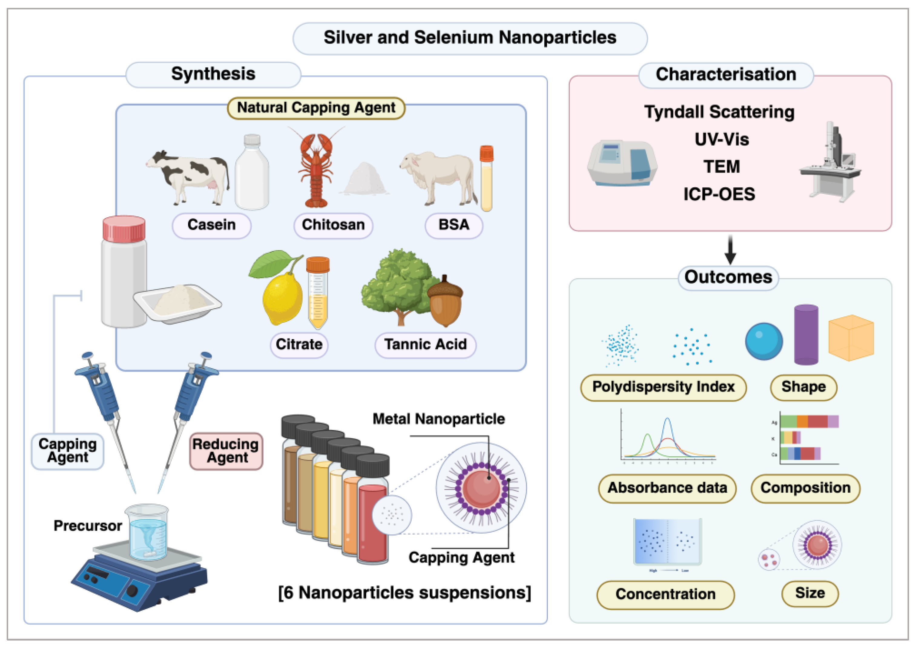
Figure 2.
Illustration of the major chemical synthesis stages of nanoparticles. (A) Diagram explaining the key stages of chemical synthesis. (B) Images of nanoparticles (NPs) after synthesis, showing a colour change due to the SPR phenomenon.
Figure 2.
Illustration of the major chemical synthesis stages of nanoparticles. (A) Diagram explaining the key stages of chemical synthesis. (B) Images of nanoparticles (NPs) after synthesis, showing a colour change due to the SPR phenomenon.
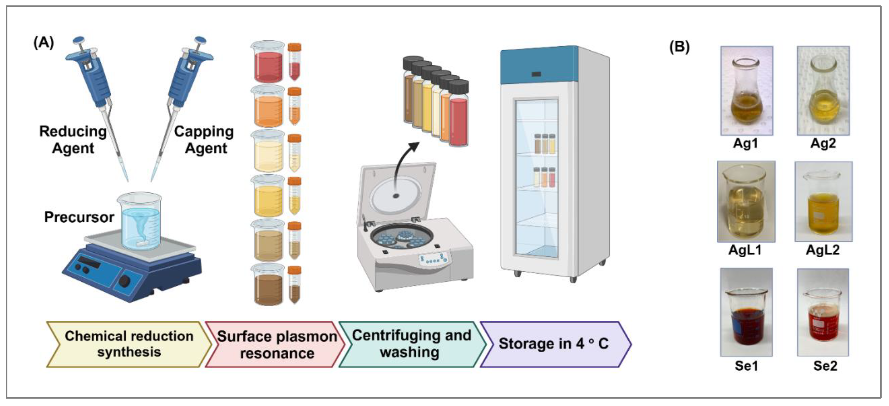
Figure 3.
Schematic representation of the Tyndall effect in a NPs suspension. A laser beam passes through a quartz cuvette containing NPs, producing visible scattering that confirms their presence.
Figure 3.
Schematic representation of the Tyndall effect in a NPs suspension. A laser beam passes through a quartz cuvette containing NPs, producing visible scattering that confirms their presence.
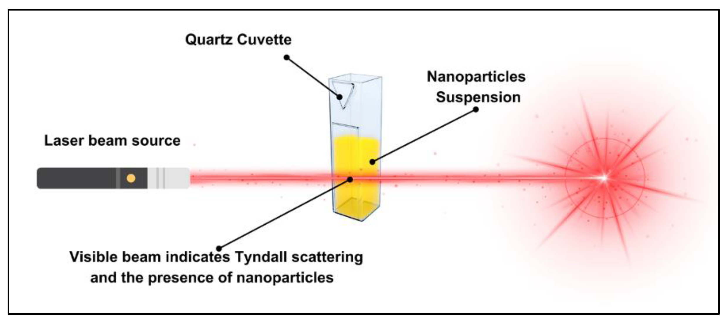
Figure 4.
Tyndall scattering in NP suspensions using different laser wavelengths. Scattering was observed in all samples except the DI water control, with intensity variations suggesting differences in particle properties.
Figure 4.
Tyndall scattering in NP suspensions using different laser wavelengths. Scattering was observed in all samples except the DI water control, with intensity variations suggesting differences in particle properties.
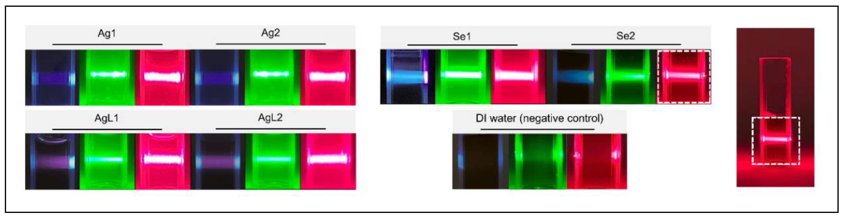
Figure 5.
Absorbance curves of the synthesised NPs.
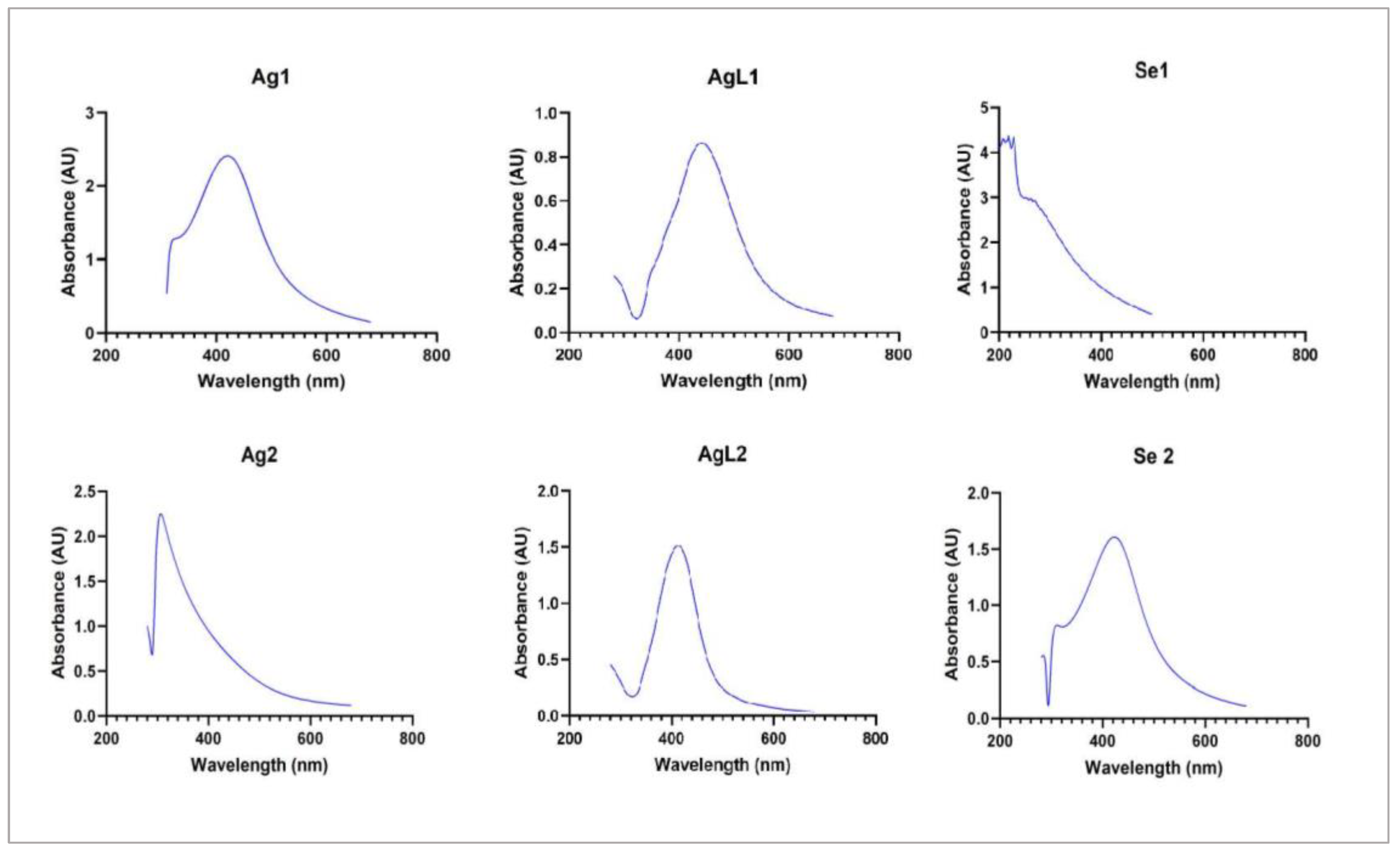
Figure 6.
TEM microscopic images of the synthesised NPs, along with histograms illustrating their size distribution. (A) NPs are capped which appears as a metal core structure with a definite external layer. (B) is uncapped NPs, displaying a bare metal core with no outer membrane.
Figure 6.
TEM microscopic images of the synthesised NPs, along with histograms illustrating their size distribution. (A) NPs are capped which appears as a metal core structure with a definite external layer. (B) is uncapped NPs, displaying a bare metal core with no outer membrane.


Figure 7.
Elemental composition of AgNPs and SeNPs determined by ICP-OES.
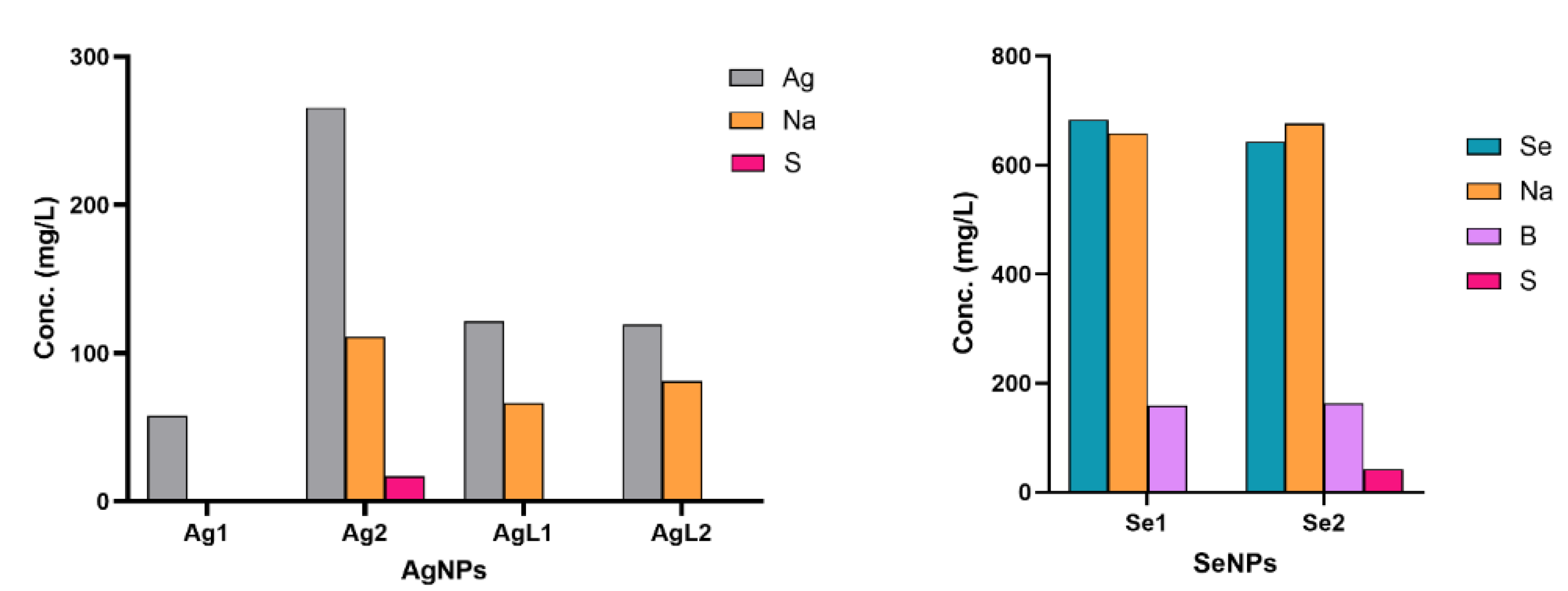

Table 1.
Custom-synthesised metal NPs and their starting concentrations.
| NPs | Synthesis concentration (μg/mL) | |
|---|---|---|
| 1 | Silver - casein capped (Ag1) | 55.4 |
| 2 | Silver - BSA capped (Ag2) | 265.8 |
| 3 | Silver - citrate capped (AgL1) | 121.6 |
| 4 | Silver - citrate and tannic acid capped (AgL2) | 119.2 |
| 5 | Selenium - chitosan capped (Se1) | 683.1 |
| 6 | Selenium - BSA capped (Se2) | 642.8 |
Table 2.
Elemental analysis of AgNPs and selenium SeNPs suspensions determined using ICP-OES. The table shows the measured concentrations of Ag and Se in different samples. <LOR> indicates values below the limit of reporting.
Table 2.
Elemental analysis of AgNPs and selenium SeNPs suspensions determined using ICP-OES. The table shows the measured concentrations of Ag and Se in different samples. <LOR> indicates values below the limit of reporting.
| Analyte | Ag1 | Ag2 | AgL1 | AgL2 | Se1 | Se2 | Limit of Reporting (mg/L) |
| Conc. (mg/L) | Conc. (mg/L) | Conc. (mg/L) | Conc. (mg/L) | Conc. (mg/L) | Conc. (mg/L) | ||
| Ag | 57.44 | 265.8 | 121.6 | 119.2 | 0.678 | 0.113 | 0.050 |
| Al | <LOR | 0.0761 | 0.0728 | 0.0718 | 0.0707 | 0.069 | 0.050 |
| B | <LOR | <LOR | <LOR | <LOR | 159 | 162 | 0.050 |
| Ba | <LOR | <LOR | <LOR | <LOR | <LOR | <LOR | 0.010 |
| Be | <LOR | <LOR | <LOR | <LOR | <LOR | <LOR | 0.010 |
| Ca | <LOR | 0.0796 | 1.569 | 1.321 | 2.706 | 0.1536 | 0.050 |
| Cd | <LOR | <LOR | <LOR | <LOR | <LOR | <LOR | 0.050 |
| Co | <LOR | <LOR | <LOR | <LOR | <LOR | <LOR | 0.050 |
| Cr | <LOR | <LOR | <LOR | <LOR | 0.0519 | <LOR | 0.050 |
| Cu | <LOR | <LOR | <LOR | <LOR | <LOR | <LOR | 0.050 |
| Fe | <LOR | <LOR | <LOR | <LOR | 0.2791 | <LOR | 0.050 |
| K | 3.913 | <LOR | <LOR | <LOR | <LOR | <LOR | 0.250 |
| Li | <LOR | <LOR | <LOR | <LOR | <LOR | <LOR | 0.050 |
| Mg | <LOR | <LOR | <LOR | <LOR | 2.774 | 0.9465 | 0.250 |
| Mn | <LOR | <LOR | <LOR | <LOR | <LOR | <LOR | 0.050 |
| Mo | <LOR | <LOR | <LOR | <LOR | <LOR | <LOR | 0.250 |
| Na | <LOR | 111 | 66.07 | 81.34 | 658.8 | 675.9 | 0.250 |
| Ni | <LOR | <LOR | <LOR | <LOR | <LOR | <LOR | 0.050 |
| P | <LOR | <LOR | <LOR | <LOR | <LOR | <LOR | 0.500 |
| Pb | <LOR | <LOR | <LOR | <LOR | <LOR | <LOR | 0.250 |
| S | <LOR | 16.9 | <LOR | <LOR | 0.5252 | 42.16 | 0.500 |
| Se | <LOR | <LOR | <LOR | <LOR | 683.1 | 642.8 | 0.250 |
| Si | <LOR | 1.689 | <LOR | <LOR | 0.3339 | <LOR | 0.250 |
| Sr | <LOR | <LOR | <LOR | <LOR | 0.0169 | <LOR | 0.010 |
| Ti | <LOR | <LOR | <LOR | <LOR | <LOR | <LOR | 0.050 |
| V | <LOR | <LOR | <LOR | <LOR | <LOR | <LOR | 0.050 |
| Zn | <LOR | 0.9173 | <LOR | <LOR | <LOR | <LOR | 0.050 |
| Zr | <LOR | <LOR | AA_01 | <LOR | <LOR | <LOR | 0.050 |
Disclaimer/Publisher’s Note: The statements, opinions and data contained in all publications are solely those of the individual author(s) and contributor(s) and not of MDPI and/or the editor(s). MDPI and/or the editor(s) disclaim responsibility for any injury to people or property resulting from any ideas, methods, instructions or products referred to in the content. |
© 2025 by the authors. Licensee MDPI, Basel, Switzerland. This article is an open access article distributed under the terms and conditions of the Creative Commons Attribution (CC BY) license (http://creativecommons.org/licenses/by/4.0/).
Copyright: This open access article is published under a Creative Commons CC BY 4.0 license, which permit the free download, distribution, and reuse, provided that the author and preprint are cited in any reuse.


