Submitted:
11 July 2025
Posted:
14 July 2025
You are already at the latest version
Abstract
Pulmonary carcinoids (PCs) are rare tumors, with an incidence ranging from 0.2 to 2 cases per 100,000 population per year. They account for 1–2% of all invasive pulmonary malignancies and represent approximately one-fourth to one-third of all well-differentiated neuroendocrine tumors (NETs) in the body. PCs are generally classified as low- to intermediate-grade malignant tumors, further subdivided into typical carcinoid (TC) and atypical carcinoid (AC), respectively. These tumors exhibit neuroendocrine morphology and differentiation, originating from mature cells of the pulmonary diffuse neuroendocrine system. Traditionally, they are categorized as central or peripheral based on their location relative to the bronchial tree; however, they can arise anywhere within the lung parenchyma. Over 40% of cases may be detected incidentally on a standard chest X-ray, although contrast-enhanced computed tomography (CT) remains the diagnostic gold standard. Surgical resection is the treatment of choice for PC, with the goal of complete tumor removal while preserving as much healthy lung tissue as possible. In contrast, advanced cases are typically not amenable to surgery, and medical management is focused on controlling hormone-related symptoms and limiting tumor progression. This review aims to provide an overview of the current diagnostic and therapeutic approaches to pulmonary carcinoids.
Keywords:
carcinoid
; lung
; neuroendocrine
; typical carcinoid
; atypical carcinoid
; lung resection
1. Introduction
Pulmonary carcinoids (PCs) account for 1–2% of all malignant pulmonary tumors and represent approximately one-fourth to one-third of all well-differentiated neuroendocrine tumors (NETs). PCs are classified as rare tumors, with an age-adjusted incidence ranging from 0.2 to 2 per 100,000 population per year [1]. They show a slight predominance in women over men and are more common in White individuals than in Black or other ethnic groups. PCs typically occur between the fourth and sixth decades of life, with a mean age at diagnosis of 45 years for typical carcinoids (TCs) and 55 years for atypical carcinoids (ACs). Notably, PCs are the most frequent primary lung cancer diagnosed in children and late adolescents, with TC being the predominant subtype [2]. They are generally found in never-smokers or current light smokers, although ACs are more frequently diagnosed in current or former smokers than TCs. PCs usually present as isolated lesions, but up to 5% of patients with multiple endocrine neoplasia type 1 (MEN1) may develop multiple PCs, which require differentiation from multiple pulmonary hamartomas [3,4]. Atypical carcinoids are less common than typical carcinoids, with a TC-to-AC ratio of approximately 8–10:1 [1].
2. Pathology
Pulmonary carcinoids (PCs) are tumors exhibiting neuroendocrine morphology and differentiation. They originate from mature cells within the diffuse neuroendocrine system of the lungs and are classified as low-grade (typical carcinoids, TCs) or intermediate-grade (atypical carcinoids, ACs) malignancies. Unlike more aggressive neuroendocrine tumors—such as small cell lung cancer (SCLC) and large cell neuroendocrine carcinoma (LCNEC)—PCs do not share a common genetic background or direct causal link [5]. Typical carcinoids are characterized by fewer than 2 mitoses per 2 mm² and the absence of necrosis, whereas atypical carcinoids display 2–10 mitoses per 2 mm² and/or areas of focal necrosis [1]. Distinguishing between these subtypes in small biopsy or cytology specimens is notably challenging and demands careful evaluation of morphological and immunohistochemical characteristics. While TCs and ACs are not reliably distinguishable in limited samples, the presence of mitotic figures or necrosis may suggest an AC diagnosis. The Ki-67 proliferation index can help exclude high-grade tumors like SCLC or LCNEC when showing low proliferative activity, but it lacks diagnostic utility for differentiating between TC and AC in standard biopsies [6].
3. Diagnosis
Pulmonary carcinoids often present with respiratory symptoms such as persistent cough, hemoptysis, chest pain, wheezing, dyspnea, and recurrent pulmonary infections. These symptoms are typically associated with centrally located tumors, whereas peripheral lesions are frequently asymptomatic. In rare instances, hormonal imbalances may lead to the diagnosis of a neuroendocrine tumor.
3.1. Biochemestry
Initial laboratory workup should include assessment of renal function, calcium and glucose levels, as well as chromogranin A. If a functional syndrome is suspected, a 24-hour urinary measurement of 5-hydroxyindoleacetic acid (5-HIAA) is advised. Carcinoid syndrome occurs in approximately 2–5% of pulmonary carcinoids, generally in the presence of hepatic metastases. Cushing’s syndrome has been documented in 1–6% of cases, and when suspected, testing should include serum cortisol, 24-hour urinary free cortisol, and adrenocorticotropic hormone (ACTH) levels. Notably, pulmonary carcinoids account for up to 40% of cases presenting with ectopic ACTH production. Conversely, syndrome of inappropriate antidiuretic hormone secretion (SIADH) is rare in pulmonary carcinoids but is more commonly seen in SCLC, where it occurs in about 5% of patients [7].
3.2. Radiology
The diagnostic imaging gold standard for pulmonary carcinoids (PCs) is contrast-enhanced computed tomography (CT). Typically, a standard lung CT protocol is employed. However, the radiologic features of PCs can be nonspecific and may resemble those of other primary lung malignancies, such as adenocarcinoma or squamous cell carcinoma. They usually appear as round or ovoid central or peripheral nodules with smooth or lobulated borders [8,9], being the central location more frequent than the peripheral [10]. PCs are often highly vascular, demonstrating high homogeneous contrast-enhancement after iodinated contrast medium administration. Calcifications are rare, and when present they make it more difficult a differential diagnosis with hamartomas. Compared to other lung cancers, they usually exhibit a slow growth rate. In centrally located tumors, indirect signs of bronchial obstruction may be observed on CT imaging, including atelectasis, air trapping, obstructive pneumonitis, and, less commonly, bronchiectasis or pulmonary abscess [11]. Diffuse Idiopathic Pulmonary Neuroendocrine Cell Hyperplasia (DIPNECH) is a rare preinvasive condition characterized by diffuse proliferation of pulmonary neuroendocrine cells. It is recognized as a potential precursor to carcinoid tumors. DIPNECH is more frequently diagnosed in non-smoking, middle-aged women and typically presents with symptoms such as chronic cough, dyspnea, and wheezing. High-resolution CT, particularly with expiratory imaging, is helpful in identifying characteristic findings such as mosaic attenuation, air trapping, and the presence of multiple small nodules, corresponding to tumorlets and carcinoid tumors [12].
Magnetic Resonance (MR) is not a primary lung imaging modality, however the presence of lung nodules, especially when solid and rounded as PCs, can be an incidental finding on MRs performed for different reasons, including screening, staging or follow-up of other malignancies, including prostate, ovarian, breast and bone marrow cancers [13,14,15,16]. When present, PCs on MR are usually isointense to muscle on T1 weighted images and hyperintense on T2 weighted images. After gadolinium based contrast medium injection, they resemble their hypervascular enhancement seen also on CT scans [9,17].
3.3. Nuclear Medicine
Nuclear medicine imaging offers higher specificity than conventional imaging modalities for detecting TC and atypical carcinoids AC. It also enables comprehensive whole-body staging and can assist in predicting response to peptide receptor radionuclide therapy (PRRT) [18]. Whole-body somatostatin receptor scintigraphy (SRS) combined with thoracic single-photon emission computed tomography (SPECT)/CT can be valuable during the preoperative phase to assess nodal and distant metastases. Approximately 80% of primary tumors, especially TC, can be detected using this technique [18,19]. Positron emission tomography with fludeoxyglucose (FDG-PET) typically shows higher standardized uptake values (SUVs) in AC, correlating with increased proliferative activity. Therefore, FDG-PET may provide insight into tumor biology. A study by Pattenden et al. reported the sensitivity and specificity of 18F-FDG PET-CT in diagnosing mediastinal lymph node involvement in 207 patients with TC and AC as 33% and 94%, respectively—highlighting that a negative PET-CT does not exclude nodal metastasis in TC [20]. When lymph node status, particularly N2 involvement, influences therapeutic decisions, further invasive staging—such as endobronchial ultrasound-guided fine-needle aspiration (EBUS-FNA), endoscopic ultrasound (EUS), or mediastinoscopy—may be necessary [21].Where available, Gallium-68-labeled 1,4,7,10-tetraazacyclododecane-1,4,7,10-tetraacetic acid (DOTA)-somatostatin analog PET imaging offers greater sensitivity than SRS and allows for improved differentiation from rare tumors with distinct enzymatic profiles [22,23]. Other emerging techniques, such as C11-5-hydroxytryptophan PET and 64Cu-DOTATATE PET, show promise in lung neuroendocrine tumor imaging but remain limited to specialized centers [24].
3.4. Bronchoscopy
Bronchoscopy is recommended for all centrally located pulmonary carcinoids to obtain diagnostic tissue samples. Flexible bronchoscopy is generally preferred; however, in some instances, rigid bronchoscopy may be advantageous—not only for tissue acquisition but also for performing therapeutic interventions such as tumor ablation [25,26]. Central airway tumors frequently produce symptoms like dyspnea, coughing, and hemoptysis. In such cases, rigid bronchoscopy can help alleviate these symptoms while simultaneously providing a means for diagnosis. Given the indolent nature of typical carcinoids (TC), bronchoscopic tumor removal may be a suitable alternative to surgical resection in elderly individuals or those with limited functional reserve [27,28]. Biopsy of bronchial carcinoids often carries a risk of bleeding. While the safety of flexible bronchoscopy for biopsy remains a topic of debate, severe bleeding episodes have been reported. More recently, cryobiopsy has emerged as a diagnostic tool that can yield larger and better-preserved tissue samples compared to conventional forceps; however, this method may also carry a higher risk of bleeding complications [29,30]. Rigid bronchoscopy, performed under general anesthesia, allows for better control of bleeding through the use of hemostatic tools like balloons and eliminates patient coughing during the procedure, making it more comfortable. For peripheral carcinoid lesions, tissue diagnosis is usually achieved via endoscopic transbronchial biopsy or, more commonly, CT-guided transthoracic biopsy. It is important to note that small biopsy samples can complicate the differential diagnosis between typical and atypical carcinoids [27,28].
4. Treatment
4.1. Surgery
Surgical resection remains the primary treatment for PCs, with the goal of removing the tumor while preserving as much lung tissue as possible. The specific surgical approach depends on the tumor’s size, location, and histological type. Standard treatment typically involves complete anatomical resection along with systematic lymph node dissection. For peripheral lesions, preferred procedures include lobectomy or segmentectomy [Figure 1 and Figure 2]. Lymph node assessment should require evaluation of at least six lymph nodes or stations—three of which must be mediastinal, including the subcarinal station [1]. In patients with compromised lung function, segmentectomy is generally favored over wedge resection due to better outcomes. In cases of peripheral AC, sublobar resections may carry a higher risk of local recurrence. For central tumors, surgical strategies should prioritize lung preservation. Bronchial sleeve resections or sleeve lobectomies are often preferred over pneumonectomy when anatomically feasible. If the tumor causes distal pneumonitis or destroyed lung, an initial endobronchial procedure to reopen the airway may be beneficial prior to reassessment for parenchyma-sparing surgery. In cases of liver metastasis, surgical resection may be performed for curative purposes, symptom relief, or tumor debulking, particularly when more than 90% of the tumor burden can be removed. Complete resection of liver metastases has been associated with 5-year survival rates exceeding 70% [31,32]. For centrally located PCs without extraluminal invasion, bronchoscopic treatment can be an effective, tissue-sparing alternative to surgery. In such cases, the pattern of tumor growth (intraluminal versus extraluminal) may be more relevant to treatment decisions than whether the tumor is classified as typical or atypical carcinoid [33,34,35]. Laser bronchoscopy may serve as a curative approach for endobronchial PCs, offering advantages such as speed, immediate effectiveness, and repeatability. Additionally, it may be combined with other treatments like radiotherapy when there is extensive intramural spread or extraluminal involvement [36,37,38].
4.2. Advanced Stages: Medical Management and Therapeutic Strategies
Advanced AC are more aggressive than TC and require a multidisciplinary approach to treatment. The main objectives are symptom control—particularly hormone-related symptoms—and slowing tumor progression. Given the variability in prognosis and the lack of curative therapies at the metastatic stage, maintaining quality of life becomes a central concern. Key considerations for medical management include the natural course of the disease without treatment, extent of metastasis, SRS uptake levels, and effectiveness of symptom control [39].
Hormone Secretion Management
Approximately 30% of patients with advanced pulmonary carcinoids experience symptoms due to hormonal hypersecretion. Carcinoid syndrome is the most commonly associated endocrine disorder in this context. Somatostatin analogues (SSAs) are considered the first-line treatment for managing hormone-related symptoms. In rare cases (1–2%), patients may present with Cushing’s syndrome, which can be managed using medications like ketoconazole, metyrapone, etomidate, or mifepristone. If hormonal symptoms are not adequately controlled, additional therapeutic interventions—such as liver-directed procedures, combined SSA and interferon therapy, or PRRT in selected cases—should be considered. To minimize the risk of carcinoid crisis during surgical or locoregional treatments, appropriate SSA prophylaxis is recommended [40,41].
Adjuvant Therapy Post-Surgery
There is currently no established standard for adjuvant therapy following complete surgical resection of pulmonary carcinoids. However, in patients with atypical carcinoids and lymph node involvement—especially those with a high proliferative index—postoperative adjuvant therapy may be warranted. Such decisions should be made through multidisciplinary team discussions [1].
Tumor Control in Palliative Settings
For patients with advanced TC or AC with low tumor burden and proliferation rate, a conservative “watch-and-wait” strategy may be appropriate, with periodic imaging every 3–6 months. SSAs have been shown to stabilize disease in 30–70% of patients with well-differentiated neuroendocrine tumors, including pulmonary carcinoids. Long-acting formulations such as octreotide and lanreotide administered every four weeks are commonly used due to their favorable safety profiles. SSAs are recommended as first-line systemic therapy in cases with low proliferation indices and positive SRS scans. However, caution is advised in patients with aggressive disease features, and early imaging (within 2–3 months) is advised to monitor response [42]. In patients with indolent but progressive disease, various loco-regional interventions targeting the primary lung tumor and metastatic sites (liver, bone, bronchus) may be beneficial. Surgical resection of visible metastases can be considered in slowly advancing typical carcinoids or low-proliferation atypical carcinoids, particularly when local symptoms arise [43,44].
Locoregional Therapies
Metastases to the liver, bones, or lungs can be effectively targeted using locoregional treatments such as radiofrequency ablation, with treatment success largely influenced by lesion size and anatomical location. Since liver metastases primarily receive their blood supply via the hepatic artery, targeted arterial interventions have proven effective. These include transarterial embolization (TAE) using inert particles and transarterial chemoembolization (TACE), which incorporates chemotherapeutic agents like doxorubicin. Both approaches have demonstrated radiologic response rates ranging from 33% to 73% [45]. However, current evidence does not clearly favor TACE over TAE alone in terms of clinical outcomes [46]. Additionally, novel treatments using radioactive microspheres—such as Yttrium-90—are showing promise for hepatic metastases. Combining locoregional strategies with surgery or systemic therapies may provide added benefit, especially in cases of disease progression [47,48,49].
Peptide Receptor Radiotargeted Therapy
Well-differentiated pulmonary carcinoids (PCs) often express somatostatin receptor subtype 2, which can be detected through imaging techniques such as Indium-111-labeled somatostatin analog scintigraphy or Gallium-68 PET scans. These imaging modalities are valuable in predicting patient response to PRRT. This therapeutic approach has shown efficacy in treating metastatic typical and atypical carcinoids, particularly using radiolabeled compounds like 90Y-DOTA-octreotide and 177Lu-DOTA-octreotide in patients exhibiting strong tracer uptake on SRS. While much of the available data is derived from single-center experiences, a large retrospective analysis of 1109 metastatic neuroendocrine tumor cases—84 of which were PCs—reported a 28% objective response rate and a 38.1% clinical improvement, with an average overall survival of 40 months. However, treatment is associated with grade 3–4 toxicities in approximately 10–33% of patients, primarily involving renal and hematologic systems. Notably, irreversible renal damage has been observed in about 9.2% of cases [50].
Systemic Chemotherapy
Systemic chemotherapy may be appropriate for patients with advanced, unresectable, and progressive PCs. However, its overall effectiveness has been limited. The reported outcomes should be interpreted cautiously, as many studies include small, heterogeneous cohorts, outdated treatment criteria, and often lack confirmation of tumor progression at the time of enrollment. Response rates to various chemotherapeutic agents—such as 5-fluorouracil, dacarbazine, and temozolomide, whether administered alone or in combination—typically remain below 30%. Combinations including 5-fluorouracil with streptozotocin or oxaliplatin have also been explored [51,52,53]. Temozolomide, in particular, is considered a reasonable palliative option for PCs due to its relatively favorable safety profile and the extent of available data in lung NETs. It may also be useful in patients with brain metastases [54]. In one randomized study comparing 5-fluorouracil with streptozotocin versus 5-fluorouracil with doxorubicin in symptomatic carcinoids—including 22 PC cases—the overall response rate was 16%, with a median duration of response of five months. The streptozotocin-based regimen was associated with better survival outcomes, while doxorubicin offered no significant benefit [51]. A retrospective study that included 79 patients with progressive NETs—eight of whom had PCs—assessed the combination of 5-fluorouracil, streptozotocin, and cisplatin. The response rate among non-pancreatic NETs was approximately 25%, with a median time to progression of 9.1 months. Notably, one PC patient achieved partial remission, enabling surgical resection of both primary and nodal disease [55]. More recently, a randomized phase II trial involving 85 NET patients evaluated the same regimen and concluded that the addition of cisplatin did not improve outcomes [56]. Given its higher toxicity profile, cisplatin should be reserved for patients with more aggressive and advanced forms of PC.
mTOR Inhibitors
Everolimus represents a potential therapeutic option for patients with typical (TC) and atypical carcinoids (AC) following the failure of prior treatments. The mammalian target of rapamycin (mTOR), a kinase within the PI3K signaling cascade, has been found to be activated in lung neuroendocrine tumors (NETs) [57]. Recent research has also identified PI3CA mutations in both TC and AC subtypes. The efficacy of everolimus was evaluated in the phase III RADIANT-2 trial, which included 429 patients with non-pancreatic, functioning NETs exhibiting carcinoid syndrome. Participants received either everolimus (10 mg daily) combined with long-acting octreotide (LAR) or a placebo plus octreotide LAR. The results indicated a clinically meaningful extension in median progression-free survival (PFS) by 5.1 months [58]. Further evidence was provided by the RADIANT-4 trial, a double-blind, placebo-controlled, randomized phase III study. Adult patients (aged ≥18) with progressive, well-differentiated, non-functional NETs of the lung or gastrointestinal tract were assigned in a 2:1 ratio to receive either everolimus (10 mg/day) or a matched placebo, alongside best supportive care. The primary endpoint was PFS, assessed via central radiology review and analyzed on an intention-to-treat basis, while overall survival served as a key secondary measure. The study found that everolimus reduced the estimated risk of disease progression or death by 52%. Although the initial interim analysis did not demonstrate a statistically significant survival benefit, it suggested a trend toward reduced mortality risk. The investigators concluded that everolimus significantly prolonged progression-free survival in patients with advanced NETs of lung and gastrointestinal origin. Additionally, the safety profile aligned with the known tolerability of the drug. Everolimus was the first targeted therapy to demonstrate consistent antitumor efficacy with manageable side effects across various NET subtypes, including pancreatic, lung, and gastrointestinal tumors [59].
Antiangiogenic Agents
The therapeutic role of antiangiogenic agents in pulmonary carcinoids PCs is still under investigation. Sunitinib, an orally administered multi-targeted tyrosine kinase inhibitor, acts on several receptors including vascular endothelial growth factor (VEGFR)-1, -2, -3, and platelet-derived growth factor receptors (PDGFR-α and PDGFR-β) [60]. In a phase II trial involving patients with carcinoid tumors, sunitinib demonstrated a modest overall response rate of 2.4%. However, a significant proportion of patients (83%) achieved disease stabilization, with a median time to progression of 10.2 months and a 1-year survival rate of 83.4% [61]. The PAZONET study evaluated the use of pazopanib, another antiangiogenic agent, as a subsequent therapy in patients with progressive metastatic NETs. It reported clinical benefit in 85% of treated individuals, which included those with pulmonary carcinoids [62]. Bevacizumab, a monoclonal antibody targeting VEGF, was assessed in a phase II study that compared it to pegylated interferon. Results showed that the bevacizumab group experienced partial tumor responses, suggesting potential activity in this setting [63].
EGFR Inhibitors and Erlotinib
Although the epidermal growth factor (EGF) signaling pathway is active in pulmonary carcinoid (PC) tumors, no activating mutations have been identified. Studies have shown EGFR expression in approximately 45.8% of typical carcinoids and 28.6% of atypical carcinoids. However, ErbB2 expression is absent in all cases, while ErbB3 and ErbB4 exhibit moderate to strong staining in all tumors analyzed. Genetic analysis of exons 18–21 of the EGFR gene in these tumors revealed no mutations within the tyrosine kinase domain. Nevertheless, a synonymous single nucleotide polymorphism (SNP) in exon 20 was found in 80.6% of cases. In vitro studies using lung carcinoid cell lines that express EGFR demonstrated that erlotinib, an EGFR inhibitor, can suppress tumor cell proliferation by disrupting EGFR-mediated signaling [64]. Additional pathways under investigation include the fibroblast growth factor and MET pathways, along with emerging therapeutics targeting the VEGF and PDGF signaling cascades [65].
5. A Brief Overview of Other Neuroendocrine Non-Carcinoid Lung Tumors
5.1. Small Cell Lung Cancer (SCLC)
Small cell lung cancer (SCLC) is recognized as the most aggressive form of lung cancer, with a particularly poor prognosis. The median overall survival typically ranges from 15 to 20 months, and the two-year survival rate is estimated at approximately 5%. SCLC is distinguished by its rapid progression and early metastatic spread, along with a high rate of recurrence following initial treatment. Consequently, surgery has historically played a limited role in the management of SCLC, with the current standard of care being a combination of platinum-based chemotherapy and radiotherapy. Today, surgical resection for limited-stage SCLC is rare, accounting for only 0–6.1% of all surgically treated lung tumors. However, recent large-scale prospective cohort studies have suggested that surgery might offer benefits in carefully selected patients with early-stage disease. In contrast, locally advanced disease (stage IIIA) is generally not considered suitable for surgical intervention, as reflected in most clinical guidelines. Nevertheless, some evidence indicates that extensive lymph node dissection in N2-stage patients may positively impact survival outcomes. Despite growing interest, the precise role of surgery in limited-stage SCLC remains uncertain due to inconsistent findings across studies and the lack of recent randomized clinical trials. Given SCLC’s high propensity for metastasis and its marked sensitivity to chemotherapy, most current guidelines recommend non-surgical treatment approaches. These include platinum-based chemotherapy combined with mediastinal radiotherapy or chemotherapy alone, with prophylactic cranial irradiation reserved for more advanced cases. Surgery in early-stage SCLC may offer benefits not only in improving survival but also in securing an accurate histopathological diagnosis. This is particularly relevant when there is suspicion of mixed histology, non-small cell lung cancer (NSCLC), or rarer tumor types, allowing clinicians to adjust the therapeutic strategy accordingly and refine prognosis. More promising results for surgical intervention have been reported when surgery is integrated into a multimodal treatment regimen that includes chemotherapy and/or radiotherapy in patients with resectable disease [66]. For instance, the National Comprehensive Cancer Network (NCCN) guidelines recommend adjuvant chemotherapy even for patients staged as N0 before surgery. In cases with lymph node involvement (N+), the guidelines support either sequential or concurrent chemoradiotherapy, highlighting the more pronounced benefit of radiotherapy in pN2 disease compared to isolated N1 involvement [67].
5.2. Large Cell Neuroendocrine Carcinoma (LCNEC)
Large cell neuroendocrine carcinoma (LCNEC) represents a rare subtype of lung cancer, accounting for less than 1% of all cases. Approximately 40% of patients are diagnosed at a metastatic stage. Histopathological differentiation between LCNEC and small cell lung cancer (SCLC) can be challenging due to several overlapping characteristics, including the presence of necrosis, neuroendocrine morphology, positive immunohistochemical staining for neuroendocrine markers, and a high mitotic index. Compared to other large cell non-neuroendocrine carcinomas, LCNEC generally carries a significantly poorer prognosis. Some studies have noted potential sex-based differences in overall survival, although these findings have not been consistently replicated. Similar to non-small cell lung cancer (NSCLC), the presence of mediastinal lymph node involvement in LCNEC is associated with a worse prognosis. Despite the absence of randomized controlled trials and the predominantly retrospective nature of existing literature, there is growing recognition that LCNEC tends to respond favorably to platinum-based neoadjuvant chemotherapy. Evidence suggests that chemotherapy may also offer a survival benefit in early-stage disease, although the data remain somewhat inconsistent. Given the biological similarities and treatment responsiveness shared with SCLC, it is considered reasonable to extend platinum-based chemotherapy regimens to both advanced and early-stage LCNEC cases [66]. Current National Comprehensive Cancer Network (NCCN) guidelines recommend surgical resection for patients with non-metastatic LCNEC. Adjuvant chemotherapy, following protocols used in SCLC, is advised postoperatively. For early-stage disease (stages I to IIB), upfront surgical intervention is typically indicated. In cases of locally advanced LCNEC (stage III), a multimodal treatment approach incorporating surgery, chemotherapy, and/or radiotherapy is suggested. However, surgical management is generally not recommended for patients with stage IV disease [67].
6. Conclusions
PCs are neuroendocrine neoplasms characterized by distinct morphological and functional features, arising from differentiated cells of the diffuse pulmonary neuroendocrine system. These tumors are histologically classified into two categories based on their biological behavior: low-grade typical carcinoids (TCs) and intermediate-grade atypical carcinoids (ACs). Surgical excision remains the standard first-line therapy, aiming to achieve complete tumor removal while preserving optimal lung function. Atypical carcinoids, particularly in advanced stages, demonstrate more aggressive clinical behavior than typical carcinoids and often necessitate a multidisciplinary treatment strategy. Management in such cases primarily focuses on controlling hormone-mediated symptoms and mitigating disease progression. The surgical approach to limited disease-SCLC should be standard lobectomy with lymphadenectomy which provides the best overall survival, in particular when compared to lesser resection such as wedge resection (39); on the other hand, the role of pneumonectomy in SCLC is unclear and, taking into consideration the disease’s biology and the high risk postoperative course, it should be avoided even in salvage settings.
Author Contributions
Conceptualization, FP . methodology AC, EMC .; software, LL.; validation, EP, FR; investigation, AT resources, MCS.; data curation, SR.; writing—original draft preparation, FP; SR.; writing—review and editing,FP, SR; supervision FP.; project administrationAll authors have read and agreed to the published version of the manuscript.
Funding
This research received no external funding.
Institutional Review Board Statement
Not applicable.
Informed Consent Statement
Not applicable.
Data Availability Statement
Data are available on request.
Conflicts of Interest
The authors declare no conflicts of interest.
Abbreviations
The following abbreviations are used in this manuscript:
| PC | Pulmonay Carcinoid |
| TC | Typical carcinoid |
| AC | Atypical Carcinoid |
| CT | Computed tomography |
| MEN1 | Multiple endocrine neoplasia type 1 |
| SCLC | Small cell lung cancer |
| LCNEC | Large cell neuroendocrine carcinoma |
| 5-HIAA | 5-hydroxyindoleacetic acid |
| ACTH | adrenocorticotropic hormone |
| SIADH | syndrome of inappropriate antidiuretic hormone secretion |
| DIPNECH | Diffuse Idiopathic Pulmonary Neuroendocrine Cell Hyperplasia |
| PRRT | peptide receptor radionuclide therapy |
| SRS | somatostatin receptor scintigraphy |
| SPECT | single-photon emission computed tomography |
| FDG-PET | Positron emission tomography with fludeoxyglucose |
| EBUS-FNA | endobronchial ultrasound-guided fine-needle aspiration |
| EUS | endoscopic ultrasound |
| DOTA | Gallium-68-labeled 1,4,7,10-tetraazacyclododecane-1,4,7,10-tetraacetic acid |
| TAE | transarterial embolization |
| TACE | transarterial chemoembolization |
| SSA | Somatostatin analogue |
| VEGF | Vascular endothelial growth factor |
| PDGFR | platelet-derived growth factor receptors |
| EGF | epidermal growth factor |
| SNP | single nucleotide polymorphism |
References
- Caplin ME, Baudin E, Ferolla P, Filosso P, Garcia-Yuste M, Lim E, Oberg K, Pelosi G, Perren A, Rossi RE, Travis WD; ENETS consensus conference participants. Pulmonary neuroendocrine (carcinoid) tumors: European Neuroendocrine Tumor Society expert consensus and recommendations for best practice for typical and atypical pulmonary carcinoids. Ann Oncol. 2015 Aug;26(8):1604-20. [CrossRef]
- Brisset C, Roumy M, Lacour B, Hescot S, Bras ML, Dijoud F, Brisse H, Delehaye F, Desandes E, Philippe-Chomette P, Sarnacki S, Irtan S, Drabent P, Pellier I, Fresneau B, Pire A, Réguerre Y, Orbach D, Mallebranche C. Bronchial Carcinoid Tumors in Children and Adolescents. Pediatr Blood Cancer. 2025 May 30:e31822. [CrossRef]
- Hemminki K, Li X. Familial carcinoid tumors and subsequent cancers: a nationwide epidemiologic study from Sweden. Int J Cancer 2001; 94: 444–448.
- Bini A, Grazia M, Petrella F, Chittolini M. Multiple chondromatous hamartomas of the lung. Interact Cardiovasc Thorac Surg. 2002 Dec;1(2):78-80. [CrossRef]
- Li Y, Linnoila I. Multi-directional differentiation of Ascl1-defined progenitors in lung development and injury repair. Am J Respir Cell Mol Biol 2012; 47:768–775.
- Ahuja G, Iyer A, Harwood R, Balata H, Craig C, Crosbie PAJ, Hewitt K, Peplow K, Hutchings D, Sharman A, Bishop P, Joseph L, Paiva-Correia A, Chaturvedi A, Barr J, Leek A, Backen A, Nuttall C, Kennedy O, Williamson A, Weaver J, Mansoor W, Evison M. Pathological & radiological variables in the diagnosis of bronchopulmonary carcinoids (BPCs) with a focus on Antigen Kiel 67 (Ki-67) proliferation index. Lung Cancer. 2025 Apr;202:108493. [CrossRef]
- Öberg K, Hellman P, Ferolla P, Papotti M, ESMO Guidelines Working Group. Neuroendocrine bronchial and thymic tumors: ESMO Clinical Practice Guidelines for diagnosis, treatment and follow-up. Ann Oncol 2012; 23(S7): vii120–3.
- Aung NH, Hlaing WY, Thwe H, Aung HA. The Importance of Advanced Imaging in Diagnosing and Differentiating Lung Carcinoid Tumors From Pulmonary TB and Upper Respiratory Infections. Cureus. 2024 Oct 22;16(10):e72158. [CrossRef]
- Reznek, RH. CT/MRI of neuroendocrine tumours. Cancer Imaging. 2006 Oct 31;6(Spec No A):S163-77. [CrossRef]
- Meisinger QC, Klein JS, Butnor KJ, Gentchos G, Leavitt BJ. CT features of peripheral pulmonary carcinoid tumors. AJR Am J Roentgenol. 2011 Nov;197(5):1073-80. [CrossRef]
- Strange CD, Strange TA, Erasmus LT, Patel S, Ahuja J, Shroff GS, Agrawal R, Truong MT. Imaging in Lung Cancer Staging. Clin Chest Med. 2024 Jun;45(2):295-305. [CrossRef]
- Baettig E, Molina-Centelles MF, Amr-Rey A, Mancheño-Franch N, Muñoz-Núñez C. Pulmonary neuroendocrine cells: Spectrum of diseases and their radiological-pathological correlations. Radiologia (Engl Ed). 2025 May-Jun;67(3):357-364. [CrossRef]
- Genovese E, Canì A, Rizzo S, Angeretti MG, Leonardi A, Fugazzola C. Comparison between MRI with spin-echo echo-planar diffusion-weighted sequence (DWI) and histology in the diagnosis of soft-tissue tumours. Radiol Med. 2011 Jun;116(4):644-56. English, Italian. [CrossRef]
- Rizzo S, Summers P, Raimondi S, Belmonte M, Maniglio M, Landoni F, Colombo N, Bellomi M. Diffusion-weighted MR imaging in assessing cervical tumour response to nonsurgical therapy. Radiol Med. 2011 Aug;116(5):766-80.
- Druskin SC, Macura KJ. MR Imaging for Prostate Cancer Screening and Active Surveillance. Radiol Clin North Am. 2018 Mar;56(2):251-261. [CrossRef]
- Park GE, Jee WH, Lee SY, Sung JK, Jung JY, Grimm R, Son Y, Paek MY, Min CK, Ha KY. Differentiation of multiple myeloma and metastases: Use of axial diffusion-weighted MR imaging in addition to standard MR imaging at 3T. PLoS One. 2018 Dec 17;13(12):e0208860. [CrossRef]
- Kurihara Y, Matsuoka S, Yamashiro T, Fujikawa A, Matsushita S, Yagihashi K, Nakajima Y. MRI of pulmonary nodules. AJR Am J Roentgenol. 2014 Mar;202(3):W210-6. [CrossRef]
- Aggarwal P, Satapathy S, Kaur G, Sood A, Bhadada SK, Walia R, Gupta R, Mittal BR. Safety and Efficacy of Peptide Receptor Radionuclide Therapy in Multiple Endocrine Neoplasia Syndrome: A Single-center Experience. Clin Nucl Med. 2025 Jul 1;50(7):605-611. [CrossRef]
- Fanti S, Farsad M, Battista G, Monetti F, Montini GC, Chiti A, Savelli G, Petrella F, Bini A, Nanni C, Romeo A, Franchi R, Bombardieri E, Canini R, Monetti N. Somatostatin receptor scintigraphy for bronchial carcinoid follow-up. Clin Nucl Med. 2003 Jul;28(7):548-52. [CrossRef]
- Pattenden H, Beddow E, Dusmet M et al. Test performance of PET-CT for mediastinal lymph node staging of pulmonary carcinoid tumors. J Clin Oncol 2013; 31: (suppl; abstr 7544).
- Guarize J, Casiraghi M, Donghi S, Diotti C, Vanoni N, Romano R, Casadio C, Brambilla D, Maisonneuve P, Petrella F, Spaggiari L. Endobronchial Ultrasound Transbronchial Needle Aspiration in Thoracic Diseases: Much More than Mediastinal Staging. Can Respir J. 2018 Mar 4;2018:4269798. [CrossRef] [PubMed]
- Ambrosini V, Nanni C, Zompatori M et al. (68)Ga-DOTA-NOC PET/CT in comparison with CT for the detection of bone metastasis in patients with neuroendocrine tumours. Eur J Nucl Med Mol Imaging 2010; 37: 722–727.
- Pelosi G, Petrella F, Sandri MT, Spaggiari L, Galetta D, Viale G. A primary pure yolk sac tumor of the lung exhibiting CDX-2 immunoreactivity and increased serum levels of alkaline phosphatase intestinal isoenzyme. Int J Surg Pathol. 2006 Jul;14(3):247-51. [CrossRef]
- Pfeifer A, Knigge U, Mortensen J et al. Clinical PET of neuroendocrine tumors using 64Cu-DOTATATE: first-in-humans study. J Nucl Med 2012; 53: 1207–1215.
- Brisset C, Roumy M, Lacour B, Hescot S, Bras ML, Dijoud F, Brisse H, Delehaye F, Desandes E, Philippe-Chomette P, Sarnacki S, Irtan S, Drabent P, Pellier I, Fresneau B, Pire A, Réguerre Y, Orbach D, Mallebranche C. Bronchial Carcinoid Tumors in Children and Adolescents. Pediatr Blood Cancer. 2025 Aug;72(8):e31822. [CrossRef]
- Petrella F, Borri A, Casiraghi M, Cavaliere S, Donghi S, Galetta D, Gasparri R, Guarize J, Pardolesi A, Solli P, Tessitore A, Venturino M, Veronesi G, Spaggiari L. Operative rigid bronchoscopy: indications, basic techniques and results. Multimed Man Cardiothorac Surg. 2014 May 27;2014:mmu006. [CrossRef]
- Messina G, Pica DG, Vicario G, Bove M, Natale G, Di Filippo V, Capasso F, Mirra R, Panini D'Alba F, Conzo G, Posta TD, Giorgiano NM, Vicidomini G, Capaccio D, Peritore V, Teodonio L, Andreetti C, Rendina EA, Fiorelli A. Advances in Endoscopic Management of Endobronchial Carcinoid. J Clin Med. 2023 Aug 16;12(16):5337. [CrossRef]
- Torii A, Oki M, Ishii Y, Yamada A, Shigematsu F, Ishida A, Niwa H, Kogure Y, Kitagawa C, Saka H. The Role of Rigid Bronchoscopic Intervention for Bronchial Carcinoid. Tohoku J Exp Med. 2021 Oct;255(2):105-110. [CrossRef]
- Boyd M, Sahebazamani M, Ie S, Rubio E. The safety of cryobiopsy in diagnosing carcinoid tumors. J Bronchology Interv Pulmonol. 2014 Jul;21(3):234-6. [CrossRef]
- Gao Y, Moua T, Midthun DE, Mullon JJ, Decker PA, Ryu JH. Diagnostic Yield and Bleeding Complications Associated With Bronchoscopic Biopsy of Endobronchial Carcinoid Tumors. J Bronchology Interv Pulmonol. 2020 Jul;27(3):184-189. [CrossRef]
- Glazer ES, Tseng JF, Al-Refaie W, Solorzano CC, Liu P, Willborn KA, Abdalla EK, Vauthey JN, Curley SA. Long-term survival after surgical management of neuroendocrine hepatic metastases. HPB (Oxford). 2010 Aug;12(6):427-33. [CrossRef]
- Peri M, Botteri E, Pisa E, De Marinis F, Ungaro A, Spada F, Grana CM, Gasparri R, Spaggiari L, Romentz N, Badalamenti G, Russo A, Fazio N. A single-institution retrospective analysis of metachronous and synchronous metastatic bronchial neuroendocrine tumors. J Thorac Dis. 2018 Jul;10(7):3928-3939. [CrossRef]
- Reuling EMBP, Dickhoff C, Plaisier PW, Coupé VMH, Mazairac AHA, Lely RJ, Bonjer HJ, Daniels JMA. Endobronchial Treatment for Bronchial Carcinoid: Patient Selection and Predictors of Outcome. Respiration. 2018;95(4):220-227. [CrossRef]
- Petrella F, Guarize J, Spaggiari L. The Role of Endobronchial Treatment for Bronchial Carcinoid: Considerations from the Thoracic Surgeon's Point of View. Respiration. 2018;96(2):204. [CrossRef]
- van der Heijden EHFM. Bronchial Carcinoid? Interventional Pulmonologist First! Respiration. 2018;95(4):217-219. [CrossRef]
- Guarino C, Mazzarella G, De Rosa N, Cesaro C, La Cerra G, Grella E, Perrotta F, Curcio C, Guerra G, Bianco A. Pre-surgical bronchoscopic treatment for typical endobronchial carcinoids. Int J Surg. 2016 Sep;33 Suppl 1:S30-5. [CrossRef]
- Cetinkaya E, Aras G, Sökücü SN, Ozgül A, Altin S. Treatment of endoluminal typical carcinoid tumor with bronchoscopic techniques. Tuberk Toraks. 2009;57(4):427-30.
- Bertoletti L, Elleuch R, Kaczmarek D, Jean-François R, Vergnon JM. Bronchoscopic cryotherapy treatment of isolated endoluminal typical carcinoid tumor. Chest. 2006 Nov;130(5):1405-11. [CrossRef]
- Aronowitz DI, Hyman K. Emerging therapies and current standards in pulmonary carcinoid management. Curr Opin Pulm Med. 2025 Jul 1;31(4):321-325. [CrossRef]
- Vaca R, Shah NA. Finding the Culprit: Cushing Syndrome Secondary to Lung Carcinoid Tumor. AACE Clin Case Rep. 2024 Sep 12;11(1):10-13. [CrossRef]
- Aldrete K, Shahla L. Severe Ectopic Adrenocorticotropic Hormone Syndrome Due to Pulmonary Carcinoid Tumor: A Case Report and Literature Review. AACE Clin Case Rep. 2024 Aug 13;10(6):232-235. [CrossRef]
- Lauricella E, Vilisova S, Chaoul N, Giglio A, D'Angelo G, Porta C, Cives M. The current status of somatostatin analogs in the treatment of neuroendocrine tumors and future perspectives. Expert Rev Neurother. 2025 Feb;25(2):245-258. [CrossRef]
- Elsheikh A, Harbuz-Miller I, Vates E, Nead M, Shafiq I. Complete Tumor Resection and Radical Lymphadenectomy: Potential Cure for Adrenocorticotropic Hormone (ACTH)-Dependent Pulmonary Carcinoid. Cureus. 2024 Nov 11;16(11):e73438. [CrossRef]
- Liu C, Cong Z, Luo J, Wang Q, Qiang Y, Wu H, Shen Y. Implications of surgical intervention in patients with metastatic pulmonary carcinoid tumors: a SEER-based population study and propensity score matching comparative analysis. J Thorac Dis. 2025 Apr 30;17(4):2248-2264. [CrossRef]
- Grozinsky-Glasberg S, Kaltsas G, Kaltsatou M, Lev-Cohain N, Klimov A, Vergadis V, Uri I, Bloom AI, Gross DJ. Hepatic intra-arterial therapies in metastatic neuroendocrine tumors: lessons from clinical practice. Endocrine. 2018 Jun;60(3):499-509. [CrossRef]
- Pelage JP, Fohlen A, Mitry E, Lagrange C, Beauchet A, Rougier P. Chemoembolization of Neuroendocrine Liver Metastases Using Streptozocin and Tris-acryl Microspheres: Embozar (EMBOsphere + ZAnosaR) Study. Cardiovasc Intervent Radiol. 2017 Mar;40(3):394-400. Epub 2016 Dec 29. Erratum in: Cardiovasc Intervent Radiol. 2017 Mar;40(3):480. [CrossRef]
- Hou S, Deng M, Hou Z, Wang Z, Wang H, Fan H. Y-90 Selective Internal Radiation Therapy for Inoperable, Chemotherapy-Resistant Liver Metastases: A Meta-analysis. Acad Radiol. 2025 Mar 26:S1076-6332(25)00099-6. [CrossRef]
- Ward C, Scott S, Wesson W, Mazurek J, Kozlowski I, Werner G, Dehbozorgi A, Phadnis M, Walter C, Rohr A, Collins Z. Dosimetry Assessment in Predicting Treatment Outcomes Following Yttrium-90 Transarterial Radioembolization of Hepatic Tumors. Cancer Biother Radiopharm. 2025 May;40(4):244-253. [CrossRef]
- Ronot M, Loffroy R, Arnold D, Greget M, Sengel C, Pinaquy JB, Pellerin O, Maleux G, Peynircioglu B, Pelage JP, Schaefer N, Sangro B, de Jong N, Zeka B, Urdaniz M, Helmberger T, Vilgrain V. Transarterial Radioembolisation with Y90 Resin Microspheres and the Effect of Reimbursement Criteria in France: Final Results of the CIRT-FR Prospective Observational Study. Cardiovasc Intervent Radiol. 2025 Feb;48(2):205-220. [CrossRef]
- Imhof A, Brunner P, Marincek N, Briel M, Schindler C, Rasch H, Mäcke HR, Rochlitz C, Müller-Brand J, Walter MA. Response, survival, and long-term toxicity after therapy with the radiolabeled somatostatin analogue [90Y-DOTA]-TOC in metastasized neuroendocrine cancers. J Clin Oncol. 2011 Jun 10;29(17):2416-23. [CrossRef]
- Sun W, Lipsitz S, Catalano P et al. Phase II/III study of doxorubicin with fluorouracil compared with streptozocin with fluorouracil or dacarbazine in the treatment of advanced carcinoid tumors: Eastern Cooperative Oncology Group Study E1281. J Clin Oncol 2005; 23: 4897–4904.
- Brizzi MP, Berruti A, Ferrero A et al. Continuous 5-fluorouracil infusion plus long acting octreotide in advanced well-differentiated neuroendocrine carcinomas. A phase II trial of the Piemonte oncology network. BMC Cancer 2009; 9: 388.
- Bajetta E, Catena L, Procopio G et al. Are capecitabine and oxaliplatin (XELOX) suitable treatments for progressing low-grade and high-grade neuroendocrine tumors? Cancer Chemother Pharmacol 2007; 59: 637–642.
- Pavel M, Grossman A, Arnold R et al. ENETS consensus guidelines for the management of brain, cardiac and ovarian metastases from neuroendocrine tumors. Neuroendocrinology 2010; 91: 326–332.
- Turner NC, Strauss SJ, Sarker D et al. Chemotherapy with 5-fluorouracil, cisplatin and streptozocin for neuroendocrine tumours. Br J Cancer 2010; 102:1106–1112.
- Meyer T, Qian W, Caplin ME et al. Capecitabine and streptozocin ± cisplatin in advanced gastro-entero-pancreatic neuroendocrine tumours. Eur J Cancer 2014;50: 902–911.
- Hay, N. The Akt-mTOR tango and its relevance to cancer. Cancer Cell 2005; 8:179–183.
- Yao JC, Shah MH, Ito T et al. Everolimus for advanced pancreatic neuroendocrine tumors. N Engl J Med 2011; 364(6): 514–523.
- Yao JC, Fazio N, Singh S, Buzzoni R, Carnaghi C, Wolin E, Tomasek J, Raderer M, Lahner H, Voi M, Pacaud LB, Rouyrre N, Sachs C, Valle JW, Fave GD, Van Cutsem E, Tesselaar M, Shimada Y, Oh DY, Strosberg J, Kulke MH, Pavel ME; RAD001 in Advanced Neuroendocrine Tumours, Fourth Trial (RADIANT-4) Study Group. Everolimus for the treatment of advanced, non-functional neuroendocrine tumours of the lung or gastrointestinal tract (RADIANT-4): a randomised, placebo-controlled, phase 3 study. Lancet. 2016 Mar 5;387(10022):968-977. [CrossRef]
- Raymond E, Dahan L, Raoul JL et al. Sunitinib malate for the treatment of pancreatic neuroendocrine tumors. N Engl J Med 2011; 364: 501–513.
- Kulke MH, Lenz HJ, Meropol NJ et al. Activity of sunitinib in patients with advanced neuroendocrine tumors. J Clin Oncol 2008; 26: 3403–3410.
- Grande E, Castellano D, García-Carbonero R et al. PAZONET: a phase II trial of pazopanib as a sequencing treatment in progressive metastatic neuroendocrine tumors (NETs) patients ( pts), on behalf of the Spanish Task Force for NETs (GETNE). In Paper Presented at ESMO, Vienna, 2012. Abstract 11570.
- Yao JC, Phan A, Hoff PM et al. Targeting vascular endothelial growth factor in advanced carcinoid tumor: a random assignment phase II study of depot octreotide with bevacizumab and pegylated interferon alpha-2b. J Clin Oncol 2008; 26: 1316–1323.
- Rickman OB, Vohra PK, Sanyal B, Vrana JA, Aubry MC, Wigle DA, Thomas CF Jr. Analysis of ErbB receptors in pulmonary carcinoid tumors. Clin Cancer Res. 2009 May 15;15(10):3315-24. [CrossRef]
- Karpathakis A, Caplin M, Thirlwell C. Hitting the target: where do molecularly targeted therapies fit in the treatment scheduling of neuroendocrine tumours? Endocr Relat Cancer 2012; 19: R73–R92.
- Petrella F, Bardoni C, Casiraghi M, Spaggiari L. The Role of Surgery in High-Grade Neuroendocrine Cancer: Indications for Clinical Practice. Front Med (Lausanne). 2022 Mar 25;9:869320. [CrossRef]
- Network NCC. Small Cell Lung Cancer - NCCN Guidelines, 2020 (2020). Available online at: https://www.nccn.org/professionals/physician_gls/ (accessed on 9 July 2025).
Figure 1.
Typical carcinoid (white arrow) of the right upper lobe treated by videoassisted right upper lobectomy and lymphadenectomy.
Figure 1.
Typical carcinoid (white arrow) of the right upper lobe treated by videoassisted right upper lobectomy and lymphadenectomy.
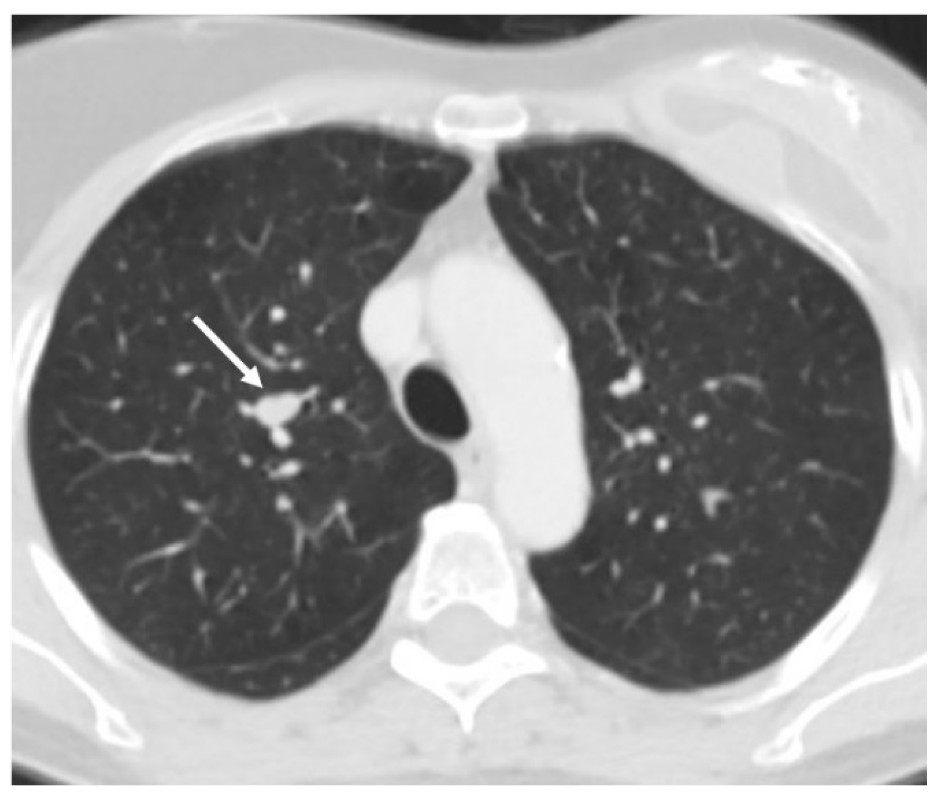
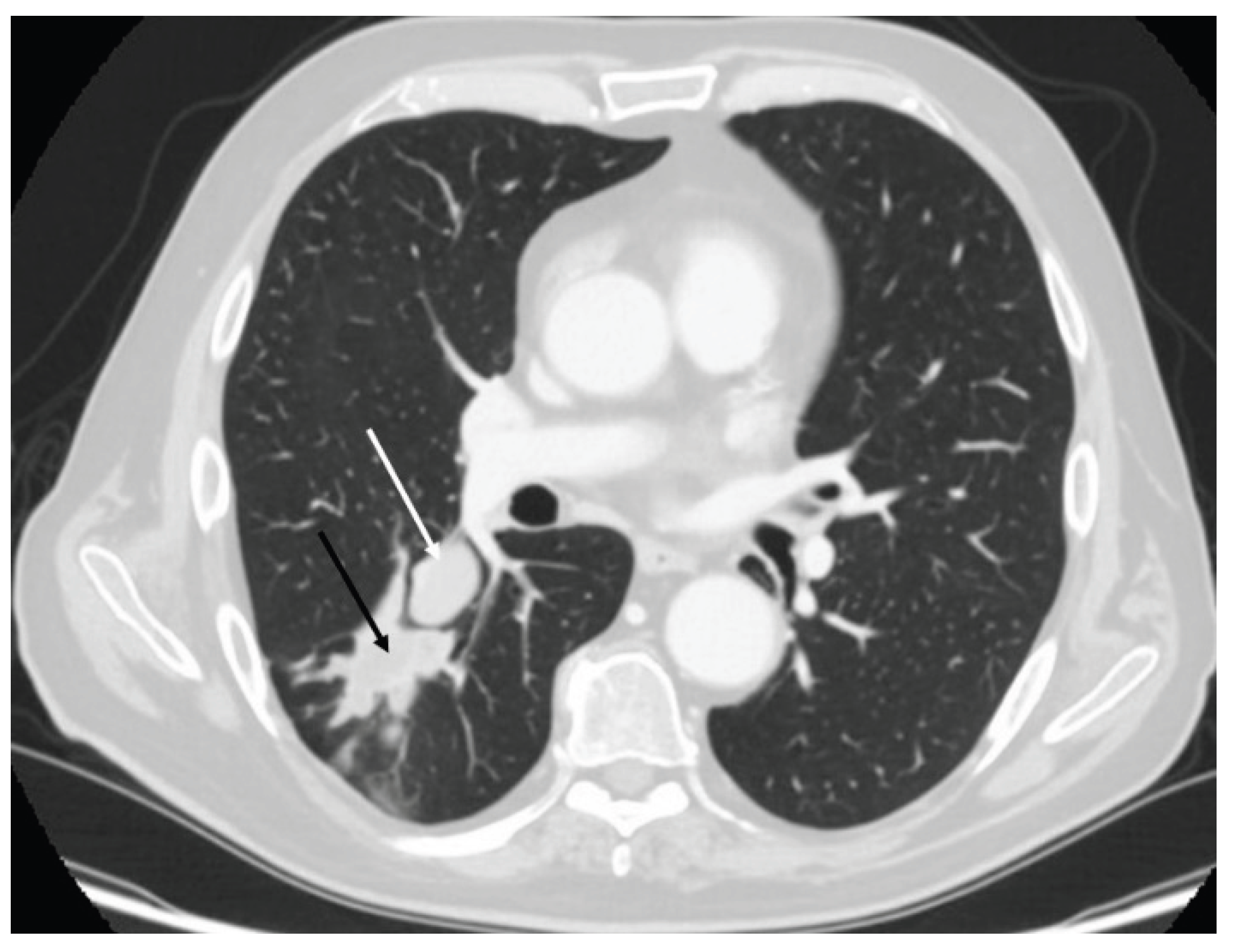
Figure 2.
Atypical carcinoid (white arrow) of the right lower lobe compli cated by post-obstructive pneumonia (black arrow) , requiring right lower bilobectomy.
Figure 2.
Atypical carcinoid (white arrow) of the right lower lobe compli cated by post-obstructive pneumonia (black arrow) , requiring right lower bilobectomy.
Disclaimer/Publisher’s Note: The statements, opinions and data contained in all publications are solely those of the individual author(s) and contributor(s) and not of MDPI and/or the editor(s). MDPI and/or the editor(s) disclaim responsibility for any injury to people or property resulting from any ideas, methods, instructions or products referred to in the content. |
© 2025 by the authors. Licensee MDPI, Basel, Switzerland. This article is an open access article distributed under the terms and conditions of the Creative Commons Attribution (CC BY) license (http://creativecommons.org/licenses/by/4.0/).
Copyright: This open access article is published under a Creative Commons CC BY 4.0 license, which permit the free download, distribution, and reuse, provided that the author and preprint are cited in any reuse.


