Submitted:
23 June 2025
Posted:
24 June 2025
You are already at the latest version
Abstract
Preterm labour (PTL)affects about 11% of all deliveries world-wide. It is a major cause of perinatal morbidity and mortality. Although the precise cause is unknown in about 50% of cases, infections are thought to be a major contributing factor. These infections are more common in earlier preterm deliveries. While some women with these infections will manifest the classical features such as fever, tachycardia (maternal and/or fetal), leucocytosis, raised biomarkers of infections and uterine tenderness/irritation others will be asymptomatic. Some of the women may develop a short/dilating cervix without any obvious contractions. Identifying such women is potentially challenging. Evidence has shown that a condensation of echogenic particles just above the cervix -the amniotic fluid (AF) sludge identified by ultrasound is a marker for microbial invasion of the amniotic cavity (MIAC) and preterm birth (PTB) in both asymptomatic and symptomatic women (including those with a short or normal cervix). Those with a short cervix with the AF sludge have a significantly greater risk of progression to PTB. Treatment with antibiotics has been shown in some but not all case series to result in a resolution of the sludge and either a delay or prevention of PTB. The widely varied results from treatment could be related to the antibiotics used and route of administration. The use of the parenteral combination of clindamycin, a cephalosporin and metronidazole has been shown to be more effective compared to azithromycin. Here we review the literature on the relationship between the sludge and PTB and conclude (1) that the AF sludge is an ultrasound marker of MIAC and PTL and (2) that following its diagnosis, appropriate counselling should be offered and the triple antibiotic combination offered. We suggest that randomised trials should be undertaken to determine the most efficacious antibiotic combination.
Keywords:
Ultrasound scan preterm birth
; infection/inflammation
; antibiotic fluid sludge
; antibiotic
1. Introduction
Preterm birth (PTB), defined as delivery before 37 completed weeks of gestation is the leading cause of severe neonatal morbidity and mortality world-wide and accounts for about 50% of neonatal deaths [1,2]. Of the estimated 15 million babies born preterm globally, 5% are before 28 weeks, 10% between 28 and 32 weeks and 84% are between 32 and 36 weeks [3]. It is estimated that of all the deaths in the first 5 years of life, about a million (18%) are in children born preterm [4] The overall PTB rate is about 11% but this varies from 4-20% depending on the WHO Region/Country - the highest being in low-income countries. [3,5,6]
Although the exact cause of spontaneous preterm birth (SPTB) is unknown in as many as 50% of cases, it is generally accepted that it is multifactorial in a large number [3,7]. Infections appear to be a common pathway in approximately 25% of cases (and probably much higher in deliveries before 23 weeks at 79% and then declining to about 11% for deliveries between 21 and 34 weeks) [8,9]. The presentation of these infections varies from obvious features of choroamnionitis (such as fever, uterine tenderness, maternal leucocytosis and tachycardia, fetal tachycardia and foul smelling/malodourous vagina discharge) to no obvious clinical features. Confirmation of microbial invasion of the amniotic cavity (MIAC) has traditionally been either from cultures of amniotic fluid and tissues and/or histologically on examination of fetal membranes, umbilical cord (funisitis) and the decidua [10,11,12]. Several studies have demonstrated the presence of infections in the amniotic fluid, fetal membranes and the cervico-vaginal secretions in women presenting in spontaneous preterm labour (SPTL) [11,12,13]. In those with a short cervix, the evidence is overwhelming [14,15] with typical organisms identified including Ureaplasma urealyticum, Gardnerella vaginalis, Candida albicans and Fusibacterium spp [16,17]. For diagnosing intra-amniotic infections, opinions vary on whether amniocentesis should be routinely performed on all those presenting in SPTL for this [13] or to only screening the vagina/cervix for infections. Tests on the amniotic fluid, when obtained, include those able to generate rapid results (such as the quantification of glucose, interleukin 6 and MMP-8 levels, white blood cells and microscopy) that could guide management and cultures that may require time to generate results. The increasing use of molecular biology diagnostic approaches (such as multiplex PCR test) has improved the rapidity of diagnosis and increased the isolation of micro-organisms that would otherwise not have been identified from routine cultures [17,18].
While the evidence is robust for the role of infections in those in SPTL with or without a short cervix [17] this is less so in those who are asymptomatic and with a normal cervix who may eventually progress to deliver preterm. Some of these women may have sub-clinical chorioamnionitis. Early identification of these women and timely institution of interventions such as (antibiotics) may reduce the risk of progression to preterm labour, not only in the symptomatic but also in the asymptomatic women. A key challenge for clinicians is whether there are features that may be indicative of a possible infection prior to shortening and dilatation of the cervix or contractions in this population. An ideal setup will be one where these women at risk are identified and timely interventions to interrupt the process of preterm labour instituted. This can only be achieved with reliable and specific markers of the causes of SPTB. Ultrasound scan can identify a population with cervical changes and allow/enable interruptions (e.g. cervical cerclage) that have indeed been shown to reduce/delay SPTB. Another possible ultrasound marker, that for MIAC- a process that has been shown to predispose to SPTL, is the amniotic sludge [19]. This review brings together the evidence for considering the AF sludge as a marker for MIAC and the need to treat this with antibiotics.
2. The Amniotic Fluid (AF) Sludge and Its Constitution
Particulate materials in the amniotic cavity are a common ultrasound scan finding. They are in general evenly distributed in the amniotic cavity and are thought to represent desquamated fetal cells, vernix caseosa, meconium and in some cases blood [20,21,22,23]. This can also be pathological, where there is excessive desquamation as in the case of congenital ichthyosis [24]. These particulate materials have been reported in about 4% of scans performed between the first and second trimesters [25,26] and rising to about 88% by 35 weeks [27]. The AF sludge on the other hand is a dense aggregate of highly echogenic material accumulating above the cervix, which is present in about 1% of uncomplicated term pregnancies [19] and rising to about 23.5% in high risk-populations (spontaneous preterm labouring women with intact membranes) [28]. Espinoza and colleagues proposed this term to describe this free-floating hyperechogenic material and associated it with an increased risk of SPTB [19].
The location of the AF sludge and its association with microbial invasion of the amniotic cavity is highly suggestive of an infective/inflammatory process (involved in its formation) [29]. Microorganisms may reach the amniotic cavity by breaching the membranes (when they ascend from the lower genital tract) or transplacentally reaching through a haematogenous spread [30]. On reaching the amniotic cavity, the micro-organisms provoke an inflammatory response in the epithelial cells of the fetal skin, the membranes and the umbilical cord [31]. As a result, there is a significant increase in pro-inflammatory cytokines (typically IL-1, IL-6 and IL-8 and TNF-a), prostaglandins, chemokines, and an increased expression of matrix metalloproteinases [29,31]. The increase expression of cytokines/chemokines in the amniotic cavity stimulates the migration of neutrophils across the decidua and the chorioamniotic membrane into the amniotic cavity resulting in an increased white cell population and enhanced antimicrobial activity [29,31,32]. The fact that some studies have failed to isolate micro-organisms and that the AF sludge has also been found in pregnancies that progress to term, suggest that the infective pathogenesis may not be applicable in all cases. Some have suggested that intra-amniotic inflammation can be as a result of exposure to "danger signals" produced by cells undergoing stress/damage or death [33,34,35,36]. This is supported by the fact that sterile inflammation is more common in women presenting in SPTL and pre-term premature rupture of fetal membranes (PPROM) compared to the classical microbiological inflammation [33,34,35,36]. Of note is also the fact that conventional cultures are only able to accurately identify infection in a proportion of cases. The increasing use of modern approaches to identify infective organisms (PCR techniques) has increased isolation rates [18,19].
In an interesting report, Romero et al. not only performed an amniocentesis from the sludge but were able to physically observe it in-vitro. The sludge was described as resembling pus on naked -eye examination. Gram stain from this and other studies has shown the presence of a variety of organisms (Mycoplasma hominis, Streptococcus mutans and Aspergillus flavus). The key question therefore is what is the precise composition of this AF sludge? It has been postulated that progressive infection induces an intense inflammatory response and that the inflammatory cells from this response (neutrophils) combine with the micro-organisms to form the particulate material that is visible as the AF sludge [36].
When micro-organisms breach the feto-maternal barrier, the consequence depends on how large the dose of micro-organisms reaching the fetal membranes is - with the risk of infection/inflammation greatest with the highest micro-organism load [36]. These micro-organisms (bacteria) can exist in one of two forms-singly (in the planktonic form) or organised in biofilms or both [36]. Following invasion of the amniotic cavity, bacteria change their phenotype to protect themselves from the host response (which includes the generation of inflammatory cells (neutrophils and monocytes) and the production of anti-microbial peptides and other mediators which can kill or injure the bacteria [36]. The bacteria achieve this by aggregating themselves in building-like structures known as biofilms. These biofilms make the micro-organisms resistant to attack by the macrophages, natural or synthetic antibiotics and anti-inflammatory mediators [37,38,39,40]. The bacteria in these biofilms are less likely to elicit an inflammatory response [28]. The formation of these intra-amniotic biofilms would partly explain why the intra-amniotic infection tends to be chronic and perhaps why they are difficult to treat as these biofilms are relatively resistant to antibiotics treatment [37,38,39,40,41]. The balance between the proportion of bacteria that are in the planktonic form and those in the biofilms likely determines the course of the infection and the likelihood of positive cultures from amniotic fluid sampling. Planktonic bacteriuria are more likely to be culture positive as opposed to those in the biofilm.
3. Imaging for the AF Sludge
The best approach to identifying the AF sludge is with a transvaginal ultrasound scan with the woman lying in the lithotomy position after emptying her bladder. The probe should be placed in the anterior fornix and adjusted to obtain a sagittal view of the entire cervical length. The length of the cervix should be measured preferably thrice and the average obtained. AF sludge is diagnosed from the presence of dense aggregate of highly echogenic particulate matter in close proximity to the internal cervical os [19,42]. This material may scatter with fetal movements or abdominal pressure but should re-accumulate within a few seconds. Where facilities are available the VOCAL software can be used to measure the volume of the AF sludge. We recommend that at least 3 minutes is spent assessing the sludge because of the dynamic nature of this marker. Figure 1 shows the AF sludge in four of our patients.
4. The AF Sludge and Intra-Amniotic Infections
An investigation of the AF sludge aspirated by amniocentesis showed this to be positive for the micro-organisms Streptococcus mutans, Mycoplasma hominis, Ureplasma urealyticum and Aspergillus flavus [43]. Interestingly in a study by Yoneda and colleagues [44] using polymerase chain reaction (PCR), AF "sludge" was present in 18.1% (19/105) of patients. However, there was a similar positive microorganisms rate in the women with a sludge and those without (31.6% versus 38.4%), but a significantly higher level of amniotic fluid interleukin-8 (15.2ng/ml vs 5.8ng/ml; p=0.005) and a higher frequency of histological chorioamnionitis in those with a sludge (52.6% vs 23.3%; p=0.01). In another study of 25 women with an AF sludge, examination of amniotic fluid collected after amniotomy showed that it was frequently associated with an inflammatory process [45]. Gill et al. in a cohort of 62 women with a short cervix and an AF sludge (AFS) who had undergone amniocentesis showed the rate of intramniotic inflammation to be 31.4% vs 3.7% in those who delivered <32 weeks compared to those who delivered after and furthermore, histological chorioamnionitis was significantly more common in the former group (75% vs 32%) [46]. Interleukin-8 was shown to have the highest sensitivity and specificity for intraamniotic inflammation and histological chorioamnionitis in this cohort. In an earlier study, Kusanovic and colleagues [28] found a higher positive culture rate in those with a sludge (33.3% vs 2.5%; P=0.003) and a higher frequence of histological chorioamnionitis (77.8% vs 19%; P<0.001). The findings of histological chorioamnionitis and funisitis were in 4 cases presenting with cervical insufficiency and an AF sludge [47]. Despite all these studies showing a high frequency of infection/inflammation in the presence of AF sludge, Ventura et al in a study of 16 cases failed to show any difference in these parameters although the women with an AF sludge delivered earlier [48]. Taken together, these case series appear to suggest that the AF sludge is a proxy or indeed a marker for microbial invasion of the amniotic cavity which in some cases may manifest as intra-amniotic infection and or inflammation and therefore a risk factor for PTB. There is need for more studies to investigate just how good a marker of preterm birth AF sludge is. Table 1 summarises the studies that have investigated the association between the AF sludge and intra-amniotic infection/inflammation (19,28,44,47,48,49,50.
5. The AF Sludge and an Ultrasound Marker for Spontaneous Preterm Labour?
Since intra-amniotic infections are generally associated with SPTB, and the AF sludge is thought to represent MIAC, the key question is whether the AF sludge could be an ultrasound marker of PTB? Identification of the AF sludge in the first half of pregnancy has been shown to be associated with inflammation/infection while in late pregnancy, it is thought to reflect maturation of the fetus (i.e. dominated by vernix caseosa, fetal squames and meconium) [20,21,51,52,53]. Espinoza et al [19] in a retrospective study of 84 women (19 or 22.4%) with intact membranes and spontaneous preterm labour, and 298 (1% with AF sludge) at term with intact membranes, concluded that the presence of AF ‘sludge’ in the spontaneous preterm labour and intact membranes group is a risk factor for MIAC, histological chorioamnionitis and impending spontaneous preterm delivery. Several other case series (mostly retrospective) have not only shown that those with the AF sludge are at greater risk of SPTL but that this risk is much greater in those with a short cervix. In a large retrospective study of 281 asymptomatic women who underwent cervical length measurement and screening for the AF sludge, Kusanovic et al [28] showed the AF sludge to be present in 66 cases. The shorter the cervical length, the higher the presence of AF sludge. The SPTB rate was higher in those with an AF sludge and this significant difference was maintained for PTB <28, <32 and <35 weeks. The combination of a cervical length of <25mm and the presence of an AF sludge conferred an odds ratio of 14.8 and 9.9 for spontaneous delivery at <28 and <32 weeks respectively [28]. Although some of studies have concluded that the presence of an AF sludge is an independent risk factor for the occurrence of SPTB, these observations have not been universal. There has to the best of our knowledge been only one prospective study [49]. Table 2 is a summary of the reports on the association between amniotic fluid sludge and SPTB [19,25,26,28,42,48,55,56,57].
6. Will Treatment Improve Outcome?
Since there is strong evidence associating the AF sludge with infection/inflammation, it follows that treatment with antibiotics may reduce the risk of PTB. This was the basis of several studies that have investigated the effect of antibiotics in women at risk of SPTB based on the ultrasound finding of an AF sludge. The results from various studies on the efficacy of antibiotics in this regard have been inconclusive. These studies (reviewed below) include case reports and retrospective and prospective studies. The first report of the use of antibiotics in women with AF sludge was reported by Himaya et al [49]. They describe a woman diagnosed with a sludge at 15+6 weeks with a cervical length of 33mm treated with intravenous ampicillin-gentamicin and oral azithromycin combination following an amniocentesis and culture of Staphylococcus warneri at 22 weeks. Following treatment, a second amniocentesis was performed, and culture was negative. She progressed to deliver at term. A historical controlled observational study by Hatanaka et al. [58] was reported in which women with an AF sludge diagnosed before 2012 and who were not treated were compared with those diagnosed after 2012 -2015 and treated with antibiotics. The women were divided into 2 groups - those at low risk were given oral clindamycin (300mg every 6 hours) and cephalexin (500 mg every 6 hours) for 7 days while the high-risk group was given intravenous clindamycin 600 mg every 8 hours and cefazolin (1g every 8 hours) for 5 days followed by oral treatment for 5 days. They showed that there was a reduction in the PTB rate in the treated group compared to the untreated group (13.2% vs 38.5%). Cuff et al. [57] in a retrospective cohort study of 97 women with the sludge compared outcome between those who had either been given oral azithromycin 500 mg on day 1 followed by 250 mg orally for 4 days or moxifloxacin 400 mg orally taken daily for 5 days and those who had not been treated. They showed that sonographic resolution of the AF sludge occurred in 34% of those who had been treated and 43% of those who had not - a difference that was not statistically significant. Furthermore, there were no differences in the rates of SPTB in both groups. Pustotina [59] undertook a prospective study of 29 women with an AF sludge (some with a short cervix and symptomatic) in which they offered vaginal clindamycin and other combinations (cefoperazone, clavulonate+amoxicillin and in some cases combined with indomethacin, progesterone and IV sulbactam) and showed that antibiotic treatment eliminated the AF sludge and prevented SPTB in all the cases. Jin et al [60] in a retrospective study of 58 women with a sludge diagnosed at 15-23 weeks and treated with a combination of IV ceftriaxone, clarithromycin and metronidazole showed a lower level of preterm birth following treatment with disappearance of the sludge in 51.7% of cases. More recently Giles et al. [61] reported on a retrospective cohort of women with the AF sludge treated and compared outcomes to those not treated. The antibiotic used was azithromycin. Interestingly the overall spontaneous preterm birth rate was higher in the treatment group but there were no differences in neonatal morbidity. Table 3 summarises all these studies [57,58,59,60,61,62,63]. Two meta-analyses [64,65] and a review of the literature by Luca et al. [66] concluded that while the AF sludge is a marker of preterm birth, there are no robust data on the benefit of antibiotics in this group. More recently there have been reports on cases where the AF sludge was identified and antibiotics administered and followed until it disappeared, and the pregnancy progressed to term [63].
A major factor that has been suggested as a potential confounder responsible for the variable results is the different antibiotic regimens used and routes of their administration. Additionally, the studies have been very heterogenous varying from those on asymptomatic low-risk/high-risk women to those on women with symptoms (i.e. presenting with uterine activity/contractions) or a combination of both. The two studies that showed no difference in treatment used azithromycin as the main antibiotic compared to the others that showed a difference (all of which used a variety of antibiotics including intravenous clindamycin and a cephalosporin). From these data it would seem that the biofilm is less likely to be penetrated by antibiotics administered orally as these may not achieve high levels in the blood. It would therefore be reasonable to recommend that these women are offered intravenous antibiotics. We have treated a number of cases in our unit (N=21) over the last 18 months with the AF sludge with a combination of intravenous clindamycin, metronidazole and ceftriaxone (given for one weeks) and in 14 out of the 21 (67%) cases, the AF sludge resolved, and pregnancies progressed to term or delivery was delayed by an average of 2 weeks in those who delivered preterm. We believe that considering how common this finding is in women at risk of SPTL, there should be randomised controlled trials on the efficacy of intravenous clindamycin combined with a cephalosporin and metronidazole to determine if such a regimen will reduce the risk of SPTB and therefore neonatal morbidity and mortality. This is the combination that appears to be most effective from the case series and covers most of the spectrum of organisms that have been isolated from the various studies.
Finally, it could be argued that leaving the fetus in-utero with MIAC would increase morbidity. The fact that the antibiotics led to resolution of the AF sludge in many cases and that the case series reviewed have not reported increased morbidity in the neonates (if anything delaying birth was associated with better outcomes) is reassuring in this context. Prior to commencing women on broad spectrum antibiotics, the potential of bacteria resistance must be discussed.
7. Conclusion
There is no doubt that intra-amniotic infections are central to a significant proportion of preterm births. Some of these manifests as obvious clinical infections and in most cases with uterine contractions and a short/dilated cervix. In some cases, however, the infections may be subclinical/asymptomatic. Identifying such cases is challenging but the presence of an AF sludge has been shown in case series to be a marker of such infections. The evidence linking the amniotic sludge with SPTB although predominantly from case series is moderately robust although more data are required. What is uncertain is whether treatment with antibiotics does indeed lead to the resolution of the AF sludge and prevention /delay of PTB. The potency of the antibiotics depends on the type of antibiotics and how they are administered. The data reviewed here suggests that oral azithromycin is not effective while a combination of parenteral ceftriaxone, metronidazole and clindamycin appear to be effective. While we feel there is enough to suggest treating these women with this combination, there is a need for randomized controlled trials on the efficacy of this regimen to generate robust evidence. Until such studies are undertaken, clinicians must continue to counsel women diagnosed with an AF sludge on the pros and cons of antibiotics and the limited data on efficacy.
Author Contributions
JCK conceived the idea. MA wrote the first draft which was reviewed by all the authors. All the authors reviewed and approved the final version.
Conflicts of Interest
None.
References
- Blencowe H, Cousens S, Chou D. et al. Born too soon, - the global epidemiology of 15 million preterm births. Reprod Health 2013; 10(suppl 1):S2)-.
- World Health Organization, Children: improving survival and well-being, 8 Sep 2020, https://www.who.int/news-room/fact-sheets/detail/children-reducing-mortality, last accessed 23rd December 2024.
- Ahmed B, Abushama M, Konje JC. Prevention of spontaneous preterm delivery - an update on where we are today. J Matern Fetal Neonatal Med. 2023 Dec;36(1):2183756. [CrossRef]
- Kinney & Rhoda 2019 - Lancet Global Health, Chawanpaiboon S, Vogel JP, Moller AB, et al. Global, regional, and national estimates of levels of preterm birth in 2014: a systematic review and modelling analysis. Lancet Glob Health 2019; 7: e37–46.).
- Chawanpaiboon S, Vogel JP, Moller AB, Lumbiganon P, Petzold M, Hogan D, Landoulsi S, Jampathong N, Kongwattanakul K, Laopaiboon M, Lewis C, Rattanakanokchai S, Teng DN, Thinkhamrop J, Watananirun K, Zhang J, Zhou W, Gülmezoglu AM. Global, regional, and national estimates of levels of preterm birth in 2014: a systematic review and modelling analysis. Lancet Glob Health. 2019 Jan;7(1):e37-e46. [CrossRef]
- Muglia LJ, Katz M. The enigma of spontaneous preterm birth. N Engl J Med. 2010 Feb 11;362(6):529-35. [CrossRef]
- Preterm labour and birth. NICE Guideline (NG25). Published : 25 November 2015. Last updated : 10 June 2022.
- Watts DH, Krohn MA, Hiler SL, Eschenbach DA. The association of occult amniotic Fluid infection with gestational age and neonatal outcome in women in preterm labor. Obstet Gynecolol 1992;79:351-357).
- Agrawal V, Hirsch E. Intrauterine infection and preterm labor. Semin Fetal Neonatal Med. 2012 Feb;17(1):12-9. [CrossRef]
- Romero R, Sirtori M, Oyarzun E, Avila C, Mazor M, Callahan R, Sabo V, Athanassiadis AP, Hobbins JC. Infection and labor. V. Prevalence, microbiology, and clinical significance of intraamniotic infection in women with preterm labor and intact membranes. Am J Obstet Gynecol. 1989 Sep;161(3):817-24. [CrossRef]
- Goldenberg RL, Andrews WW. Intrauterine infection and why preterm prevention programs have failed. Am J Public Health. 1996 Jun;86(6):781-3. [CrossRef]
- Kim CJ, Romero R, Chaemsaithong P, Chaiyasit N, Yoon BH, Kim YM. Acute chorioamnionitis and funisitis: definition, pathologic features, and clinical significance. Am J Obstet Gynecol. 2015 Oct;213(4 Suppl):S29-52. [CrossRef]
- Romero R, Salafia CM, Athanassiadis AP, Hanaoka S, Mazor M, Sepulveda W, Bracken MB. The relationship between acute inflammatory lesions of the preterm placenta and amniotic fluid microbiology. Am J Obstet Gynecol. 1992 May;166(5):1382-8. [CrossRef]
- Lee SM, Park KH, Jung EY, Jang JA, Yoo HN. Frequency and clinical significance of short cervix in patients with preterm premature rupture of membranes. PLoS One. 2017 Mar 30;12(3):e0174657. [CrossRef]
- Kiefer DG, Keeler SM, Rust OA, Wayock CP, Vintzileos AM, Hanna N. Is midtrimester short cervix a sign of intraamniotic inflammation? Am J Obstet Gynecol. 2009 Apr;200(4):374.e1-5. [CrossRef]
- Cassell GH, Davis RO, Waites KB, Brown MB, Marriott PA, Stagno S, Davis JK. Isolation of Mycoplasma hominis and Ureaplasma urealyticum from amniotic fluid at 16-20 weeks of gestation: potential effect on outcome of pregnancy. Sex Transm Dis. 1983 Oct-Dec;10(4 Suppl):294-302.
- Daskalakis G, Psarris A, Koutras A, Fasoulakis Z, Prokopakis I, Varthaliti A, Karasmani C, Ntounis T, Domali E, Theodora M, Antsaklis P, Pappa KI, Papapanagiotou A. Maternal Infection and Preterm Birth: From Molecular Basis to Clinical Implications. Children (Basel). 2023 May 22;10(5):907. [CrossRef]
- Romero R, Miranda J, Chaiworapongsa T, Chaemsaithong P, Gotsch F, Dong Z, Ahmed AI, Yoon BH, Hassan SS, Kim CJ, Korzeniewski SJ, Yeo L. A novel molecular microbiologic technique for the rapid diagnosis of microbial invasion of the amniotic cavity and intra-amniotic infection in preterm labor with intact membranes. Am J Reprod Immunol. 2014 Apr;71(4):330-58. [CrossRef]
- Espinoza J, Gonçalves LF, Romero R, Nien JK, Stites S, Kim YM, Hassan S, Gomez R, Yoon BH, Chaiworapongsa T, Lee W, Mazor M. The prevalence and clinical significance of amniotic fluid 'sludge' in patients with preterm labor and intact membranes. Ultrasound Obstet Gynecol. 2005 Apr;25(4):346-52. [CrossRef]
- Benacerraf BR, Gatter MA, Ginsburgh F. Ultrasound diagnosis of meconium-stained amniotic fluid. AmJ Obstet Gynecol 1984;149:570–572. [CrossRef]
- DeVore GR, Platt LD. Ultrasound appearance of particulate matter in amniotic cavity: vernix or meconium? J Clin Ultrasound 1986;14:229–230.
- Sepulveda WH, Quiroz VH. Sonographic detection of echogenic amniotic fluid and its clinical significance. J Perinat Med 1989;17:333–335.
- Sherer DM, Abramowicz JS, Smith SA, Woods JR Jr. Sonographically homogeneous echogenic amniotic fluid in detecting meconium-stained amniotic fluid. Obstet Gynecol 1991;78:819–822.
- Vohra N, Rochelson B, Smith-Levitin M. Three-dimensional sonographic findings in congenital (harlequin) ichthyosis. J Ultrasound Med 2003;22:737–739. [CrossRef]
- Bujold E, Pasquier JC, Simoneau J, Arpin MH, Duperron L, Morency AM, Audibert F. Intra-amniotic sludge, short cervix, and risk of preterm delivery. J Obstet Gynaecol Can. 2006 Mar;28(3):198-202. [CrossRef]
- Adanir I, Ozyuncu O, Gokmen Karasu AF, Onderoglu LS. Amniotic fluid "sludge"; prevalence and clinical significance of it in asymptomatic patients at high risk for spontaneous preterm delivery. J Matern Fetal Neonatal Med. 2018 Jan;31(2):135-140. [CrossRef]
- Parulekar SG. Ultrasonographic demonstration of floating Ultrasonographic demonstration of floating particles in amniotic fluid. J Ultrasound Med 1983; 2: 107–110.
- Kusanovic JP, Espinoza J, Romero R, Gonçalves LF, Nien JK, Soto E, Khalek N, Camacho N, Hendler I, Mittal P, Friel LA, Gotsch F, Erez O, Than NG, Mazaki-Tovi S, Schoen ML, Hassan SS. Clinical significance of the presence of amniotic fluid 'sludge' in asymptomatic patients at high risk for spontaneous preterm delivery. Ultrasound Obstet Gynecol. 2007 Oct;30(5):706-14. [CrossRef]
- Bearfield C, Davenport ES, Sivapathasundaram V, Allaker RP. Possible association between amniotic fluid microorganism infection and microflora in the mouth. BJOG. 2002; 109(5):527– 33.
- Gomez-Lopez N, Romero R, Xu Y, et al. Neutrophil Extracellular Traps in the Amniotic Cavity of Women with Intra-Amniotic Infection: A New Mechanism of Host Defense. Reprod Sci. 2017; 24(8):1139-1153. [CrossRef]
- Gomez-Lopez N, Romero R, Garsia-Flores V , et al. Amniotic fluid neutrophils can phagocytize bacteria: A mechanism for microbial killing in the amniotic cavity. Am J Reprod Immunol. 2017; 78(4):. [CrossRef]
- Chen GY, Nunez G. Sterile inflammation: sensing and reacting to damage. Nat Rev Immunol. 2010; 10(12):826–37. [CrossRef]
- Romero R, Miranda J, Chaiworapongsa T, et al. Prevalence and clinical significance of sterile intra-amniotic inflammation in patients with preterm labor and intact membranes. Am J Reprod Immunol. 2014; 72(5):458–74. [CrossRef]
- Romero R, Miranda J, Chaemsaithong P, et al. Sterile and microbial-associated intra- amniotic inflammation in preterm prelabor rupture of membranes. J Matern Fetal Neonatal Med. 2014:1–16. [CrossRef]
- Romero R, Miranda J, Chaiworapongsa T, et al. Sterile intra- amniotic inflammation in asymptomatic patients with a sonographic short cervix: prevalence and clinical significance. J Matern Fetal Neonatal Med. 2014:1–17. [CrossRef]
- Romero R, Kusanovic JP, Espinoza J, Gotsch F, Nhan-Chang CL, Erez O, Kim CJ, Khalek N, Mittal P, Goncalves LF, Schaudinn C, Hassan SS, Costerton JW. What is amniotic fluid 'sludge'? Ultrasound Obstet Gynecol. 2007 Oct;30(5):793-8. [CrossRef]
- Donlan RM, Costerton JW. Biofilms: survival mechanisms of clinically relevant microorganisms.Clin Microbiol Rev 2002;15:167–193. [PubMed: 11932229]. [CrossRef]
- Costerton W, Veeh R, Shirtliff M, Pasmore M, Post C, Ehrlich G. The application of biofilm science to the study and control of chronic bacterial infections. J Clin Invest 2003;112:1466–1477. [CrossRef]
- Donlan RM. Role of biofilms in antimicrobial resistance. ASAIO J 2000;46:S47–S52. [CrossRef]
- Jensen ET, Kharazmi A, Lam K, Costerton JW, Hoiby N. Human polymorphonuclear leukocyte response to Pseudomonas aeruginosa grown in biofilms. Infect Immun 1990;58:2383–2385. [CrossRef]
- Jensen ET, Kharazmi A, Hoiby N, Costerton JW. Some bacterial parameters influencing theneutrophil oxidative burst response to Pseudomonas aeruginosa biofilms. APMIS 1992;100:727–733. [CrossRef]
- Hatanaka AR, Mattar R, Kawanami TE, França MS, Rolo LC, Nomura RM, Araujo Júnior E, Nardozza LM, Moron AF. Amniotic fluid "sludge" is an independent risk factor for preterm delivery. J Matern Fetal Neonatal Med. 2016;29(1):120-5. [CrossRef]
- Romero R , Kusanovic JP, Espinoza J, Gotsch F, Nhan-Chang CL. Erez O, Kim CJ, Khalek N, Mittal P, Goncalves LF, Schaudinn C, Hassan SS, Costerton JW. What is amniotic fluid sludge? Ultrasound Obstet Gynecol 2007;30:793-398.
- Yoneda N, Yoneda S, Niimi H, Ito M, Fukuta K, Ueno T, Ito M, Shiozaki A, Kigawa M, Kitajima I, Saito S. Sludge reflects intra-amniotic inflammation with or without microorganisms. Am J Reprod Immunol. 2018 Feb;79(2). [CrossRef]
- Kusanovic JP, Jung E, Romero R, Mittal Green P, Nhan-Chang CL, Vaisbuch E, Erez O, Kim CJ, Gonçalves LF, Espinoza J, Mazaki-Tovi S, Chaiworapongsa T, Diaz-Primera R, Yeo L, Suksai M, Gotsch F, Hassan SS. Characterization of amniotic fluid sludge in preterm and term gestations. J Matern Fetal Neonatal Med. 2022 Dec;35(25):9770-9779. [CrossRef]
- Gill, N.; Romero, R.; Pacora, P.; Tarca, A.L.; Benshalom-Tirosh, N.; Pacora, P.; Kabiri, D.; Tirosh, D.; Jung, E.J.; Yeo, L.; et al. 467:Patients with Short Cervix and Amniotic Fluid Sludge Delivering ≤32 Weeks Have Stereotypic Inflammatory Signature. Am. J.Obstet. Gynecol. 2019, 220, S312. [CrossRef]
- Paules C, Moreno E, Gonzales A, Fabre E, González de Agüero R, Oros D. Amniotic fluid sludge as a marker of intra-amniotic infection and histological chorioamnionitis in cervical insufficiency: a report of four cases and literature review. J Matern Fetal Neonatal Med. 2016;29(16):2681-4. [CrossRef]
- Ventura W, Nazario C, Ingar J, Huertas E, Limay O, Castillo W. Risk of impending preterm delivery associated with the presence of amniotic fluid sludge in women in preterm labor with intact membranes. Fetal Diagn Ther. 2011;30(2):116-21. [CrossRef]
- Himaya E, Rhalmi N, Girard M, Tétu A, Desgagné J, Abdous B, Gekas J, Giguère Y, Bujold E. Midtrimester intra-amniotic sludge and the risk of spontaneous preterm birth. Am J Perinatol. 2011 Dec;28(10):815-20. [CrossRef]
- Pedregosa JP, Ruiz CM, Medina TB, Rascin AG, del GalloJ, de la Fuente JL, Alonso MJT. Amniotic sludge and short cervix as inflammation and intraamniotic infection markers Obstet Gynecolo Int J 2017;7:00239. [CrossRef]
- Sepulveda WH, Quiroz VH. Sonographic detection of echogenic amniotic fluid and its clinical significance. J Perinat Med 1989; 17: 333–335.
- Sherer DM, Abramowicz JS, Smith SA, Woods JR, Jr. Sonographically homogeneous echogenic amniotic fluid in detecting meconium-stained amniotic fluid. Obstet Gynecol 1991; 78:819–822.
- Zimmer EZ, Bronshtein M. Ultrasonic features of intraamniotic ‘unidentified debris’ at 14–16 weeks’ gestation. Ultrasound Obstet Gynecol 1996; 7: 178–181. 2.
- Tsunoda Y, Fukami T, Yoneyama K, Kawabata I, Takeshita T. The presence of amniotic fluid sludge in pregnant women with a short cervix: an independent risk of preterm delivery. J Matern Fetal Neonatal Med. 2020 Mar;33(6):920-923. [CrossRef]
- Yasuda S, Tanaka M, Kyozuka H, Suzuki S, Yamaguchi A, Nomura Y, Fujimori K. Association of amniotic fluid sludge with preterm labor and histologic chorioamnionitis in pregnant Japanese women with intact membranes: A retrospective study. J Obstet Gynaecol Res. 2020 Jan;46(1):87-92. [CrossRef]
- Pahlavan, F.; Niknejad, F.; Irani, S.; Niknejadi, M. Does Amniotic Fluid Sludge Result in Preterm Labor in Pregnancies after Assisted Reproduction Technology? A Nested Case—Control Study. J. Matern. Fetal Neonatal Med. 2022, 35, 7153–7157. [CrossRef]
- Cuff RD, Carter E, Taam R, Bruner E, Patwardhan S, Newman RB, Chang EY, Sullivan SA. Effect of Antibiotic Treatment of Amniotic Fluid Sludge. Am J Obstet Gynecol MFM. 2020 Feb;2(1):100073. [CrossRef]
- Hatanaka AR, Franca MS, Hamamoto TENK, Rolo LC, Mattar R, Moron AF. Antibiotic treatment for patients with amniotic fluid "sludge" to prevent spontaneous preterm birth: A historically controlled observational study. Acta Obstet Gynecol Scand. 2019 Sep;98(9):1157-1163. [CrossRef]
- Pustotina O. Effects of antibiotic therapy in women with the amniotic fluid "sludge" at 15-24 weeks of gestation on pregnancy outcomes. J Matern Fetal Neonatal Med. 2020 Sep;33(17):3016-3027. [CrossRef]
- Jin W H, Ha Kim Y, Kim J W, Kim T Y, Kim A, Yang Y. Antibiotic treatment of amniotic fluid “sludge” in patients during the second or third trimester with uterine contraction. Int J Gynaecol Obstet. 2021;153(01):119–124.
- Giles ML, Krishnaswamy S, Metlapalli M, Roman A, Jin W, Li W, Mol BW, Sheehan P, Said J. Azithromycin treatment for short cervix with or without amniotic fluid sludge: A matched cohort study. Aust N Z J Obstet Gynaecol. 2023 Jun;63(3):384-390. [CrossRef]
- Fuchs F, Boucoiran I, Picard A, Dube J, Wavrant S, Bujold E, Audibert F. Impact of amniotic fluid "sludge" on the risk of preterm delivery. J Matern Fetal Neonatal Med. 2015 Jul;28(10):1176-80. [CrossRef]
- Yeo L, Romero R, Chaiworapongsa T, Para R, Johnson J, Kmak D, Jung E, Yoon BH, Hsu CD. Resolution of acute cervical insufficiency after antibiotics in a case with amniotic fluid sludge. J Matern Fetal Neonatal Med. 2022 Dec;35(25):5416-5426. [CrossRef]
- Sapantzoglou I, Pergialiotis V, Prokopakis I, Douligeris A, Stavros S, Panagopoulos P, Theodora M, Antsaklis P, Daskalakis G. Antibiotic therapy in patients with amniotic fluid sludge and risk of preterm birth: a meta-analysis. Arch Gynecol Obstet. 2024 Feb;309(2):347-361. [CrossRef]
- Pannain GD, Pereira AMG, Rocha MLTLFD, Lopes RGC. Amniotic Sludge and Prematurity: Systematic Review and Meta-analysis. Rev Bras Ginecol Obstet. 2023 Aug;45(8):e489-e498. [CrossRef]
- Luca ST, Săsăran V, Muntean M, Mărginean C. A Review of the Literature: Amniotic Fluid "Sludge"-Clinical Significance and Perinatal Outcomes. J Clin Med. 2024 Sep 7;13(17):5306. [CrossRef]
Figure 1.
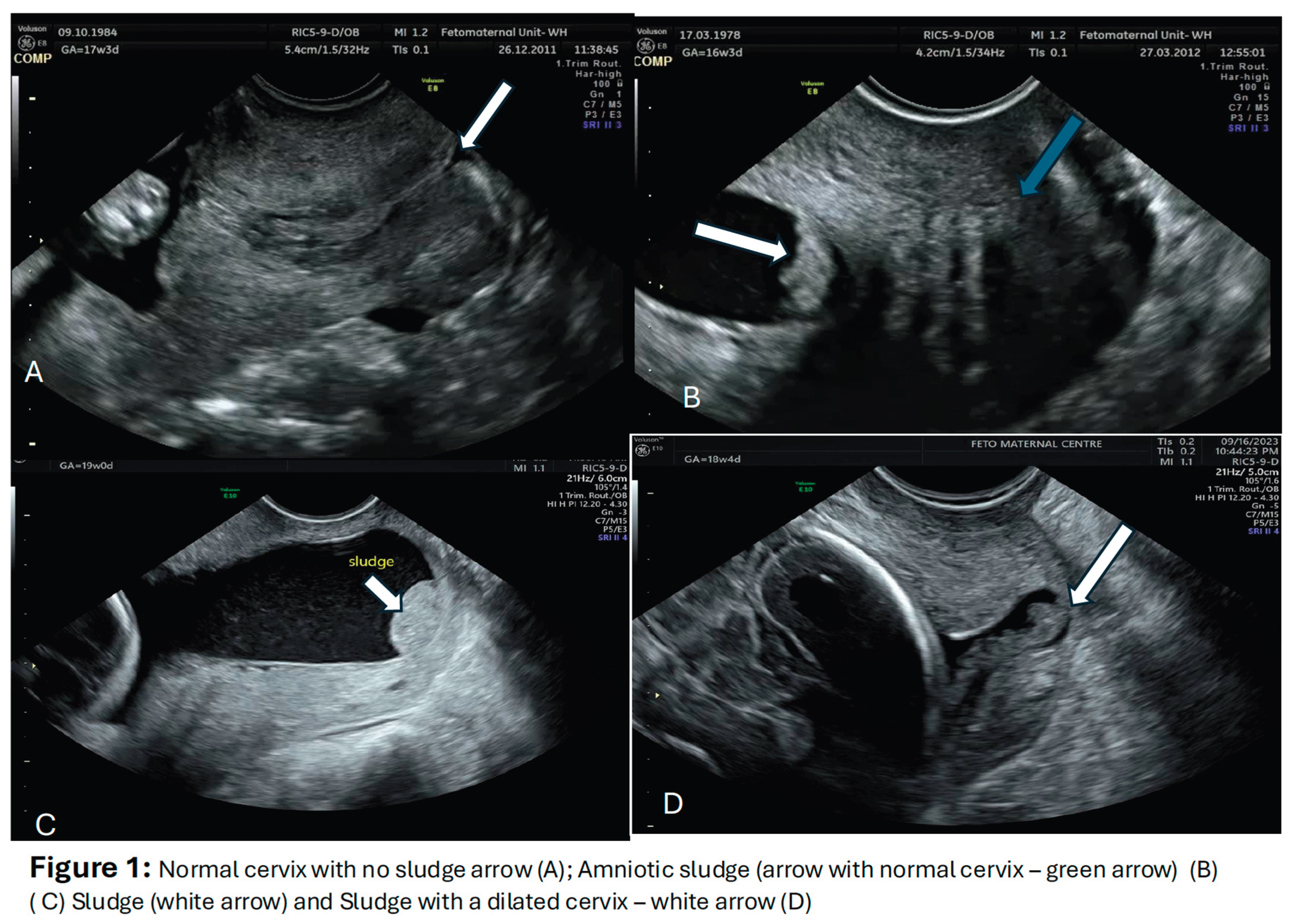

Table 1.
Summary of studies that have investigated intra-amniotic infection/inflammation in women with an amniotic sludge.
Table 1.
Summary of studies that have investigated intra-amniotic infection/inflammation in women with an amniotic sludge.
| Study details (authors and Year) | Type of study | Population studied and gestation at study | Method of investigation | Principal findings | Organisms isolated from culture/PCR |
|---|---|---|---|---|---|
| Espinoza et al. 2005 [19] | Retrospective | 84 women in preterm labour at 20-35 weeks or 298 in term labour. | Amniocentesis of the preterm group (N=84) for culture. 19 had a sludge and 65 did not. Histological examination of chorion-amnion and placenta | Histological chorioamnionitis in those with and without sludge. 77.8% (14/18) vs 19% (11/58); p<0.001 and positive AF culture 33.3% (6/18) vs 2.5% (1/40); p=0.003 | Ureplasma urealyticum, Fusobacterium nucleatum, Candida albicans, Peptostreptococcus spp, Gardnerella vaginalis |
| Kusanovic et al. 2007 [28] | Retrospective case control | 281 asymptomatic women at 13-29 weeks with a short cervix | 66 had a sludge and 215 did not. Amniocentesis performed on 51 (23 with sludge and 28 without) for AF culture and WBC in AF of >50 cells/mm3 Histology of membranes and cord |
MIAC rates of 21.7% (5/23) in AF sludge vs 0% (0/28) in non-sludge group and 27.3% (6/23) vs 3.6% (1/28) for intra-amniotic inflammation | Urealasma urealyticum, Staphylococcus aureus and Fusobacterium nucleatum |
| Himaya et al. 2011 [49] | Prospective | 310 women undergoing karyotyping by amniocentesis at 14-24 weeks | Quantification of amniotic fluid concentration of MMP-8 (MMP-8), glucose and lactate from 310 women (94 with free floating particles; 19 with dense amniotic sludge and 200 with no particles/no sludge). CL normal in all cases except 1 with a CL of <15mm | No significant differences in MMP-8, lactate and glucose in the groups. No differences in markers of MIAC in all groups. Woman with CL<15mm had higher MMP-8, and lower glucose. 2 women who delivered <32 weeks had higher mean lactate | Staphylococcus warneri in one case |
| Ventura et al. 2011 [48] | Retrospective case control | 58 women in preterm labour at 22-34 weeks. |
Two groups - 16 with sludge and 42 without. Histological examination and Histological chorioamnionitis was based on the presence of inflammatory cells in the chorionic plate and/or chorioamniotic membranes. |
No difference in histological chorioamnionitis between those with and without sludge (18.8% vs 14.3; p=0.067) | Organisms not characterised |
| Paules et al. 2016 [47] | Case report at 21-24 weeks | 4 cases of cervical weakness and bulging membranes with an amniotic fluid sludge | Amniocentesis in 3/4 cases (one of the cases refused amniocentesis) | All had histological chorioamnionitis and 2 had funisitis | Fusobacterium nucleatum (in 2/3 cdases) |
| Pedregosa et al. 2017 [50] | Retrospective & Prospective | Asymptomatic/symptomatic women with a short CL <25mm at 16-32 weeks | Amniocentesis in 15 cases - 12 with sludge. PCR, culture, gram stain and WBC and glucose level Microbiological study of placenta, membranes and umbilical cord |
From 15 amnios, 8 had MIAC and 6 with sterile inflammation (without any isolated organism) and 1 negative 10 positive cultures of placenta, membranes & cord |
Genital mycoplasma (Ureaplasma urealyticium, Mycomplasma hominis - most common organisms |
| Yoneda et al. 2017 [44] | Retrospective | 105 women in preterm labour at 20-29 weeks | Amniocentesis from 105 women in preterm labour (19 with sludge and 86 without) for culture, PCR (Positive AF cultures -examined using a nucleotide sequence- based analysis of bacterial genome DNA or 16S rRNA metagenomics) and IL-8 and placental histology of placenta. |
Women with vs without sludge PCR - no difference 31.6% (6/19) vs 38.4% (33/86) P>0.05 Funisitis 31.6% (6/10) vs 23.2% (20/86), p=0.447 Histological chorioamnionitis 52.6%(10/19) vs 23.3% - P=0.01 IL-8 - 15.2ng/ml vs 5.8ng/ml; P=0.005 |
Streptococcus parvum, Streptococcus agalactiae, Ureaplasma parvum, Flavobacterium succinicans, Ureaplasma urealyticum |
| Gill et al. 2019 [46] | Cohort | 62 asymptomatic women with a short cervix (≤25mm) at 16-22 weeks | Amniocentesis for concentrations of 33 proteins and histological examination of chorioamnion. Cohort was divided into those who delivered ≤ 32 weeks (35) and those who delivered >32 weeks (27) and variables compared (>1.5fold change in protein concentration considered significant) |
Intra-amniotic inflammatory rate higher in <32 weeks group (31.4% vs 3.7%; p=0.008); acute histological chorioamnionitis greater (75% vs 32%; p=0.002); higher mean concentration of 8/13 proteins - with IL-8 showing the highest difference (4.1 fold) | No organisms investigated |
CL = cervical length MIAC - microbial invasion of the amniochorion.
Table 2.
Summary of studies that have investigated the association between the amniotic fluid sludge and preterm labour.
Table 2.
Summary of studies that have investigated the association between the amniotic fluid sludge and preterm labour.
| Authors and Year of Study | Type of study | Population studied and gestation of study | Cervical assessment | Outcome -in terms of rates/risk of preterm birth |
|---|---|---|---|---|
| Espinoza et al. 2005 [19] | Retrospective | Women recruited at 20-35 weeks who went into preterm labour (N=84) and delivered at term controls (N=298) Sludge present in 19 of the preterm cohort (i.e. 19/84) |
CL ≤ 25mm in all those with sludge (19) and 49/65 of those without | Risk of PTB significantly greater in those with sludge at 48 hours, 7 days of delivery from diagnosis and at 32 and 35 weeks: by 48 hrs - 42.9% vs 4.4% by 7 days - 71.4% vs 15.6% <32 weeks 75.0 vs 25.8% <35 weeks 92.9% vs 37.8% |
| Burjold et al. 2006 [25] | Retrospective | 89 women at risk of preterm birth recruited for cervical length measurement at 18-32 weeks’ gestation-14 with sludge and 75 without | CL significantly shorter in those with sludge - 34.0±10 in those with no sludge vs 23±11 and 16±14 in those with light and dense sludge | Spontaneous PTB in <34 weeks - 8/9 (88.9%) vs 5/75 (6.7%) in those with and without sludge |
| Kusanovic et al. 2007 [28] | Retrospective case-control | 281 patients between 13 and 29 weeks-Sludge N=66, controls N=216 | Cervical length measured and groups into <5mm, <15mm, <25mm and >30mm | Sludge present in 69% (20/29), 49% (33/68), 35% (49/142) and 12% (12/99) respectively for CL<5mm, <15mm; <25mm & >30mm Spontaneous PTB - no sludge vs sludge - <28 weeks 9.4%vs 54.3%; <32 weeks 14.6% vs 60% & <35 weeks - 19.8% vs 42.3% Odd of SPTB if combined sludge and CL<25mm - 14.8for delivery <28 weeks and 9.9 for delivery <35 weeks |
| Ventura et al. 2011 [48] | Retrospective cohort | 58 women with threatened preterm labour at 22-34 weeks - 16 with amniotic fluid sludge and 42 without | Of the 16 with AFS, 75% had CL ≤25mm & 37.5% CL≤15mm |
SPTB greater in those with AFS 25% vs 2.4 within 48 hours 37.5% vs 11.9% within 7 days 75% vs 23.9% within 14 days USS to delivery interval 21.7+_30.1 vs 49.4+137.8 days |
| Hatanaka et al. 2016 [42] | Prospective cohort | 195 women at 16-26 weeks, 49 with sludge and 146 without | CL<25mm - 38.8% 19/49 (sludge) vs 17.5% (23/146) | Gestational age at delivery in sludge vs no sludge group - 35.8+_5.4 weeks vs 37.8+_3.6 weeks SPTB rates differed at up to <35 weeks (at<28 weeks - 12.2% vs 3.4%; at<32 weeks - 17.1% vs 5.1% & at<35 weeks 26.8% vs 8.5%) |
| Adanir et al. 2018 [26] | Prospective | 92 women at high risk of preterm delivery between 20-34 weeks';-18 with sludge and 74 without | CL≤25mm in 8/18 (sludge) vs 9/74 (no sludge) | SPTB rate of 66.7% in those with sludge vs 27% (20/74) in those without sludge |
| Tsunoda et al. 2020 [54] | Retrospective cohort | 110 patients at 14-30 weeks - TVS measurement of cervical length and sludge. 29 with sludge and 81 without | 29 delivered <34 weeks and 51<37 weeks. 16/29 and 21/51 had sludge. CL<20mm - 24/29 vs 33/51 and <15mm - 17/29 vs 21/51 | Risk of SPTB increased with the presence of AFS Odd ratio for delivery <35 weeks - 6.44 and <37 week - 4.46 |
| Yasuda et al. 2020 [55] | Retrospective | 54 women presenting in preterm labour at 22-36+6 weeks. Cervical length measured and sludge identified | Sludge present in 11 cases | AFS cohort delivered at 28.3±4.5 weeks vs 31.7±4.3 weeks |
| Pahlavan et al. 2022 [56] | Nested case control | 110 women who underwent ART in the form of IVF-ET - 63 with sludge and 67 without | CL<30mm in control group - 10.4% and 28.6% in the study groups | SPTB prevalence of 23.6% in case and 10.4% in control |
| Suff et al. 2023 [57] | retrospective cohort | 147 women - 54 with sludge and 93 without | Women with the sludge more likely to have a short CL (19mm vs 14mm) | Women with AFS + short CL, more likely to have a mid-trimester loss and delivery <24 weeks (RR 3.4; 95%CI 3.4-12-20.3) |
SPTB=spontaneous preterm birth; PTB= preterm birth; CL = cervical length; IVF-ET = in-vitro fertilisation and embryo transfer; AFS=amniotic fluid sludge.
Table 3.
Summary of studies that have investigated antibiotics in women with amniotic fluid and its impact on the risk of preterm birth.
Table 3.
Summary of studies that have investigated antibiotics in women with amniotic fluid and its impact on the risk of preterm birth.
| Authors and year of study | Type of study | No of cases studied included | Antibiotic regimen and duration | Outcome (in terms of risk of preterm birth) |
|---|---|---|---|---|
| Fuchs et al. 2014 [62] | Retrospective case control | 77 asymptomatic women at 15-32 weeks - 63 Rx and 14 untreated Cervical length measured |
Azithromycin 500 mg on day 1 and then 250 mg IV and oral for 4 days | Overall SPTB rates - 57% (36/63) vs 29% (4/14) , P=0.05 in the treated and untreated groups; PTB<28 weeks - 11.1%vs 28.6 P=0.1 PTB<32 weeks - 17.5%vs42.9% - P=0.07 PTB<34 weeks - 19.1% vs 57.1% P=0.006 Conclusion: Use of azithromycin reduced the risk of PTB <34 weeks |
| Hatanaka et al. 2019 [58] | Observational historical controlled | Cohort of 86 - 64 asymptomatic diagnosed with AFS at 16-26 weeks (divided into high and low risk) and 22 controls with AFS Cervical length measured |
Two groups High risk (CL<25mm/other risk factors) IV clindamycin+ ceftazolin for 5 days and then oral for 5 days Low risk CL>25mm Clindamycin (oral) + cephalexin for 7 days - low risk group |
Risk of SPTB <34 weeks in high-risk group - 13.2% vs 38.5% (P=0.047) in treated vs untreated No difference in SPTB rate at all gestations in both groups together (i.e. combined high and low risk - r=treated vs untreated) Conclusion: In high risk group, antibiotics reduce risk of SPTB <34 weeks |
| Cuff et al. 2020 [57] | Retrospective cohort | 97 asymptomatic women with AFS diagnosed at 15-25 weeks- 51 treated and 46 untreated CL measured in both groups |
Mixed treatment 46 Rx with oral Azithromycin x5 days 5 Rx with oral Moxifloxacin x 5 days |
Overall SPTB rate <37 weeks- 49.5% and 22.7% <28 weeks CL measurements same in treated and untreated groups SPTB <37 weeks - 53% vs 45.7% in treated vs untreated (P=0.47) SPTB <228 weeks - 21.6% vs 19.6% (P=0.81) Conclusion: Treatment made no difference in outcome |
| Pustotina 2019 [59] | Prospective | 29 asymptomatic women with AFS diagnosed at 14-24 weeks divided into three groups 14 with CL<25mm & symptomatic 7 with Cl >25mm and asymptomatic 8 with CL>25mm |
All 29 received Vaginal clindamycin and probiotics and plus 16- IV cefoperazone/sulbactam 8 - oral amoxicillin/clavulanate IV butoconazole to 18 Progesterone and indomethacin given to all those with CL<25mm |
Intravenous antibiotics prevented SPTB in all women with CL>25mm and. asymptotic women with CL<25mm and 70% in those with symptoms and CL<25mm Conclusion: Intravenous antibiotics delayed delivery or prevented SPTB |
| Jin et al. 2021 [60] | Retrospective cohort | 58 women at 15-32 weeks symptomatic women with intact membranes and AFS | IV Ceftriaxone 1 g daily, Clarithromycin 500 mg BD orally and metronidazole 500 mg tds - all for 4 weeks | AFS disappeared in 30/58 (51.7%) USS to delivery interval - 67.7+-35.7 days vs 28.4+-35.7 in those without AFS and with persisting AFS after treatment SPTB <28, <32 & <34 weeks was greater in the persistent group Conclusion: Antibiotics may cause AFS to disappear in women presenting in PTL and this is associated with improved outcome |
| Giles et al. 2023 [61] | Retrospective cohort | 374 asymptomatic high-risk women at 13-24 weeks and CL ≤ 15mm - 129 Rx and 245 not Rx Cervical cerclage. performed on >60% of cases and vaginal progesterone given to most |
Azithromycin - IV or oral or both for 7 days | SPTB rates - 51.2% vs 50.6% in the azithromycin and un-treated groups No difference in SPTB <28, <34 weeks and PPROM. Conclusion: The data do no support the routine use of azithromycin in women with a short cervix and AFS |
| Yeo et al 2021 [63] | Case report | Symptomatic woman presenting at 20+6 weeks and sludge - treatment started at 22 weeks | Short cervix, amniocentesis (sterile inflammation), 11 days treatment with IV ceftriaxone (1 gm daily), IV metronidazole 500 mg 8 hourly and oral Clarithromycin 500 mg daily for up | Short cervix progressively became normal and sludge disappeared. Elective delivery at 36+2 weeks |
AFS - amniotic fluid sludge; PPROM= preterm prelabour rupture of fetal membranes' SPTB= spontaneous preterm birth;.Rx=treatment; IV=intravenous.
Disclaimer/Publisher’s Note: The statements, opinions and data contained in all publications are solely those of the individual author(s) and contributor(s) and not of MDPI and/or the editor(s). MDPI and/or the editor(s) disclaim responsibility for any injury to people or property resulting from any ideas, methods, instructions or products referred to in the content. |
© 2025 by the authors. Licensee MDPI, Basel, Switzerland. This article is an open access article distributed under the terms and conditions of the Creative Commons Attribution (CC BY) license (http://creativecommons.org/licenses/by/4.0/).
Copyright: This open access article is published under a Creative Commons CC BY 4.0 license, which permit the free download, distribution, and reuse, provided that the author and preprint are cited in any reuse.


