Submitted:
13 April 2025
Posted:
14 April 2025
You are already at the latest version
Abstract
Background/Objectives: Proximal junctional kyphosis (PJK) is one of the most frequently dis-cussed complications following corrective surgery in patients with neuromuscular scoliosis (NMS). Despite its clinical relevance, the etiology of PJK remains incompletely understood and appears to be multifactorial. Biomechanical and limited clinical studies have suggested that pre-operative hyperkyphosis, resection of the spinous processes with consequent disruption of poste-rior ligamentous structures, and rod contouring parameters may contribute as risk factors. Meth-ods: To validate these findings, we retrospectively analyzed 99 NMS patients who underwent posterior spinal fusion using a standardized screw-rod system between 2009 and 2017. Radio-graphic parameters were assessed preoperatively (preOP), postoperatively (postOP), and at an average follow-up (FU) of 29 months. Clinical data included age, weight, height, gender, and Risser sign. Radiological measurements comprised Cobb angles, thoracic kyphosis (TK), lumbar lordosis, levels of the upper (UIV) and lower (LIV) instrumented vertebrae, the number of fused levels, sagittal alignment parameters, the rod contour angle (RCA), and the mismatch between RCA and the proximal junctional angle (PJA). Patients were stratified into a PJK group and a non-PJK group. Results: The overall incidence of PJK was 23.2%. In line with previous biome-chanical findings, spinous process resection was significantly associated with PJK development. Furthermore, the PJK group demonstrated significantly higher preoperative TK (59.3° ± 29.04° vs. 34.5° ± 26.76°, p < 0.001), greater RCA (10.2° ± 4.01° vs. 7.7° ± 4.34°, p = 0.021), and a larger postop-erative mismatch between PJA and RCA (PJA−RCA: 3.8° ± 6.76° vs. −1.8° ± 6.55°, p < 0.001) com-pared to the non-PJK group. Conclusions: Spinous process resection, a pronounced mismatch between postoperative PJA and RCA (odds ratio [OR] = 1.19, p = 0.002), excessive rod bending (i.e., high RCA), and severe preoperative thoracic hyperkyphosis are significant risk factors for PJK. These variables should be carefully considered during the surgical planning and execution of de-formity correction in NMS patients.
Keywords:
neuromuscular scoliosis
; neuromyopathic scoliosis
; proximal junctional kyphosis
; complication
; risk factor
; spinous process
; sagittal alignment
; rod contouring
; hyperkyphosis
1. Introduction
Neuromuscular scoliosis (NMS) is a complex three-dimensional spinal deformity caused by neuromyopathic disorders that impair muscle strength and control [1,2]. It is commonly associated with conditions such as spinal muscular atrophy, cerebral palsy, and muscular dystrophy, leading to progressive spinal misalignment and instability [3]. Treatment strategies for NMS include conservative approaches and surgical interventions, such as posterior spinal correction and fusion [4], as well as the use of growth-friendly devices like VEPTR™ or MAGEC™ rods in younger patients [5,6,7,8].
Surgical correction plays a central role in halting progression, preventing complications, and improving quality of life in patients with NMS [1,9,10]. However, these procedures are associated with a high rate of complications, necessitating close postoperative monitoring [11,12,13]. A meta-analysis by Sharma et al. (2012) demonstrated a significantly higher complication rate in patients with NMS compared to those with adolescent idiopathic scoliosis (AIS) [14,15,16].
One notable postoperative complication is proximal junctional kyphosis (PJK), which can result in pain, spinal imbalance, implant-related problems, and even neurological deficits [17,18]. Reported incidence rates for PJK vary widely between 7% and 61%, likely due in part to differing definitions used across studies [9,19]. While numerous investigations have examined PJK risk factors in adult deformity and AIS populations, dedicated studies on NMS remain limited [13,20,21].
In NMS patients, particularly children undergoing long spinal fusions to the sacrum and pelvis, PJK is recognized as a frequent and serious complication [22,23,24]. The spectrum of the condition ranges from mild PJK to proximal junctional failure (PJF), underlining the importance of a comprehensive understanding of its causes and prevention [23,25]. Contributing factors discussed in the literature include surgical techniques, choice of instrumentation (e.g., pedicle screws vs. hooks or sublaminar bands), and rod material stiffness. In addition, sagittal profile parameters, particularly preoperative thoracic hyperkyphosis, have been linked to PJK development [26,27,28,29,30,31,32].
More recently, specific radiological parameters related to rod contouring have gained attention. In particular, the rod contour angle (RCA) and its mismatch with the proximal junctional angle (PJA) have been proposed as influential factors [33,34,35,36,37,38]. A pronounced curvature at the proximal rod end (i.e., high RCA) and a significant postoperative discrepancy between PJA and RCA (PJA–RCA) are suspected to contribute to junctional instability. However, these relationships remain poorly understood and are scarcely studied in the NMS population [34].
Furthermore, resection of spinous processes—commonly performed during surgery to harvest bone for fusion—inevitably disrupts the supra- and interspinous ligaments. This destabilization may increase flexibility in the adjacent cranial segments and has been identified in recent biomechanical studies as a potential risk factor for PJK [39].
Given the surgical complexity of NMS, especially in pediatric patients, the identification and understanding of such risk factors are critical. Incorporating them into preoperative planning and intraoperative decision-making may help to reduce the incidence of PJK and improve long-term outcomes [24].
Proximal junctional kyphosis remains a significant challenge in the surgical treatment of neuromuscular scoliosis. This single-center study aims to identify modifiable risk factors—such as spinous process resection and inadequate rod contouring—to support the development of preventive strategies. Ultimately, addressing these factors may contribute to reducing PJK incidence and enhancing surgical outcomes in this vulnerable patient population.
2. Materials and Methods
This single-center retrospective study included 99 patients with neuromuscular scoliosis (NMS) who underwent corrective posterior spinal fusion between 2009 and 2017 using a standardized screw-rod system (titanium polyaxial pedicle screws, titanium and/or cobalt-chromium rods; Expedium®, DePuy Synthes) and a uniform surgical technique. In 12 patients, bilateral transverse process hooks were utilized at the upper instrumented vertebra (UIV). Spinal bands were used in 10 patients: in 7 cases, three bands were applied on the concave side at the apex of the thoracic curve to assist curve correction in the presence of pedicle hypoplasia; in 3 cases, spinal bands were placed at the UIV. No additional prophylactic strategies—such as vertebroplasty or sublaminar wiring—were employed to prevent proximal junctional kyphosis (PJK).
Given the retrospective nature of the study, which was based on existing radiographic (hospital PACS) and clinical (patient chart) data, patient consent was not required in accordance with institutional regulations.
Full-spine standing or sitting radiographs (anteroposterior and lateral views) were obtained preoperatively (preOP), immediately postoperatively (postOP), and at a mean follow-up (FU) of 29 months. All images were digitally stitched. Radiographic measurements were performed by a single observer (K.B.), while clinical data were extracted from patient records.
Collected clinical data included age at surgery, sex, weight, height, and Risser sign. Radiographic assessments comprised pre- and postOP Cobb angles, thoracic kyphosis (T4–T12), lumbar lordosis (L1–S1), UIV and lower instrumented vertebra (LIV) levels, number of instrumented vertebrae, rod contour angle (RCA, defined as the angle between UIV and UIV–1), and mismatch between RCA and the proximal junctional angle (PJA, measured as the angle between the inferior endplate of UIV and the superior endplate of UIV+2). Additional sagittal spinopelvic parameters included pelvic tilt (PT), sacral slope (SS), pelvic incidence (PI), and sagittal vertical axis (SVA).
PJK was defined according to established criteria [40,41], requiring both of the following: (1) a segmental kyphosis of ≥10° between UIV and UIV+2, and (2) a postoperative increase in this segmental kyphosis of at least 10° compared to the preoperative value (Figure 1).
The study cohort was divided into two groups: patients with PJK (n = 23) and those without PJK (n = 76).
Statistical analysis was performed using SPSS Statistics, version 28.0.1.1 (IBM Corp., Armonk, NY, USA). The incidence of PJK was calculated using cross-tabulations. Group comparisons were conducted using the Student’s t-test or Mann–Whitney U-test for continuous variables, and the chi-square test for categorical variables. Pearson correlation analysis was used to explore relationships between significant risk factors. Variables with a statistically significant association with PJK were further analyzed using multivariate logistic regression. In cases of high intercorrelation between variables (|r| > 0.7), only the parameter with the stronger correlation to the dependent variable (PJK) was included in the final model. Odds ratios (OR) were calculated to quantify the influence of individual predictors on the likelihood of developing PJK. Descriptive data are reported as mean ± standard deviation (SD). Statistical significance was defined as p < 0.05.
The study was approved by the Ethics Committee of the Westfalen-Lippe Medical Association on 08/23/2017 (no: 2017-427-f-S) and complies with the guidelines of the Declaration of Helsinki.
3. Results
A total of 99 patients with neuromuscular scoliosis (NMS) were included in this study. The underlying diagnoses were as follows: cerebral palsy (n = 54), muscular dystrophy (n = 17), spinal muscular atrophy (n = 13), spina bifida (n = 10), neurofibromatosis (n = 4), and other etiologies (n = 1). The mean age at surgery was 17.0 ± 7.46 years, and 51.5% (n = 51) of patients were female.
Proximal junctional kyphosis (PJK) developed in 23 patients (23.2%), while 76 patients (76.8%) did not develop PJK during the follow-up period. The mean follow-up duration was 29 months. Among the PJK group, 78% (n = 18) developed PJK within the first 12 months, whereas 22% (n = 5) were diagnosed after more than 24 months.
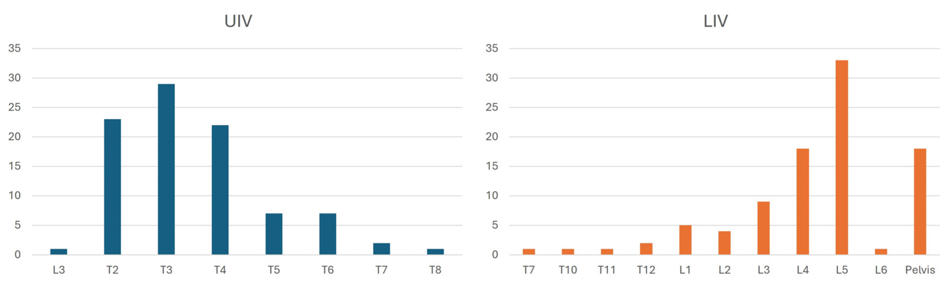
The distribution of the upper instrumented vertebra (UIV) was as follows: T2 (22.2%), T3 (29.3%), T4 (22.2%), T5 (7.1%), T6 (6.1%), and T7 (2.0%). The lowest instrumented vertebra (LIV) was distributed as follows: T12 (2.0%), L1 (5.1%), L2 (4.0%), L3 (9.1%), L4 (19.2%), L5 (32.3%), and S2/Ilium (18.2%). On average, 13.8 vertebrae and 12.8 segments were instrumented per patient.
Figure 2.
Distribution of upper (UIV) and lower (LIV) instrumented vertebrae.
Demographic, radiographic, and surgical parameters were compared between groups (Table 1).
No significant differences were found for age, sex, BMI, number of instrumented vertebrae, or pre- and postoperative Cobb angles. However, significant differences were observed in thoracic kyphosis (TK), the rate of spinous process resection, rod contour angle (RCA), and mismatch between RCA and proximal junctional angle (PJA).
While 47.5% (n = 47) of patients received bilateral titanium rods, 24.2% (n = 24) cobalt–chromium rods, and 15.2% (n = 15) a hybrid construct, rod material was not significantly associated with PJK incidence. Similarly, the use of transverse process hooks (n = 12) or spinal bands at the UIV (n = 3) did not affect the frequency of PJK.
Spinous process resection showed a significant association with PJK: 87.0% of patients in the PJK group underwent this procedure, compared to 59.2% in the non-PJK group (p = 0.036).
Preoperative thoracic kyphosis was significantly higher in the PJK group (59.3° ± 29.04° vs. 34.5° ± 26.76°, p < 0.001) and remained elevated postoperatively and at 12-month follow-up. Moreover, the reduction in TK from preOP to postOP was more pronounced in the PJK group (−21.3° ± 19.28° vs. −10.7° ± 20.2°, p = 0.032).
Immediately postoperatively, both PJA and RCA differed significantly between groups (PJA: 14.0° ± 6.31° vs. 5.9° ± 6.49°, p < 0.001; RCA: 10.2° ± 4.01° vs. 7.7° ± 4.34°, p = 0.021). Notably, the mismatch between PJA and RCA (postOP PJA–RCA) was significantly higher in the PJK group (3.78° ± 6.75° vs. −1.8° ± 6.55°, p < 0.001), indicating a potential biomechanical risk factor for PJK.
To assess potential predictors of PJK, Pearson correlation analysis was conducted (Table 2). No predictors showed multicollinearity (r > |0.7|), and thus all were included in the multivariate logistic regression model.
Spinous process resection, preoperative thoracic kyphosis (TK), and the postoperative mismatch between PJA and RCA were significant predictors in the regression model (Table 3). Spinous process resection increased the likelihood of developing PJK by 6.3-fold (OR = 6.35, p = 0.036). A one-degree increase in PJA–RCA mismatch raised the PJK risk by 19% (OR = 1.19, p = 0.002), while each degree increase in preoperative TK raised the risk by 6.5% (OR = 1.07, p = 0.017).
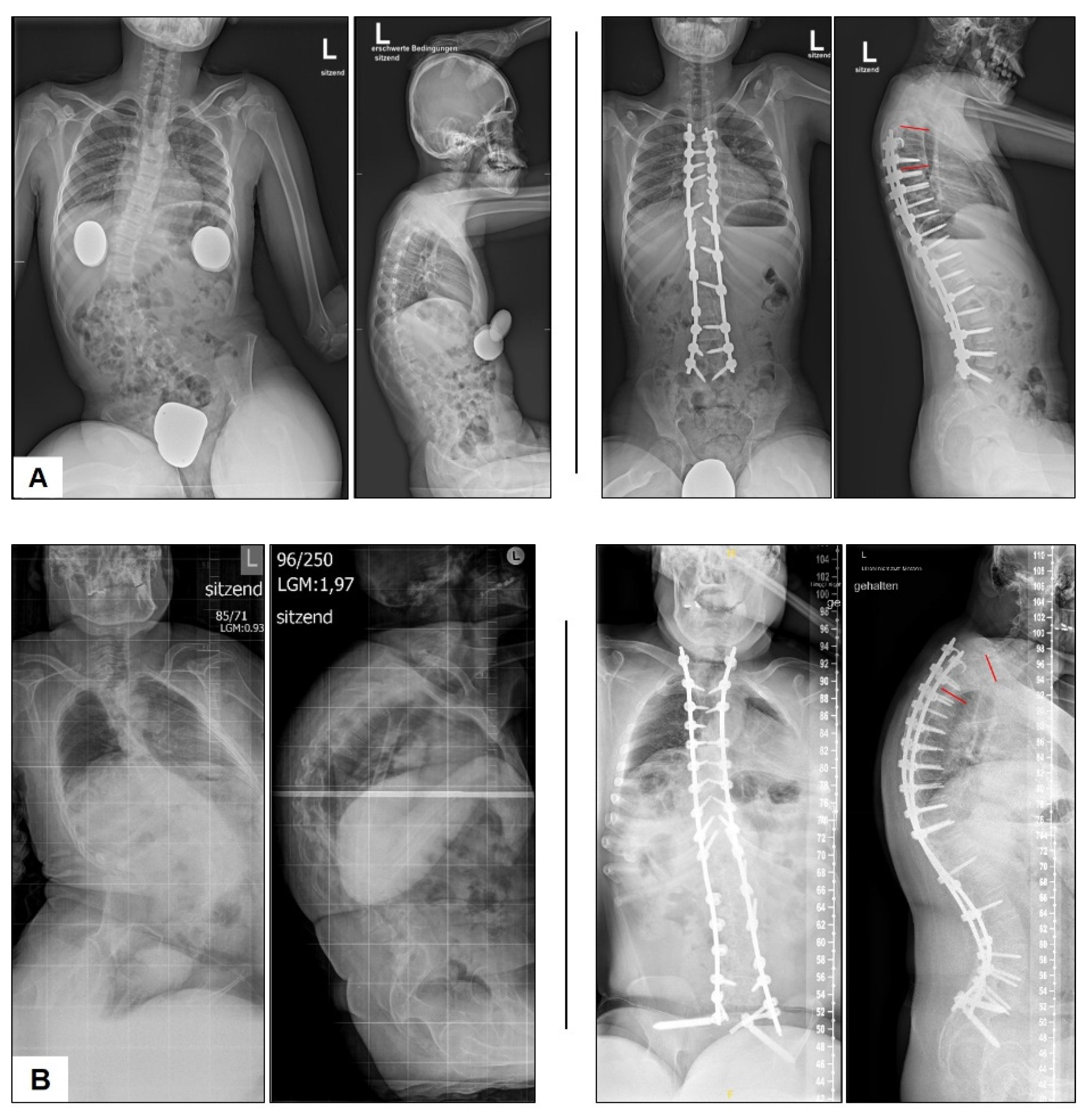
Figure 3.
Representative radiographs from the non-PJK (A) and PJK (B) groups. PJA indicated by red lines.
Figure 3.
Representative radiographs from the non-PJK (A) and PJK (B) groups. PJA indicated by red lines.
4. Discussion
Proximal junctional kyphosis (PJK), particularly its more severe manifestation—proximal junctional failure (PJF)—is a well-documented complication following corrective spinal surgery with posterior instrumentation in patients with neuromuscular scoliosis (NMS). PJK carries the potential risk of necessitating revision surgery and may significantly impact patients’ overall health and quality of life. Depending on the definition applied in prior studies, the reported incidence of PJK varies widely, ranging from 7% to 61% [9,19]. These discrepancies are largely attributable to differences in study populations, which include varying scoliosis types such as NMS, adolescent idiopathic scoliosis (AIS), and adult spinal deformity.
In contrast to adult and AIS populations, where numerous risk factors have been well established, fewer studies have focused on PJK in NMS, resulting in limited data. Furthermore, a lack of consensus regarding the definition of PJK complicates the interpretation of findings. For example, Sacramenta et al. assessed the reproducibility and interobserver reliability of the proximal junctional angle (PJA) measured at UIV/UIV+1 or UIV/UIV+2, concluding that using the first two vertebrae above the upper instrumented vertebra (UIV) yielded high reproducibility and agreement [42].
In our study, 23% of patients developed PJK, a rate consistent with findings reported in the literature. Nevertheless, the heterogeneity in definitions remains a significant challenge. Akosman et al. (2024) likened the state of PJK research to the Tower of Babel, reflecting the confusion caused by a lack of standardized terminology and criteria [43]. Future research must address both the precise definition and underlying mechanisms of PJK and PJF to enable meaningful comparisons across studies and to guide the development of preventive strategies.
Well-established risk factors for PJK include compromised posterior ligamentous structures, poor bone quality, and sagittal imbalance [26,44,45]. Additional studies have implicated combined anterior-posterior instrumentation, halo gravity traction [21], aggressive flattening of TK [30], preoperative hyperkyphosis, and thoracoplasty as contributing factors [46,47,48,49].
Lange et al. demonstrated in a biomechanical study that rupture or dissection of the supra- and interspinous ligaments, particularly in the cranial segments adjacent to the instrumentation or even below, is associated with a higher incidence of PJK. This may be explained by the fact that these ligaments span multiple levels from one spinous process to another [39]. Similarly, a finite element study by Cammarata et al. revealed a positive correlation between ligament dissection and increased stress at the junctional area, ultimately leading to PJK [44,50].
Our data support these findings, showing a sixfold increase in PJK risk following resection of spinous processes. Such resections, often performed to harvest autograft or facilitate spinal release, should therefore be approached with caution—especially in patients deemed at high risk for PJK [21,22]. Several studies also suggest that augmentation of the posterior tension band using Mersilene tape or hybrid constructs (e.g., sublaminar polyester bands) at the proximal instrumentation level may reduce PJK risk [29,51,52].
In adult spinal deformity, stretching or rupture of posterior ligaments and spinous process fractures are described as potential failure mechanisms. However, unlike modifiable intraoperative factors, these risks are largely outside the surgeon’s control [23].
Rod contouring at the proximal end of the instrumentation is another frequently discussed risk factor. Parameters such as PJA and rod contour angle (RCA), as well as the mismatch between them, have been proposed as relevant indicators of risk. Intraoperative rod contouring directly affects adjacent segments [47]. Our findings confirm the biomechanical results of Cammarata et al., showing that increasing the RCA from 10° to 20°, 30°, and 40° raises the PJA by 6%, 13%, and 19%, respectively—highlighting that over-contouring of the rod increases the likelihood of PJK [44].
Additionally, our study reveals that each degree of RCA increase is associated with a 1.15-fold increase in PJK risk. Both excessive and insufficient contouring at the proximal end—particularly when not aligned with the patient’s native sagittal profile—appear problematic. Over-contouring results in an elevated PJA, which was significantly higher in the PJK group postoperatively. Conversely, a rod that is too straight also poses a risk, as demonstrated by a pronounced mismatch between PJA and RCA (i.e., RCA significantly lower than PJA). Our logistic regression analysis showed that each degree of mismatch increases the risk of PJK by 19%.
It is important to note that RCA is influenced by individual factors, including rod material, diameter, and screw angulation. Nonetheless, our results are consistent with those of Wang et al., who identified inadequate rod contouring relative to thoracic kyphosis as a major risk factor. They emphasize the need for individualized and precise intraoperative rod shaping to mitigate PJK risk [33,53].
We initially investigated thoracic kyphosis (TK) to understand the impact of sagittal alignment on PJK. Preoperative TK was significantly higher in the PJK group compared to non-PJK patients, and the risk of developing PJK increased by 6.5% for each degree of TK increase. These findings align with those reported by Kim et al. and Lonner et al. [46,47]. Consequently, surgeons should be cautious in cases of preoperative hyperkyphosis. Kim et al. also found that aggressive TK correction during surgery significantly elevates PJK risk. While our data showed a similar trend, statistical significance was not reached. However, the extent of TK correction did differ significantly between PJK and non-PJK patients [46].
Regarding the sagittal vertical axis (SVA), no significant difference was found between groups in our cohort, consistent with the findings of Burton et al. [54]. In contrast, Wang et al. reported preoperative SVA differences between PJK and non-PJK groups [28]. In our study, baseline SVA values were similar, potentially due to the characteristic mobility impairments of NMS patients, which complicate standardized radiographic imaging. Differences in X-ray protocols across institutions may further contribute to conflicting findings [55,56].
This study has several limitations. The lack of standardized radiographic acquisition, combined with varying patient positioning due to physical impairments, introduces potential sources of error. While whole-body imaging systems like EOS® could help address these challenges, such technologies are not universally available [57,58]. Additionally, the retrospective design introduces inherent biases. Ideally, radiographic assessments should be complemented by clinical data to evaluate the functional significance of PJK and potential PJF. Our study focused exclusively on radiographic parameters and does not allow conclusions regarding clinical outcomes.
Rod contouring and its relationship to native kyphosis—quantified by RCA–PJA mismatch—may influence clinical outcomes, but further prospective studies are needed. Tools such as the Hart-ISSG severity scale may help correlate radiographic findings with clinical relevance [59]. Furthermore, the literature lacks consensus on which Cobb angle defines clinically significant PJK. Bridwell et al. proposed a 20° threshold in adults, yet found no corresponding difference in clinical outcome scores. Interestingly, they observed that patients with shorter instrumented segments ending in the lower thoracic spine, and those with iliac screws extending to the pelvis, had higher PJK angles, which may also constitute risk factors—at least in adult populations [60].
However, our study presents one of the largest single-center NMS cohorts analyzed to date. By including only complete and recent patient data, and avoiding inter-center variability in surgical technique, we provide a robust basis for future research focused specifically on NMS-related PJK.
5. Conclusions
This study identifies several significant risk factors for the development of proximal junctional kyphosis (PJK) in patients with neuromuscular scoliosis (NMS). These include intraoperative resection of the spinous processes, elevated preoperative thoracic kyphosis, excessive absolute rod contour angle (RCA), and—most critically—a pronounced mismatch between the proximal junctional angle (PJA) and RCA. This mismatch reflects suboptimal rod contouring that does not align with the patient’s native thoracic sagittal profile.
To minimize the risk of PJK, surgeons should avoid destabilizing adjacent segments through aggressive resection of the spinous processes, particularly when harvesting autologous bone for spondylodesis. Moreover, careful attention must be given to rod contouring—especially at the proximal end of the instrumentation—to ensure it accurately reflects the patient’s individual sagittal alignment. This is particularly important in cases of preoperative thoracic hyperkyphosis, where precise adaptation to the natural high-thoracic profile, as indicated by the PJA, is essential for preventing junctional complications.
Supplementary Materials
none
Author Contributions
Conceptualization, T.L.; methodology, T.L. and K.B.; software, K.B. and T.L.; formal analysis, T.L.; investigation, K.B. and T.L.; resources, G.G. and A.S.B.; data curation, K.B. and T.L.; writing—original draft preparation, T.L.; writing—review and editing, S.B. and A.S.B.; visualization, T.L.; supervision, A.S.B.; project administration, T.L. All authors have read and agreed to the published version of the manuscript.
Funding
This research received no external funding.
Institutional Review Board Statement
The study was conducted according to the guidelines of the Declaration of Helsinki and approved by the Ethics Committee of Westphalia-Lippe Medical Association and the University of Muenster (reference no.: 2017-427-f-S).
Informed Consent Statement
Patient consent was waived due to its retrospective design.
Data Availability Statement
Data collected for this study, including individual patient data, will not be made available.
Conflicts of Interest
The authors declare no conflict of interest.
References
- Yaszay, B.; Coe, K.M.; Scannell, B.P. Neuromuscular Scoliosis: An Overview. 2022. 2022. [Google Scholar] [CrossRef]
- Allam, A.M.; Schwabe, A.L. Neuromuscular scoliosis. PM R 2013, 5, 957–963. [Google Scholar] [CrossRef]
- Duncan, C.; Maenza, S.; Schmid, C.; Segal, E.; Couto, J. Gait Disorders in Patients with Instrumented Neuromuscular Scoliosis. Coluna/Columna 2019, 18, 272–275. [Google Scholar] [CrossRef]
- Modi, H.N.; Suh, S.W.; Song, H.R.; Fernandez, H.M.; Yang, J.H. Treatment of neuromuscular scoliosis with posterior-only pedicle screw fixation. J Orthop Surg Res 2008, 3, 23. [Google Scholar] [CrossRef] [PubMed]
- Montero, C.; Meneses, D.; Godoy, W.; Alvarado, F.; Acosta, M. Evaluation of the VEPTR (Vertical Expandable Prosthetic Titanium Rib) Device in the Treatment of Patients with Congenital and Neuromuscular Spinal Deformities. Global Spine Journal 2017, 5, s-0035-1554432-s-1550035-1554432. [Google Scholar] [CrossRef]
- White, K.K.; Song, K.M.; Frost, N.; Daines, B.K. VEPTR growing rods for early-onset neuromuscular scoliosis: feasible and effective. Clin Orthop Relat Res 2011, 469, 1335–1341. [Google Scholar] [CrossRef] [PubMed]
- Piazzolla, A.; Solarino, G.; De Giorgi, S.; Mori, C.M.; Moretti, L.; De Giorgi, G. Cotrel-Dubousset instrumentation in neuromuscular scoliosis. Eur Spine J 2011, 20 Suppl 1, S75–84. [Google Scholar] [CrossRef]
- Lampe, L.P.; Schulze Bovingloh, A.; Gosheger, G.; Schulte, T.L.; Lange, T. Magnetically Controlled Growing Rods in Treatment of Early-Onset Scoliosis: A Single Center Study With a Minimum of 2-Year-Follow up and Preliminary Results After Converting Surgery. Spine (Phila Pa 1976) 2019, 44, 1201–1210. [Google Scholar] [CrossRef]
- Kim, H.S.; Kwon, J.W.; Park, K.B. Clinical Issues in Indication, Correction, and Outcomes of the Surgery for Neuromuscular Scoliosis: Narrative Review in Pedicle Screw Era. Neurospine 2022, 19, 177–187. [Google Scholar] [CrossRef]
- Protopsaltis, T.S.; Boniello, A.J.; Schwab, F.J. Management of Spinal Deformity in Adult Patients With Neuromuscular Disease. J Am Acad Orthop Surg 2016, 24, 634–644. [Google Scholar] [CrossRef]
- Turturro, F.; Montanaro, A.; Calderaro, C.; Labianca, L.; Di Sanzo, V.; Ferretti, A. Rate of complications due to neuromuscular scoliosis spine surgery in a 30-years consecutive series. Eur Spine J 2017, 26, 539–545. [Google Scholar] [CrossRef]
- Cognetti, D.; Keeny, H.M.; Samdani, A.F.; Pahys, J.M.; Hanson, D.S.; Blanke, K.; Hwang, S.W. Neuromuscular scoliosis complication rates from 2004 to 2015: a report from the Scoliosis Research Society Morbidity and Mortality database. Neurosurg Focus 2017, 43, E10. [Google Scholar] [CrossRef] [PubMed]
- Brooks, J.T.; Sponseller, P.D. What's New in the Management of Neuromuscular Scoliosis. J Pediatr Orthop 2016, 36, 627–633. [Google Scholar] [CrossRef] [PubMed]
- Sharma, S.; Wu, C.; Andersen, T.; Wang, Y.; Hansen, E.S.; Bunger, C.E. Prevalence of complications in neuromuscular scoliosis surgery: a literature meta-analysis from the past 15 years. Eur Spine J 2013, 22, 1230–1249. [Google Scholar] [CrossRef] [PubMed]
- Deveza, L.R.; Chhabra, B.N.; Heydemann, J.; Hung, C.; Vanorny, D.; Birhiray, D.; Dahl, B. Comparison of baseline characteristics and postoperative complications in neuromuscular, syndromic and congenital scoliosis. J Pediatr Orthop B 2023, 32, 350–356. [Google Scholar] [CrossRef]
- Beckmann, K.; Lange, T.; Gosheger, G.; Bovingloh, A.S.; Borowski, M.; Bullmann, V.; Liljenqvist, U.; Schulte, T.L. Surgical correction of scoliosis in patients with severe cerebral palsy. Eur Spine J 2016, 25, 506–516. [Google Scholar] [CrossRef]
- Ha, Y.; Maruo, K.; Racine, L.; Schairer, W.W.; Hu, S.S.; Deviren, V.; Burch, S.; Tay, B.; Chou, D.; Mummaneni, P.V.; et al. Proximal junctional kyphosis and clinical outcomes in adult spinal deformity surgery with fusion from the thoracic spine to the sacrum: a comparison of proximal and distal upper instrumented vertebrae. J Neurosurg Spine 2013, 19, 360–369. [Google Scholar] [CrossRef]
- Cho, S.K.; Kim, Y.J.; Lenke, L.G. Proximal Junctional Kyphosis Following Spinal Deformity Surgery in the Pediatric Patient. J Am Acad Orthop Surg 2015, 23, 408–414. [Google Scholar] [CrossRef]
- Passias, P.G.; Krol, O.; Williamson, T.K.; Lafage, V.; Lafage, R.; Smith, J.S.; Line, B.; Vira, S.; Lipa, S.; Daniels, A.; et al. The Benefit of Addressing Malalignment in Revision Surgery for Proximal Junctional Kyphosis Following ASD Surgery. Spine (Phila Pa 1976) 2023, 48, 1581–1587. [Google Scholar] [CrossRef]
- Lonstein, J.E.; Koop, S.E.; Novachek, T.F.; Perra, J.H. Results and complications after spinal fusion for neuromuscular scoliosis in cerebral palsy and static encephalopathy using luque galveston instrumentation: experience in 93 patients. Spine (Phila Pa 1976) 2012, 37, 583–591. [Google Scholar] [CrossRef]
- Toll, B.J.; Gandhi, S.V.; Amanullah, A.; Samdani, A.F.; Janjua, M.B.; Kong, Q.; Pahys, J.M.; Hwang, S.W. Risk Factors for Proximal Junctional Kyphosis Following Surgical Deformity Correction in Pediatric Neuromuscular Scoliosis. Spine (Phila Pa 1976) 2021, 46, 169–174. [Google Scholar] [CrossRef]
- Kim, H.J.; Iyer, S. Proximal Junctional Kyphosis. J Am Acad Orthop Surg 2016, 24, 318–326. [Google Scholar] [CrossRef] [PubMed]
- Kim, H.J.; Yang, J.H.; Chang, D.G.; Suk, S.I.; Suh, S.W.; Kim, S.I.; Song, K.S.; Park, J.B.; Cho, W. Proximal Junctional Kyphosis in Adult Spinal Deformity: Definition, Classification, Risk Factors, and Prevention Strategies. Asian Spine J 2022, 16, 440–450. [Google Scholar] [CrossRef] [PubMed]
- Menger, R.; Park, P.J.; Bixby, E.C.; Marciano, G.; Cerpa, M.; Roye, D.; Roye, B.D.; Vitale, M.; Lenke, L. Complications in ambulatory pediatric patients with nonidiopathic spinal deformity undergoing fusion to the pelvis using the sacral-alar-iliac technique within 2 years of surgery. J Neurosurg Pediatr 2021, 28, 13–20. [Google Scholar] [CrossRef] [PubMed]
- Glassman, S.D.; Coseo, M.P.; Carreon, L.Y. Sagittal balance is more than just alignment: why PJK remains an unresolved problem. Scoliosis Spinal Disord 2016, 11, 1. [Google Scholar] [CrossRef]
- Denis, F.; Sun, E.C.; Winter, R.B. Incidence and risk factors for proximal and distal junctional kyphosis following surgical treatment for Scheuermann kyphosis: minimum five-year follow-up. Spine (Phila Pa 1976) 2009, 34, E729–734. [Google Scholar] [CrossRef]
- Lee, G.A.; Betz, R.R.; Clements, D.H., 3rd; Huss, G.K. Proximal kyphosis after posterior spinal fusion in patients with idiopathic scoliosis. Spine (Phila Pa 1976) 1999, 24, 795–799. [Google Scholar] [CrossRef]
- Wang, J.; Yang, N.; Luo, M.; Xia, L.; Li, N. Large Difference Between Proximal Junctional Angle and Rod Contouring Angle is a Risk Factor for Proximal Junctional Kyphosis. World Neurosurg 2020, 136, e683–e689. [Google Scholar] [CrossRef]
- Lange, T.; Schmoelz, W.; Gosheger, G.; Eichinger, M.; Heinrichs, C.H.; Boevingloh, A.S.; Schulte, T.L. Is a gradual reduction of stiffness on top of posterior instrumentation possible with a suitable proximal implant? A biomechanical study. Spine J 2017, 17, 1148–1155. [Google Scholar] [CrossRef]
- Erkilinc, M.; Baldwin, K.D.; Pasha, S.; Mistovich, R.J. Proximal junctional kyphosis in pediatric spinal deformity surgery: a systematic review and critical analysis. Spine Deform 2022, 10, 257–266. [Google Scholar] [CrossRef]
- Han, S.; Hyun, S.J.; Kim, K.J.; Jahng, T.A.; Kim, H.J. Comparative Study Between Cobalt Chrome and Titanium Alloy Rods for Multilevel Spinal Fusion: Proximal Junctional Kyphosis More Frequently Occurred in Patients Having Cobalt Chrome Rods. World Neurosurg 2017, 103, 404–409. [Google Scholar] [CrossRef]
- Liu, F.Y.; Wang, T.; Yang, S.D.; Wang, H.; Yang, D.L.; Ding, W.Y. Incidence and risk factors for proximal junctional kyphosis: a meta-analysis. Eur Spine J 2016, 25, 2376–2383. [Google Scholar] [CrossRef]
- Wang, J.; Yang, N.; Luo, M.; Xia, L.; Li, N. Large Difference Between Proximal Junctional Angle and Rod Contouring Angle is a Risk Factor for Proximal Junctional Kyphosis. World Neurosurgery 2020. [Google Scholar] [CrossRef]
- Yan, P.; Bao, H.; Qiu, Y.; Bao, M.; Varghese, J.J.; Sun, X.; Liu, Z.; Zhu, Z.; Qian, B.; Zheng, M.; et al. Mismatch Between Proximal Rod Contouring and Proximal Junctional Angle: A Predisposed Risk Factor for Proximal Junctional Kyphosis in Degenerative Scoliosis. Spine 2017, 42, E280–E287. [Google Scholar] [CrossRef] [PubMed]
- Yang, B.; Xu, L.; Wang, M.; Wang, B.; Zhu, Z.; Qiu, Y.; Sun, X. Unmatched rod contouring at the proximal end predisposes to occurrence of junctional kyphosis in early-onset scoliosis patients undergoing traditional growing rods treatment. BMC Musculoskelet Disord 2022, 23, 624. [Google Scholar] [CrossRef]
- Cao, J.; Zhu, W.; Zhang, X.; Bai, Y.; Guo, D.; Yao, Z.; Gao, R. Benefits of fixing 3 proximal vertebral bodies vs. 2 in the treatment of early-onset scoliosis with growing rods. J Pediatr Orthop B 2023, 32, 342–349. [Google Scholar] [CrossRef]
- Boeckenfoerde, K.; Schulze Boevingloh, A.; Gosheger, G.; Bockholt, S.; Lampe, L.P.; Lange, T. Risk Factors of Proximal Junctional Kyphosis in Adolescent Idiopathic Scoliosis – The Spinous Processes and Proximal Rod Contouring. 2022. [CrossRef]
- Yang, B.; Xu, L.; Qiu, Y.; Wang, M.; Du, C.; Wang, B.; Zhu, Z.; Sun, X. Mismatch Between Proximal Rod Contour Angle and Proximal Junctional Angle: A Risk Factor Associated With Proximal Junctional Kyphosis After Growing Rods Treatment for Early-Onset Scoliosis. 2020. [CrossRef]
- Lange, T.; Schulte, T.L.; Gosheger, G.; Schulze Boevingloh, A.; Mayr, R.; Schmoelz, W. Effects of multilevel posterior ligament dissection after spinal instrumentation on adjacent segment biomechanics as a potential risk factor for proximal junctional kyphosis: a biomechanical study. BMC Musculoskelet Disord 2018, 19, 57. [Google Scholar] [CrossRef] [PubMed]
- Glattes, R.C.; Bridwell, K.H.; Lenke, L.G.; Kim, Y.J.; Rinella, A.; Edwards 2nd, C. Proximal junctional kyphosis in adult spinal deformity following long instrumented posterior spinal fusion: incidence, outcomes, and risk factor analysis. Spine 2005, 30, 1643–1649. [Google Scholar] [CrossRef] [PubMed]
- Mika, A.P.; Mesfin, A.; Rubery, P.T.; Molinari, R.; Kebaish, K.M.; Menga, E.N. Proximal Junctional Kyphosis: A Pediatric and Adult Spinal Deformity Surgery Dilemma. JBJS Rev 2019, 7, e4. [Google Scholar] [CrossRef]
- Sacramento-Dominguez, C.; Vayas-Diez, R.; Coll-Mesa, L.; Parrilla, A.P.; Machado-Calvo, M.; Pinilla, J.A.; Sosa, A.J.; Lopez Gde, L. Reproducibility measuring the angle of proximal junctional kyphosis using the first or the second vertebra above the upper instrumented vertebrae in patients surgically treated for scoliosis. Spine (Phila Pa 1976) 2009, 34, 2787–2791. [Google Scholar] [CrossRef]
- Akosman, I.; Hirase, T.; Chow, J.L.; Subramanian, T.; Uzzo, R.; Jones, C.H.; Persaud, S.G.; Demopoulos, B.; Tuma, O.; Cunningham, M.; et al. Heterogeneity in the Definitions of Proximal Junctional Kyphosis and Failure in Spinal Deformity Literature: A Tower of Babel. Spine (Phila Pa 1976) 2024. [Google Scholar] [CrossRef] [PubMed]
- Cammarata, M.; Aubin, C.E.; Wang, X.; Mac-Thiong, J.M. Biomechanical risk factors for proximal junctional kyphosis: a detailed numerical analysis of surgical instrumentation variables. Spine (Phila Pa 1976) 2014, 39, E500–507. [Google Scholar] [CrossRef]
- Kim, Y.J.; Bridwell, K.H.; Lenke, L.G.; Kim, J.; Cho, S.K. Proximal junctional kyphosis in adolescent idiopathic scoliosis following segmental posterior spinal instrumentation and fusion: minimum 5-year follow-up. Spine (Phila Pa 1976) 2005, 30, 2045–2050. [Google Scholar] [CrossRef] [PubMed]
- Kim, Y.J.; Lenke, L.G.; Bridwell, K.H.; Kim, J.; Cho, S.K.; Cheh, G.; Yoon, J. Proximal junctional kyphosis in adolescent idiopathic scoliosis after 3 different types of posterior segmental spinal instrumentation and fusions: incidence and risk factor analysis of 410 cases. Spine 2007, 32, 2731–2738. [Google Scholar] [CrossRef]
- Lonner, B.S.; Ren, Y.; Newton, P.O.; Shah, S.A.; Samdani, A.F.; Shufflebarger, H.L.; Asghar, J.; Sponseller, P.; Betz, R.R.; Yaszay, B. Risk Factors of Proximal Junctional Kyphosis in Adolescent Idiopathic Scoliosis—The Pelvis and Other Considerations. Spine deformity 2017, 5, 181–188. [Google Scholar] [CrossRef] [PubMed]
- Rhee, J.M.; Bridwell, K.H.; Won, D.S.; Lenke, L.G.; Chotigavanichaya, C.; Hanson, D.S. Sagittal plane analysis of adolescent idiopathic scoliosis: the effect of anterior versus posterior instrumentation. Spine (Phila Pa 1976) 2002, 27, 2350–2356. [Google Scholar] [CrossRef]
- Koller, H.; Schulte, T.L.; Meier, O.; Koller, J.; Bullmann, V.; Hitzl, W.; Mayer, M.; Lange, T.; Schmucker, J. The influence of isolated thoracoplasty on the evolution of pulmonary function after treatment of severe thoracic scoliosis. Eur Spine J 2017, 26, 1765–1774. [Google Scholar] [CrossRef]
- Korkmaz, M.; Akgul, T.; Sariyilmaz, K.; Ozkunt, O.; Dikici, F.; Yazicioglu, O. Effectiveness of posterior structures in the development of proximal junctional kyphosis following posterior instrumentation: A biomechanical study in a sheep spine model. Acta Orthop Traumatol Turc 2019, 53, 385–389. [Google Scholar] [CrossRef]
- Rodnoi, P.; Le, H.; Hiatt, L.; Wick, J.; Barber, J.; Javidan, Y.; Roberto, R.; Klineberg, E.O. Ligament Augmentation With Mersilene Tape Reduces the Rates of Proximal Junctional Kyphosis and Failure in Adult Spinal Deformity. Neurospine 2021, 18, 580–586. [Google Scholar] [CrossRef]
- Battista, C.; Wild, C.; Kreul, S.; Albert, M. Prevention of Proximal Junctional Kyphosis & Failure Using Sublaminar Bands in a Hybrid Construct in Pediatric Kyphosis Deformity. Int J Spine Surg 2018, 12, 644–649. [Google Scholar] [CrossRef]
- Clement, J.L.; Pesenti, S.; Ilharreborde, B.; Morin, C.; Charles, Y.P.; Parent, H.F.; Violas, P.; Szadkowski, M.; Boissiere, L.; Solla, F. Proximal junctional kyphosis is a rebalancing spinal phenomenon due to insufficient postoperative thoracic kyphosis after adolescent idiopathic scoliosis surgery. Eur Spine J 2021, 30, 1988–1997. [Google Scholar] [CrossRef]
- Burton, D.A.; Karkenny, A.J.; Schulz, J.F.; Hanstein, R.; Gomez, J.A. Sagittal spinopelvic changes after posterior spinal fusion in adolescent idiopathic scoliosis. Journal of Children's Orthopaedics 2020, 14, 544–553. [Google Scholar] [CrossRef] [PubMed]
- Ha, A.S.; Lee, N.; Blake, R.; Mathew, J.; Cerpa, M.; Lenke, L.G. Can spinal deformity patients maintain proper arm positions while undergoing full-body X-ray? Spine Deform 2021, 9, 387–394. [Google Scholar] [CrossRef] [PubMed]
- Xue, R.; Liu, D.; Shen, Y. The differences in whole-body sagittal alignment between different postures in young, healthy adults. BMC Musculoskelet Disord 2020, 21, 696. [Google Scholar] [CrossRef]
- Shakeri, M.; Mahdavi, S.M.; Rikhtehgar, M.; Soleimani, M.; Ghandhari, H.; Jafari, B.; Daneshmand, S. EOS(R) is reliable to evaluate spinopelvic parameters: a validation study. BMC Med Imaging 2024, 24, 35. [Google Scholar] [CrossRef] [PubMed]
- Basques, B.A.; Long, W.D., 3rd; Golinvaux, N.S.; Bohl, D.D.; Samuel, A.M.; Lukasiewicz, A.M.; Webb, M.L.; Grauer, J.N. Poor visualization limits diagnosis of proximal junctional kyphosis in adolescent idiopathic scoliosis. Spine J 2017, 17, 784–789. [Google Scholar] [CrossRef]
- Hart, R.; McCarthy, I.; O'Brien, M.; Bess, S.; Line, B.; Adjei, O.B.; Burton, D.; Gupta, M.; Ames, C.; Deviren, V.; et al. Identification of decision criteria for revision surgery among patients with proximal junctional failure after surgical treatment of spinal deformity. Spine (Phila Pa 1976) 2013, 38, E1223–1227. [Google Scholar] [CrossRef]
- Bridwell, K.H.; Lenke, L.G.; Cho, S.K.; Pahys, J.M.; Zebala, L.P.; Dorward, I.G.; Cho, W.; Baldus, C.; Hill, B.W.; Kang, M.M. Proximal junctional kyphosis in primary adult deformity surgery: evaluation of 20 degrees as a critical angle. Neurosurgery 2013, 72, 899–906. [Google Scholar] [CrossRef]
Figure 1.
Measurement of proximal junctional kyphosis (PJK) and rod contour angle (RCA).
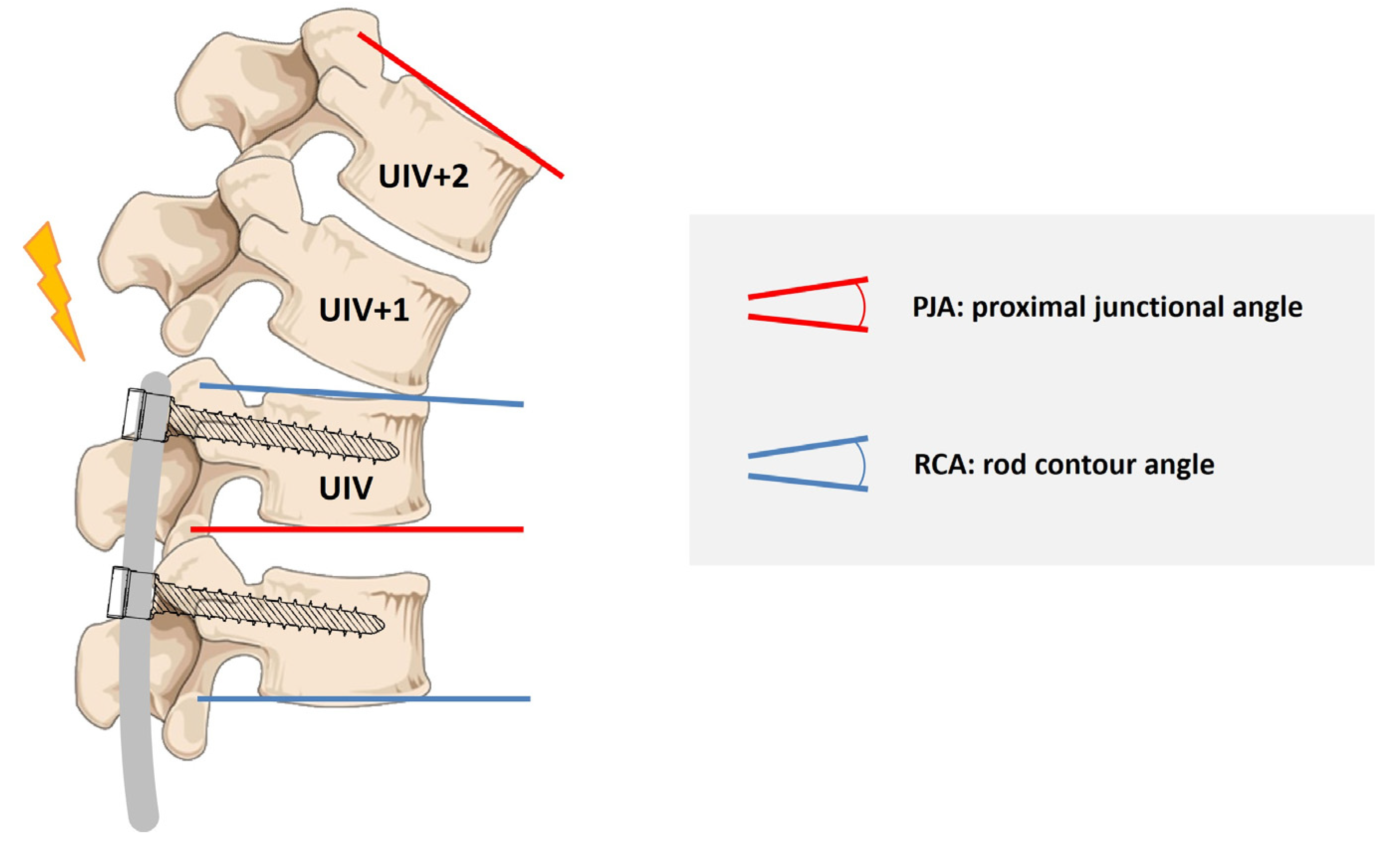

Table 1.
Demographic, radiographic, and surgical parameters. Values are mean ± standard deviation (SD) unless otherwise stated.
Table 1.
Demographic, radiographic, and surgical parameters. Values are mean ± standard deviation (SD) unless otherwise stated.
| non-PJK Group | PJK Group | p | |
| age | 16.6 ± 6.78 | 18.5 ± 9.36 | 0.260 |
| BMI | 18.7 ± 5.18 | 17.4 ± 4.78 | 0.282 |
| UIV (median) | T3 | T3 | 0.308 |
| LIV (median) | L5 | L5 | 0.244 |
| instrumented vertebra (n) | 13.8 ± 2.72 | 14.0 ± 2.47 | 0.770 |
| fused segments (n) | 12.7 ± 2.71 | 13.5 ± 2.09 | 0.190 |
| percentage of patients with resected spinous processes | 59.2% | 87.0% | 0.036 |
| resected spinous processes (n) | 7.2 ± 5.65 | 9.4 ± 4.26 | 0.101 |
| Cobb preOP (°) | 81.7 ± 26.38 | 80.7 ± 26.41 | 0.874 |
| Δ Cobb preOP vs. postOP (°) | -42.3 ± 17.84 | -44.3 ± 17.6 | 0.645 |
| TK preOP (°) | 34.5 ± 26.76 | 59.3 ± 29.04 | <0.001 |
| TK postOP (°) | 24.1 ± 14.11 | 38.0 ± 16.48 | <0.001 |
| TK 12 m FU (°) | 24.4 ± 15.15 | 39.2 ± 18.51 | <0.001 |
| Δ TK preOP v. postOP (°) | -10.7 ± 20.2 | −21.3 ± 19.28 | 0.032 |
| LL preOP (°) | 37.9 ± 28.11 | 41.4 ± 31.91 | 0.625 |
| LL postOP (°) | 37.1 ± 16.59 | 44.0 ± 11.58 | 0.067 |
| LL 12 m FU (°) | 37.7 ± 18.64 | 42.6 ± 17.23 | 0.286 |
| PI (°) | 54.6 ± 22.3 | 48.4 ± 10.55 | 0.486 |
| PT (°) | 10.2 ± 8.76 | 7.3 ± 3.35 | 0.387 |
| SL (°) | 44.4 ± 20.51 | 41.1 ± 11.74 | 0.691 |
| SVA preOP (cm) | 0.4 ± 5.98 | 0.6 ± 2.85 | 0.919 |
| SVA postOP (cm) | 0.1 ± 3.82 | 1.2 ± 5.09 | 0.671 |
| SVA 12 m FU (cm) | 1.0 ± 6.35 | -2.4 ± 2.85 | 0.139 |
| PJA preOP (°) | 3.7 ± 7.93 | 4.2 ± 7.22 | 0.777 |
| PJA postOP (°) | 5.9 ± 6.49 | 14.0 ± 6.31 | <0.001 |
| PJA 12 m FU (°) | 7.3 ± 8,91 | 21.3 ± 10.02 | <0.001 |
| RCA (°) | 7.7 ± 4.34 | 10.2 ± 4.01 | 0.021 |
| postOP PJA-RCA (°) | -1.8 ± 6.55 | 3.78 ± 6.75 | <0.001 |
Note. UIV, upper instrumented vertebra; LIV, lowest instrumented vertebra; TK, thoracic kyphosis (T4-12); LL, lumbar lordosis (L1-S1); PI, pelvic incidence; PT, pelvic tilt; SL, sacral slope; SVA, sagittal vertical axis; PJA, proximal junctional angle; RCA, rod contour angle; postOP PJA-RCA, mismatch between postOP PJA and RCA.
Table 2.
Pearson correlation matrix among predictors.
| 1. | 2. | 3. | 4. | 5. | 6. | |
| 1. spinous process resection | 1 | |||||
| 2. TK (preOP) |
0.044 | 1 | ||||
| 3. ΔTK (pre vs. postOP) |
0.088 | 0.862 ** | 1 | |||
| 4. RCA (postOP) |
0.001 | 0.338 ** | -0.218 * | 1 | ||
| 5. ΔPJA-RCA (postOP) |
0.142 | 0.005 | 0.014 | -0.240 * | 1 | |
| 6. PJK | 0.224 * | 0.377 ** | -0.235 * | 0.248 * | 0.355 ** | 1 |
* p < 0.05, ** p < 0.01. Note. ΔPJA-RCA (postOP), mismatch between postOP PJA and rod contour angle; RCA, rod contour angle; TK, thoracic kyphosis T4-T12; ΔTK, thoracic kyphosis preOP–thoracic kyphosis postOP.
Table 3.
Multivariate logistic regression analysis of risk factors for PJK.
| Estimate Coefficient | Odds Ratio(Exp(B)) | p | 95% Confidence Interval | ||
| spinous process resection | 1.848 | 6.346 | 0.036 | 0.769 | 52.396 |
| TK (preOP) | 0.063 | 1.065 | 0.017 | 1.011 | 1.121 |
| ΔTK (pre vs. postOP) | 0.043 | 1.043 | 0.175 | 0.981 | 1.110 |
| RCA (postOP) | 0.137 | 1.147 | 0.094 | 0.977 | 1.346 |
| ΔPJA-RCA (postOP) | 0.174 | 1.190 | 0.002 | 1.065 | 1.331 |
Note. ΔPJA-RCA (postOP), mismatch between postOP PJA and rod contour angle; RCA, rod contour angle; TK, thoracic kyphosis T4-T12; ΔTK, thoracic kyphosis preOP–thoracic kyphosis postOP.
Disclaimer/Publisher’s Note: The statements, opinions and data contained in all publications are solely those of the individual author(s) and contributor(s) and not of MDPI and/or the editor(s). MDPI and/or the editor(s) disclaim responsibility for any injury to people or property resulting from any ideas, methods, instructions or products referred to in the content. |
© 2025 by the authors. Licensee MDPI, Basel, Switzerland. This article is an open access article distributed under the terms and conditions of the Creative Commons Attribution (CC BY) license (http://creativecommons.org/licenses/by/4.0/).
Copyright: This open access article is published under a Creative Commons CC BY 4.0 license, which permit the free download, distribution, and reuse, provided that the author and preprint are cited in any reuse.


