Preprint
Article

This version is not peer-reviewed.

Interplay of PAK1 and CAMKII in Pancreatic Beta Cell Insulin Secretion

A peer-reviewed article of this preprint also exists.

Submitted:

08 March 2025

Posted:

11 March 2025

You are already at the latest version

Abstract

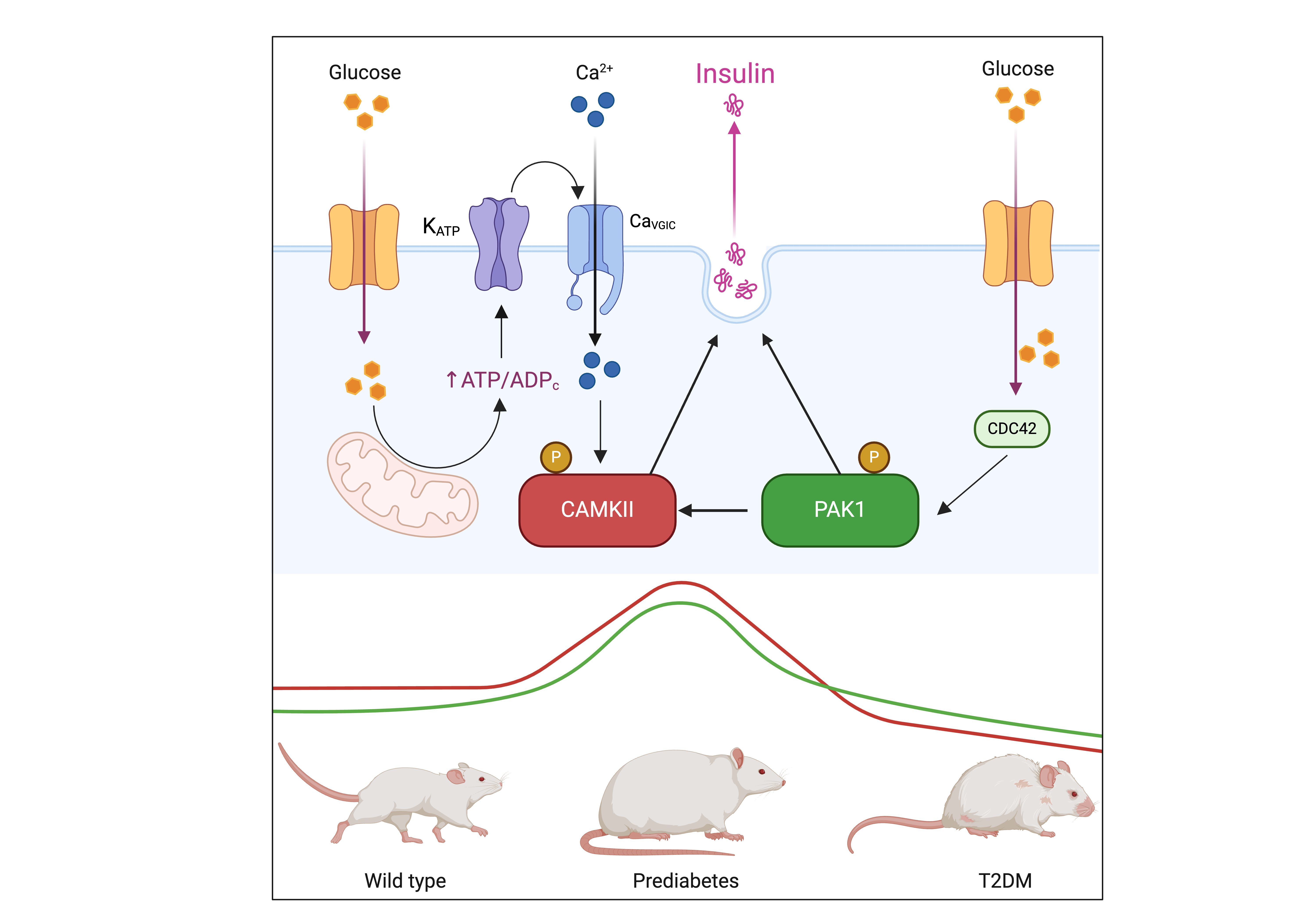
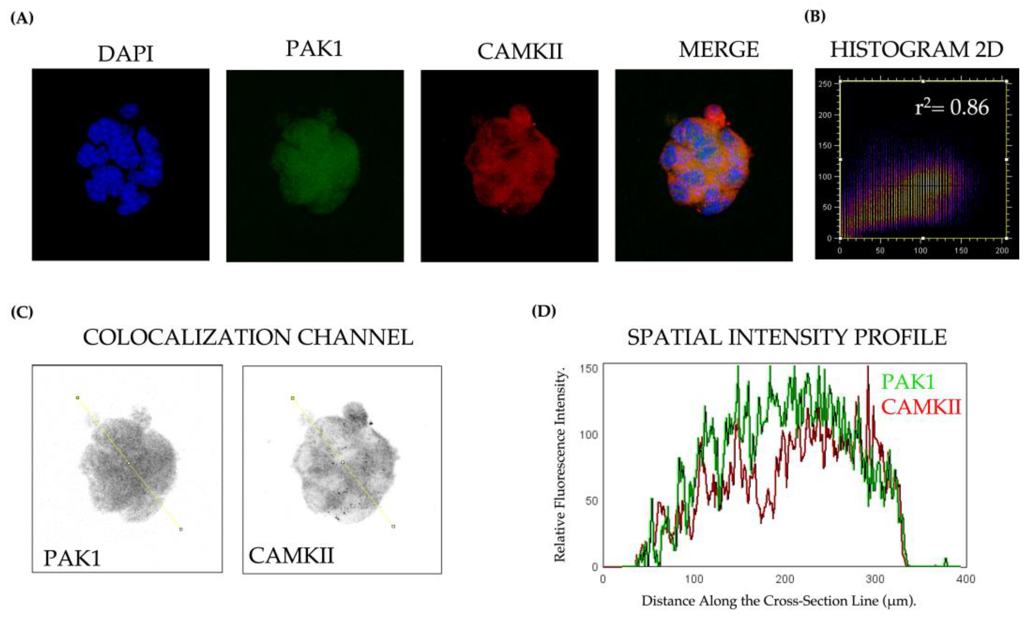
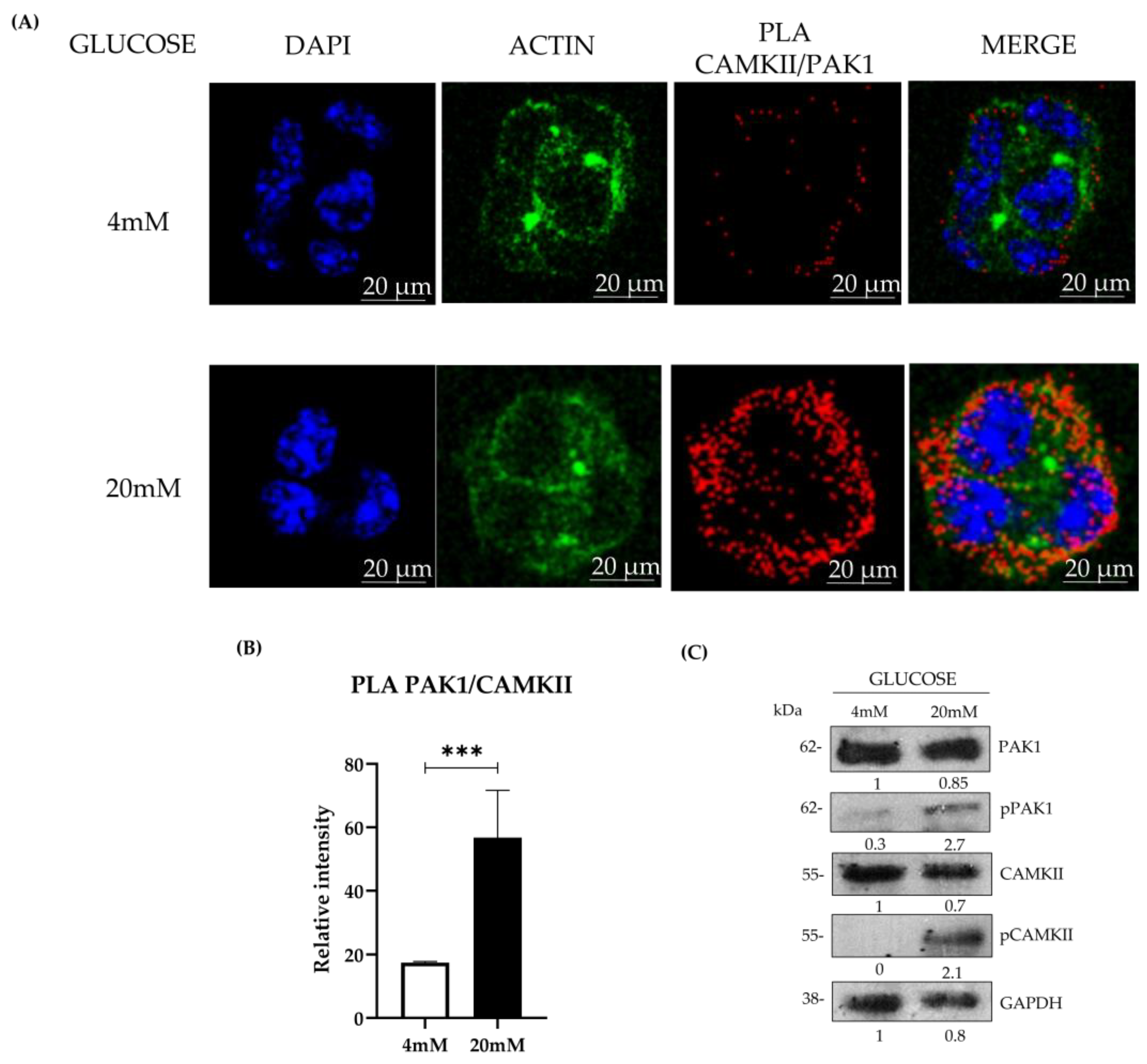
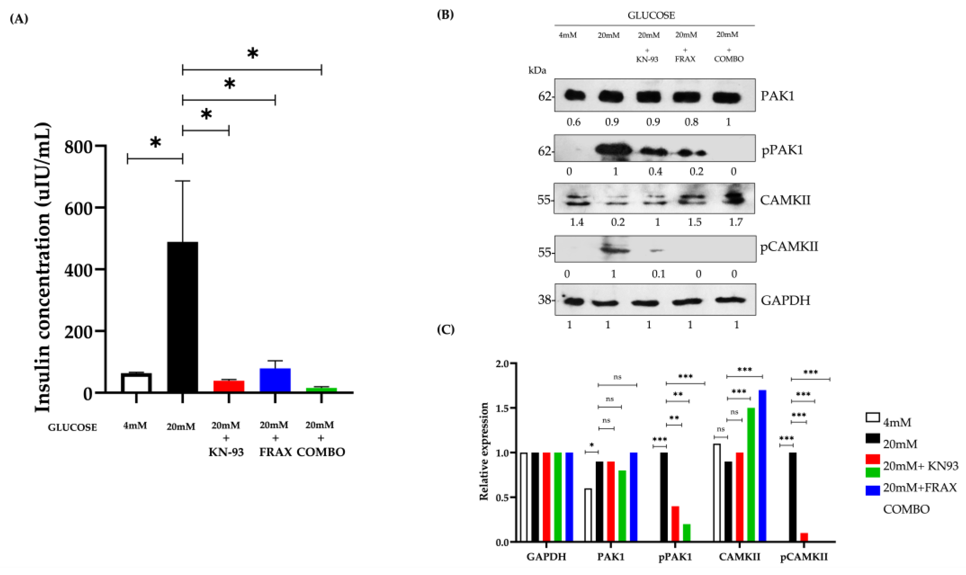
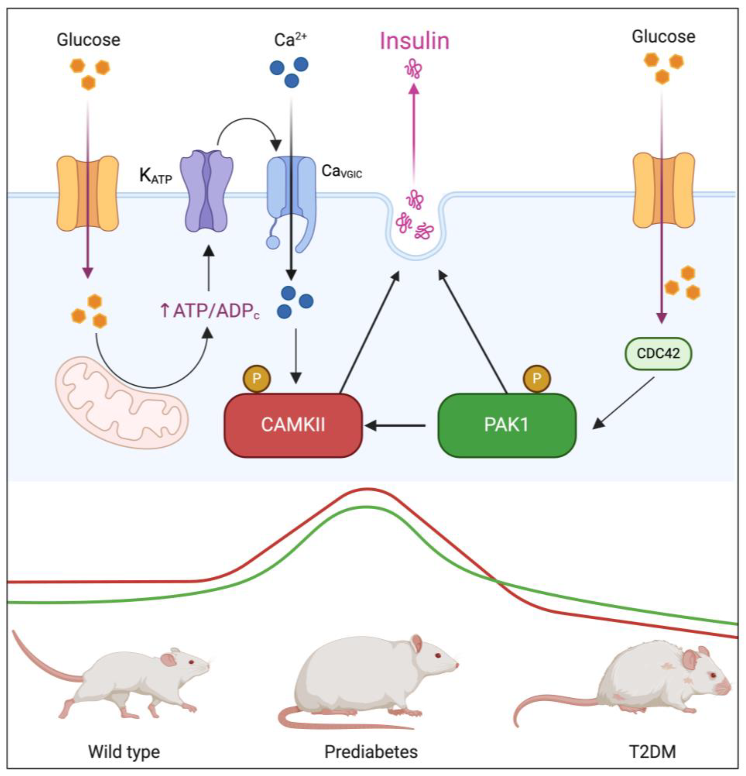
Background/Objectives: Type 2 diabetes mellitus (T2DM) is a major global health challenge, primarily driven by insulin resistance and beta-cell dysfunction. This study investigated the roles of p21-activated kinase 1 (PAK1) and calci-um/calmodulin-dependent protein kinase II (CAMKII) in insulin secretion, aiming to elucidate their involvement in this process and their implications in T2DM pathophysi-ology. Methods: Using the Beta-TC-6 insulinoma cell line, we assessed colocalization and interaction of PAK1 and CAMKII under glucose stimulation through indirect immuno-fluorescence (IFI) and proximity ligation assays (PLA). To examine their expression dynamics in a physiological context, we performed immunohistochemistry (IHC) on pancreatic sections from wild-type (WT), prediabetic, and T2DM murine models. Addi-tionally, bioinformatic analysis of publicly available RNA sequencing (RNA-Seq) data from human islets of healthy donors, prediabetic individuals, and T2DM patients pro-vided translational validation. Results: High glucose conditions significantly increased PAK1-CAMKII colocalization, correlating with enhanced insulin secretion. Pharmaco-logical inhibition of these kinases reduced insulin release, confirming their regulatory roles. Murine and human islet analyses showed a progressive increase in kinase ex-pression from prediabetes to T2DM, highlighting their relevance in disease progression. Conclusions: The coordinated function of PAK1 and CaMKII in insulin secretion suggests their potential as biomarkers and therapeutic targets in T2DM. Further studies are warranted to explore their mechanistic roles and therapeutic applications in preserving beta-cell function.

Keywords: 
;  ;  ;  ;  ;  

1. Introduction

Type 2 Diabetes Mellitus (T2DM) remains one of the most significant public health challenges globally, driven by rising obesity rates and sedentary lifestyles. Characterized by chronic hyperglycemia due to insulin resistance and progressive beta-cell dysfunction, which involves endoplasmic reticulum (ER) stress, mitochondrial dysfunction, oxidative stress, and chronic low-grade inflammation. These factors contribute to impaired insulin synthesis, vesicular trafficking, and increased beta-cell apoptosis, T2DM affects hundreds of millions worldwide and is associated with severe complications, including cardiovascular disease, renal failure, and blindness. Despite advances in diabetes management, the progressive decline in beta-cell function remains a critical barrier to long-term glycemic control, highlighting the need for novel therapeutic strategies [1,2,3].
Insulin secretion from pancreatic beta-cells is tightly regulated through a biphasic mechanism. The first phase involves a rapid release of pre-stored insulin granules in response to glucose intake, while the second phase consists of sustained insulin secretion, necessary for prolonged glucose homeostasis. Several well-characterized proteins orchestrate these processes, including ATP-sensitive potassium channels (K_ATP), SNARE complex proteins (Syntaxin-1, SNAP-25, VAMP2), and L-type and P/Q-type voltage-gated calcium channels, which regulate vesicle docking, priming, and fusion. However, recent evidence suggests that kinases involved in cytoskeletal remodeling and intracellular signaling, such as p21-activated kinase 1 (PAK1) and calcium/calmodulin-dependent protein kinase II (CAMKII), may also play fundamental roles in insulin granule mobilization and secretion [4,5,6,7].
PAK1 is a serine/threonine kinase activated by the small GTPases Cdc42 and Rac1, which regulates various physiological processes, including cytoskeletal remodeling, cell motility, and gene transcription [8,9,10]. Dysregulation of PAK1 has been implicated in oncogenesis, cardiovascular diseases, neurological disorders and, more recently, its role has begun to be explored in the exocrine pancreas, where it remains significantly under-studied, but its role in pancreatic beta-cells has only recently been explored [11,12,13,14]. Emerging evidence suggests that Pak1 is particularly important for the second phase of insulin secretion, where it regulates cytoskeletal remodeling and actin-dependent vesicle trafficking, facilitating insulin granule exocytosis in response to prolonged glucose stimulation. This has been demonstrated in PAK1 knockout mice, which exhibit selective impairments in the second phase of insulin release [7,15,16,17].
PAK1 knockout (PAK1⁻/⁻) mice exhibit significant impairments in glucose-stimulated insulin secretion, primarily affecting the sustained second phase. These mice develop fasting hyperglycemia, glucose intolerance, and a reduction in beta-cell mass, underscoring the essential role of PAK1 in beta-cell function. At the molecular level, PAK1 promotes insulin biogenesis by enhancing the transcription of PDX1, NEUROD1, and INS, contributing to increased insulin content and beta-cell survival under metabolic stress [18,19].
Calcium/calmodulin-dependent protein kinase II (CAMKII) is a multifunctional serine/threonine kinase that plays a central role in various cellular processes, including synaptic plasticity, vascular homeostasis, and metabolic regulation. In pancreatic beta-cells, CAMKII serves as a calcium sensor, amplifying Ca²⁺ influx triggered by glucose and other secretagogues, thereby enhancing insulin granule exocytosis [20,21,22]. CAMKII has been extensively studied in first-phase insulin secretion, where it facilitates the rapid fusion of insulin-containing vesicles with the plasma membrane. Pharmacological inhibition or genetic deletion of CAMKII in beta-cells significantly reduces insulin release and impairs glucose tolerance, highlighting its role in glucose-stimulated insulin secretion [23,24].
In the context of T2DM, chronic hyperglycemia is associated with persistent CAMKII activation in pancreatic islets and metabolic tissues. This prolonged activation contributes to beta-cell dysfunction, oxidative stress, and impaired insulin secretion, exacerbating diabetes progression. Moreover, CAMKII hyperactivation in the vascular system leads to vascular remodeling, endothelial dysfunction, and inflammation, further aggravating diabetes-related complications [25]. Our research group has demonstrated that PAK1 directly phosphorylates and activates CAMKII, suggesting a functional link between these kinases in insulin secretion [26]. Given that PAK1 is primarily implicated in the second phase of insulin secretion, while CAMKII is a key regulator of the first phase, their interaction may represent a previously unrecognized mechanism coordinating insulin granule mobilization across both phases. Understanding the PAK1-CAMKII axis could provide novel insights into beta-cell adaptation under metabolic stress and uncover potential therapeutic targets for preserving insulin secretion in T2DM.

2. Materials and Methods

2.1. Cell Culture and Glucose-Stimulated Insulin Secretion Assay (GSIS)

Beta-TC-6 murine insulinoma cells (ATCC, Manassas, VA, USA) were cultured in Dulbecco's Modified Eagle Medium High Glucose (DMEM, Gibco, Thermo Fisher Scientific, Waltham, MA, USA) supplemented with 15% fetal bovine serum (FBS), 50 U/mL penicillin, and 50 μg/mL streptomycin. Cells were maintained at 37°C in a humidified atmosphere containing 5% CO₂. For glucose-stimulated insulin secretion (GSIS) assays cells were preincubated for 4 hours in high-glucose DMEM (15% FBS) containing the kinase inhibitors FRAX-1036 (PAK1 inhibitor, 5 μM, MedChemExpress, Monmouth Junction, NJ, USA) and KN93 (CAMKII inhibitor, 20 μM, Sigma-Aldrich, St. Louis, MO, USA). Cell viability was assessed using the MTT assay after 24 hours of inhibitor treatment, confirming no significant reduction in viability compared to vehicle-treated controls (DMSO 0.1%) (p > 0.05, Student’s t-test). After preincubation, cells were washed with Krebs-Ringer buffer containing 0 mM glucose, and a supernatant sample was collected (baseline secretion). Subsequently, cells were incubated for 10 minutes in Krebs-Ringer buffer with 4 mM glucose, and another supernatant sample was collected (basal secretion). Finally, cells were exposed to either 4 mM or 20 mM glucose for 60 minutes, and a final supernatant sample was collected to measure glucose-stimulated insulin secretion. Insulin secretion was normalized to basal insulin levels. Experiments were performed using 50,000 cells per well to ensure appropriate protein content for ELISA normalization.

2.2. Immunofluorescence and Confocal Microscopy

Beta-TC-6 cells (5,000 per well) were plated onto sterile glass coverslips previously treated with poly-L-lysine (Sigma-Aldrich) to promote cell adhesion and maintained overnight in high-glucose DMEM supplemented with 15% FBS at 37°C in a humidified 5% CO₂ incubator. Cells were then fixed with 4% paraformaldehyde (PFA) for 15 minutes and permeabilized using 0.2% Triton X-100 for 10 minutes. After blocking with 5% bovine serum albumin (BSA) in phosphate-buffered saline (PBS) for 1 hour, cells were incubated with primary antibodies at room temperature for 2 hours. The following primary antibodies were used: PAK1 (1:500, Cat# 2602, Cell Signaling Technology, Boston, MA, USA), Phospho-PAK1 (Ser199/204, 1:500, Cat# 2601, Cell Signaling Technology), CAMKII (1:500, Cat# AB134041, Abcam, Cambridge, UK), Phospho-CAMKII (Thr287, 1:500, Cat# 3361, Cell Signaling Technology). After PBS washes, cells were incubated with Alexa Fluor 488 or 594-conjugated secondary antibodies (1:500, Thermo Fisher Scientific, Waltham, MA, USA) for 1 hour in the dark. Nuclei were counterstained with DAPI (Sigma-Aldrich), and images were captured using a Leica TCS SP8x confocal microscope (Leica Microsystems, Wetzlar, Germany). Colocalization analysis was performed using ImageJ software (NIH, Bethesda, MD, USA) with the Coloc2 plugin

2.3. Proximity Ligation Assay (PLA)

To assess Pak1-CaMKII interactions, Duolink® In Situ Detection Reagents (Sigma-Aldrich) were used per the manufacturer’s instructions. Beta-TC-6 cells (5,000 per well) were disposed in 16-well chamber slides (Nunc Lab-Tek, Thermo Scientific, Waltham, MA, 131 USA) previously treated with poly-L-lysine (Sigma-Aldrich) to promote cell adhesion and maintained overnight in high-glucose DMEM supplemented with 15% FBS at 37°C in a humidified 5% CO₂ incubator. Cells were then stimulated following the glucose-stimulated insulin secretion (GSIS) protocol. After glucose stimulation, cells were fixed, permeabilized, and blocked as described above. Cells were incubated overnight at 4°C with anti-Pak1 and anti-CaMKII primary antibodies. The following day, samples were incubated with species-specific PLA probes (anti-rabbit PLUS and anti-mouse MINUS, Olink Bioscience, Sigma-Aldrich) for 1 hour at 37°C, followed by ligation and amplification steps per the manufacturer’s protocol. For each experimental condition, at least 100 cells per field were analyzed from five randomly selected fields, and a minimum of 300 total cells were counted per condition. All experiments were conducted in three independent biological replicates (n = 3) to ensure reproducibility. Fluorescent signals were visualized using a Leica TCS SP8 confocal microscope, and PLA-positive signals were quantified using MetaMorph software (Molecular Devices, San Jose, 138 CA, USA).

2.4. Enzyme-Linked ImmunoSorbent Assay

Insulin secretion was quantified using a Mouse Insulin ELISA Kit (Thermo Scientific, USA) according to the manufacturer’s instructions. Absorbance was measured at 450 nm using a BioTek Epoch2 microplate reader (BioTek, Winooski, VT, USA). Insulin concentrations were normalized to total protein content, determined via quantification of total insulin per well using ELISA.

2.5. Western Blotting

Beta-TC-6 cells (50,000 per well) were preincubated with inhibitors for 4 hours in high-glucose DMEM (15% FBS) and subsequently stimulated following the glucose-stimulated insulin secretion (GSIS) protocol. After stimulation, cells were lysed in RIPA buffer (20 mM Tris-HCl pH 7.4, 150 mM NaCl, 1 mM EDTA, 1% Triton X-100, 0.5% SDS, 1% sodium deoxycholate) supplemented with protease (Sigma-Aldrich) and phosphatase inhibitors (PhosSTOP, Roche, Basel, Switzerland). Proteins (30 μg) were separated via SDS-PAGE and transferred onto Immobilon-P PVDF membranes (Millipore, Burlington, MA, USA). Membranes were blocked in 5% nonfat dried milk or 1% BSA in TBS-T, incubated with primary antibodies overnight at 4°C, followed by HRP-conjugated secondary antibodies for 1 hour at room temperature. Protein bands were visualized using the SuperSignal West Femto Maximum Sensitivity Substrate (Thermo Scientific, USA).

2.6. Animal Model of T2DM and Immunohistochemistry

All animal experiments were conducted following the ethical guidelines established by the Institutional Animal Care and Use Committee (IACUC) of Facultad de Estudios Superiores Iztacala, UNAM, with approval code CE/FESI/012022/1472. Male C57BL/6 mice (8 weeks old, n = 5 per group) were used to establish a murine model of Type 2 Diabetes Mellitus (T2DM). Mice in the wild-type (WT) control group were maintained on a normal chow diet, while those in the prediabetic (PD) group were fed a high-fat diet (HFD, 60% kcal from fat, 4.9 kcal/g) for 8 weeks to induce a prediabetic state. To further impair β-cell function and induce a T2DM model, a separate group of mice was fed an HFD for 12 weeks, followed by a single intraperitoneal injection of streptozotocin (STZ, 40 mg/kg). Diabetes onset was confirmed by fasting glucose evaluation (>200 mg/dL) and increased homeostatic model assessment of insulin resistance (HOMA-IR) values. After euthanasia, pancreata were collected, fixed with Bouin’s solution, and embedded in paraffin for histological analysis.

2.7. Immunohistochemistry.

Pancreatic tissue sections were deparaffinized, rehydrated, and subjected to antigen retrieval in sodium citrate buffer (0.01 M, pH 6.0). Endogenous peroxidase activity was blocked with 3% hydrogen peroxide, and sections were incubated overnight with anti-PAK1 (1:750) and anti-CAMKII (1:1000) antibodies. Visualization was performed using DAB substrate, counterstained with hematoxylin, and analyzed using ImageJ software.

2.8. Bioinformatic Analysis of RNA-Sequencing Data from Human Pancreatic Islets

Publicly available RNA-seq data from human pancreatic islets were retrieved from the Gene Expression Omnibus (GEO) database (Accession No: GSM5009229) as reported by Wigger et al. [27]. The dataset included samples from healthy donors (n = 15), prediabetic individuals (n = 11), and patients diagnosed with Type 2 Diabetes Mellitus (T2DM, n = 39). Differential gene expression analysis was performed using DESeq2 (R Bioconductor) to assess transcriptional changes associated with pancreatic β-cell function and diabetes progression. Raw read counts were normalized using the Trimmed Mean of M-values (TMM) method, and transcript abundances were expressed as transcripts per million (TPM) and reads per kilobase per million mapped reads (RPKM). Principal component analysis (PCA) was conducted to visualize sample clustering and assess batch effects. The analysis included the Pak kinase family genes (PAK1, ENSG00000149269; PAK2, ENSG00000180370; PAK3, ENSG00000077232; PAK4, ENSG00000163902; PAK5, ENSG00000101349; PAK6, ENSG00000137843) and the CAMKII isoforms (CAMK2A, ENSG00000070831; CAMK2B, ENSG00000152492; CAMK2G, ENSG00000157388; CAMK2D, ENSG00000182484). To further contextualize these findings, we examined the expression of genes critical to pancreatic β-cell function, glucose metabolism, and insulin signaling, including INS (ENSG00000254647), INSR (ENSG00000171105), MAFA (ENSG00000133048), PDX1 (ENSG00000139515), GCK (ENSG00000106633), NEUROD1 (ENSG00000100461), FOXO1 (ENSG00000150907), and PKM (ENSG00000067225). Gene expression levels were compared across non-diabetic, prediabetic, and T2DM groups to assess progressive transcriptional changes.

2.9. Statistical Analysis

Data are presented as mean ± standard error of the mean (SEM). Statistical significance was determined using one-way ANOVA followed by Dunnett’s post hoc test for multiple comparisons, or Student’s t-test for pairwise comparisons, as appropriate. Prior to statistical testing, normality was assessed using the Shapiro-Wilk test, and homogeneity of variances was evaluated with Levene’s test to ensure the validity of parametric analyses. For non-normally distributed data, the Kruskal-Wallis test followed by Dunn’s post hoc test was used instead of ANOVA. To evaluate the correlation between Pak1 and CaMKII colocalization, Pearson’s correlation coefficient (r²) was used for normally distributed data, whereas Spearman’s rank correlation (ρ) was applied when normality assumptions were not met. The statistical significance of the correlation was assessed for both tests. A p< 0.05 was considered statistically significant. All results shown are representative of three independent biological experiments (n = 3 per condition), with technical triplicates performed for each experimental group. Statistical analyses were conducted using GraphPad Prism 9 (GraphPad Software, San Diego, CA, USA).

3. Results

3.1. PAK1 and CAMKII Colocalization and Interaction in Response to Glucose Stimulation

To investigate the spatial and functional relationship between PAK1 and CAMKII in pancreatic beta cells, we first examined their colocalization under different glucose conditions using immunofluorescence microscopy. Beta-TC-6 cells were stained for PAK1 and CAMKII, revealing a strong overlap in fluorescence signals under high glucose conditions (Figure 1A). Quantification of colocalization through a 2D fluorescence intensity histogram confirmed a significant correlation between PAK1 and CAMKII signals (r² = 0.86, p<0.001, Figure 1B), suggesting a coordinated interaction within the cytoplasm of beta cells. Spatial intensity profiling further supported this observation, demonstrating co-enrichment of both kinases in specific intracellular compartments (Figure 1C,D). To determine whether the colocalization of PAK1 and CAMKII corresponds to a direct protein-protein interaction, we performed a Proximity Ligation Assay (PLA). Under basal glucose conditions (4 mM), PAK1-CAMKII interactions were minimal. However, exposure to high glucose (20 mM) significantly increased the PLA signal (Figure 2A), indicating a glucose-dependent enhancement of PAK1-CAMKII interactions. Quantitative analysis confirmed a robust increase in PLA puncta under hyperglycemic conditions, with a 3.2-fold increase (p< 0.001, one-way ANOVA) compared to basal glucose conditions (4 mM). The mean PLA signal intensity per cell was 18.7 ± 2.1 at 4 mM glucose vs. 62.5 ± 7.4 at 20 mM glucose, indicating a significant glucose-dependent enhancement of PAK1-CAMKII interactions. Given that PAK1 is a known kinase with diverse phosphorylation targets, we hypothesized that glucose stimulation might modulate PAK1 and CAMKII activity through phosphorylation events. Western blot analysis revealed a marked increase in the phosphorylation of PAK1 (pSer199/204) and CAMKII (pThr287) at 20 mM glucose, whereas total protein levels of these kinases remained unchanged (Figure 2C). These results suggest that high glucose not only promotes the physical interaction between PAK1 and CAMKII but also enhances their phosphorylation, potentially linking their activity to the regulation of insulin secretion regulation.

3.2. Dual Kinase Inhibition Disrupts Glucose-Stimulated Insulin Secretion

Given that glucose stimulation promotes the interaction and phosphorylation of PAK1 and CAMKII, we next investigated whether these kinases are functionally required for insulin secretion. We pharmacologically inhibited PAK1 and CAMKII using FRAX-1036 (4uM) and KN93 (20uM), respectively, and assessed their impact on insulin secretion and kinase activation. Insulin secretion was measured in cells incubated under basal (4 mM) and high (20 mM) glucose conditions, with or without kinase inhibitors. As expected, control cells exhibited a robust increase in insulin secretion at 20 mM glucose compared to 4 mM (Figure 3A). However, inhibition of either PAK1 or CAMKII significantly attenuated glucose-stimulated insulin release. Notably, combined inhibition of both kinases (COMBO) abolished insulin secretion, suggesting a cooperative role of these kinases in regulating beta-cell function. Western blot analysis (Figure 3B) revealed that high glucose significantly increased phosphorylation levels of PAK1 and CAMKII, consistent with their activation in response to metabolic stimulation. Treatment with KN93 specifically reduced pCAMKII levels, while FRAX-1036 suppressed PAK1 phosphorylation. Importantly, dual inhibition led to a near-complete loss of phosphorylation for both kinases, suggesting that PAK1 and CAMKII function as part of a coordinated signaling network. Densitometric quantification of protein levels (Figure 3C) confirmed that the effects of kinase inhibition were statistically significant. ANOVA with Dunnett’s post-hoc test demonstrated that while inhibition of PAK1 or CAMKII alone resulted in partial suppression of insulin secretion, combined inhibition led to near-complete abrogation. These findings underscore the essential role of PAK1 and CAMKII in glucose-stimulated insulin release and suggest that their interaction is critical for maintaining beta-cell functionality.

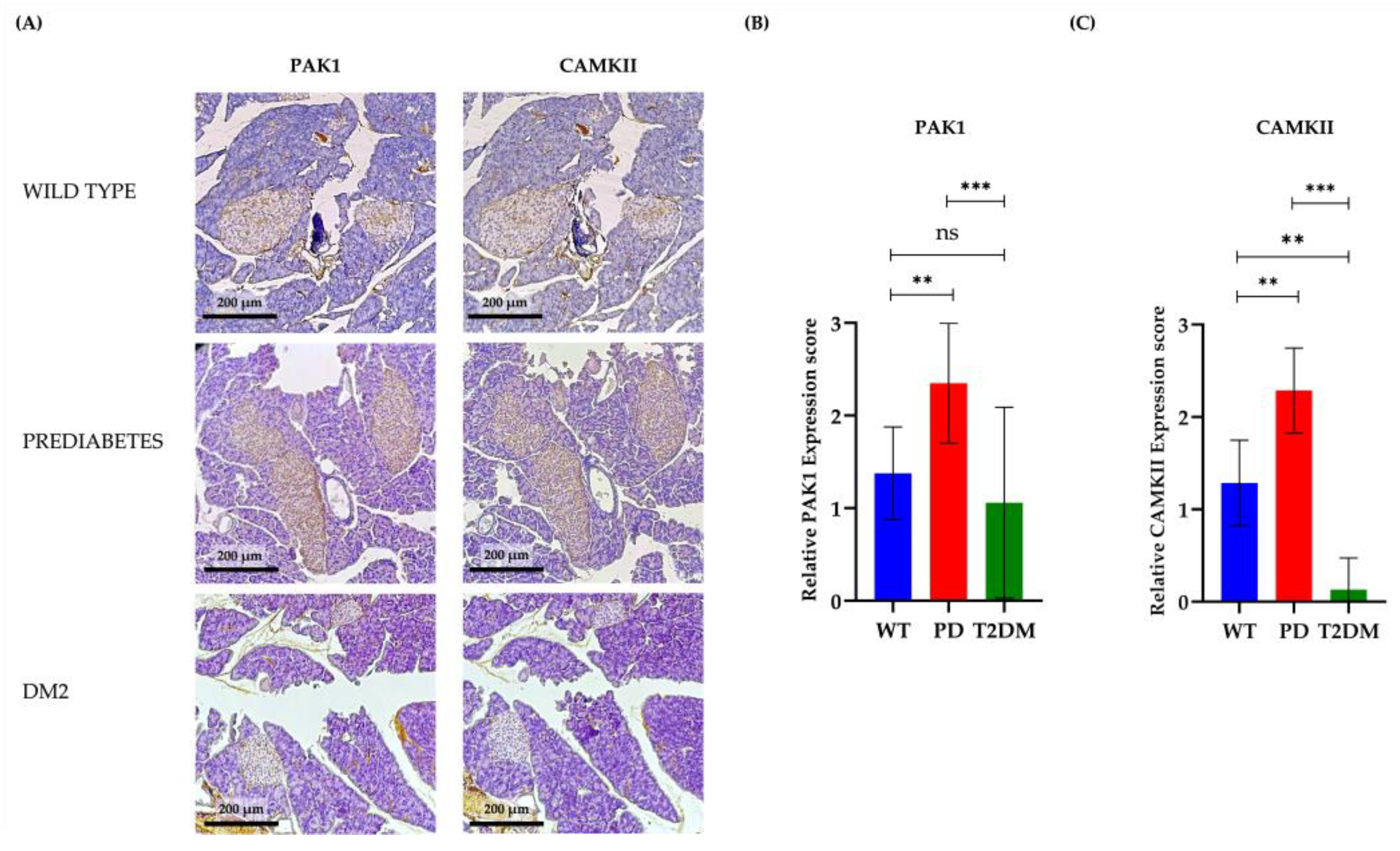
3.3. PAK1 and CAMKII Expression in Pancreatic Islets During Diabetes Progression

To determine the in vivo relevance of PAK1 and CAMKII in pancreatic islets during diabetes progression, we performed immunohistochemical (IHC) analysis on pancreatic sections from wild-type (WT), prediabetic (PD), and Type 2 Diabetes Mellitus (T2DM) murine models. Representative images of PAK1 and CAMKII staining are shown in Figure 4A. The IHC staining intensity of pancreatic sections stained for PAK1 and CaMKII was significantly elevated in PD compared to WT, reaching peak expression at this intermediate stage. This upregulation likely reflects a compensatory response to increased insulin demand during the hyperinsulinemic phase of diabetes progression. However, in T2DM mice, PAK1 and CAMKII expression were markedly reduced, falling below WT levels and coinciding with the decline in insulin secretion observed in advanced disease stages. These findings suggest that chronic metabolic stress and progressive β-cell dysfunction disrupt the PAK1/CAMKII regulatory axis, contributing to the transition from a compensatory to a decompensatory state in pancreatic islets.
Quantitative analysis of immunostaining intensity revealed a significant increase in PAK1 expression from WT to PD, with peak levels observed in the prediabetic state (Figure 4B). However, in T2DM, PAK1 expression declined sharply, indicating that its upregulation is transient and may be essential for β-cell compensation during early metabolic stress. A similar trend was observed for CAMKII (Figure 4C), where expression levels peaked in PD but were significantly downregulated in T2DM, further reinforcing the hypothesis that these kinases contribute to β-cell adaptation but become dysregulated in later disease stages.
The biphasic expression pattern of PAK1 and CAMKII suggests that these kinases play a crucial role in early β-cell adaptation to metabolic stress, sustaining insulin secretion under hyperglycemic conditions. However, their subsequent downregulation in T2DM implies that chronic hyperglycemia and disease progression impair their regulatory functions, ultimately contributing to β-cell failure. These findings highlight PAK1 and CAMKII as potential biomarkers of β-cell resilience and dysfunction in diabetes, offering novel insights into disease progression and therapeutic targets.

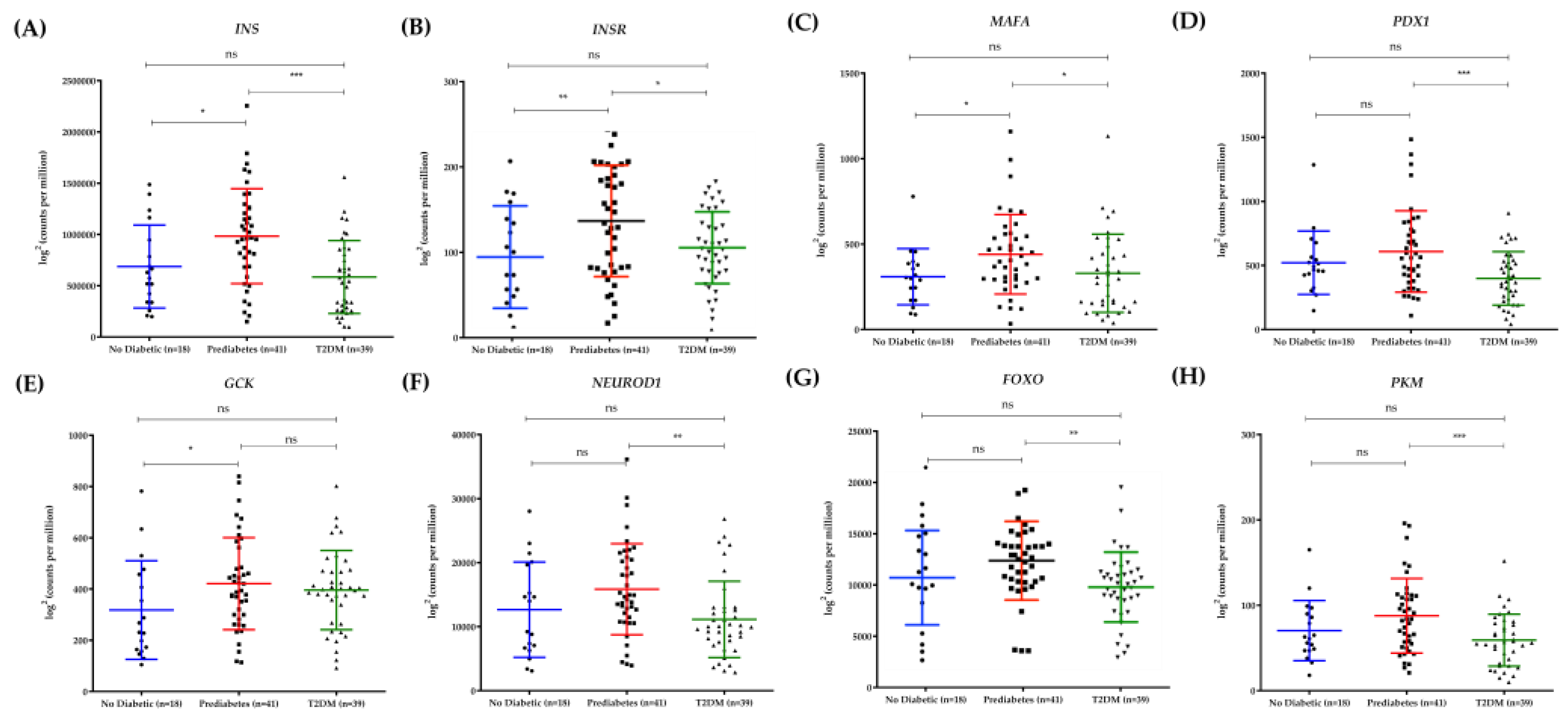
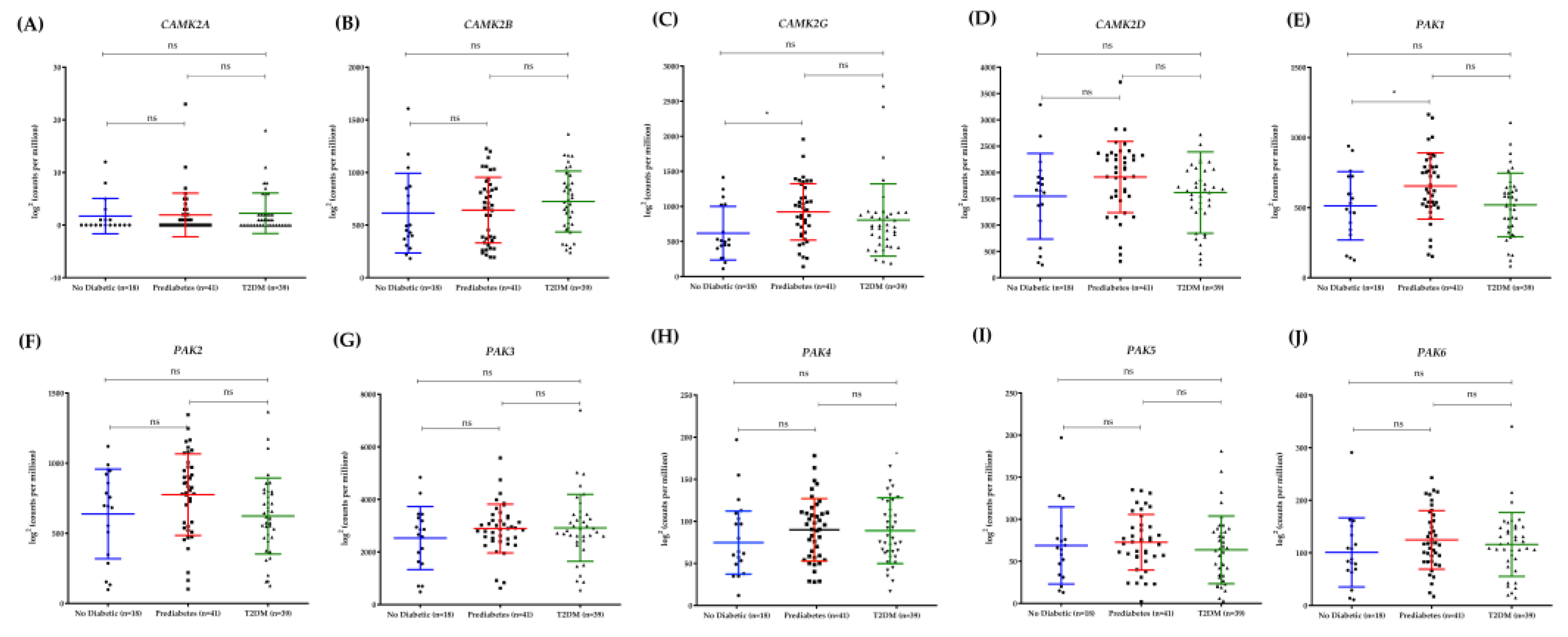
3.4. Differential Expression of PAK1 and CAMK2G in Human Pancreatic Islets Reflects Diabetes Progression

To extend our findings from murine models to human pathology, we analyzed publicly available RNA-seq data from pancreatic islets of non-diabetic individuals, prediabetic subjects, and patients with T2DM. Figure 5 illustrates the expression patterns of classical β-cell regulatory genes, including INS, INSR, MAFA, PDX1, GCK, NEUROD1, FOXO, and PKM (A-H). As expected, genes essential for β-cell function, such as INS and PDX1, exhibited a progressive decline in expression as hyperglycemia increased, reinforcing their critical role in diabetes early stage. Next, we examined the expression profiles of the CAMK2 (CAMK2A, CAMK2B, CAMK2G, and CAMK2D) and PAK (PAK1, PAK2, PAK3, PAK4, PAK5, and PAK6) gene families to assess their potential involvement in diabetes progression (Figure 6). Among the CAMK2 isoforms analyzed, only CAMK2G exhibited a significant expression pattern consistent with disease progression. Similarly, within the PAK family, PAK1 was the only isoform displaying a comparable trend. Both CAMK2G and PAK1 showed increased expression in prediabetic islets, aligning with the hyperinsulinemic compensatory phase, but exhibited a significant decline in T2DM islets, paralleling the loss of β-cell function observed in advanced disease.
The expression trajectory of CAMK2G and PAK1 closely mirrored that of canonical β-cell markers such as PDX1 and INS, further supporting their involvement in β-cell adaptation and failure. The consistency between murine and human data suggests a conserved regulatory mechanism in which CAMK2G and PAK1 contribute to insulin secretion and β-cell resilience under metabolic stress. These findings reinforce the hypothesis that dysregulation of these kinases may play a role in β-cell decompensation and T2DM progression, making them promising candidates for further investigation as potential therapeutic targets. To summarize our findings, we propose a mechanistic model illustrating the dynamic regulation of PAK1 and CAMKII during insulin secretion and diabetes progression. Figure 7 provides an overview of their functional interplay in β-cells, highlighting their biphasic expression pattern and the transition from compensation to decompensation during diabetes development.

4. Discussion

Type 2 Diabetes Mellitus (T2DM) is characterized by progressive β-cell dysfunction, leading to impaired insulin secretion and chronic hyperglycemia [28]. While numerous molecular pathways governing insulin exocytosis have been extensively studied, emerging evidence suggests that kinases such as PAK1 and CAMKII may play pivotal roles in β-cell adaptation and failure during metabolic stress [15,29]. Our findings highlight a glucose-dependent interaction between PAK1 and CAMKII, positioning these kinases as potential modulators of insulin secretion. Furthermore, the observed biphasic expression pattern of PAK1 and CAMKII in murine and human pancreatic islets suggests a transient compensatory role in early diabetes, followed by dysregulation in advanced disease stages.
Our data reveal a strong colocalization and interaction between PAK1 and CAMKII in pancreatic β-cells, which is significantly enhanced under hyperglycemic conditions. The increase in PLA signals under high glucose suggests a direct functional interplay between these kinases, potentially coordinating cytoskeletal remodeling and vesicle trafficking during insulin secretion [15]. Compared to other proteins involved in insulin granule mobilization, PAK1 shares functional similarities with Cdc42 and Rac1, which regulate actin polymerization and vesicle translocation [5]. CAMKII, on the other hand, operates in a complementary manner to synaptotagmins and Rab3, which modulate Ca²⁺-dependent vesicle priming and fusion with the plasma membrane [23]. This suggests that PAK1 and CAMKII act as integrators between cytoskeletal remodeling and Ca²⁺ signaling, a mechanism distinct from SNARE proteins that primarily facilitate vesicle docking.
Further supporting this notion, our western blot analyses indicate that glucose stimulation leads to the phosphorylation of both PAK1 and CAMKII, correlating with increased insulin secretion. The inhibition of either kinase significantly blunted glucose-stimulated insulin release, reinforcing their critical roles in β-cell function. Quantitative analysis demonstrated that individual inhibition of CaMKII (KN-93) or PAK1 (FRAX-1036) led to a ~75% and ~70% reduction in insulin secretion, respectively (p < 0.01). However, dual inhibition resulted in an ~90% suppression of insulin release (p < 0.001), a reduction greater than the expected additive effect. This suggests a synergistic relationship between these kinases in β-cell insulin secretion, rather than a simple parallel contribution of independent pathways (Figure 3A). These results align with previous findings showing that Pak1 knockout mice develop glucose intolerance and defective insulin secretion due to impaired cytoskeletal reorganization and granule trafficking [18]. Likewise, pharmacological inhibition of CaMKII has been associated with attenuated first-phase insulin secretion and β-cell dysfunction [30]. Our study extends these findings by providing direct evidence that simultaneous inhibition of PAK1 and CAMKII leads to a more severe defect in insulin secretion than single inhibition, reinforcing the concept that both kinases work in a coordinated manner to regulate β-cell physiology. Further investigation is required to determine whether this synergy involves direct interaction between the kinases or a shared downstream effector controlling insulin granule exocytosis.
The immunohistochemical analysis of murine pancreatic islets revealed a striking biphasic expression pattern of PAK1 and CAMKII during diabetes progression. In prediabetic mice, expression levels were significantly elevated compared to wild-type controls, suggesting an adaptive response to increased insulin demand during the hyperinsulinemic phase [31,32]. However, in T2DM mice, both kinases were markedly downregulated, correlating with β-cell failure and reduced insulin secretion. This pattern was mirrored in our analysis of human pancreatic islets, where PAK1 and CAMKII expression peaked in prediabetic individuals before declining in T2DM patients. These findings align with previous reports demonstrating that β-cell compensation during early diabetes involves transient upregulation of key signaling pathways, which eventually become dysfunctional as metabolic stress progresses. While most functional studies on PAK1 and CAMKII in insulin secretion have been conducted in murine models and insulinoma cell lines, our RNA-Seq analysis of human pancreatic islets demonstrated that PAK1 is differentially expressed in prediabetes and T2DM, suggesting that its regulation is also relevant in humans. However, the functional role of PAK1 in human β-cells remains largely unexplored. Future studies should aim to validate these findings in primary human islets and evaluate whether PAK1 modulation can restore insulin secretion in T2DM. Additionally, the interplay between PAK1, CAMKII, and other key β-cell regulators such as GLP-1 signaling warrants further investigation to determine its therapeutic potential. [27,33].
The observed decline in PAK1 and CAMKII expression in T2DM may be attributed to chronic hyperglycemia-induced β-cell exhaustion. Sustained metabolic stress has been shown to impair kinase signaling pathways, leading to defects in insulin secretion and β-cell apoptosis [34]. Additionally, oxidative stress and glucotoxicity have been implicated in the degradation of cytoskeletal proteins and calcium signaling components, which may further contribute to the observed downregulation of these kinases [35]. Understanding the molecular mechanisms underlying this transition from compensation to decompensation could provide valuable insights into β-cell failure in T2DM.
Given the critical roles of PAK1 and CAMKII in insulin secretion, their modulation may represent a novel therapeutic avenue for preserving β-cell function in T2DM. Previous studies have explored the potential of kinase activators in enhancing insulin secretion, with promising results in preclinical models [36,37]. Pharmacological activation of PAK1 has been shown to improve β-cell survival and insulin release under metabolic stress conditions [18]. Likewise, selective CAMKII modulators have been proposed to restore β-cell calcium homeostasis and insulin granule mobilization in diabetic models [23]. However, chronic CAMKII hyperactivation has been linked to β-cell apoptosis and oxidative stress, underscoring the need for precise therapeutic modulation [38]. Future studies should investigate the feasibility of targeting these kinases with selective agonists or inhibitors to fine-tune their activity and restore β-cell function in T2DM.
Additionally, the potential of PAK1 and CAMKII as biomarkers for β-cell resilience warrants further exploration. Their early upregulation in prediabetes suggests that they may serve as indicators of β-cell compensatory capacity, helping to identify individuals at risk of disease progression before β-cell failure becomes irreversible. Longitudinal studies analyzing kinase expression in relation to metabolic parameters could provide valuable predictive insights into diabetes progression.
While our study provides compelling evidence for the functional interplay between PAK1 and CAMKII in β-cell physiology, several limitations should be acknowledged. First, although we demonstrated kinase colocalization and interaction using PLA and immunofluorescence, further biochemical analyses such as co-immunoprecipitation are needed to confirm direct protein-protein interactions. Second, our study primarily relies on pharmacological inhibition, which may have off-target effects; future investigations using β-cell-specific knockout models for PAK1 and CAMKII would provide more definitive insights. Finally, while our human islet analysis highlights the clinical relevance of these kinases, functional studies in human β-cells are necessary to validate their roles in insulin secretion and diabetes pathophysiology.
Future research should explore the molecular mechanisms underlying PAK1-mediated CAMKII activation, particularly the downstream targets involved in β-cell survival and insulin exocytosis. Additionally, studies investigating the effects of targeted kinase modulation in in vivo diabetes models could pave the way for novel therapeutic strategies aimed at preserving β-cell function in T2DM.

5. Conclusions

Our findings identify PAK1 and CAMKII as key regulators of insulin secretion, exhibiting a biphasic protein profile during diabetes progression. Their early upregulation suggests a role in β-cell compensation, while their subsequent decline in T2DM implicates them in β-cell dysfunction. The glucose-dependent interaction between these kinases highlights a novel regulatory mechanism integrating cytoskeletal remodeling and calcium dynamics in insulin secretion. Targeting PAK1 and CAMKII may represent a promising strategy for preserving β-cell function and improving metabolic outcomes in T2DM.

Author Contributions

Conceptualization, Jazmín García-Machorro, Luis Arias-Romero and Héctor Saldívar-Cerón; Data curation, Nely López-Desiderio, Luis Arias-Romero and Héctor Saldívar-Cerón; Formal analysis, Nely López-Desiderio, Genaro Patiño-López, Citlaltépetl Salinas-Lara, Carlos Sánchez-Garibay, Olga Villamar-Cruz, Alonso Vilches-Flores and Héctor Saldívar-Cerón; Funding acquisition, Luis Arias-Romero and Héctor Saldívar-Cerón; Investigation, Nely López-Desiderio, Genaro Patiño-López, Citlaltépetl Salinas-Lara, Carlos Sánchez-Garibay, Olga Villamar-Cruz, Alonso Vilches-Flores, Leonel Armas-López, Luis Arias-Romero and Héctor Saldívar-Cerón; Methodology, Nely López-Desiderio, Genaro Patiño-López, Citlaltépetl Salinas-Lara, Carlos Sánchez-Garibay, Olga Villamar-Cruz, Alonso Vilches-Flores and Héctor Saldívar-Cerón; Project administration, Luis Arias-Romero and Héctor Saldívar-Cerón; Resources, Jazmín García-Machorro, Luis Arias-Romero and Héctor Saldívar-Cerón; Software, José Peralta-Romero and Héctor Saldívar-Cerón; Supervision, Luis Arias-Romero and Héctor Saldívar-Cerón; Validation, Nely López-Desiderio, Luis Arias-Romero and Héctor Saldívar-Cerón; Visualization, Genaro Patiño-López, Citlaltépetl Salinas-Lara, Carlos Sánchez-Garibay, Olga Villamar-Cruz, Alonso Vilches-Flores and Héctor Saldívar-Cerón; Writing – original draft, Nely López-Desiderio, José Peralta-Romero, Leonel Armas-López and Héctor Saldívar-Cerón; Writing – review & editing, Jazmín García-Machorro, Luis Arias-Romero and Héctor Saldívar-Cerón.

Funding

This research was supported by the Consejo Mexiquense de Ciencia y Tecnología (COMECYT) under grant FICDTEM-2023-131 and by the Universidad Nacional Autónoma de México (UNAM) through PAPIIT IA201725 and PAPIME 203825, awarded to HISC. Additionally, funding was provided by COMECYT (grant FICDTEM-2023-130) and UNAM (PAPIIT IN211022) to LEAR.

Institutional Review Board Statement

The animal study protocol was approved by the Institutional Animal Care and Use Committee (IACUC) of Facultad de Estudios Superiores Iztacala, UNAM, with approval code CE/FESI/012022/1472, approved in January 2022.

Data Availability Statement

Data will be made available upon reasonable request by accredited researchers

Acknowledgments

We sincerely thank Luz María Del Razo Jiménez for generously providing the Beta-TC-6 cell line used in this study. Additionally, we extend our gratitude to Luz del Carmen Sánchez Peña for her valuable technical and administrative support. Their contributions were instrumental in the successful execution of our experiments.

Conflicts of Interest

The authors declare no conflicts of interest

Abbreviations

The following abbreviations are used in this manuscript:
T2DM Type 2 Diabetes Mellitus
PAK1 P21-activated kinase 1
CAMKII Calcium/calmodulin-dependent protein kinase II
GSIS Glucose-stimulated insulin secretion
PLA Proximity ligation assay
PD Prediabetes
INS Insulin gene
INSR Insulin receptor gene
MAFA Maf bZIP transcription factor A gene
PDX1 Pancreatic and duodenal homeobox 1 gene
GCK Glucokinase gene
NEUROD1 Neurogenic differentiation factor 1 gene
FOXO Forkhead box O gene
PKM Pyruvate kinase muscle isoform gene

References

  1. DeFronzo, R.A.; Ferrannini, E.; Groop, L.; Henry, R.R.; Herman, W.H.; Holst, J.J.; Hu, F.B.; Kahn, C.R.; Raz, I.; Shulman, G.I.; et al. Type 2 diabetes mellitus. Nature reviews. Disease primers 2015, 1, 15019. [CrossRef]
  2. Rutter, G.A.; Hodson, D.J. Beta cell connectivity in pancreatic islets: a type 2 diabetes target? Cellular and molecular life sciences : CMLS 2015, 72, 453-467. [CrossRef]
  3. Nauck, M.A.; Meier, J.J. The incretin effect in healthy individuals and those with type 2 diabetes: physiology, pathophysiology, and response to therapeutic interventions. The lancet. Diabetes & endocrinology 2016, 4, 525-536. [CrossRef]
  4. Rorsman, P.; Eliasson, L.; Renström, E.; Gromada, J.; Barg, S.; Göpel, S. The Cell Physiology of Biphasic Insulin Secretion. News in physiological sciences : an international journal of physiology produced jointly by the International Union of Physiological Sciences and the American Physiological Society 2000, 15, 72-77. [CrossRef]
  5. Wang, Z.; Thurmond, D.C. Mechanisms of biphasic insulin-granule exocytosis - roles of the cytoskeleton, small GTPases and SNARE proteins. Journal of cell science 2009, 122, 893-903. [CrossRef]
  6. Nie, J.; Sun, C.; Faruque, O.; Ye, G.; Li, J.; Liang, Q.; Chang, Z.; Yang, W.; Han, X.; Shi, Y. Synapses of amphids defective (SAD-A) kinase promotes glucose-stimulated insulin secretion through activation of p21-activated kinase (PAK1) in pancreatic β-Cells. The Journal of biological chemistry 2012, 287, 26435-26444. [CrossRef]
  7. Kalwat, M.A.; Yoder, S.M.; Wang, Z.; Thurmond, D.C. A p21-activated kinase (PAK1) signaling cascade coordinately regulates F-actin remodeling and insulin granule exocytosis in pancreatic β cells. Biochemical pharmacology 2013, 85, 808-816. [CrossRef]
  8. Kumar, R.; Sanawar, R.; Li, X.; Li, F. Structure, biochemistry, and biology of PAK kinases. Gene 2017, 605, 20-31. [CrossRef]
  9. Semenova, G.; Chernoff, J. Targeting PAK1. Biochemical Society transactions 2017, 45, 79-88. [CrossRef]
  10. Radu, M.; Semenova, G.; Kosoff, R.; Chernoff, J. PAK signalling during the development and progression of cancer. Nature reviews. Cancer 2014, 14, 13-25. [CrossRef]
  11. Kichina, J.V.; Goc, A.; Al-Husein, B.; Somanath, P.R.; Kandel, E.S. PAK1 as a therapeutic target. Expert opinion on therapeutic targets 2010, 14, 703-725. [CrossRef]
  12. Wang, Y.; Wang, S.; Lei, M.; Boyett, M.; Tsui, H.; Liu, W.; Wang, X. The p21-activated kinase 1 (Pak1) signalling pathway in cardiac disease: from mechanistic study to therapeutic exploration. British journal of pharmacology 2018, 175, 1362-1374. [CrossRef]
  13. Kreis, P.; Barnier, J.V. PAK signalling in neuronal physiology. Cellular signalling 2009, 21, 384-393. [CrossRef]
  14. Ramos-Alvarez, I.; Jensen, R.T. The Important Role of p21-Activated Kinases in Pancreatic Exocrine Function. Biology 2025, 14. [CrossRef]
  15. Wang, Z.; Oh, E.; Thurmond, D.C. Glucose-stimulated Cdc42 signaling is essential for the second phase of insulin secretion. The Journal of biological chemistry 2007, 282, 9536-9546. [CrossRef]
  16. Sylow, L.; Kleinert, M.; Pehmøller, C.; Prats, C.; Chiu, T.T.; Klip, A.; Richter, E.A.; Jensen, T.E. Akt and Rac1 signaling are jointly required for insulin-stimulated glucose uptake in skeletal muscle and downregulated in insulin resistance. Cellular signalling 2014, 26, 323-331. [CrossRef]
  17. Sylow, L.; Jensen, T.E.; Kleinert, M.; Højlund, K.; Kiens, B.; Wojtaszewski, J.; Prats, C.; Schjerling, P.; Richter, E.A. Rac1 signaling is required for insulin-stimulated glucose uptake and is dysregulated in insulin-resistant murine and human skeletal muscle. Diabetes 2013, 62, 1865-1875. [CrossRef]
  18. Ahn, M.; Yoder, S.M.; Wang, Z.; Oh, E.; Ramalingam, L.; Tunduguru, R.; Thurmond, D.C. The p21-activated kinase (PAK1) is involved in diet-induced beta cell mass expansion and survival in mice and human islets. Diabetologia 2016, 59, 2145-2155. [CrossRef]
  19. Kořánová, T.; Dvořáček, L.; Grebeňová, D.; Röselová, P.; Obr, A.; Kuželová, K. PAK1 and PAK2 in cell metabolism regulation. Journal of cellular biochemistry 2022, 123, 375-389. [CrossRef]
  20. Yasuda, R.; Hayashi, Y.; Hell, J.W. CaMKII: a central molecular organizer of synaptic plasticity, learning and memory. Nature reviews. Neuroscience 2022, 23, 666-682. [CrossRef]
  21. Reyes Gaido, O.E.; Nkashama, L.J.; Schole, K.L.; Wang, Q.; Umapathi, P.; Mesubi, O.O.; Konstantinidis, K.; Luczak, E.D.; Anderson, M.E. CaMKII as a Therapeutic Target in Cardiovascular Disease. Annual review of pharmacology and toxicology 2023, 63, 249-272. [CrossRef]
  22. Richter, E.A.; Hargreaves, M. Exercise, GLUT4, and skeletal muscle glucose uptake. Physiological reviews 2013, 93, 993-1017. [CrossRef]
  23. Dadi, P.K.; Vierra, N.C.; Ustione, A.; Piston, D.W.; Colbran, R.J.; Jacobson, D.A. Inhibition of pancreatic β-cell Ca2+/calmodulin-dependent protein kinase II reduces glucose-stimulated calcium influx and insulin secretion, impairing glucose tolerance. The Journal of biological chemistry 2014, 289, 12435-12445. [CrossRef]
  24. Santos, G.J.; Ferreira, S.M.; Ortis, F.; Rezende, L.F.; Li, C.; Naji, A.; Carneiro, E.M.; Kaestner, K.H.; Boschero, A.C. Metabolic memory of ß-cells controls insulin secretion and is mediated by CaMKII. Molecular metabolism 2014, 3, 484-489. [CrossRef]
  25. Hegyi, B.; Bers, D.M.; Bossuyt, J. CaMKII signaling in heart diseases: Emerging role in diabetic cardiomyopathy. Journal of molecular and cellular cardiology 2019, 127, 246-259. [CrossRef]
  26. Saldivar-Cerón, H.I.; Villamar-Cruz, O.; Wells, C.M.; Oguz, I.; Spaggiari, F.; Chernoff, J.; Patiño-López, G.; Huerta-Yepez, S.; Montecillo-Aguado, M.; Rivera-Pazos, C.M.; et al. p21-Activated Kinase 1 Promotes Breast Tumorigenesis via Phosphorylation and Activation of the Calcium/Calmodulin-Dependent Protein Kinase II. Frontiers in cell and developmental biology 2021, 9, 759259. [CrossRef]
  27. Wigger, L.; Barovic, M.; Brunner, A.D.; Marzetta, F.; Schöniger, E.; Mehl, F.; Kipke, N.; Friedland, D.; Burdet, F.; Kessler, C.; et al. Multi-omics profiling of living human pancreatic islet donors reveals heterogeneous beta cell trajectories towards type 2 diabetes. Nature metabolism 2021, 3, 1017-1031. [CrossRef]
  28. Seino, S.; Shibasaki, T.; Minami, K. Dynamics of insulin secretion and the clinical implications for obesity and diabetes. The Journal of clinical investigation 2011, 121, 2118-2125. [CrossRef]
  29. Illario, M.; Monaco, S.; Cavallo, A.L.; Esposito, I.; Formisano, P.; D'Andrea, L.; Cipolletta, E.; Trimarco, B.; Fenzi, G.; Rossi, G.; et al. Calcium-calmodulin-dependent kinase II (CaMKII) mediates insulin-stimulated proliferation and glucose uptake. Cellular signalling 2009, 21, 786-792. [CrossRef]
  30. Li, G.; Hidaka, H.; Wollheim, C.B. Inhibition of voltage-gated Ca2+ channels and insulin secretion in HIT cells by the Ca2+/calmodulin-dependent protein kinase II inhibitor KN-62: comparison with antagonists of calmodulin and L-type Ca2+ channels. Molecular pharmacology 1992, 42, 489-488. [CrossRef]
  31. Keane, K.N.; Cruzat, V.F.; Carlessi, R.; de Bittencourt, P.I., Jr.; Newsholme, P. Molecular Events Linking Oxidative Stress and Inflammation to Insulin Resistance and β-Cell Dysfunction. Oxidative medicine and cellular longevity 2015, 2015, 181643. [CrossRef]
  32. Luc, K.; Schramm-Luc, A.; Guzik, T.J.; Mikolajczyk, T.P. Oxidative stress and inflammatory markers in prediabetes and diabetes. Journal of physiology and pharmacology : an official journal of the Polish Physiological Society 2019, 70. [CrossRef]
  33. Dor, Y.; Brown, J.; Martinez, O.I.; Melton, D.A. Adult pancreatic beta-cells are formed by self-duplication rather than stem-cell differentiation. Nature 2004, 429, 41-46. [CrossRef]
  34. Weir, G.C.; Bonner-Weir, S. Five stages of evolving beta-cell dysfunction during progression to diabetes. Diabetes 2004, 53 Suppl 3, S16-21. [CrossRef]
  35. Khalid, M.; Alkaabi, J.; Khan, M.A.B.; Adem, A. Insulin Signal Transduction Perturbations in Insulin Resistance. International journal of molecular sciences 2021, 22. [CrossRef]
  36. Kaihara, K.A.; Dickson, L.M.; Jacobson, D.A.; Tamarina, N.; Roe, M.W.; Philipson, L.H.; Wicksteed, B. β-Cell-specific protein kinase A activation enhances the efficiency of glucose control by increasing acute-phase insulin secretion. Diabetes 2013, 62, 1527-1536. [CrossRef]
  37. Oakie, A.; Wang, R. β-Cell Receptor Tyrosine Kinases in Controlling Insulin Secretion and Exocytotic Machinery: c-Kit and Insulin Receptor. Endocrinology 2018, 159, 3813-3821. [CrossRef]
  38. Dixit, S.S.; Wang, T.; Manzano, E.J.; Yoo, S.; Lee, J.; Chiang, D.Y.; Ryan, N.; Respress, J.L.; Yechoor, V.K.; Wehrens, X.H. Effects of CaMKII-mediated phosphorylation of ryanodine receptor type 2 on islet calcium handling, insulin secretion, and glucose tolerance. PloS one 2013, 8, e58655. [CrossRef]
Figure 1. Colocalization and Interaction Analysis of PAK1 and CAMKII in Beta-TC-6 Cells. Panel A displays immunofluorescence labeling with DAPI (blue) for nuclei, anti-PAK1 (green), and anti-CAMKII (red), highlighting their distribution within the cells; the merge illustrates significant colocalization. Panel B shows a 2D histogram of fluorescence intensity with a Pearson correlation coefficient (r² = 0.86), indicating a strong overlap between PAK1 and CAMKII signals. Panel C presents grayscale images of PAK1 and CAMKII, with a marked line indicating the region analyzed for spatial intensity profiling. Panel D offers a spatial intensity profile along the indicated line in Panel C, where peaks represent regions of high protein concentration, demonstrating the interaction zones of PAK1 and CAMKII.
Preprints 151702 g001
Figure 2. Influence of Glucose Concentration on PAK1 and CAMKII Interaction and Phosphorylation. Panel A. Immunofluorescence and Proximity Ligation Assay (PLA) Visualization: Cells cultured under low (4 mM) and high (20 mM) glucose conditions stained with DAPI (blue) for nuclei, Actin (green) to outline cell structure, and PLA for PAK1/CAMKII interaction (red). Merged images show the localization and interaction between CAMKII and PAK1 within the cells. Panel B. Quantitative Analysis of PLA Signals (***p < 0.001). Panel C. Western Blot Analysis of Protein Expression and Phosphorylation. GAPDH is used as a loading control to confirm equal protein loading across samples.
Preprints 151702 g002
Figure 3. Dual Kinase Inhibition Attenuates Insulin Secretion in Response to High Glucose. Panel A. Insulin Secretion Response: Insulin secretion in Beta-TC-6 cells is shown under low (4 mM) and high (20 mM) glucose conditions, with the application of kinase inhibitors KN-93 (20 μM), FRAX-1036 (5 μM), and their combination (COMBO). Panel B. Western Blot Analysis: The bands quantify protein levels with respect to GAPDH as a loading control. Panel C. Quantitative Analysis of Protein Expression: Bar charts present the relative expression of PAK1, pPAK1, CAMKII, and pCAMKII under various conditions. Statistically significant differences, analyzed by ANOVA with Dunnett’s post-hoc test, (ns = no significant, *p < 0.05, **p < 0.01, ***p < 0.001).
Preprints 151702 g003
Figure 4. Expression Levels of PAK1 and CAMKII in Pancreatic Tissue Across Different Stages of Diabetes in Murine Models. Panel A. Immunohistochemical Analysis: Pancreatic tissue sections from wild-type (WT, n=5), prediabetic (PD, n=5), and Type 2 Diabetes Mellitus (T2DM, n=5) murine models immunostained for PAK1 and CAMKII. Panel B. Quantitative Analysis of PAK1 Expression: The bar graph shows the relative expression scores of PAK1 in pancreatic tissues of WT, PD, and T2DM mice. Panel C. Quantitative Analysis of CAMKII Expression. The relative expression scores in Panels B and C are based on staining intensity, where 3 represents strong expression, 2 medium expression, 1 low expression, and 0 indicates null expression. The expression levels are analyzed using ANOVA, (**p < 0.01, ***p < 0.001).
Preprints 151702 g004
Figure 5. Differential Gene Expression Analysis in Human Pancreatic Islets Across Various Glycemic States. Analysis of gene expression changes in human pancreatic islets, comparing non-diabetic individuals (n=15), those with prediabetes (n=11), and patients diagnosed with Type 2 Diabetes Mellitus (T2DM, n=39). The genes analyzed, including INS, INSR, MAFA, PDX1, GCK, NEUROD1, FOXO, and PKM, are critical to the development and metabolic regulation associated with diabetes. Data sourced from a public database were analyzed to quantify expression levels across these groups. Statistical differences were evaluated using ANOVA (*p < 0.05, **p < 0.01, ***p < 0.001) [27].
Preprints 151702 g005
Figure 6. Differential Expression of PAK1 and CAMK2G Reflects Diabetes Progression in Human Pancreatic Islets. Expression levels of PAK1 and CAMK2 family genes in human pancreatic islets, categorized into three groups based on glycemic status: non-diabetic (n=15), prediabetes (n=11), and Type 2 Diabetes Mellitus (T2DM, n=39). The scatter plots from (A) to (J) display the expression of CAMK2A, CAMK2B, CAMK2G, CAMK2D, PAK1, PAK2, PAK3, PAK4, PAK5, and PAK6. Data analysis involved ANOVA to discern statistically significant differences across the groups, marked by *p < 0.05 [27].
Preprints 151702 g006
Figure 7. Schematic representation of PAK1 and CAMKII involvement in insulin secretion across different metabolic states. Under physiological conditions, glucose metabolism leads to ATP production, closure of K_ATP channels, calcium influx through voltage-gated calcium channels (Ca_V), and subsequent insulin release. CAMKII primarily regulates the first phase of insulin secretion, whereas PAK1 is involved in cytoskeletal remodeling and vesicle trafficking, facilitating the second phase. During prediabetes, increased insulin demand leads to upregulation of PAK1 and CAMKII expression, enhancing insulin secretion as a compensatory response. However, in advanced T2DM, chronic hyperglycemia, oxidative stress, and β-cell dysfunction lead to a marked downregulation of these kinases, correlating with impaired insulin secretion and β-cell failure. In the lower panel, the red line represents CAMKII expression, and the green line represents PAK1 expression across different metabolic states (wild-type, prediabetes, and T2DM). Both kinases exhibit a biphasic expression pattern, peaking in prediabetes as a compensatory response before declining in T2DM, reflecting β-cell dysfunction. Created in BioRender. Saldivar Ceron, H. (2025) https://BioRender.com/y00n751.
Preprints 151702 g007
Disclaimer/Publisher’s Note: The statements, opinions and data contained in all publications are solely those of the individual author(s) and contributor(s) and not of MDPI and/or the editor(s). MDPI and/or the editor(s) disclaim responsibility for any injury to people or property resulting from any ideas, methods, instructions or products referred to in the content.
Copyright: This open access article is published under a Creative Commons CC BY 4.0 license, which permit the free download, distribution, and reuse, provided that the author and preprint are cited in any reuse.
Prerpints.org logo

Preprints.org is a free preprint server supported by MDPI in Basel, Switzerland.

Subscribe

Disclaimer

Terms of Use

Privacy Policy

Privacy Settings

© 2025 MDPI (Basel, Switzerland) unless otherwise stated