Submitted:
12 February 2025
Posted:
13 February 2025
You are already at the latest version
Abstract
The propagation of membrane tension perturbations is a potentially critical mechanism in the mechanical signal transduction across the surfaces of living cells. Tethered proteins in the cell membrane play a crucial role in the propagation of the membrane tension. Intact cell membranes in eukaryotic cells possess unique characteristics, such as transmembrane proteins bound to the underlying cortex, rendering them immobile over timescales of minutes to hours. These immobile obstacles significantly alter the dynamics of lipid flow. While existing simplified lipid flow models provide fundamental insights into membrane tension dynamics, they fall short in accounting for complex phenomena like vesicle crumpling. To address this, we propose a more sophisticated model of lipid bilayers, solving the Stokes equations in a two-dimensional framework with embedded obstacles. We employ the finite element method and the FEniCS library to solve the weak form of the Stokes equations, providing a more accurate representation of membrane behaviour under physiological conditions.
Keywords:
cell membrane
; Stokes equations
; finite element method
Introduction
The deformability of the membrane, the outermost surface of living cells, allows cells to be dynamic, mobile and flexible. Factors that affect this deformability, such as tension on the membrane, can regulate a myriad of cellular functions, including membrane resealing, cell motility, polarization, shape maintenance, membrane area control, and endocytic vesicle trafficking [1]. Houk and coworkers showed how membrane tension regulated cell polarity in HL-60 cells [2]. Plasma membrane tension regulates many key cellular processes. It is modulated by and can modulate membrane trafficking [3]. It is widely assumed that membrane flow transmits local changes in membrane tension across the cell in milliseconds, mediating long-range signalling. Propagation of membrane tension occurs quickly in cell-attached blebs but is suppressed mainly in intact cells [4]. Fused vesicle membranes undergo actomyosin-mediated folding and retention, preventing them from incorporating into the apical surface. In addition, the diffusion of proteins and lipids between the fused vesicle and the apical surface is limited [5]. Membrane tension affects cell migration, vesicle fusion and recycling, the cell cycle, cell signalling, and mechanosensation [6,7,8]. However, there has been controversy over the speed and degree to which local changes in membrane tension propagate in cells [9]. In artificial lipid bilayers, changes in membrane tension propagate across a cell-sized region in milliseconds [10]. Cell migration and spreading involve the coordination of membrane trafficking, actomyosin contraction, and modifications to plasma membrane tension and area [6]. Cell membranes are not smooth, continuous surfaces. They contain immobile obstacles like transmembrane proteins anchored to the cytoskeleton. These obstacles influence the flow of lipids and proteins, creating non-uniform tension across the membrane. In this study, we solve Stokes equations with embedded obstacles, which will help to understand the model of how irregularities in the membrane affect the overall membrane tension, providing a more realistic representation of cellular behaviour.
Problem Formulation
The Stokes equations, fundamental in fluid dynamics, describe the flow of incompressible fluids. The Stokes equation is applied to describe lipid flow through a medium comprising randomly dispersed immobile obstacles [11]. These equations take the form:
along with suitable boundary conditions. Here, we present a FEniCS implementation of a mixed finite element method to solve the Stokes equations in 2D. For this purpose, we need to derive the weak form of the Stokes equations.
Weak Form Derivation
To derive the weak form of the Stokes equations, we multiply the momentum equation by a test function and the continuity equation by a test function , then integrate over the domain . Applying Green’s formula, we obtain:
We seek a finite element approximation such that:
for all test functions .
Here,
Boundary Conditions
We divide the boundary into , with the boundary conditions:
For , the test function v vanishes. With , the boundary term is zero, simplifying the residual form:
Finite Element Approximation
We use inf-sup stable Taylor-Hood approximation spaces for the finite element method. To implement the velocity boundary conditions, a penalty formulation is employed with a penalty parameter , where is a constant and h is the local mesh size. A "do nothing" stress-free boundary condition is applied at the outflow boundary.
FEniCS Implementation
The FEniCS implementation involves defining the mesh, function spaces, boundary conditions, and the variational problem. The solution is computed and saved as PVD files, which can be visualized using ParaView. The velocity and pressure fields are also plotted using FEniCS plotting functions.
Results
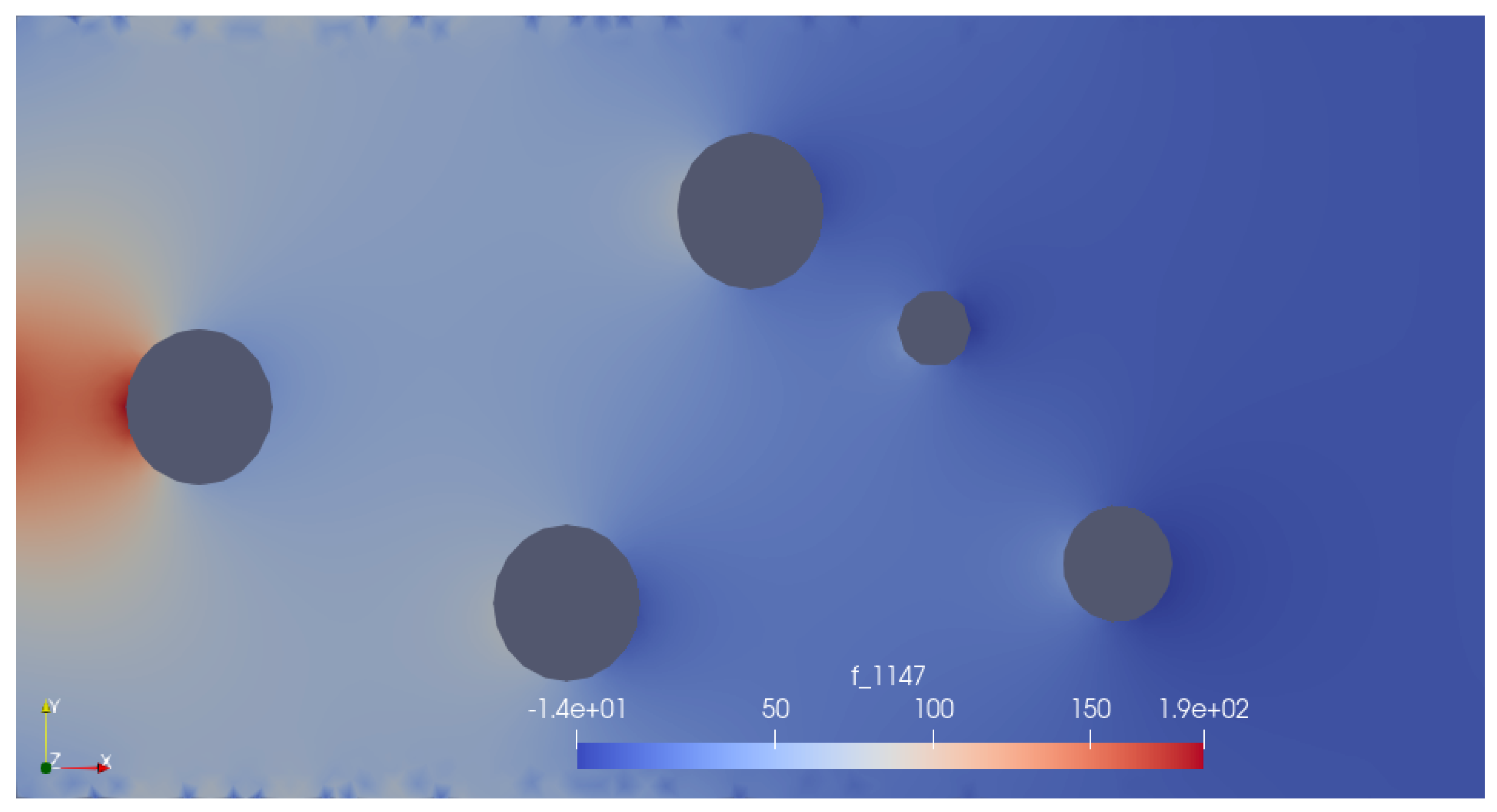
Initial simulations in a rectangular domain with circular obstacles demonstrate smooth and continuous velocity fields. The pressure distribution aligns well with theoretical predictions. See Figure 2 and Figure 3. Figure 6–8 are captured using ParaView software. Code is accessible via GitHub in [12].
Figure 1.
Mesh.
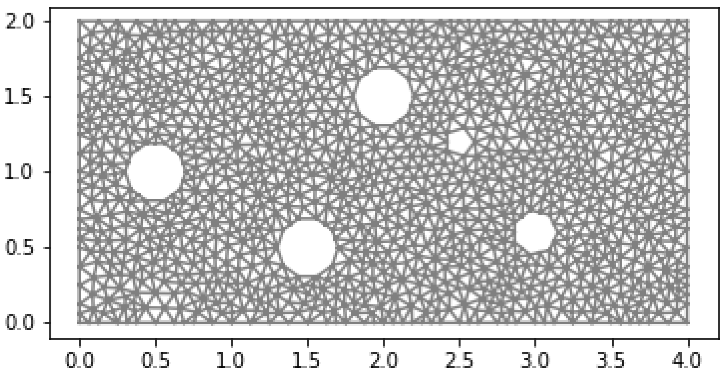
Figure 2.
Velocity.
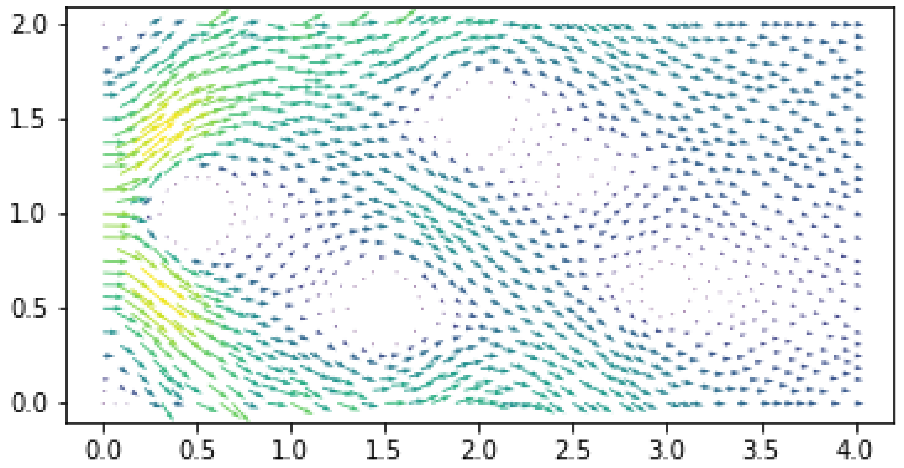
Figure 3.
Pressure.
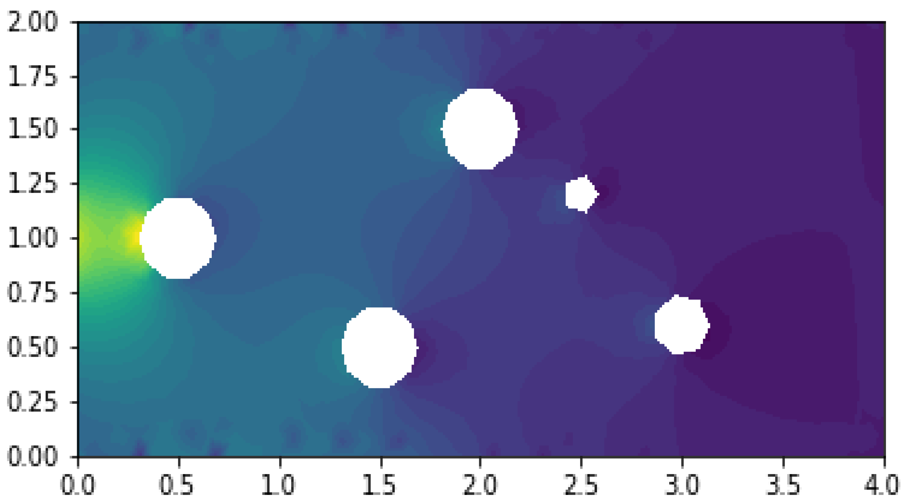
Figure 4.
Mesh.
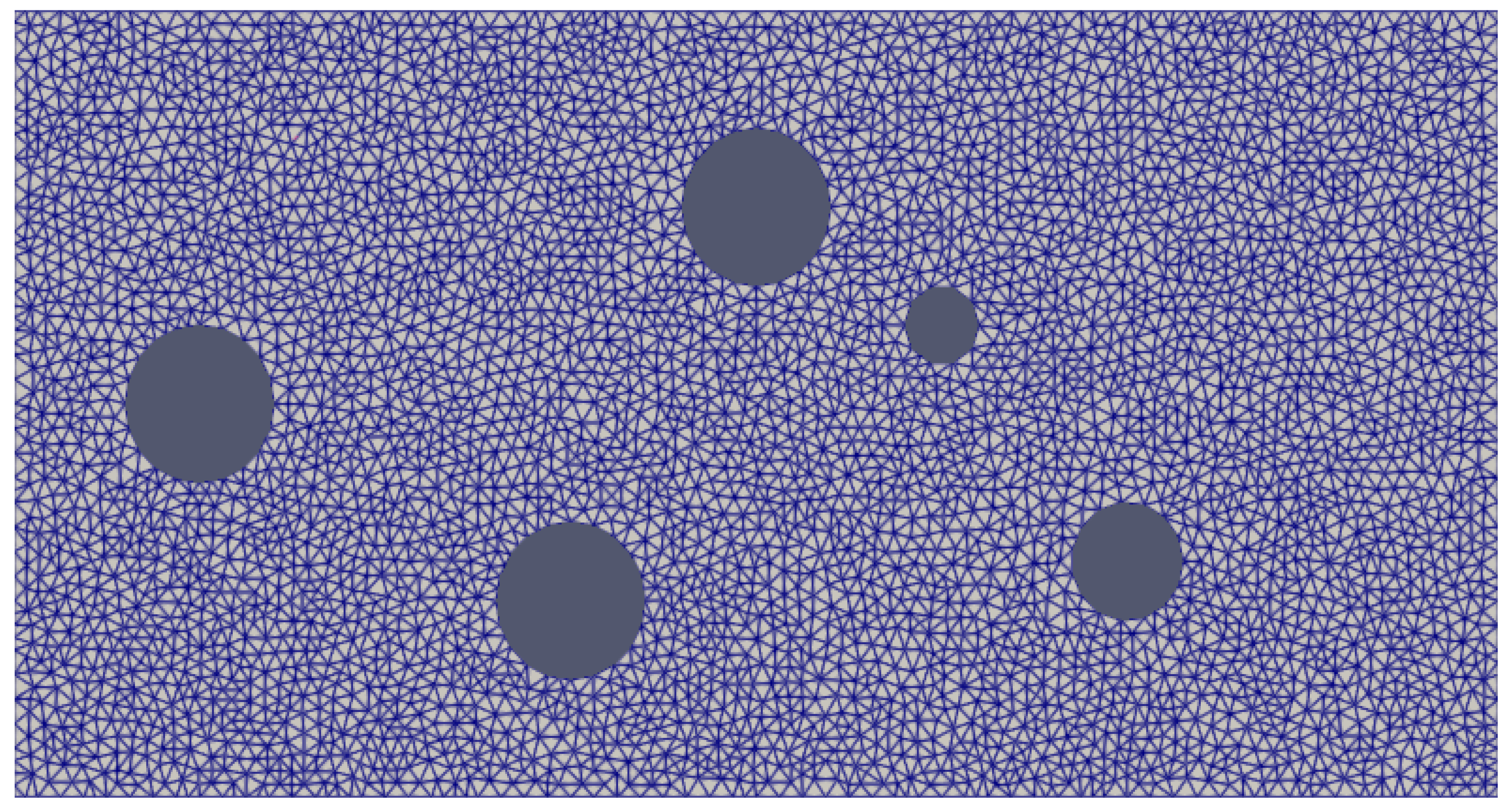
Figure 5.
Velocity.
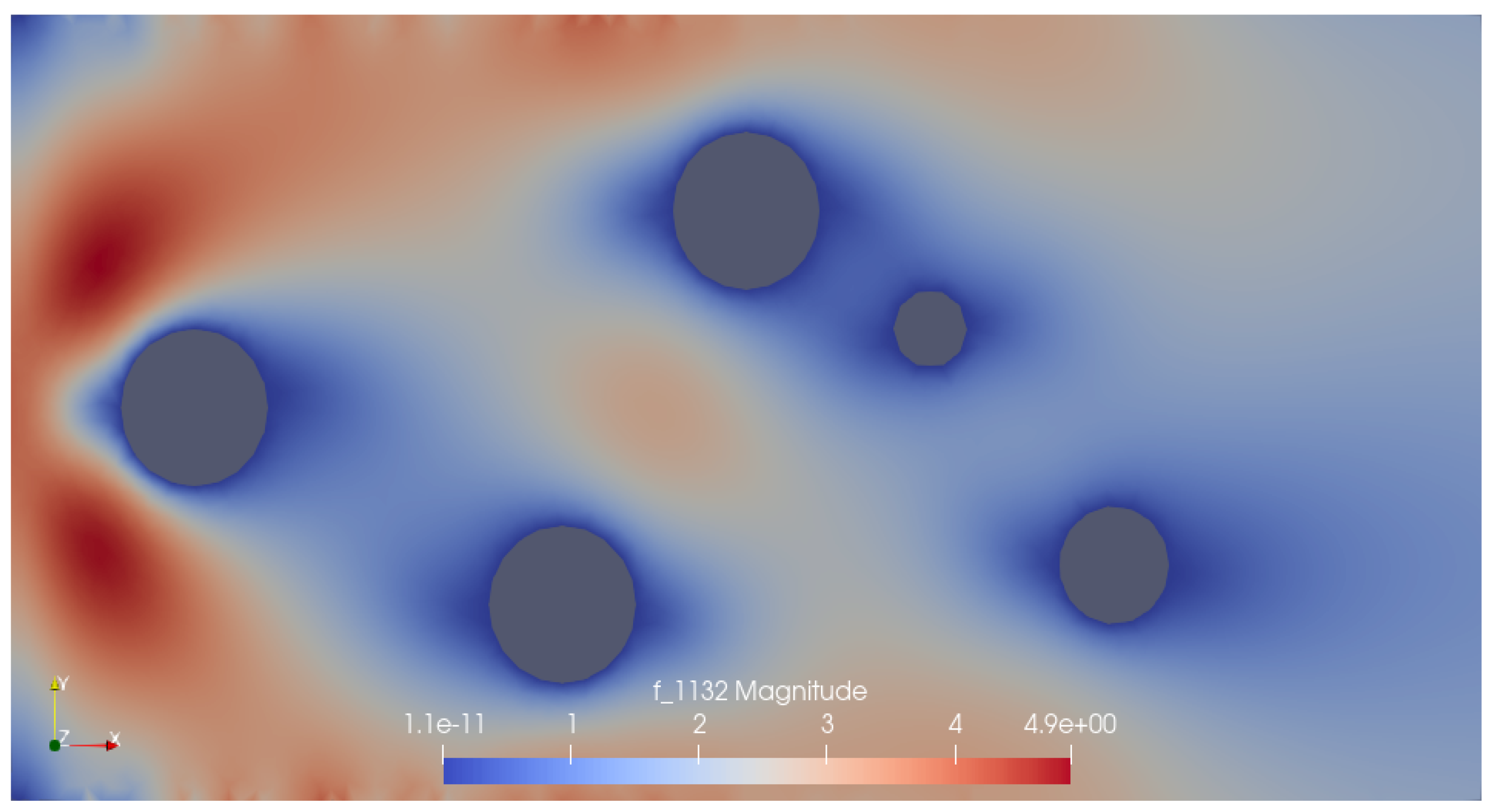
Figure 6.
Pressure.
Conclusions and Way Forward
In this study, we have successfully solved the Stokes equations in a 2D framework with embedded obstacles, applying no-slip and periodic boundary conditions. This approach will be helpful to simulate the complex flow dynamics and membrane tension distribution in a simplified model of the cellular membrane environment. The results of our simulations will provide valuable insights to study further how obstacles, such as tethered proteins, can influence the behaviour of lipid flow and membrane tension propagation. Pressure drop boundary conditions will be implied to test whether the flow is Darcy. The paper [11] will help implement the pressure boundary conditions. It is reasonable to consider the code written in [13] to move the circular solids with the fluid velocity and to calculate the density of the circular obstacles in the fluid. To understand the code [13], one has to understand the Arbitrary Lagrangian Eulerian (ALE) formulation.
References
- Djakbarova, U.; Madraki, Y.; Chan, E.; Kural, C. Dynamic interplay between cell membrane tension and clathrin-mediated endocytosis. Biol Cell 2021, 113, 344–73.
- Houk, A.; Jilkine, A.; Mejean, C.; Boltyanskiy, R.; Dufresne, E.; Angenent, SB, e.a. Membrane tension maintains cell polarity by confining signals to the leading edge during neutrophil migration. Cell 2012, 148, 175–88.
- Thottacherry, J.; Kosmalska, A.; Kumar, A.; Vishen, A.; Elosegui-Artola, A.; Pradhan, S, e.a. Mechanochemical feedback control of dynamin independent endocytosis modulates membrane tension in adherent cells. Nat Commun 2018, 9, 4217.
- Shi, Z.; Graber, Z.; Baumgart, T.; Stone, H.; Cohen, A. Cell membranes resist flow. Cell 2018, 175, 1769–79.e13.
- Kamalesh, K.; Scher, N.; Biton, T.; Schejter, E.; Shilo, E.Z.; Avinoam, O. Exocytosis by vesicle crumpling maintains apical membrane homeostasis during exocrine secretion. Dev Cell 2021, 56, 1603–1616.e6.
- Gauthier, N.; Fardin, M.; Roca-Cusachs, P.; Sheetz, M. Temporary increase in plasma membrane tension coordinates the activation of exocytosis and contraction during cell spreading. Proc Natl Acad Sci U S A 2011, 108, 14467–72.
- Boulant, S.; Kural, C.; Zeeh, J.; Ubelmann, F.; Kirchhausen, T. Actin dynamics counteract membrane tension during clathrin-mediated endocytosis. Nat Cell Biol 2011, 13, 1124–31.
- Basu, R.; Whitlock, B.; Husson, J.; Le Floc’h, A.; Jin, W.; Oyler-Yaniv, A, e.a. Cytotoxic T cells use mechanical force to potentiate target cell killing. Cell 2016, 165, 100–10.
- Diz-Muñoz, A.; Fletcher, D.; Weiner, O. Use the force: Membrane tension as an organizer of cell shape and motility. Trends Cell Biol 2013, 23, 47–53.
- Shi, Z.; Baumgart, T. Membrane tension and peripheral protein density mediate membrane shape transitions. Nat Commun 2015, 6, 5974.
- Bertoluzza, S.; Chabannes, V.; Prud’Homme, C.; Szopos, M. Boundary conditions involving pressure for the Stokes problem and applications in computational hemodynamics. Comput Methods Appl Mech Eng 2017, 322, 58–80.
- SMubasshar. new-rep. GitHub repository 2025.
- Hruza. Fluid-Particle-ALE. GitHub repository 2025.
Disclaimer/Publisher’s Note: The statements, opinions and data contained in all publications are solely those of the individual author(s) and contributor(s) and not of MDPI and/or the editor(s). MDPI and/or the editor(s) disclaim responsibility for any injury to people or property resulting from any ideas, methods, instructions or products referred to in the content. |
© 2025 by the authors. Licensee MDPI, Basel, Switzerland. This article is an open access article distributed under the terms and conditions of the Creative Commons Attribution (CC BY) license (http://creativecommons.org/licenses/by/4.0/).
Copyright: This open access article is published under a Creative Commons CC BY 4.0 license, which permit the free download, distribution, and reuse, provided that the author and preprint are cited in any reuse.


