Preprint
Article

This version is not peer-reviewed.

Metabolomics, Genetics, and Environmental Factors: Intersecting Paths in Abdominal Aortic Aneurysm Risk

A peer-reviewed article of this preprint also exists.

Submitted:

09 December 2024

Posted:

10 December 2024

You are already at the latest version

Abstract

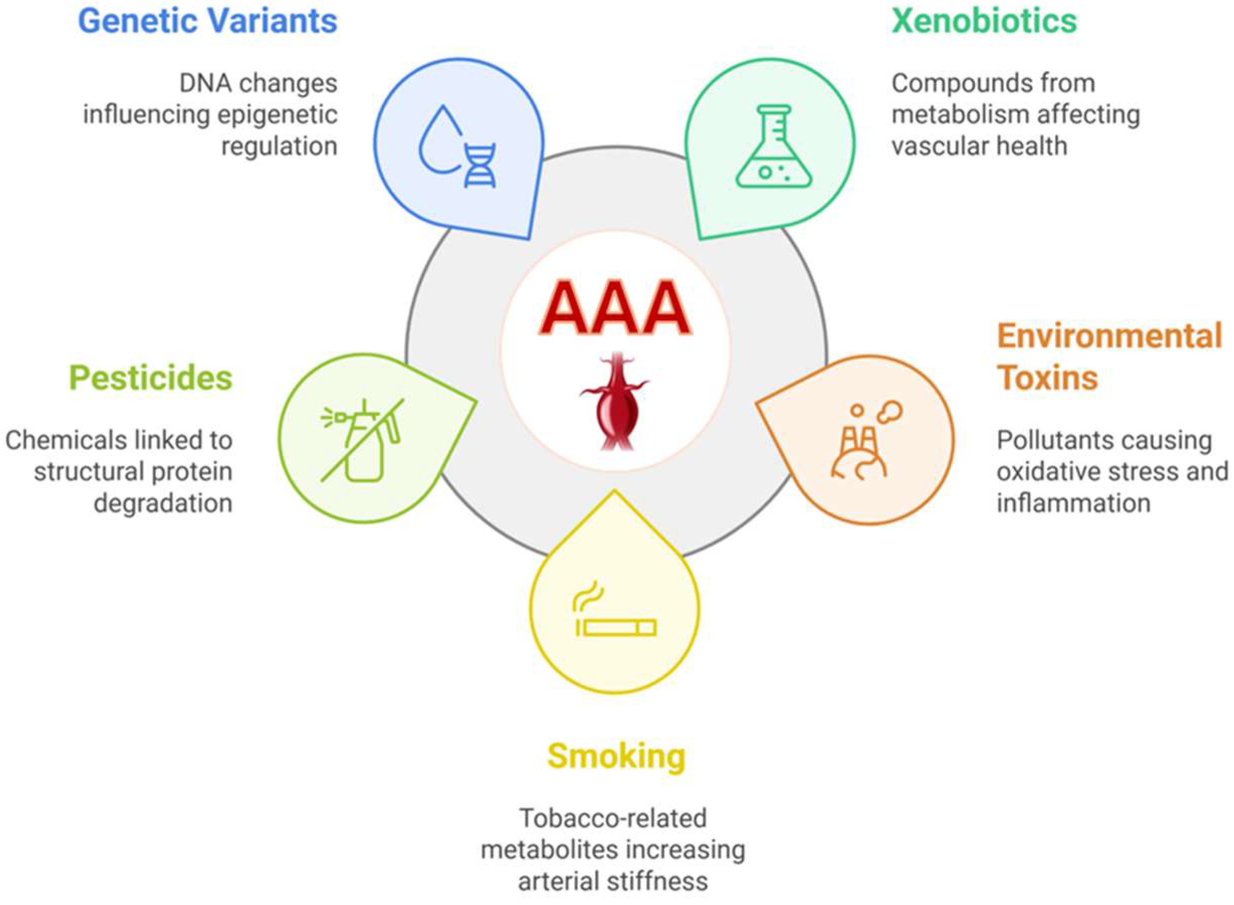
Abdominal aortic aneurysm (AAA) represents a significant public health concern, particularly in men aged 55 to 64, where it occurs in about 1%. We investigated metabolomics and genetics of AAA by analyzing a cohort consisting of 76 patients diagnosed with AAA and 228 matched controls. Utilizing the Metabolon DiscoveryHD4 platform for non-targeted metabolomics profiling, we identified 11 novel metabolites that significantly increased the risk of AAA. These metabolites were primarily associated with environmental and lifestyle factors, notably smoking and pesticide exposure, which underscores the influence of external factors on the progression of AAA. Additionally, several genetic variants were associated with xenobiotics, highlighting a genetic predisposition that may exacerbate the effects of these environmental exposures. The integration of metabolomic and genetic data provides compelling evidence that lifestyle, environmental, and genetic factors are intricately linked to the etiology of AAA. The results of our study not only deepen the understanding of the complex pathophysiology of AAA but also pave the way for the development of targeted therapeutic strategies.

Keywords: 
;  ;  ;  

1. Introduction

Abdominal aortic aneurysm (AAA), characterized by an aortic diameter exceeding 3 cm, constitutes a substantial global healthcare challenge [1]. The risk of AAA escalates notably after 60 years old. Clinically relevant aneurysms, exceeding 4 cm in diameter, are present in approximately 1% of men aged from 55 to 64, with prevalence escalating by 2% to 4% per subsequent decade [2,3]. AAAs manifest four to six times more frequently in men than in women [4,5] and is more prevalent in white individuals compared to Black individuals [6]. AAA is characterized by localized structural deterioration of the wall of the aorta, resulting in progressive dilation and rupture [7]. AAA pathogenesis is closely related to the progressive depletion and dysfunction of vascular smooth muscle cells, and includes proteolysis, oxidative stress, inflammatory immune response, and apoptosis [8]. These processes cause the loss of elasticity and resistance of the artery wall.
Smoking and hypertension are significant risk factors for AAA. Additionally, AAAs are more prevalent in individuals with atherosclerosis, with an approximate 5% prevalence in those with coronary artery disease [9,10]. Positive family history substantially increases the risk of AAA [11,12], suggesting that genetic factors play an important role in the development of AAA [13]. AAA appears less common in individuals with diabetes [6].
The core of AAA management requires longitudinal surveillance until the aneurysm reaches a size where the risk of rupture surpasses the risk of repair [14]. Inflammatory processes [15,16] and gut microbiota [17] have important roles in the pathogenesis of AAA. A recent study based on 449 463 participants from the UK Biobank reported that exposure to long-term air pollutants increased the risk of AAA [18]. Similarly, exposure to environmental toxins, polycyclic aromatic hydrocarbons (PAH), has been implicated in cardiovascular diseases and in AAA [19].
In recent years there has been a growing interest in exploring the metabolic and genetic profiles associated with AAA to understand its multifactorial nature. Previous metabolomics studies on AAA have been limited by small sample sizes and a small number of metabolites analysed, leading to inconsistent results [20,21,22,23,24,25,26,27,28,29]. Metabolites are crucial for cellular functions, influencing various physiological processes and signalling pathways. Given the several risk factors and metabolic pathways affected in patients with AAA, metabolomics approach is likely to identify pathological processes in AAA expansion and allow to find novel therapeutic strategies.
The identification of circulating biomarkers having diagnostic and prognostic value for the diagnosis of AAA is challenging. Biomarkers should have sufficient specificity and sensitivity to be used in clinical practice. Based on the pathophysiology of an AAA, circulating biomarkers can be classified according to their relationship with prothrombotic activity, degradation of the extracellular matrix of the vascular wall or the immunoinflammatory. To comprehend the impact of the genetic background and associated pathways in the development of AAA, we applied a comprehensive high-throughput liquid chromatography–tandem mass spectroscopy (LC-MS/MS) in the patients with AAA compared to matched controls in the Finnish population-based METSIM study.

2. Results

2.1. Baseline Characteristics of AAA Cases and Controls

Table 1 shows the baseline characteristics of the participants with AAA and matched controls. Participants with AAA had higher levels of triglycerides, lower insulin sensitivity (Matsuda), increased levels of hs-CRP, a higher percentage of smokers, and a higher percentage of the participants on statin medication.

2.2. Differences in the Metabolite Abundances Between the AAA Cases and Controls

We found statistically significant differences in 12 metabolites between the participants with AAA and the controls. The participants with AAA had an increased abundance of xenobiotics (n=9), a carbohydrate N-acetylneuraminate (n=1), and a lipid 3beta, 7alpha-dihydroxy-5-cholestenoate (n=1) compared to the controls (Table 2). The most significant metabolite differences among the xenobiotics were for 2-naphtol sulfate (p=4.7x10⁻⁹), methyl naphthyl sulfate (p =1.2x10⁻⁷), and 4-vinyl phenol sulfate (p =2.4x10⁻⁷). Abundance of biliverdin was decreased in the participants with AAA compared to the controls.
Suppl. Table 1 presents the results for all 229 metabolites nominally associated with the risk of developing AAA. Suppl. Table 2 presents result for a subset of metabolites that demonstrated a significant association with AAA under a rigorous threshold. Initially, we identified 229 metabolites with nominal significance (p<0.05), including 136 that increased and 93 that decreased the risk of AAA, predominantly among lipids (n=90), amino acids (n=55), and xenobiotics (n=41). To focus on more significant associations, we applied a more rigorous cutoff for nominal significance p<9.9×10⁻⁴. Using this approach, the metabolites most significantly associated with increased risk of AAA included mannose (p=2.4×10⁻⁴), 3-amino-2-piperidone (p=9.5×10⁻⁵), and 4-vinylcatechol sulfate (p=7.2×10⁻⁵). The most significant metabolites reducing the risk of AAA were hippurate (p=6.5×10⁻⁵), bilirubin (E, E) (p=9.8×10⁻⁵), eicosanoid carnitine (C20:1) (p=1.1×10⁻⁴), and bilirubin degradation product C17H18N2O4 (1) (p=3.9×10⁻⁴). Using this stringent threshold, we were able to replicate findings in previous studies only for two metabolites, hippurate and bilirubin, both known to decrease the risk of AAA.

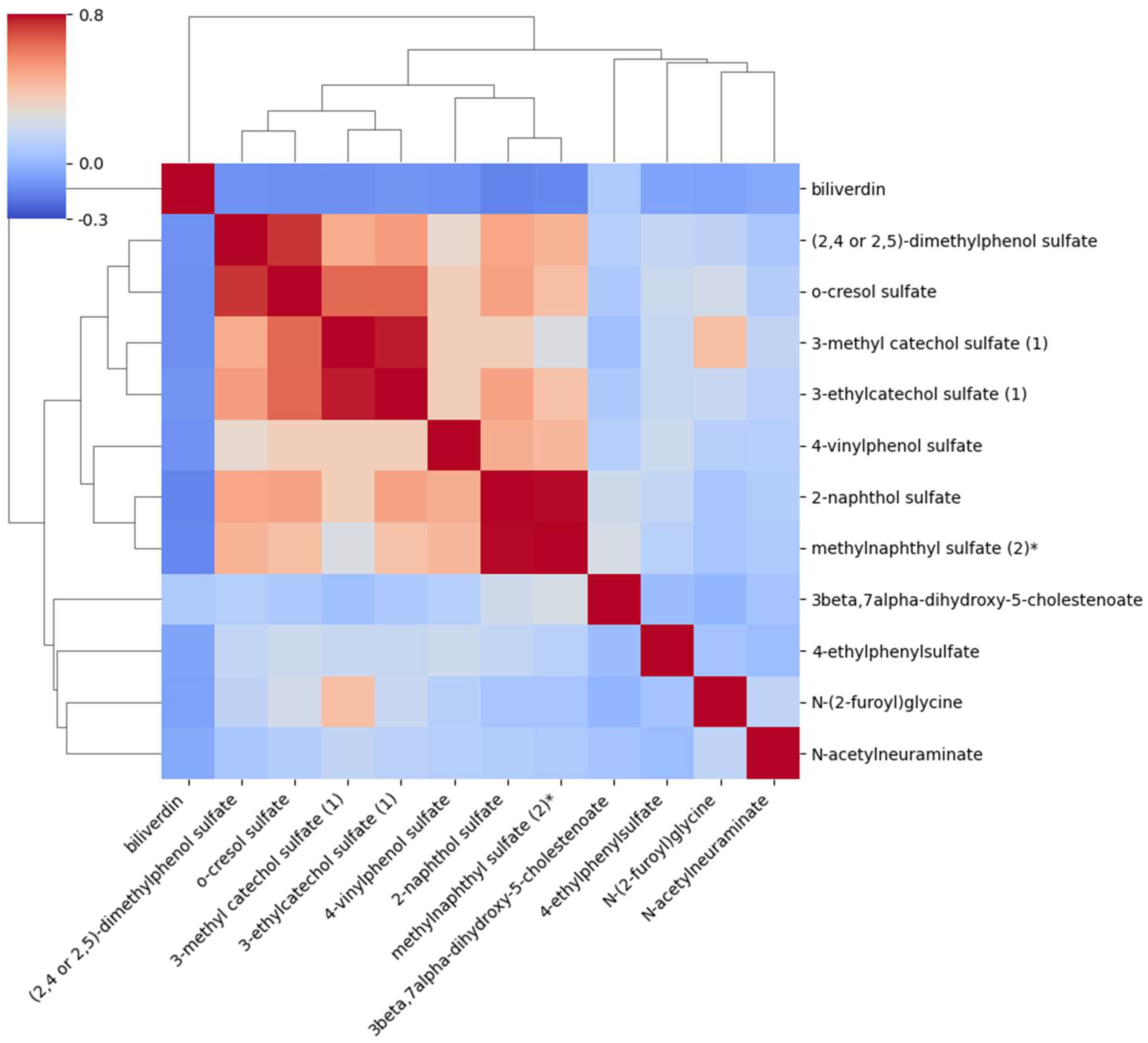
2.3. Correlations Between the Metabolites Associated with AAA

Figure 1 shows the intercorrelations between the 12 metabolites. Xenobiotics had the highest intercorrelations. (2.4 or 2.5)-dimethylphenol sulfate had the high correlations with o-cresol sulfate (0.74), 3-methyl catechol sulfate with 3-ethylcatechol sulfate (0.78), 4-vinulphenol sulfate with 2-naphtol sulfate (0,46), and methylnaphtyl sulfate (2) with 2-naphtol sulfate (0.78). Correlations of xenobiotics with N-acetylneuraminate, and 3beta, 7 alpha-dihygxy-5-cholestenoate were low. Correlations of N-acetylneuraminate, 3beta, 7alpha-dihydroxy-5-cholostenoate and biliverdin were also low with other metabolites.

2.4. Association of the Genetic Variants with Metabolites in Patients with AAA

We utilized the GWAS database () to identify the genes and genetic variants significantly associated with the metabolites and AAA. Table 3 gives the genome-wide statistically significant genetic variants (p<5x10⁻⁸), and Suppl. Table 1 shows the statistically significant variants (p<5x10⁻⁶) given the 1009 metabolites included in the statistical analysis.
Seven of the nine xenobiotics were significantly associated with genetic variants and two of them methyl naphthyl sulfate, and (2,4 or 2.5)-dimethylphenol sulfate were not associated with any of the genetic variants. A variant of rs169828 of the ARSL gene was significantly associated with 2-naphthol sulfate (p=2 x 10-28. The ARSL gene is a sulfatase and makes an enzyme arylsulfatase. This enzyme is part of a group known as sulfatases. The function of this enzyme is not known [30].
4-vinylphenol sulfate was also significantly associated with a variant of the ARSL gene, and additionally with a variant of the voltage-driven transporter SLC17A1 gene (rs9461218, p=1 x 10-19). 4-eyhylphenoylsulgate was associated with the variants of the SLC17A1 and SLC17A3 transported genes, and a variant (rs144597325, p=4x10⁻⁸) of the MBD2 (a methyl-CpG binding domain) gene which belongs to a family of DNA methylation gene capable of binding specifically to methylated DNA [31].
O-cresol sulfate was associated with a variant of the SGF29 gene (rs480400, p=8x10-12). SGF29 specifically recognizes and binds methylated 'Lys-4' of histone H3, and non-histone proteins that are methylated on Lys residues [32]. 3-methyl catechol sulfate was associated with genetic variants in SLC51A (rs2342307, p=5x10-12), PCYT1A, and SLC17A1 (rs9461218, p=2x10-11) genes. SLC51A is a protein coding gene and regulates bile acid and bile salt metabolism. PCYT1A is involved in the regulation of phosphatidylcholine biosynthesis.
N-(2-furoyl) glycine is an acyl glycine and associated with a variant in the CREG2 gene (rs6751877, p=6x10-24). CREG binds to mannose-6-phosphate/insulin-like growth factor-2 receptor and may enhance autophagy [33]. CREG2 is a secreted N-glycoprotein expressed specifically in the brain. Compound 3-ethylcatechol sulfate was associated with the genetic variants of SLC51A (rs6795511, p=1x10-11), and SLC17A3 (rs1186313, p=5x10-11) genes.
Biliverdin is a byproduct of hemoglobin breakdown. The resulting biliverdin is reduced to bilirubin. Biliverdin has significant antioxidant properties. UGT1-UDP glucuronosyltransferase family gene encodes a UDP-glucuronosyltransferase, an enzyme of the glucuronidation pathway that transforms small lipophilic molecules, including bilirubin, into water-soluble, excretable metabolites. This gene is a part of a complex locus that encodes several UDP-glucuronosyltransferases [34]. The most significant variants of the UGT1-UDR genes were for genetic variants rs1976391 (p=2x10-802), rs887829 (p=3x10-403), and rs35754645 (p=2x10-250). Biliverdin was also significantly associated with genetic variant rs111366223 (p=4x10⁻⁸) of FYB1 and rs201662188 (p=3x10-23) of SLCO1B1.
N-acetylneuraminate was associated with genetic variants of four genes, LAMC1 (rs116448311, p=8x10-84), which regulates glycosphingolipid binding activity, NPL (N-acetylneuraminate lyases) (rs78799057, p=4x10-78), which regulates cellular concentrations of N-acetyl-neuraminic acid, ARHGEF3 (rs1354034, p=4x10-78), which regulates skeletal muscle regeneration, and SNHG16 (rs2109101, p=2x10-16) which regulates cell proliferation and apoptosis. 3beta, 7alpha-dihydroxy-5-cholestenoate was associated with the FBXL19 gene (rs7206511, p= 5x10-11) which plays a role in cell migration and cell proliferation.

3. Discussion

Our study integrates metabolomics and genetics to explore potential biomarkers associated with AAA in the METSIM cohort. Our study reports several novel findings. We found that among the 12 metabolites identified, 9 xenobiotics were significantly associated with an increased risk of AAA. Xenobiotics are chemical substances including plants, drugs, pesticides, food additives, chemicals, and environmental pollutants [35]. Four of the xenobiotics belonged to benzoate metabolism.
We observed that xenobiotics were significantly increased in the participants with AAA compared to the controls, suggesting that an altered xenobiotic metabolism contributes to the pathophysiological processes resulting in aneurysm formation. We conclude that exposure to environmental toxins, including PAHs, pesticides, and herbicides is the major risk factor for the development of AAA [19,36,37].
Five metabolites were associated with tobacco smoking (2-naphthol sulfate, methylnaphthyl sulfate, 4-vinylphenol sulfate, o-cresol sulfate, 4-ethylphenylsulfate) [38]. Exposure to PAHs increases the risk of cardiovascular diseases, including AAA [19,39]. PAH compounds are a group of organic compounds found in tobacco and tobacco smoke, formed primarily during the incomplete combustion of organic materials [40,41,42,43]. PAHs induce endothelial dysfunction, oxidative stress, and inflammation, which weaken the aortic wall resulting in vascular remodelling, arterial stiffness, and plaque formation [40,41,42,43]. Chronic PAHs exposure increases the risk of AAA, especially in individuals having several risk factors for AAA.
We found two metabolites that are derivatives of pesticides/herbicides (2,4 or 2,5)-dimethylphenol sulfate and o-cresol sulfate. Pesticides and herbicides increase the risk of AAA by causing oxidative stress, inflammation, and arterial stiffness by degrading structural proteins, such as elastin and collagen which are important for the integrity of the aorta [36,37]. Additionally, pesticides disrupt normal metabolic and immune processes that regulate vascular tissue remodelling, particularly in individuals with several risk factors for AAA [36,37].
The significant metabolite differences we observed between the AAA cases and matched controls, particularly in the xenobiotics class, suggest a potential dysregulation in the body's ability to process and eliminate these compounds in the patients with AAA. Increased abundances of 2-naphthol sulfate, methylnaphthyl sulfate, 4-vinylphenol sulfate, and o-cresol sulfate indicate an altered xenobiotic metabolism which could contribute to pathophysiological processes leading to aneurysm formation and progression. These findings support the hypothesis that exposure to environmental toxins increases the risk of AAA, as suggested by the studies linking cardiovascular diseases and environmental pollutants PAHs, pesticides, and herbicides to an increased risk of AAA [44,45,46].
N-(2-furoyl) glycine is a metabolite generated by microbiota and found in food prepared by strong heat. This metabolite belongs to the class of N-acyl-alpha amino acids and is a product of fatty acid catabolism and regulates mitochondrial fatty acid beta-oxidation [47]. N-(2-furoyl) glycine participates in the pathways increasing oxidative stress, inflammation, and mitochondrial dysfunction, which are risk factors for cardiovascular diseases [48].
We found an association between the carbohydrate conjugate N-acetyl-alpha-neuraminate, a sialic acid found on the surface of various cell types, and an increased risk of AAA. Sialic acids play a vital role in mediating cell-cell and cell-molecule interactions in eukaryotes, and they can be used by pathogens like E. coli to evade host immune responses [49]. Infections of bacterial and fungal origin are known to contribute to the development of infectious AAA, which is associated with an elevated risk of aneurysm rupture [50]. Biliverdin, a product of heme catabolism, is known for its anti-inflammatory and antioxidant properties. Increased concentration of biliverdin in the controls in our study suggests that it reduces the risk of AAA [51].
We investigated the association of the genetic variants with the metabolites utilized the GWAS database. Seven out of the nine xenobiotics were associated with genetic variants, and two metabolites, methylnaphthyl sulfate, and (2,4 or 2,5)-dimethylphenol sulfate did not associate significantly with any of the genetic variants. Two variants of the ARSL gene were associated with 2-naphthol sulfate and 4-vinylphenol sulfate. We found several genetic variants associated with metabolites which could serve as biomarkers for the risk of AAA. Additionally, we discovered that two genes regulate epigenetic changes, the MBD2 (a methyl-CpG binding domain) gene which belong to a family of DNA associated with 4-vinylphenol sulfate, and the SGF29 gene which binds methylated Lys-4 of histone H3 and is associated with O-cresol sulfate. This finding gives additional evidence that the risk of AAA is regulated by environmental factors.
Previous studies have reported associations of several metabolites with AAA, including hippurate, bilirubin, proline, glycerol, aspartate, glutamate, proline, citric acid, 2-oxoglutaric acid, and succinic acid [20,21,22,23,24,25,26,27,28,29]. However, these findings have not been replicated in other studies. These studies have applied p<0.05 as a statistically significant association without Bonferroni correction for multiple comparisons. We applied a stricter threshold for nominal significance (p<9.9×10⁻⁴) and found several metabolites across different classes of metabolites, including carbohydrates, amino acids, and lipids, which were nominally associated with an increased risk of AAA. This suggests that by increasing the sample size we can identify several new metabolites significantly associated with AAA.
Figure 2 summarizes our findings and underscores the critical links between xenobiotics in the development of AAA, influenced by lifestyle and environmental toxins. We identified key metabolites and genetic variants contributing to the pathophysiology of AAA. However, more studies with larger sample sizes and diverse populations are needed to validate our findings and clarify the metabolic pathways involved in the risk of AAA to enhance predictive models and preventative strategies for AAA.
In summary, our study provides a comprehensive analysis of the metabolomics and genetic risk factors associated with AAA in the METSIM cohort, offering new insights into the pathophysiology of AAA. We identified 12 metabolites, including nine xenobiotics significantly associated with the risk of AAA, highlighting the role of environmental toxins such as PAHs, pesticides, and herbicides increasing vascular remodelling, oxidative stress, and inflammation. Genetic analyses revealed associations between the key metabolites, and specific genetic variants emphasizing an independent effect of genetic predisposition on the risk of AAA. The preventive role of the metabolites, especially biliverdin, suggests potential therapeutic avenues.
Future research should focus on expanding metabolomic and genetic analyses to larger, more diverse populations to validate the findings observed in this study and enhance our understanding of AAA’s complex ethicology. Longitudinal studies tracking metabolic changes over time in individuals at high risk for AAA may help to identify early biomarkers, providing an opportunity for preventive interventions. Given the strong associations between xenobiotics and the risk of AAA, studies into environmental and lifestyle modifications could offer practical applications to reduce the incidence of AAA. Further exploration of the metabolic pathways involved in xenobiotic metabolism may lead to the development of targeted therapies aimed at mitigating the harmful effects of environmental toxins. The identification of potential biomarkers, such as 2-naphthol sulfate, also opens avenues for precision medicine approaches, where metabolic profiling could guide personalized monitoring and treatment strategies for individuals at high risk of AAA.

4. Materials and Methods

4.1. Study Population

The METSIM study includes 10 197 men, aged from 45 to 73 years at baseline, and randomly selected from the population register of Kuopio, Eastern Finland. The METSIM study was approved by the Ethics Committee at the Kuopio University Hospital, Finland. All participants provided written informed consent. The design and methods of the METSIM study have been previously described in detail [52,53]. A total of 304 men from the METSIM study were included in the current study, 76 participants having AAA and 228 controls matched for age, BMI, and systolic blood pressure. BMI was calculated as weight divided by height squared. Smoking status was defined as current smoking. Other laboratory measurements have been previously reported [52]. Estimated glomerular filtration rate (eGFR) was calculated using the CKD-Epi equation [53].

4.2. Clinical and Laboratory Measurements

Height was measured without shoes to the nearest 0.5 cm. Weight was measured in light clothing with a calibrated digital scale (Seca 877, Hamburg, Germany). Laboratory studies after 12 h fasting included the following measurements, plasma glucose and insulin, lipids, lipoproteins, and mass spectrometry metabolomics (Metabolon, Durham, NC). An oral glucose tolerance test was performed to evaluate glucose tolerance (75 g of glucose). Clinical and laboratory measurement methods have been previously published [41]. Briefly, plasma glucose was measured by enzymatic hexokinase photometric assay (Konelab Systems Reagents, Thermo Fischer Scientific, Vantaa, Finland). Insulin was determined by immunoassay (ADVIA Centaur Insulin IRI, no 02230141, Siemens Medical Solutions Diagnostics, Tarrytown, NY, USA). Serum alanine aminotransferase (ALT) was measured by an enzymatic photometric test (Konelab Reagent System, Thermo Fisher Scientific, Vantaa, Finland).

4.3. Metabolomics

Metabolites were measured by using Metabolon Inc.’s untargeted Discovery HD4 platform based on Ultrahigh Performance Liquid Chromatography-Tandem Mass Spectroscopy (UPLC-MS/MS) (Metabolon, Morrisville, NC, USA). Samples stored at −80 C prior analysis were prepared using the automated MicroLab STAR® system from Hamilton Company. Several recovery standards were added prior to the first step in the extraction process for quality control (QC) purposes. A pooled matrix sample generated by taking a small volume of each experimental sample served as a technical replicate throughout the data set. Extracted water samples served as process blanks, and QC standards that were carefully chosen not to interfere with the measurement of endogenous compounds were spiked into every analysed sample, allowing instrument performance monitoring, and aiding chromatographic alignment. Overall process variability was determined by calculating the median relative standard deviation for all endogenous metabolites present in 100% of the pooled matrix samples. Data normalization step was performed to correct variation resulting from instrument inter-day tuning differences in studies spanning multiple days. Experimental samples were randomized across the platform run with QC samples spaced evenly. Raw data was extracted, peak-identified and QC processed using Metabolon’s hardware and software, and peaks were quantified using area-under-the-curve. Compounds were identified by comparison to library entries of purified standards or recurrent unknown entities. Library matches for each compound were checked for each sample and corrected if necessary. Each metabolite was rescaled to set the median equal to 1.
The Metabolon DiscoveryHD4 platform identified a total of 1540 metabolites. From this initial set, only metabolites with at least 40% complete data across the dataset were retained, while all metabolites lacking identification information were excluded, resulting in 1009 metabolites for statistical analysis. All samples were processed together for peak quantification and data scaling. Evaluation of overall process variability by the median relative standard deviation for endogenous quantified raw mass spectrometry peaks for each metabolite using the area under the curve and metabolites present in all 20 technical replicates in each batch was performed. Variation was adjusted for day-to-day instrument tuning differences and columns used for biochemical extraction by scaling the raw peak quantifications to the median for each metabolite by the Metabolon batch.

4.4. Selection of Genetic Variants Associated with Increased Risk of AAA

We identified genetic variants associated with an increased risk of AAA from previous publications and the GWAS Catalog (The NHGRI-EBI Catalog of human genome-wide association studies), (https://www.ebi.ac.uk/gwas/) in individuals of European ancestry. Among the genetic variants for each gene, we selected the variant having the most significant association with AAA.

4.5. Statistical Analysis

We conducted statistical analyses using IBM SPSS Statistics, version 29. We log-transformed all continuous variables except for age to correct for their skewed distribution. We performed association analyses between the genetic variants and metabolites using linear regression analysis adjusted for known risk factors for AAA (age, BMI, smoking and systolic blood pressure). We give the results as standardized beta coefficients and p values with the metabolite as a dependent variable. We used one-way ANOVA to assess the differences in clinical traits and metabolites between the two groups, participants with AAA and controls matched for age, BMI, and systolic blood pressure. The threshold for statistical significance is ≤ 5.0x10⁻⁵. Correlations between the metabolites were calculated using the Pearson correlation.

5. Conclusions

We applied metabolomics and genetics to identify novel metabolites associated with AAA in the METSIM cohort. Among the 12 metabolites significantly increasing the risk of AAA, nine were xenobiotics linked to lifestyle and environmental exposures, including substances from benzoate metabolism, smoking, and pesticides. Genetic analyses highlighted several genetic variants associated with metabolite abundances and increasing the risk of AAA. Our comprehensive analysis, combining metabolomics with genetic data, robustly demonstrates that lifestyle and environmental influences and genetic factors play significant roles in the aetiology of AAA.

Supplementary Materials

The following supporting information can be downloaded at the website of this paper posted on Preprints.org, Table S1: Nominally significant metabolites associated with abdominal aortic aneurysm in the METSIM cohort.

Author Contributions

Conceptualization: L.F.S., A.J.L., and M.L.; Methodology: J.V., L.F.S., and M.L.; Investigation: L.F.S., J.V., A.O., A.J.L., and M.L.; Visualization: L.F.S.; Funding acquisition: M.L.; Project administration: M.L.; Supervision: M.L.; Writing—original draft: L.F.S., and M.L. Writing review: L.F.S., J.V., A.O., A.J.L., and M.L. All authors have read and agreed to the published version of the manuscript.

Funding

Academy of Finland grant no. 321428 (M.L.); Centre of Excellence of Cardiovascular and Metabolic Diseases, the Academy of Finland, grant no. 271961 (M.L.); Sigrid Juselius Foundation grant (M.L.); Finnish Foundation for Cardiovascular Research grant (M.L.); and Kuopio University Hospital grant (M.L.).

Institutional Review Board Statement

The METSIM study was approved by the Ethics Committee of the Kuopio University Hospital, Finland. All participants provided written informed consent.

Informed Consent Statement

Informed consent was obtained from all subjects involved in the study.

Data Availability Statement

The data that support the findings of this study are available from the corresponding authors [M.L. and L.F.S.] upon reasonable request.

Conflicts of Interest

The authors declare no conflicts of interest..

References

  1. Lozano R, Naghavi M, Foreman K, Lim S, Shibuya K, et al. Global and regional mortality from 235 causes of death for 20 age groups in 1990 and 2010: a systematic analysis for the Global Burden of Disease Study 2010. Lancet. 2012; 380: 2095–2128. [CrossRef]
  2. Singh K, Bonaa KH, Jacobsen BK, et al. Prevalence of and risk factors for abdominal aortic aneurysms in a population-based study: The Tromso Study. Am J Epidemiol. 2001; 154: 236–244.
  3. Powell JT, Greenhalgh RM. Clinical practice. Small abdominal aortic aneurysms. N Engl J Med. 2003; 348: 1895–1901.
  4. Scott RA, Wilson NM, Ashton HA, et al. Influence of screening on the incidence of ruptured abdominal aortic aneurysm: 5-year results of a randomized controlled study. Br J Surg. 1995; 82: 1066–1070. [CrossRef]
  5. Lederle FA, Johnson GR, Wilson SE. Aneurysm detection and management veterans affairs cooperative study. Abdominal aortic aneurysm in women. J Vasc Surg. 2001; 34: 122–126.
  6. Lederle FA, Johnson GR, Wilson SE, et al. The aneurysm detection and management study screening program: Validation cohort and final results. Arch Intern Med. 2000; 160: 1425–1430.
  7. Khan H, Abu-Raisi M, Feasson M, Shaikh F, Saposnik G, et al. Current prognostic biomarkers for abdominal aortic aneurysm: a comprehensive scoping review of the literature. Biomolecules. 2024; 14: 661. [CrossRef]
  8. Torres-Fonseca M, Galanb M, Martinez-Lopez D, Canes L, Roldan-Monteroa R, et al. Pathophysiology of abdominal aortic aneurysm: Pathophysiology of abdominal aortic aneurysm. Atherosclerosis. 2019; 31: 166–177.
  9. Bengtsson H, Ekberg O, Aspelin P, et al. Ultrasound screening of the abdominal aorta in patients with intermittent claudication. Eur J Vasc Surg. 1989; 3: 497–502. [CrossRef]
  10. Cabellon S, Jr, Moncrief CL, Pierre DR, et al. Incidence of abdominal aortic aneurysms in patients with atheromatous arterial disease. Am J Surg. 1983; 146: 575–576. [CrossRef]
  11. Fleming C, Whitlock EP, Beil TL, et al. Screening for abdominal aortic aneurysm: A best-evidence systematic review for the US Preventive Services Task Force. Ann Intern Med. 2005; 142: 203–211. [CrossRef]
  12. Salo JA, Soisalon-Soininen S, Bondestam S, et al. Familial occurrence of abdominal aortic aneurysm. Ann Intern Med. 1999; 130: 637–642. [CrossRef]
  13. Jones GT, Tromp G, Kuivaniemi H, Gretarsdottir S, Baas AF, Giusti B, et al. Meta-analysis of genome-wide association studies for abdominal aortic aneurysm identifies four new disease-specific risk loci. Circ Res. 2017; 120: 341–353. [CrossRef]
  14. Chaikof EL, et al. The Society for Vascular Surgery practice guidelines on the care of patients with an abdominal aortic aneurysm. J Vasc Surg. 2018; 67: 2–77. [CrossRef]
  15. Ramprasath T, Han Y-M, Zhang D, Yu C-J, Ming-Hui MH. Tryptophan catabolism and inflammation: a novel therapeutic target for aortic diseases. Front Immunol. 2021; 12: 731701. [CrossRef]
  16. Ling X, Jie W, Qin X, Zhang S, Shi K, Li T, et al. Gut microbiome sheds light on the development and treatment of abdominal aortic aneurysm. Front Cardiovasc Med. 2022; 9: 1063683. [CrossRef]
  17. Benson TW, Conrad KA, Li XS, Wang Z, Helsley RN, Schugar RC, et al. Gut microbiota-derived trimethylamine N-oxide contributes to abdominal aortic aneurysm through inflammatory and apoptotic mechanisms. Circulation. 2023; 147(14): 1079–1096. [CrossRef]
  18. Ma Y, Li D, Cui F, Wang J, Tang L, Yang Y, Liu R, Tian Y. Air pollutants, genetic susceptibility, and abdominal aortic aneurysm risk: a prospective study. Eur Heart J. 2024; 45(12): 1030–1039. [CrossRef]
  19. Ramesh A, Prins PA, Perati PR, Rekhadevi PV, Sampson UK. Metabolism of benzo(a)pyrene by aortic subcellular fractions in the setting of abdominal aortic aneurysms. Mol Cell Biochem. 2016; 411(1-2): 383–391. [CrossRef]
  20. Rupérez FJ, Ramos-Mozo P, Teul J, Martinez-Pinna R, Garcia A, et al. Metabolomic study of plasma of patients with abdominal aortic aneurysm. Anal Bioanal Chem. 2012; 403(6): 1651–1660. [CrossRef]
  21. Ciborowski M, Teul J, Martin-Ventura JL, Egido J, Barbas C. Metabolomics with LC-QTOF-MS permits the prediction of disease stage in aortic abdominal aneurysm based on plasma metabolic fingerprint. PLoS One. 2012; 7(2): e31982. [CrossRef]
  22. Qureshi MI, Greco M, Vorkas PA, Holmes E, Davies AH. Application of metabolic profiling to abdominal aortic aneurysm research. J Proteome Res. 2017; 16(7): 2325–2332. [CrossRef]
  23. Guo Y, Wan S, Han M, Zhao Y, Li C, Cai G, Zhang S, Sun Z, Hu X, Cao H, Li Z. Plasma Metabolomics Analysis Identifies Abnormal Energy, Lipid, and Amino Acid Metabolism in Abdominal Aortic Aneurysms. Med Sci Monit. 2020; 26: e926766. [CrossRef]
  24. Lieberg J, Wanhainen A, Ottas A, Vähi M, Mihkel Zilmer M, et al. Metabolomic profile of abdominal aortic aneurysm. Metabolites. 2021; 11(8): 555. [CrossRef]
  25. Liu J, Liu M, Feng J, Zhu H, Wu J, Zhang H, et al. Alpha-ketoglutarate ameliorates abdominal aortic aneurysm via inhibiting PXDN/HOCL/ERK signaling pathways. J Transl Med. 2022; 20(1): 461. [CrossRef]
  26. Sun L-Y, Lyu Y-Y, Zhang H-Y, Zhi Shen Z, Lin G-Q, et al. Nuclear receptor NR1D1 regulates abdominal aortic aneurysm development by targeting the mitochondrial tricarboxylic acid cycle enzyme aconitase-2. Circulation. 2022; 146(21): 1591–1609. [CrossRef]
  27. Chinetti G, Carboni J, Murdaca J, Moratal C, Sibille B, Raffort J, Lareyre F, Baptiste EJ, Hassen-Khodja R, Neels JG. Diabetes-induced changes in macrophage biology might lead to reduced risk for abdominal aortic aneurysm development. Metabolites. 2022; 12(2): 128. [CrossRef]
  28. Xie T, Lei C, Song W, Wu X, Wu J, et al. Plasma lipidomics analysis reveals the potential role of lysophosphatidylcholines in abdominal aortic aneurysm progression and formation. Int J Mol Sci. 2023; 24(12): 10253. [CrossRef]
  29. Vanmaele A, Bouwens E, Hoeks SE, Kindt A, Lamont L, et al. Targeted proteomics and metabolomics for biomarker discovery in abdominal aortic aneurysm and post-EVAR sac volume. Clin Chim Acta. 2024; 554: 117786. [CrossRef]
  30. Daniele A, Parenti G, d’Addio M, Andria G, Ballabio A, Meroni G. Biochemical characterization of arylsulfatase E and functional analysis of mutations found in patients with X-linked chondrodysplasia punctata. Am J Hum Genet. 1998; 62: 562–572. [CrossRef]
  31. Baubec T, Ivanek R, Lienert F, Schubeler D. Methylation-dependent and -independent genomic targeting principles of the MBD protein family. Cell. 2013; 153: 480–929. [CrossRef]
  32. Huang L-Y, Hsu D-W, Pears CJ. Methylation-directed acetylation of histone H3 regulates developmental sensitivity to histone deacetylase inhibition. Nucleic Acids Res. 2021; 49(7): 3781–3795. [CrossRef]
  33. Kunita R, Otomo A, Joh-E Ikeda J-E. Identification and characterization of novel members of the CREG family, putative secreted glycoproteins expressed specifically in brain. Genomics. 2002; 80: 456–460.
  34. Kutsuno Y, Hirashima R, Sakamoto M, Ushikubo H, Michimae H, et al. Expression of UDP-Glucuronosyltransferase 1 (UGT1) and glucuronidation activity toward endogenous substances in humanized UGT1 mouse brain. Drug Metab Dispos. 2015; 43(7): 1071–1076. [CrossRef]
  35. Štefanac T, Grgas D, Landeka Dragičević T. Xenobiotics-Division and Methods of Detection: A Review. J Xenobiot. 2021; 11(4): 130–141.
  36. Bulka CM, Daviglus ML, Persky VW, et al. Association of occupational exposures with cardiovascular disease among US Hispanics/Latinos. Heart. 2019; 105: 439–448. [CrossRef]
  37. Mostafalou S, Abdollahi M. Pesticides and human chronic diseases: evidences, mechanisms, and perspectives. Toxicol Appl Pharmacol. 2013; 268(2): 157–177. [CrossRef]
  38. Lorenz DR, Misra V, Chettimada S, Uno H, Wang L, Blount BC, De Jesús VR, Gelman BB, Morgello S, Wolinsky SM, Gabuzda D. Acrolein and other toxicant exposures in relation to cardiovascular disease among marijuana and tobacco smokers in a longitudinal cohort of HIV-positive and negative adults. EClinicalMedicine. 2021; 31: 100697. [CrossRef]
  39. Alhamdow A, Lindh C, Albin M, et al. Early markers of cardiovascular disease are associated with occupational exposure to polycyclic aromatic hydrocarbons. Sci Rep. 2017; 7: 9426. [CrossRef]
  40. Choi H, Harrison R, Komulainen H, et al. Polycyclic aromatic hydrocarbons. In: WHO Guidelines for Indoor Air Quality: Selected Pollutants. Geneva: World Health Organization; 2010. Available from: https://www.ncbi.nlm.nih.gov/books/NBK138709/.
  41. Patel AB, Shaikh S, Jain KR, Desai C, Madamwar D. Polycyclic Aromatic Hydrocarbons: Sources, Toxicity, and Remediation Approaches. Front Microbiol. 2020; 11: 562813. [CrossRef]
  42. Mallah MA, Li C, Mallah MA, Noreen S, Liu Y, Saeed M, et al. Polycyclic aromatic hydrocarbon and its effects on human health: An overview. Chemosphere. 2022; 296: 133948.
  43. Wu P. Association between polycyclic aromatic hydrocarbons exposure with red cell width distribution and ischemic heart disease: insights from a population-based study. Sci Rep. 2024; 14: 196. [CrossRef]
  44. Ramesh A, Prins PA, Perati PR, Rekhadevi PV, Sampson UK. Metabolism of benzo(a)pyrene by aortic subcellular fractions in the setting of abdominal aortic aneurysms. Mol Cell Biochem. 2016; 411(1-2): 383–391. [CrossRef]
  45. Bulka CM, Daviglus ML, Persky VW, et al. Association of occupational exposures with cardiovascular disease among US Hispanics/Latinos. Heart. 2019; 105: 439–448. [CrossRef]
  46. Mostafalou S, Abdollahi M. Pesticides and human chronic diseases: evidences, mechanisms, and perspectives. Toxicol Appl Pharmacol. 2013; 268(2): 157–177. [CrossRef]
  47. Nizioł J, Ossoliński K, Płaza-Altamer A, et al. Untargeted urinary metabolomics for bladder cancer biomarker screening with ultrahigh-resolution mass spectrometry. Sci Rep. 2023; 13: 9802. [CrossRef]
  48. Wishart DS, Guo AC, Oler E, et al. HMDB 5.0: the Human Metabolome Database for 2022. Nucleic Acids Res. 2022; 50(D1): 31. [CrossRef]
  49. Burzyńska P, Sobala ŁF, Mikołajczyk K, Jodłowska M, Jaśkiewicz E. Sialic Acids as Receptors for Pathogens. Biomolecules. 2021; 11(6): 831. [CrossRef]
  50. Alvi N, Nguyen P, Liu S, et al. Infected abdominal aortic aneurysm: a rare entity. JACC. 2020; 75(11_Supplement_1): 3187. [CrossRef]
  51. Vuruşkan E, Saraçoğlu E, Düzen İV. Serum bilirubin levels and the presence and progression of abdominal aortic aneurysms. Angiology. 2017; 68(5): 428–432. [CrossRef]
  52. Laakso M, Kuusisto J, Stančáková A, Kuulasmaa T, Pajukanta P, Lusis AJ, et al. The Metabolic Syndrome in Men study: a resource for studies of metabolic and cardiovascular diseases. J Lipid Res. 2017; 58: 481–493. [CrossRef]
  53. Inker LA, Eneanya ND, Coresh J, Tighiouart H, Wang D, Sang Y, et al. New creatinine- and cystatin C–based equations to estimate GFR without race. N Engl J Med. 2021; 385: 1737–1749. [CrossRef]
Figure 1. The heatmap illustrates the intercorrelations of 12 metabolites significantly associated with AAA. Positive correlations are shown in red and negative correlations in blue.
Preprints 142303 g001
Figure 2. Overview of the contributing factors increasing the risk of AAA. The figure illustrates the associations identified in the METSIM cohort, highlighting the interplay between lifestyle, environmental toxins, and genetic factors in the development of AAA.
Preprints 142303 g002
Table 1. Baseline characteristics of AAA cases and matched controls in the METSIM study.
Variable Cases (n=76) Controls (n=228)
Mean ± SD Mean ± SD p
Age (years) 62.91 ± 5.72 62.06 ± 5.62 0.258
Body mass index (kg/m2) 27.48 ± 4.29 27.39 ± 3.91 0.957
Systolic blood pressure (mmHg) 140.93 ± 15.48 141.90 ± 15.33 0.968
Total triglycerides (mmol/l) 1.71 ± 1.52 1.31 ± 0.70 0.003
LDL cholesterol (mmol/l) 3.17 ± 0.85 3.29 ± 0.84 0.267
Fasting plasma glucose (mmol/l) 5.75 ± 0.51 5.64 ± 0.48 0.086
Matsuda ISI (mg/dl, mU/l) 5.26 ± 3,08 7.10 ± 4.79 0.004
eGFR (ml/min/1.73 m²) 81.13 ± 16.25 83.73 ± 10.89 0.117
hs-CRP (mg/l) 4.03 ± 7.09 2.75 ± 5.06 0.004
Smoking (%) 40.8 15.4 3.0x10⁻⁶
Statin medication (%) 47.4 29.4 0.004
Coronary heart disease medication (%) 30.3 17.1 0.014
Abbreviations: eGFR, estimated glomerular filtration rate; hs-CRP, high sensitivity C-reactive protein, LDL, low-density lipoprotein. p values were calculated with ANOVA and the chi-square test. Controls were matched for age, BMI, and systolic blood pressure.
Table 2. Statistically significant differences in metabolite abundances between the participants with AAA compared to matched controls.
Cases Controls*
Metabolite Sub class n Mean ± SD n Mean ± SD p
Xenobiotics
2-naphthol sulfate Chemical 71 0.67 ±1.14 200 -0.18 ± 0.97 4.7 x 10⁻⁶
Methylnaphthyl sulfate Chemical 55 0.66 ±1.11 113 -0.28 ± 1.01 1.2 x 10⁻⁷
4-vinylphenol sulfate Benzoate Metab. 76 0.60 ±1.23 227 -0.17 ± 1.06 2.4 x 10⁻⁷
4-ethylphenylsulfate Benzoate Metab. 76 0.50 ± 1.07 227 -0.17 ± 0.92 2.6 x 10⁻⁷
(2,4 or 2,5)-dimethylphenol sulfate Food/Plant 62 0.52 ± 0.88 142 -0.21 ± 0.95 8.1 x 10⁻⁷
O-cresol sulfate Benzoate Metab. 70 0.58 ± 1.05 176 -0.09 ± 0.93 1.9 x 10⁻⁶
3-methyl catecol sulfate Benzoate Metab. 76 0.55 ± 0.91 226 -0.10 ± 1.05 2.0 x 10⁻⁶
N-(2-furoyl)glycine Food/Plant 63 0.54 ± 1.19 167 -0.11 ± 0.77 2.2 x 10⁻⁶
3-ethylcatechol sulfate Food/Plant 69 0.61 ± 0.95 187 -0.05 ± 1.02 4.1 x 10⁻⁶
Cofactors/Vitamins
Biliverdin Hemoglobin/ Porphyrin Metab. 76 -0.39 ± 0.89 226 0.21 ± 1.06 1.2 x 10⁻⁵
Carbohydrate
N-acetylneuraminate Aminosugar Metab. 76 0.45 ± 1.04 227 -0.18 ± 1.11 2.4 x 10⁻⁵
Lipid
3beta,7alpha-dihydroxy-5-cholestenoate Sterol 71 0.46 ± 1.03 224 -0.10 ± 0.93 2.5 x 10⁻⁵
Abbreviation: Metab., metabolism.
Table 3. Metabolites associated with AAA and genetic variants at the genome-wide significance level.
Metabolite Genetic variant p value Beta Gene
2-naphthol sulfate rs169828-T 2 x 10-28 0.15 increase ARSL
4-vinylphenol sulfate rs211644-C 9 x 10-12 0.09 increase ARSL
rs9461218-A 1 x 10-19 0.10 increase SLC17A1
4-ethylphenylsulfate rs13200784-T 4 x 10-20 0.21 increase SLC17A1
rs556339-T 4 x 10-24 0.11 increase SLC17A3
rs144597325-T 4 x 10⁻⁸ 0.55 increase LINC01919, MBD2
o-cresol sulfate rs480400-G 8 x 10-12 0.10 decrease SGF29
3-methyl catechol sulfate rs2342307-G 5 x 10-12 0.13 decrease SLC51A, PCYT1A
rs113759232-T 5 x 10-13 0.50 decrease LINC02499
rs9461218-A 2 x 10-11 0.07 increase SLC17A1
N-(2-furoyl)glycine rs6751877-? 6 x 10-24 0.78 decrease CREG2
3-ethylcatechol sulfate rs6795511-A 1 x 10-11 0.18 increase SLC51A, PCYT1A
rs1186313-C 5 x 10-11 0.16 increase SLC17A3
Biliverdin rs10168416-?rs887829-T 3 x 10-143 x 10-403 0.49 increase0.70 increase UGT1A7, UGT1A8, UGT1A10,UGT1A9,UGT1A7,UGT1A3, UGT1A5, UGT1A6, UGT1A8, UGT1A9, UGT1A10, UGT1A4
rs4148325-T 6 x 10-19 0.27 increase UGT1A5, UGT1A8, UGT1A10, UGT1A9, UGT1A3, UGT1A7, UGT1A6, UGT1A1, UGT1A4
rs1976391-A 2 x 10-802 0.65 decrease UGT1A8, UGT1A4,UGT1A7, UGT1A9, UGT1A5, UGT1A10, UGT1A6, UGT1A3
rs35754645-A 2 x 10-250 0.51 increase UGT1A3, UGT1A5, UGT1A10, UGT1A7, UGT1A6,UGT1A4, UGT1A9, UGT1A8
rs4149056-C 1 x 10-13 0.17 increase SLCO1B1
rs111366223-A 4 x 10⁻⁸ 1.48 decrease FYB1
rs1871395-G 3 x 10-15 0.15 increase SLCO1B1
rs201662188-? 3 x 10-23 0.12 decrease SLCO1B1
N-acetylneuraminate rs116448311-T 8 x 10-84 1.31 increase LAMC1
rs78799057-A 4 x 10-78 1.11 increase NPL
rs1354034-T 1 x 10-23 0.15 decrease ARHGEF3
rs2109101-A 2 x 10-18 0.09 decrease SNHG16
3beta,7alpha-dihydroxy-5-cholestenoate rs1573558-T 3 x 10-42 0.26 increase LINC02732
rs7206511-A 5 x 10-11 0.13 increase FBXL19
Disclaimer/Publisher’s Note: The statements, opinions and data contained in all publications are solely those of the individual author(s) and contributor(s) and not of MDPI and/or the editor(s). MDPI and/or the editor(s) disclaim responsibility for any injury to people or property resulting from any ideas, methods, instructions or products referred to in the content.
Copyright: This open access article is published under a Creative Commons CC BY 4.0 license, which permit the free download, distribution, and reuse, provided that the author and preprint are cited in any reuse.
Prerpints.org logo

Preprints.org is a free preprint server supported by MDPI in Basel, Switzerland.

Subscribe

Disclaimer

Terms of Use

Privacy Policy

Privacy Settings

© 2025 MDPI (Basel, Switzerland) unless otherwise stated