

Submitted:
26 November 2024
Posted:
27 November 2024
You are already at the latest version
Abstract
Sarcoidosis and lymphoma share immunopathological characteristics that suggest a complex, interconnected relationship. This article examines the multi-faceted mechanisms linking sarcoidosis to lymphoma, a phenomenon referred to as sarcoidosis-lymphoma syndrome (SLS). SLS is hard to diagnose, requiring distinct criteria and imaging to differentiate overlapping features and histological differences. The co-occurrence of these diseases may be explained by genetic predispositions, immune dysregulation, and environmental factors that together enhance malignancy risk. In active sarcoidosis, chronic inflammation and granuloma formation induce the production of cytokines that can contribute to lymphoma development. The role of macrophage polarization is also discussed. Immunosuppressive treatment prescribed in sarcoidosis patients, particularly corticosteroids and biological agents, may inadvertently increase further the susceptibility to lymphoproliferative malignancies. These common mechanisms emphasize the need for vigilant monitoring of lymphoma in patients with sarcoidosis as this granulomatous disease can not only mimic but also promote the development of lymphoma.
Keywords:
sarcoidosis
; lymphoma
; differential diagnosis
; immunopathogenesis
; genetics
; risk factors
; immunosuppression
1. Introduction
Sarcoidosis-lymphoma syndrome was first described in 1986 by Brincker in a study of 46 cases that demonstrated a relationship between sarcoidosis and the development of lymphoproliferative disease. Lymphoma may develop either years after a sarcoidosis diagnosis or even before. Additionally, cases have been reported in which sarcoidosis coexists with lymphoma, presenting as a paraneoplastic syndrome [1]. The average interval between the onset of sarcoidosis and lymphoma is estimated to be 24 months, although cases have been documented where the interval spans decades. Middle-aged individuals with chronic active sarcoidosis have a five-fold higher incidence of lymphoproliferative diseases, with half of these cases involving low-grade lympho-mas located in the lungs. Solid tumor frequency is also elevated, particularly involving the cervix, liver, lungs, skin, testicles, and uterus [1].
The median age of sarcoidosis onset in patients who develop lymphoma is over 40 years, approximately 10 years older than in those with sarcoidosis unaccompanied by cancer. Other hematologic malignancies documented in patients with a prior sarcoidosis diagnosis exceeding one year include myeloma, acute myelogenous leukemia, hairy cell leukemia, chronic myelogenous leukemia, Epstein-Barr virus–associated lymphoproliferative disease, lymphoproliferative disorders, myeloproliferative disorder, chronic lymphocytic leukemia, mycosis fungoides, and T-cell granular lymphocytic leukemia [2].
2. Epidemiology
Various hypotheses describe the complicated relationship of sarcoidosis and cancer or lymphoproliferative diseases in particular. Notably, sarcoidosis is often diagnosed following malignancy, although it can also coincide with or even precede cancer onset [3]. In a recent study by Di Frances-co et al. (2024), 63.9% of cases showed sarcoidosis preceding cancer, while in 27.8%, both were discovered simultaneously, and in 8.3%, sarcoidosis followed cancer [4,5]. The majority of cases of sarcoidosis occurring after lymphoma seem to be of mild intensity or even self-healing [5].
A meta-analysis of studies conducted in Japan, the UK, the USA, and Scandinavia reported a pooled relative risk (RR) of 1.19 (95% CI, 1.07–1.32) for all invasive cancers. The highest observed risk occurred within the first four years following a sarcoidosis diagnosis (RR, 1.41; 95% CI, 1.27–1.56), followed by the period between five- and nine-years post-diagnosis (RR, 1.31; 95% CI, 1.15–1.48). After 10 years, the risk decreased to 1.06 (95% CI, 0.93–1.21). Notably, the analysis revealed an approximately twofold increased risk for developing hematologic cancers (lymphomas, Hodgkin’s lymphoma, leukemia), skin cancers (melanoma, nonmelanoma), and cancers of specific organs (upper digestive tract, kidney, liver, colorectal, bladder) [6]. A different study further suggested that a prior diagnosis of sarcoidosis negatively impacts cancer prognosis, particularly in younger patients [7].
In sarcoidosis-lymphoma syndrome, lymphoma typically appears 2-8 years after sarcoidosis diagnosis, primarily in chronic sarcoidosis patients [8]. A really interesting addition to current bibliography is the claim that spleen and bone marrow involvement are risk factors for hematological malignancies development [9].
Sarcoidosis is often misdiagnosed with hematological diseases, namely granulomatous-lymphocytic interstitial lung disease accompanying Common Variable Immunodeficiency and also lymphocytic granulomatosis, highlighting the affinity of both entities to the reticulo-endothelial system, innate and adaptive immunity [10].
3. Differential Diagnosis
Establishing a definitive diagnosis of sarcoidosis-lymphoma syndrome through clinical and radiological findings is challenging, and an optimal diagnostic approach has yet to be determined. Sarcoidosis is notable for its wide range of clinical presentations. Whether symptomatic or asymptomatic, acute or chronic, sarcoidosis can affect various organs, with clinical impacts ranging from benign to severe [11].
The diagnosis of sarcoidosis is based on three primary criteria: a compatible clinical presentation, evidence of non-caseating granulomas on histological examination, and the exclusion of alternative diagnoses [12]. On the other hand, the diagnosis of lymphoma is established through a lymph node biopsy, which utilizes morphological analysis, immunohistochemistry, and flow cytometry. Although fine-needle aspiration and core needle biopsy are often employed during the initial evaluation of adenopathy, these methods do not provide sufficient tissue for a definitive lymphoma diagnosis, particularly for verifying Hodgkin lymphoma, which requires the identification of Reed Sternberg cells [13].
CT imaging is the preferred method for evaluating sarcoidosis and differentiating it from other mediastinal abnormalities based on the presence of mediastinal lymph node enlargement. Previous studies have highlighted the critical role of CT in diagnosing both sarcoidosis and Hodgkin's lymphoma by assessing mediastinal lymph node enlargement. However, these studies primarily focused on qualitative analysis of imaging characteristics rather than quantitative assessments. Significant differences exist between Hodgkin's lymphoma and sarcoidosis regarding mediastinal lymph node enlargement. Sarcoidosis is characterized by non-caseating granulomas composed of epithelioid cells, whereas lymphomatous nodes primarily consist of lymphocytes and Reed Sternberg cells. Consequently, distinct absorption characteristics can be observed in imaging, reflecting the histological differences between Hodgkin's lymphoma and sarcoidosis [14].
Both sarcoidosis and lymphoma can lead to increased FDG uptake in mediastinal lymph nodes; thus, FDG-PET-CT cannot eliminate the need for histological verification [15]. In the case of Hodgkin Lymphoma, accurate interpretation of FDG-PET/CT findings is essential for determining the prognosis of the disease and selecting appropriate treatments. FDG-PET/CT scans post-chemotherapy showing persistent symmetrical bilateral mediastinal and hilar lymphadenopathy, coupled with interval progression in metabolic and morphologic features, as well as associated lung parenchymal changes, further supported the diagnosis of concurrent sarcoidosis [16].
Echo features of lymph nodes (LNs) observed through endoscopic ultrasound (EUS), combined with fine needle aspiration (FNA), provide a less invasive alternative for diagnosing the etiology of mediastinal lymphadenopathy (MLAD). While benign MLAD tends to involve smaller lymph nodes compared to other etiologies, echo features alone are not a reliable diagnostic tool. Therefore, FNA is recommended whenever feasible. However, due to the relatively low sensitivity of FNA, lymph nodes with benign FNA results should undergo further evaluation if clinical suspicion persists [17].
Previous studies have successfully utilized MRI, particularly diffusion-weighted imaging (DWI) and apparent diffusion coefficient (ADC) measurements, to differentiate between malignant and benign lymph node pathologies. Diffusion reflects the mobility of water molecules Despite its utility, MRI does not effectively differentiate between neoplasm subtypes. Findings confirm that ADC values in lymph nodes affected by malignant lymphoma are significantly lower than those in nodes affected by sarcoidosis. Diffusion measurements also revealed lower values in sarcoidosis-affected nodes compared to those with malignant lymphoma. MRI-based diffusion and T2 ratio indicators facilitate differentiation between sarcoidosis-related and lymphoma-related lymphadenopathy. While the T2 ratio demonstrated higher specificity but lower accuracy compared to ADC values, their combined application offers a valuable diagnostic advancement. This distinction is particularly significant for younger patients presenting with similar symptoms of sarcoidosis or lymphoma [18].
Accurate diagnosis is crucial, as both lymphoma and sarcoidosis can present with B symptoms, diffuse lymphadenopathy, and hepatosplenic involvement. Differentiating between these two diseases is particularly challenging, as lymphoma and sarcoidosis can, albeit rarely, coexist [19].
One of the prominent features of sarcoidosis is high Angiotensin-Converting Enzyme (ACE) levels [4]. According to a study conducted by Cerri et. al. (2019) patients with SLS exhibit even higher levels of ACE in comparison to patients with sarcoidosis or lymphoma alone. This phenomenon may be explained by the hypothesis that ACE reflects the intensity of lymphocytic activation. During the follow-up period of patients with sarcoidosis excessively high levels of ACE should raise suspicion of malignancy [20].
An associated lymphoma should be considered in all patients with suspected sarcoidosis, particularly those who fail to respond to treatment, or who exhibit persistent hematological abnormalities. In cases of splenomegaly, splenectomy should be performed to rule out lymphoma if less invasive diagnostic methods prove [21]. The persistence and progression of lymphoma following the discontinuation of prednisone may contribute to a recurrence of sarcoidosis. Conversely, effective lymphoma treatment through splenectomy has been associated with favorable sarcoidosis outcomes, with no recurrence observed seven months posttreatment. It is hypothesized that these two conditions may exhibit a synergistic relationship, necessitating comprehensive and targeted management to achieve resolution [18].
4. Immunopathogenesis
4.1. Granuloma Formation
According to published literature, sarcoidosis arises in genetically predisposed individuals as a result of a cell-mediated immunological reaction to one or more antigens, most of which are analyzed in this article. Well-formed granulomas are the outcome of this cell-mediated response to antigenic stimuli [22].
From an immunopathogenic perspective, granuloma formation represents a pathological reaction initiated by CD4+ T cells that interact with antigen-presenting cells. When antigens are encountered, they are phagocytosed and presented by cells like macrophages or dendritic cells to CD4+ T helper cells. This interaction triggers an immune response characterized by a strong Th1-type cytokine cascade, including interleukin (IL)-2, tumor necrosis factor (TNF-α), and other contributors like T regulatory cells (Tregs), which also produce interferon-gamma (γ-INF). The release of γ-IFN and TNF-α subsequently drives macrophage accumulation, activation, and clustering, leading to granulomatous inflammation. Granulomas form as a barrier around antigenic material, with a layered structure: the core contains macrophages, epithelioid cells, and multinucleated giant cells, surrounded by CD8+ and CD4+ T cells, B cells, monocytes, mast cells, and fibroblasts. These are further encased in rings of hyaline collagen. Overall, granuloma initiation and disease progression are driven by Th1 cytokines, while dysfunctional Tregs (which support immune regulation) and enhanced Th17 response may contribute to granuloma persistence [3].
Sarcoid granulomas can develop in patients with malignant tumors, such as carcinoma and malignant lymphoma, through the establishment of a cell-mediated immune response against tumor antigens produced by granulomas within the tumor. Sarcoidosis and lymphoma share numerous immunological traits, including cutaneous anergy, peripheral lymphadenopathies, and increased T-helper cell infiltration in tissues [23]. Additionally, patients with chronic sarcoidosis have a higher likelihood of developing lymphoproliferative diseases, most likely due to immunological abnormalities, such as B-cell system hyperactivity, a decrease in circulating T-helper cells, and an increase in T-helper cells in granulomatous tissues [24]. Sarcoidosis is frequently associated with hypergammaglobulinemia suggesting B-cell hyperreactivity, in up to 80% of cases [25].
4.2. Pathophysiology
Immunopathogenic pathways in both sarcoidosis and cancer involve immune dysregulation and chronic inflammation as the main triggers [3,23]. Below, some of these mechanisms are discussed and then summarized in Figure 1.
Three mechanisms have been proposed to broadly explain the relationship between sarcoidosis and cancer [3].
- Genetic Predisposition and Environmental Triggers: Genetically predisposed individuals with sarcoidosis may develop malignancies after persistent environmental exposure;
- Immunosuppressive Treatment Risks: Corticosteroid treatment, often required in sarcoidosis, can reduce immune surveillance, potentially in-creasing cancer risk (discussed further in a later chapter);
- Sarcoid-Like Reactions (SLRs): Some malignancies can trigger sarcoidosis-like responses in tissues without systemic sarcoidosis, particularly when malignancy predates sarcoidosis [3]).
Inflammatory cytokines (e.g., tumor necrosis factor-α, interleukin-6 and transforming growth factor-β, nitric oxide, and vascular endothelial growth factor) can increase the risk of malignancy when produced excessively as they may promote angiogenesis, cellular proliferation, stromal growth, and tissue remodeling. Dysfunctional myeloid dendritic cells may further impair tumor immune surveillance [26,27,28].
The role of macrophage polarization is also significant. The macrophage is a common dominator in the processes of inflammation and tumor formation and engages in both innate and adaptive immune response [29]. The states of homeostasis, chronic inflammation and disequilibrium are regulated by the dynamic switch between M1 and M2 polarization [3].
M1 (“killer”) macrophages, the first line of defense against intracellular pathogens, induce Th1 CD4+ response and complement-mediated phagocytosis. According to the stimuli the suitable level of CD64 and CD80 markers is expressed. Various antimicrobial mechanisms are also activated due to the production of proinflammatory agents e.g. cytokines, chemokines, reactive oxygen and nitrogen intermediates. Eventually, the M1-like macrophage eliminates the antigen, resolves the inflammation and induces cancer cell cytolysis [30].
M2 (“healer”) macrophages (CD163+) produce anti-inflammatory cytokines, such as IL-4, IL-10, and TGF-β, in order to modulate the inflammation and protect the organism [31]. Repair mechanisms, metabolic processes, but also granulomatosis formation are some of the other pathways that these macrophages contribute to [3]. In advanced stages of sarcoidosis, M2-activation can even cause fibrosis [32,33].
The subpopulations of these cells are of particular interest in the topic of malignancies even though their role usually involves parasitic, helminthic and fungal infections [31]. The M2d population, also referred to as Tumor Associated Macrophages (TAMs), arises from adult myeloid precursors found in circulation and may induce inflammatory response or its resolution. The different behavior of TAMs depends on the reprogramming, the continuous plasticity and the present stimuli that concludes to its self-regulating polarization. The reprogramming of an immunosuppressive microenvironment results is proliferation, invasion, and metastasis of cancer cells [34].
The specific equilibrium between M1 and M2 polarization is unique for different neoplasms and is decisive for the tumor progression [35,36].
In sarcoidosis’ microenvironment, M1- and M2- macrophages have distinct roles. M1- polarization’s anticancer properties in SLR may act as natural barrier to a neoplasm. M2-like populations on the other hand can lead to hematological lymphoproliferation, such as lymphoma, MGUS, and macroglobulinemia [3]. For lymphoma formation in particular, the activation of the lymphocyte-macrophage axis - such as the one observed in active sarcoidosis – is a main driver of the malignant proliferation [5,37].
Elevated mitotic activity and unregulated cellular proliferation can occur after a specific trigger [3] increasing simultaneously the risk for mutation and malignant transformation [2]. In sarcoidosis lymphocytes undergo multiple mitoses response to inflammation [2]. In addition, BAFF, a pro-proliferative cytokine for B lymphocytes is elevated (in correlation with ACE levels) and could act as a trigger for clonal proliferation [3,8]. Interestingly, in patients with sarcoidosis-lymphoma syndrome a higher CD4/CD8 lymphocyte ratio is observed in BALF, compared to other patients [8].
According to another hypothesis, sarcoidosis induces regulatory T cell amplification. As a result, naive and effector T cells proliferate and increase the production of IL-2, a B cell growth factor. This might explain the observation of altered B cell subtypes in the peripheral blood of sarcoidosis patients [38].
In a study conducted by Hachisu et al. (2022), the potential role of Th17-dominant immune response was discussed. Th17 balance seems to be elevated in the state of tumor immunity in patients undergoing ICI treatment [39] and in BALF from active sarcoidosis [40,41]. When sarcoidosis subsides and Th17 balance lowers, a greater likelihood of neoplasm onset was observed, implying that the remission stage of sarcoidosis may be the most susceptible to tumor development.
5. Epigenetics
The molecular and genetic foundation of any disease provides an excellent lens through which to understand the associated pathogenetic mechanisms. A comparison of the genetic background of sarcoidosis and lymphoma suggests that microRNAs, HLA status, differentially expressed proteins, epigenetic mechanisms, and oxidative stress may play a role in Sarcoidosis-Lymphoma Syndrome.
Recent research indicates that microRNAs may potentially be linked to various forms of lymphoma, in addition to sarcoidosis. MicroRNAs (miRNAs) are small noncoding RNAs that post-transcriptionally regulate mRNAs expressed in the human genome. They offer insights into disease-specific gene pathways [42].
In sarcoidosis, microRNAs have been associated with the regulation of the TGFβ and “wingless and integrase-1” pathways [43]. The latter pathway is active during embryogenesis and in processes of tissue homeostasis and healing [44]. Extracellular vesicles from bronchoalveolar lavage (BAL) of sarcoidosis samples exhibited higher levels of miR-146a and miR-150 than those in healthy controls, with even higher levels found in cases with Scadding Stage II radiographs compared to Stage I. Lung function metrics, such as vital capacity and the FEV1/FVC ratio, are inversely correlated with these microRNAs, indicating that their expression is linked to a more severe disease phenotype. The study also found that miR-146a may act as an inhibitor, with increased expression induced by TNF-α and IL-1β in the context of NF-κB activation, while miR-150 may be involved in the activation of CD3+CD4+ T cells via NOTCH3. Other microRNAs linked to inflammation and angiogenesis may further influence the disease phenotype [42].
The role of miRNAs in lymphoma has also been documented. In human B-cell non-Hodgkin lymphoma (B-NHL) xenograft mouse models, therapeutic potential has been demonstrated by altering the expression of tumor suppressor miRNAs such as miR-144/451, miR-181a, miR-27, miR-28-5p, and miR-34a, as well as the oncomiRs miR-17-92, miR-21, and miR-155. Modifying the expression of specific miRNAs has shown potential to increase sensitivity to R-CHOP chemotherapy components and B-NHL-targeted drugs like bortezomib and imatinib. Additionally, miRNAs regulate the inhibitory receptor programmed cell death-1 (PD-1) and its ligand PD-L1 [45]. Another study suggested a link between miR-155-5p expression and the prognosis of Mantle Cell Lymphoma, with high miR-155-5p levels possibly indicating a poor prognosis [46]. Therefore, the simultaneous influence of miRNAs in both sarcoidosis and lymphoma suggests a potential interaction between the two diseases.
Differentially expressed proteins (DEPs) may also contribute to Sarcoidosis-Lymphoma Syndrome. A proteomic analysis comparing the DEPs in vitreoretinal lymphoma (VRL) with those in sarcoidosis and controls identified 1,594 proteins in the vitreous humor of the samples. In VRL, 282 DEPs were found compared to controls, with 249 upregulated and 33 downregulated. Enrichment pathway analysis revealed changes in proteasome-related pathways, with 14 DEPs significantly upregulated in VRL compared to controls and sarcoidosis. Proteins like HMGB2 (related to mitochondrial energy metabolism) and PSAT1 (linked to serine biosynthesis) showed notably higher expression in VRL, suggesting a connection to tumor cell metabolism. Pathway analysis using Reactome and KEGG identified significant modifications, with the “proteasome” pathway and the “citrate cycle (TCA cycle)” among the most altered. Reactome analysis highlighted “nuclear events mediated by NFE2L2” and changes in cell proliferation pathways, such as “programmed cell death” and “cellular responses to stimuli,” which may contribute to tumor cell growth. Key proteins involved in these changes include PSMA7, PSMB6, PSMA5, PSMA6, PSMB4, PSMA3, PSMB5, PSMA4, PSMB2, and PSMA2 [47].
Over the past two decades, studies have increasingly demonstrated that persistent oxidative stress can lead to chronic inflammation, which may underlie numerous chronic diseases, including diabetes, cancer, heart disease, neurological disorders, lung conditions, and hematologic disorders like lymphoma. Oxidative stress activates transcription factors such as NF-κB, AP-1, p53, HIF-1α, PPAR-γ, β-catenin/Wnt, and Nrf2, leading to the expression of over 500 genes related to growth factors, inflammatory cytokines, chemokines, cell cycle regulation, and anti-inflammatory responses [48].
Even though oxidative stress is not directly considered as a genetic factor, genetics can significantly influence the cell’s susceptibility to oxidation. In sarcoidosis, oxidative stress appears to play a key role in pathophysiology and disease progression. Elevated markers of oxidative stress, such as oxidatively damaged macromolecules and lipid peroxidation products, have been reported in sarcoidosis patients, generating interest in non-invasive predictive tools for disease outcomes. Disruptions in antioxidant defense systems, such as glutathione, superoxide dismutase, and paraoxonase-1, are linked to disease development. Disturbed mitochondrial homeostasis, leading to reactive species, also impacts cellular health [49]. This evidence suggests that oxidative stress may connect sarcoidosis and lymphoma, explaining Sarcoidosis-Lymphoma Syndrome.
Epigenetic mechanisms, including DNA and histone methylation, as well as molecular pathways like JAK/STAT, may further contribute to sarcoidosis development, though additional research is needed [50,51]. Epigenetic mechanisms, including DNA and histone methylation, are also implicated in lymphoma. Moreover, abnormal JAK/STAT signaling is observed in various T-cell malignancies, suggesting another potential link to sarcoidosis [52,53].
5.1. Genetics
Finally, shared HLA status between sarcoidosis and lymphoma may contribute to Sarcoidosis-Lymphoma Syndrome progression. A study on HLA variation in NHL among transplant-indicated patients identified statistically significant associations, such as between Follicular Lymphoma and HLA-DRB10701 among Caucasians [54]. Similarly, HLA-DRB1 is the most common allele associated with sarcoidosis, with different alleles linked to distinct sarcoidosis phenotypes, such as DRB103 with Löfgren’s syndrome and DRB1*04 with ocular sarcoidosis [55,56]. The shared HLA-DRB1 status in both diseases points to a possible common pathogenetic mechanism.
These are the predominant genetic and molecular mechanisms that may connect sarcoidosis to lymphoma and they are summarized in Figure 2.
6. Environmental and Infectious Factors
6.1. Common Environmental Triggers
Several studies have reviewed the environmental factors that predispose individuals to or prevent the formation of lymphoma and sarcoidosis separately.
There have been described four different pathophysiological pathways by which the environment can affect the pathogenesis of sarcoidosis in particular [57].
The first mechanism involves exposure of the organs -mainly the lungs and skin- to the antigen, where it is captured and bound to the HLA II molecules of APCs. The antigen is then presented to CD4+ T-cells, resulting in a Th1/Th17 response and, ultimately, the formation of sarcoid granulomas. Various HLA polymorphisms alter this interaction and influence the disease phenotype. Certain polymorphisms can even protect the individual from the granulomatous disease [57].
The second mechanism proposes that following the exposure to the antigen and the subsequent immune system dysregulation, through molecular mimicry, autoreactive B- and T- cells are produced. Specifically for Lofgren Syndrome (a manifestation of sarcoidosis), vimentin is believed to be the autoantigen. Overlap with certain connective tissue diseases is possible as antinuclear antibodies have been detected in some cases [57].
The third mechanism, that may explain the pathogenesis of Drug Induced Sarcoidosis (DISR), can be divided into two phases. In the first phase an external factor affects the individual’s immune system in a way that in the second phase it is more susceptible to another agent that concludes to sarcoidosis formation. For instance, in DIRS, certain drugs (e.g. Immune Checkpoint Inhibitors [ICIs]) increase the risk of sarcoidosis in “susceptible” individuals [57].
Lastly, authors suggest that external factors can have an association with sarcoidosis without necessarily actually causing it [57]. These mechanisms are mainly proposed based on in vitro studies, and they may all contribute to persistent granulomatous disease.
Here, we will briefly review some of the factors that are in common between the different entities.
Firstly, the striking difference regarding the effect of smoking is worth noting. Cigarette smoking is linked to lymphoma formation (NHL and HL) and especially to the follicular form [59,60]. Not the same can be told for sarcoidosis as it was more common for non-smokers to develop the disease [62].
Sun exposure appears to protect against both lymphoma and sarcoidosis [57,60]. Decreased sun exposure causing 1,25-diydroxyvitamin D deficiency (the biologically active form of vitamin D) has been interlinked to reduced production of cathelicin, an antimicrobial peptide. This mechanism is known to predispose to infectious granulomatous diseases (e.g. tuberculosis) [57].
Moreover, those who work in the field of agriculture or live in a farm are at high risk for sarcoidosis and lymphoma. This may be due to environmental factors, such as pesticides, or infections associated with livestock farming [59]. Also, obesity and high BMI has a positive association to both sarcoidosis and lymphoma [59,60,61].
6.2. Infectious Diseases
Infectious risk factors have been associated with both sarcoidosis and lympho-ma. These factors are briefly analyzed.
6.2.1. NHL
Research suggests that the etiology of non-Hodgkin lymphoma involves infections - mainly viral - and immunosuppression. Human herpes virus 8, hepatitis C (HCV) [59,60] hepatitis B (HBV), human T-cell lymphotropic virus 1(HTLV-1) [59], Epstein-Barr virus, Human Immunodeficiency Virus (HIV), Helicobacter pyloris (linked to gastric MALT lymphoma) [59,60] are all linked to the pathogenesis of NHL. Also, some bacterial agents have been suspected, such as Campylobacter jejuni (contained in animal feces), Chlamydia psittaci (infection via contact with birds) and lastly Borrelia burgdorferi, which is associated with rare MALT lymphomas [59].
Patients infected by HIV present different kind of malignancies. Specifically, Kaposi’s sarcoma and NHL are the first and second most common type of cancers that these patients develop [60], while HIV is the most well-characterized infectious factor for NHL [59]. HIV infection can affect the pathophysiology of NHL indirectly by prolonged activation B-cells and poor immunological control of oncogenic viruses (e.g. EBV) because of the induced systemic immunosuppression [13,59], or directly by encoding HIV proteins which promote B-cell clonogenesis and chromosomal translocations [60]. AIDS related NHL lymphomas also may manifest in HIV-negative patients and they usually are heterogenous. Duration of high viral load and the current CD4+ T-cell count can serve as prediction factors for the risk of lymphoma [59].
Epstein-Barr virus (EBV), is also known to cause numerous NHL subtypes, such as Burkitt lymphoma, sinonasal, angiocentric T-cell lymphoma, and immunosuppression-related NHL, as it is also recognized as a class 1 carcinogen [60]. Approximately half of DLBCL cases associated with HIV infection are EBV-positive, while around 30% of Burkitt's lymphoma cases are linked to EBV. In immunocompetent hosts, EBV is primarily associated with rare NK/T cell subtypes, and nearly all cases (>95%) of endemic Burkitt's lymphoma in Northern Africa are EBV-positive [59]. The virus, after the primary infection, stays in a latent state in patient’s lymphocytes enabling the possibility of reactivation in cases of systemic immunosuppression or another co-infection (such as malaria). The mechanisms that provide the viruses’ onco-genic properties include the immortalization of B cells and encoding specific gene products. As a result of the latter genomic instability and cell proliferation are induced and apoptosis is blocked [13,60].
6.2.2. HL
The development of HL seems to be the outcome of the interaction of genetics, environmental factors and the likely impaired immune system. An abnormal response of the latter to various infectious agents may trigger oncogenic processes. Although the exposure to infections from a young age can be a protective factor against HL [60,61] – because of the premature maturation of cellular immunity [60] and the subsequent cytokine balance [61] –, early exposure to EBV does not follow the same path. Infectious Mononucleosis (IM) especially when it is caused by EBV increases the risk for HL and the young age of infection elevates even more the relative risk [60]. EBV seems to prevent the apoptosis of BCR-mutated GC B-cells through the expression of the Latent Membrane Protein 2A (LMP2a). In the EBV-positive Hodgkin and Reed/Sternberg (HRS) cell LMP2a is a BCR mimic, while Latent Membrane Protein 1 (LMP1) mimics CD40 receptors. This mechanism shows that the virus induces cancer cell proliferation. It is important to note that EBV-negative HL originates from different pathophysiological paths [61].
Additionally, studies have shown that EBV-positivity in children under 15 years old had better survival than individuals from 45 to 96 years old with the nodular sclerosis subtype, whose risk of death was double. The survival rate of the age group 15 to 44 years old either stays unaffected or is protected [61].
Immunosuppression plays also a role in the pathogenesis, but to a smaller extent that in NHL. HIV infection does increase the risk for HL, while the majority of the patients are also EBV positive [60]. Usually, HIV-HL is associated with more advanced stages of the disease with extranodal involvement and B-symptoms. The rise of antiretroviral therapy has decreased the number of cases caused by the virus [61].
6.2.3. Sarcoidosis
Although evidence is conflicting, mycobacteria have been linked to sarcoidosis [57,63,64] with the suggested mechanism being that, mycobacterial antigens - in the absence of viable mycobacterial organisms – may trigger the immune process of sarcoidosis [57]. This claim is supported, by the identification of different myco-bacterial components [57,63]. One of these components, mycobacterial catalase-peroxidase protein (mKatG), was found in sarcoidosis lesions, alongside with T-cell responses to mKatG in the peripheral blood of sarcoidosis patients [57]. Immune responses to mycobacteria have been reported in BAL samples as well [63,64]. It is noteworthy that a reagent that induces almost exclusively the granulomatous inflammation in sarcoidosis (Kveim-Siltzbach) imitates the physicochemical behavior of mKatG [57].
Bacteria (e.g. Borrelia [57], Propionibacterium acnes [57,63,64]) and fungi [57,63] are the other organisms that have been blamed. Propionibacterium acnes, a skin commensal bacterium, is the only documented microorganism that has been cultured from sarcoid tissues. Here also, specific immune responses to the bacterium in sarcoidosis patients have been recognized [57,64].
Some studies suggest that alterations in gut and respiratory microbiota might play a role in the development of the disease, since human microbiota contributes to immune homeostasis. In bronchoalveolar lavage (BAL) of sarcoidosis patients a less diverse and abundant microbiota was determined [57]. Other studies have also observed an increased incidence of Atopobium and Fusobacterium presence in pathological BAL fluid [57,65]. Opposite results have been though reported, where no significant difference between the lung microbiota of healthy and non-healthy subjects was observed [63].
While infectious agents play a role in the pathogenesis of both lymphoma and sarcoidosis, a common link is not evident. Lymphoma’s development is more associated to viral agents and immunosuppression, while sarcoidosis seems to have a stronger link towards bacterial and mycobacterial agents.
Weak evidence supports that some bacteria may cause rare subtypes of MALT lymphoma and Borrelia burgdorferi is mentioned [59]. Lyme borreliosis, a multisystemic disorder caused by Borrelia burgdorferi (Bb) has been related to NHL subtypes – and not overall risk for NHL – via self-reported history of infection and seropositivity for anti-Borrelia antibodies. Mantle cell lymphoma, a rare B-cell NHL, was the specific subtype where the link was determined [66].
In regions where Lyme borreliosis is endemic, anti-Borrelia antibodies were detected at a higher rate in sarcoidosis patients in relation to healthy subjects and individuals in non-endemic regions [67]. Interestingly, Lyme disease is included in the differential diagnosis of extra-pulmonary sarcoidosis. Non-specific positive test for anti-Bb has been reported again in a previous evaluation, where the researchers doubted the association [68]. A pathophysiological causation is considered possible by Ishihara et al., but cross-reactivity between Borrelia and other agents suspected to trigger the development sarcoidosis (molecular mimicry) was not excluded [67]. In another study, DNA of Bb was identified, by the use of PCR, in a minority of sarcoidosis patients and a possible etiologic role was suggested for these cases [69].
A further evaluation of infectious factors that contribute to the formation of lymphoma and sarcoidosis separately, but also the examination of possible common risk factors is needed.
7. Effects of Immunosuppressive Treatment
Immunosuppression’s role in malignancy development, especially in organ transplant recipients, is well-documented. In patients undergoing long-term immunosuppressive treatment, such as those with organ transplants, viral and non-viral agents elevate the risk of virus-related cancers and B-cell non-Hodgkin lymphoma. Newly formed neoplastic cells can no longer be detected by the patient’s immune system, and in the case of transplant recipients, the risk of malignancy depends on the intensity and the duration of immunosuppressive treatment [70]. Within the first year following solid organ transplantation, Epstein-Barr virus (EBV)-related diffuse large B-cell lymphoma (DLBCL) is the most common type of lymphoma associated with immunosuppression [71]. So, the question arises whether the immunosuppressive treatment of sarcoidosis can potentially lead to lymphoma.
7.1. Immunosuppressants Used in Sarcoidosis
Not all patients with sarcoidosis require treatment, as 30% of cases will subside without any intervention [72]. Reducing morbidity and mortality risk, and improving quality of the life (QoL) constitute the main goals of treatment and on that ground their assessment is crucial in decision-making [73]. The guidelines also differ according to the phase of the disease (acute, chronic or acute on chronic) [72] and its manifestations (pulmonary, cardiac, neurosarcoidosis) [72,73].
The main treatments prescribed include:
- Methotrexate (MTX) and anti-TNF-α antibodies (Infliximab-IFX): the second- and third-line treatment. These are used in cases of unacceptable toxicity of CS, in refractory or relapsing disease and as steroid-sparing agents [72].
- Other drugs: Azathioprine (AZA), Leflunomide and Rituximab, an anti CD20 B-lymphocyte antibody used as a third-line drug [72].
- Pulmonary Fibrosis: antifibrotic treatment (nintedanib) may be added to the therapeutic regimen and in severe cases lung transplantation might be necessary [72].
7.2. CS and Lymphoma
Although oral corticosteroids are frequently used in sarcoidosis, their role in lymphoma formation remains controversial.
Some authors have concluded that topical steroids may serve as an independent risk factor for HL that depends on the dosage, the duration and the potency of the treatment [74,75]. Others have refuted this claim [71,76], while the former study accepts the association of topical treatment only due to systemic immunosuppression. These findings are worth noting in this analysis since topical treatment may be used in cases of anterior uveitis and skin lesions [77].
Regarding the oral administration, there have been results in favor of their involvement in lymphoma formation [71], but also against it [70,78].
In a case-control study that collected data from the UK Clinical Practice Re-search Datalink (CPRD), the authors suggested that all routes of administration were associated with increased risk of lymphoma, especially HL and in patients younger than 50 years old. Intravenous and intramuscular administration affect-ed predominantly the relative risk followed by oral, topical, and then inhaled steroids [79].
7.3. MTX, Anti-TNF-α Agents and Lymphoma
The effects of MTX and anti-TNF-α were studied in patients suffering from rheumatoid arthritis and no elevation of relative risk in lymphoma formation was documented [80,81,82].
Likewise, patients with Inflammatory Bowel Disease (IBD) receiving anti-TNF-α treatment were not more at risk of lymphoma than other IBD patients [83].
In spite of these observations, Hepatosplenic T-cell Lymphoma (HSTCL), a rare and lethal disease, was associated with IFX and primarily with Thiopurine use (AZA or 6- mercaptopurine) [83].
7.4. AZA and Lymphoma
The risk of NHL and squamous cell skin cancer was found to be elevated in non-transplant patients under AZA treatment.
Azathioprine, a 6-mercaptopurine derivative, may promote cancer development via two mechanisms:
- its immunosuppressant abilities, particularly after viral exposure in post-transplant patients, can promote lymphoproliferative disorders;
- it can directly damage DNA through 6-thioguanine accumulation [70].
Moreover, as mentioned above AZA is also linked to HSTCL [83].
Given these variable risks associated with immunosuppressive therapies, ongoing assessment of lymphoma risk is crucial in sarcoidosis patients requiring long-term immunosuppression.
However, the reverse phenomenon has been also described. Systemic sarcoidosis or only local reactions, as described below, may occur after immuno-suppressants or chemotherapy, as published in a series of sarcoidosis flaring after breast cancer treatment [84].
8. Sarcoid-Like Reactions
The development of noncaseating epithelioid cell granulomas in oncologic patients that do not fulfill the criteria for systemic sarcoidosis, is defined as sarcoid-like reaction [2]. SLR develop as a result of neo-plastic proliferation.
Recent evidence suggests that oncological immunostimulating therapies (such as interferon, PDL1 inhibitors, anti-TNF-α) may be responsible for granuloma formation [3,5,39], especially in patients with hematologic malignancies, such as non-Hodgkin lymphoma, chronic myelogenous leukemia, multiple myeloma, essential thrombocytosis and predominantly Hodgkin disease [2]. When drug-induced, these reactions are known as Drug-Induced Sarcoidosis (DIRS) [3]. Alpha interferon is the most common therapeutic agent that causes sarcoidosis in oncologic patients but also poses as a risk factor for patients without malignancies [2,5].
Immune Checkpoint Inhibitor (ICI) treatment which includes PD-1, PDL-1 and CTLA-4 inhibitors [85] is known to cause Sarcoid-like granulomas [39] through the induced predomination of Th1 response. CTLA-4 and PC-1 inhibitors provoke IL-17 secretion by CD4+ cells and increase the Th17/T regulatory cells ratio. CTLA-4 blockade also contributes to the increase of Th1-related markers [39]. Figure 3 depicts the mechanisms analyzed above.
9. Conclusion
In conclusion, the complex relationship between sarcoidosis and lympho-ma, known as Sarcoidosis-Lymphoma Syndrome, reveals a multifaceted interplay of genetic, environmental, and immunological factors. Distinguishing sarcoidosis from lymphoma is challenging due to overlapping clinical and imaging features. While advanced imaging techniques like FDG-PET/CT and MRI offer valuable insights, histological confirmation remains crucial. The coexistence of both conditions complicates diagnosis and management, requiring a multidisciplinary approach to ensure accurate diagnosis and effective treatment. Evidence suggests that chronic inflammation, immune dysregulation, and specific genetic predispositions, including shared HLA alleles and microRNA patterns, may contribute to this association. Additionally, environmental exposures and infectious agents, while not conclusively linked to both conditions, appear to influence disease susceptibility. Immunosuppressive therapies, essential for managing sarcoidosis, introduce potential risks of malignancy, particularly in individuals with pro-longed exposure. Furthermore, sarcoid-like reactions (SLRs) highlight the possibility of paraneoplastic manifestations and stress the importance of distinguishing true sarcoidosis from granulomatous responses associated with malignancy. A deeper understanding of the shared molecular pathways, such as the JAK/STAT and oxidative stress mechanisms, may not only elucidate the development of Sarcoidosis-Lymphoma Syndrome but also improve therapeutic approaches, including targeted immunomodulatory treatments. This highlights the necessity for vigilant monitoring and personalized management in patients with sarcoidosis, especially those requiring long-term immunosuppression, to balance therapeutic benefits with cancer risk.
10. Key Points
- Sarcoidosis-Lymphoma Syndrome describes the link between sarcoidosis and lymphoproliferative diseases, often seen in middle-aged individuals, with Hodgkin disease being the most common lymphoma type;
- Differentiating sarcoidosis from lymphoma is challenging due to overlapping features, requiring a combination of advanced imaging techniques, histological verification, and careful clinical assessment;
- Sarcoidosis may develop before, after, or alongside cancer. Key contributing factors include genetic predisposition, immunosuppressive treatment, and sarcoid-like reactions (SLRs) triggered by tumors;
- Granulomas form in sarcoidosis due to a cell-mediated immune response, which can also occur around tumor antigens, showing immunological overlap with lymphoma;
- Chronic inflammation and immune dysregulation drive both sarcoidosis and cancer risk, with macrophage polarization (M1/ M2 types) and cytokine elevations playing significant roles;
- SLRs are granulomas in cancer patients without systemic sarcoidosis, often triggered by cancer therapies, and may indicate an immune response to cancer;
- Shared genetic markers like microRNAs and HLA alleles link sarcoidosis and lymphoma, suggesting common immunological pathways;
- Risk factors like smoking, obesity, and certain infections (e.g., EBV, HIV for lymphoma, bacteria for sarcoidosis) influence disease development in each condition;
- Long-term use of corticosteroids and other immunosuppressants in sarcoidosis may increase lymphoma risk, necessitating close monitoring in treat-ed patients. These points summarize the complex interactions between sarcoidosis, lymphoma, immune responses, genetic predispositions, and environmental influences.
Author Contributions
Conceptualization, F.D. and P.S.; methodology, F.D.; data curation, S.V. and D.E.; writing—original draft preparation, S.V. and D.E.; writing—review and editing, F.D. and IC.P; supervision, P.S.; .All authors have read and agreed to the published version of the manuscript.” Please turn to the CRediT taxonomy for the term explanation. Authorship must be limited to those who have contributed substantially to the work reported.
Funding
“This research received no external funding”.
Conflicts of Interest
“The authors declare no conflicts of interest”.
References
- García Casares, E.; Mateo Soria, L.; García Melchor, E.; Mínguez Blasco, S.; Vall-Llobera Calmet, F.; Cañellas Oliver, G.; Holgado Pérez, S. [Sarcoidosis-lymphoma syndrome]. Reumatol Clin 2009, 5, 31–33. [Google Scholar] [CrossRef] [PubMed]
- Cohen, P.R.; Kurzrock, R. Sarcoidosis and malignancy. Clin Dermatol 2007, 25, 326–333. [Google Scholar] [CrossRef] [PubMed]
- Di Francesco, A.M.; Pasciuto, G.; Verrecchia, E.; Sicignano, L.L.; Gerardino, L.; Massaro, M.G.; Urbani, A.; Manna, R. Sarcoidosis and Cancer: The Role of the Granulomatous Reaction as a Double-Edged Sword. J Clin Med 2024, 13. [Google Scholar] [CrossRef]
- Papanikolaou, I.C.; Sharma, O.P. The relationship between sarcoidosis and lymphoma. Eur Respir J 2010, 36, 1207–1209. [Google Scholar] [CrossRef] [PubMed]
- London, J.; Grados, A.; Fermé, C.; Charmillon, A.; Maurier, F.; Deau, B.; Crickx, E.; Brice, P.; Chapelon-Abric, C.; Haioun, C.; et al. Sarcoidosis occurring after lymphoma: report of 14 patients and review of the literature. Medicine (Baltimore) 2014, 93, e121. [Google Scholar] [CrossRef]
- Arkema, E.V.; Cozier, Y.C. Epidemiology of sarcoidosis: current findings and future directions. Ther Adv Chronic Dis 2018, 9, 227–240. [Google Scholar] [CrossRef]
- Shu, X.; Ji, J.; Sundquist, K.; Sundquist, J.; Hemminki, K. Survival in cancer patients with previous hospitalization for sarcoidosis: a Swedish population-based cohort study during 1964-2006. Ann Oncol 2011, 22, 1427–1434. [Google Scholar] [CrossRef]
- El Jammal, T.; Pavic, M.; Gerfaud-Valentin, M.; Jamilloux, Y.; Sève, P. Sarcoidosis and Cancer: A Complex Relationship. Front Med (Lausanne) 2020, 7, 594118. [Google Scholar] [CrossRef] [PubMed]
- Brito-Zerón, P.; Flores-Chávez, A.; González-de-Paz, L.; Feijoo-Massó, C.; de Escalante, B.; González-García, A.; Gómez-de-la-Torre, R.; Policarpo-Torres, G.; Alguacil, A.; García-Morillo, J.S.; et al. Temporal relationship between sarcoidosis and malignancies in a nationwide cohort of 1942 patients. Postgrad Med J 2024. [Google Scholar] [CrossRef]
- Crouser, E.D.; Maier, L.A.; Wilson, K.C.; Bonham, C.A.; Morgenthau, A.S.; Patterson, K.C.; Abston, E.; Bernstein, R.C.; Blankstein, R.; Chen, E.S.; et al. Diagnosis and Detection of Sarcoidosis. An Official American Thoracic Society Clinical Practice Guideline. Am J Respir Crit Care Med 2020, 201, e26–e51. [Google Scholar] [CrossRef]
- Chikamori, F.; Yorita, K.; Yoshino, T.; Ito, S.; Mizobuchi, M.; Ueta, K.; Mizobuchi, K.; Shimizu, S.; Nanjo, K.; Yukishige, S.; et al. Sarcoidosis-lymphoma syndrome with portal hypertension: diagnostic clues and approach. Radiol Case Rep 2021, 16, 2192–2201. [Google Scholar] [CrossRef] [PubMed]
- Sève, P.; Pacheco, Y.; Durupt, F.; Jamilloux, Y.; Gerfaud-Valentin, M.; Isaac, S.; Boussel, L.; Calender, A.; Androdias, G.; Valeyre, D.; et al. Sarcoidosis: A Clinical Overview from Symptoms to Diagnosis. Cells 2021, 10. [Google Scholar] [CrossRef] [PubMed]
- Lewis, W.D.; Lilly, S.; Jones, K.L. Lymphoma: Diagnosis and Treatment. Am Fam Physician 2020, 101, 34–41. [Google Scholar]
- Cao, L.; Wu, H.; Liu, Y. Value of CT spectral imaging in the differential diagnosis of sarcoidosis and Hodgkin's lymphoma based on mediastinal enlarged lymph node: A STARD compliant article. Medicine (Baltimore) 2022, 101, e31502. [Google Scholar] [CrossRef] [PubMed]
- Kis, A.; Eszes, N.; Tamasi, L.; Losonczy, G.; Csekeo, A.; Csomor, J.; Muller, V. Sarcoidosis lymphoma syndrome - the value of PET-CT in the diagnosis. World J Surg Oncol 2013, 11, 235. [Google Scholar] [CrossRef]
- Rabei, O.; Al-Rasheed, U.; Alrammahi, M.; Al-Ibraheem, A. Differentiating Hodgkin Lymphoma and Sarcoid Reaction in Subsequent FDG-PET/CT: A Case Report and Literature Review. World J Nucl Med 2023, 22, 306–309. [Google Scholar] [CrossRef]
- Santos, F.S.; Verma, N.; Marchiori, E.; Watte, G.; Medeiros, T.M.; Mohammed, T.H.; Hochhegger, B. MRI-based differentiation between lymphoma and sarcoidosis in mediastinal lymph nodes. J Bras Pneumol 2021, 47, e20200055. [Google Scholar] [CrossRef]
- Oskuei, A.; Hicks, L.; Ghaffar, H.; Hoffstein, V. Sarcoidosis-lymphoma syndrome: a diagnostic dilemma. BMJ Case Rep 2017, 2017. [Google Scholar] [CrossRef]
- Ji, B.; Khatun, N.; Mostafidi, E.; Gupta, R.; McFarlane, S.I. Sarcoidosis Versus Lymphoma: A Clinical Diagnostic Dilemma in a Patient With Extensive Lymphadenopathy. Cureus 2023, 15, e43281. [Google Scholar] [CrossRef]
- Cerri, S.; Fontana, M.; Balduzzi, S.; Potenza, L.; Faverio, P.; Luppi, M.; D'Amico, R.; Spagnolo, P.; Clini, E.; Luppi, F. Clinical differences in sarcoidosis patients with and without lymphoma: a single-centre retrospective cohort analysis. Eur Respir J 2019, 54. [Google Scholar] [CrossRef]
- Dal Lago, L.; Sarrand, J.; Woff, E.; Awada, A.; Vouche, M.; Pepersack, T. Sarcoidosis Versus Lymphoma? Eur J Case Rep Intern Med 2021, 8, 002250. [Google Scholar] [CrossRef]
- Whooley, P.D.; Dorer, R.K.; Aboulafia, D.M. The fear of lymphadenopathy: A cautionary case of sarcoidosis masquerading as recurrent diffuse large b-cell lymphoma (DLBCL). Leuk Res Rep 2018, 9, 48–53. [Google Scholar] [CrossRef] [PubMed]
- Gediz, F.; Yilmaz, A.F.; Payzin, B.; Yuksel, T.; Calli, A.O.; Kobak, S. Development of Hodgkin lymphoma in a patient with sarcoidosis. Reumatismo 2017, 69, 84–87. [Google Scholar] [CrossRef] [PubMed]
- Barata, C.; Pereira, M.I.; Barbosa, M.; Mousinho, F.; Boléo-Tomé, J.P. A curious presentation of an endobronchial Hodgkin's lymphoma in a sarcoidosis-lymphoma syndrome. Pulmonology 2023, 29, 264–265. [Google Scholar] [CrossRef]
- Papanikolaou, I.; Shigemitsu, H.; Afthinos, A. Immune status in sarcoidosis: one size does not fit all. Monaldi Arch Chest Dis 2020, 90. [Google Scholar] [CrossRef] [PubMed]
- Pabst, S.; Karpushova, A.; Diaz-Lacava, A.; Herms, S.; Walier, M.; Zimmer, S.; Cichon, S.; Nickenig, G.; Nöthen, M.M.; Wienker, T.F.; et al. VEGF gene haplotypes are associated with sarcoidosis. Chest 2010, 137, 156–163. [Google Scholar] [CrossRef]
- Sekiya, M.; Ohwada, A.; Miura, K.; Takahashi, S.; Fukuchi, Y. Serum vascular endothelial growth factor as a possible prognostic indicator in sarcoidosis. Lung 2003, 181, 259–265. [Google Scholar] [CrossRef]
- Tana, C.; Giamberardino, M.A.; Di Gioacchino, M.; Mezzetti, A.; Schiavone, C. Immunopathogenesis of sarcoidosis and risk of malignancy: a lost truth? Int J Immunopathol Pharmacol 2013, 26, 305–313. [Google Scholar] [CrossRef]
- Atri, C.; Guerfali, F.Z.; Laouini, D. Role of Human Macrophage Polarization in Inflammation during Infectious Diseases. Int J Mol Sci 2018, 19. [Google Scholar] [CrossRef]
- Mantovani, A.; Sica, A.; Sozzani, S.; Allavena, P.; Vecchi, A.; Locati, M. The chemokine system in diverse forms of macrophage activation and polarization. Trends Immunol 2004, 25, 677–686. [Google Scholar] [CrossRef]
- Ambarus, C.A.; Noordenbos, T.; de Hair, M.J.; Tak, P.P.; Baeten, D.L. Intimal lining layer macrophages but not synovial sublining macrophages display an IL-10 polarized-like phenotype in chronic synovitis. Arthritis Res Ther 2012, 14, R74. [Google Scholar] [CrossRef]
- Zhang, H.; Costabel, U.; Dai, H. The Role of Diverse Immune Cells in Sarcoidosis. Front Immunol 2021, 12, 788502. [Google Scholar] [CrossRef] [PubMed]
- Locke, L.W.; Crouser, E.D.; White, P.; Julian, M.W.; Caceres, E.G.; Papp, A.C.; Le, V.T.; Sadee, W.; Schlesinger, L.S. IL-13-regulated Macrophage Polarization during Granuloma Formation in an In Vitro Human Sarcoidosis Model. Am J Respir Cell Mol Biol 2019, 60, 84–95. [Google Scholar] [CrossRef]
- Mantovani, A.; Marchesi, F.; Malesci, A.; Laghi, L.; Allavena, P. Tumour-associated macrophages as treatment targets in oncology. Nat Rev Clin Oncol 2017, 14, 399–416. [Google Scholar] [CrossRef] [PubMed]
- Zhang, B.; Yao, G.; Zhang, Y.; Gao, J.; Yang, B.; Rao, Z.; Gao, J. M2-polarized tumor-associated macrophages are associated with poor prognoses resulting from accelerated lymphangiogenesis in lung adenocarcinoma. Clinics (Sao Paulo) 2011, 66, 1879–1886. [Google Scholar] [CrossRef] [PubMed]
- Bögels, M.; Braster, R.; Nijland, P.G.; Gül, N.; van de Luijtgaarden, W.; Fijneman, R.J.; Meijer, G.A.; Jimenez, C.R.; Beelen, R.H.; van Egmond, M. Carcinoma origin dictates differential skewing of monocyte function. Oncoimmunology 2012, 1, 798–809. [Google Scholar] [CrossRef]
- Chalayer, É.; Bachy, E.; Occelli, P.; Weiler, L.; Faurie, P.; Ghesquieres, H.; Pavic, M.; Broussolle, C.; Sève, P. Sarcoidosis and lymphoma: a comparative study. Qjm 2015, 108, 871–878. [Google Scholar] [CrossRef]
- Oda, Y.; Ishioka, K.; Ohtake, T.; Sato, S.; Tamai, Y.; Oki, R.; Matsui, K.; Mochida, Y.; Moriya, H.; Hidaka, S.; et al. Intravascular lymphoma forming massive aortic tumors complicated with sarcoidosis and focal segmental glomerulosclerosis: a case report and literature review. BMC Nephrol 2018, 19, 300. [Google Scholar] [CrossRef]
- Hachisu, Y.; Koga, Y.; Kasama, S.; Kaira, K.; Uno, S.; Yatomi, M.; Aoki-Saito, H.; Tsurumaki, H.; Jingu, A.; Sunaga, N.; et al. The Relationship between Tumor Development and Sarcoidosis in Aspects of Carcinogenesis before and after the Onset of Sarcoidosis. Medicina (Kaunas) 2022, 58. [Google Scholar] [CrossRef]
- Crouser, E.D. Role of imbalance between Th17 and regulatory T-cells in sarcoidosis. Curr Opin Pulm Med 2018, 24, 521–526. [Google Scholar] [CrossRef]
- Huang, H.; Lu, Z.; Jiang, C.; Liu, J.; Wang, Y.; Xu, Z. Imbalance between Th17 and regulatory T-Cells in sarcoidosis. Int J Mol Sci 2013, 14, 21463–21473. [Google Scholar] [CrossRef] [PubMed]
- Arger, N.K.; O'Connor, B.; Koth, L.L. Molecular profiling in sarcoidosis. Curr Opin Pulm Med 2020, 26, 562–567. [Google Scholar] [CrossRef] [PubMed]
- Crouser, E.D.; Julian, M.W.; Crawford, M.; Shao, G.; Yu, L.; Planck, S.R.; Rosenbaum, J.T.; Patrick Nana-Sinkam, S. Differential expression of microRNA and predicted targets in pulmonary sarcoidosis. Biochem Biophys Res Commun 2012, 417, 886–891. [Google Scholar] [CrossRef] [PubMed]
- Rai, N.; D'Armiento, J. Wingless/integrase-1 signaling in allergic asthma and pediatric lung diseases. Curr Opin Pediatr 2022, 34, 572–579. [Google Scholar] [CrossRef]
- Fuertes, T.; Ramiro, A.R.; de Yebenes, V.G. miRNA-Based Therapies in B Cell Non-Hodgkin Lymphoma. Trends Immunol 2020, 41, 932–947. [Google Scholar] [CrossRef]
- He, J.; Xi, Y.; Gao, N.; Xu, E.; Chang, J.; Liu, J. Identification of miRNA-34a and miRNA-155 as prognostic markers for mantle cell lymphoma. J Int Med Res 2021, 49, 3000605211016390. [Google Scholar] [CrossRef]
- Komatsu, H.; Usui, Y.; Tsubota, K.; Fujii, R.; Yamaguchi, T.; Maruyama, K.; Wakita, R.; Asakage, M.; Hamada, K.; Yamakawa, N.; et al. Vitreous Humor Proteomic Profile in Patients With Vitreoretinal Lymphoma. Invest Ophthalmol Vis Sci 2023, 64, 2. [Google Scholar] [CrossRef]
- Reuter, S.; Gupta, S.C.; Chaturvedi, M.M.; Aggarwal, B.B. Oxidative stress, inflammation, and cancer: how are they linked? Free Radic Biol Med 2010, 49, 1603–1616. [Google Scholar] [CrossRef]
- Fois, S.S.; Canu, S.; Fois, A.G. The Role of Oxidative Stress in Sarcoidosis. Int J Mol Sci 2021, 22. [Google Scholar] [CrossRef]
- Konigsberg, I.R.; Maier, L.A.; Yang, I.V. Epigenetics and sarcoidosis. Eur Respir Rev 2021, 30. [Google Scholar] [CrossRef]
- Liu, J.; Ma, P.; Lai, L.; Villanueva, A.; Koenig, A.; Bean, G.R.; Bowles, D.E.; Glass, C.; Watson, M.; Lavine, K.J.; et al. Transcriptional and Immune Landscape of Cardiac Sarcoidosis. Circ Res 2022, 131, 654–669. [Google Scholar] [CrossRef] [PubMed]
- Liu, J.; Li, J.N.; Wu, H.; Liu, P. The Status and Prospects of Epigenetics in the Treatment of Lymphoma. Front Oncol 2022, 12, 874645. [Google Scholar] [CrossRef]
- Waldmann, T.A. JAK/STAT pathway directed therapy of T-cell leukemia/lymphoma: Inspired by functional and structural genomics. Mol Cell Endocrinol 2017, 451, 66–70. [Google Scholar] [CrossRef] [PubMed]
- Zhong, C.; Gragert, L.; Maiers, M.; Hill, B.T.; Garcia-Gomez, J.; Gendzekhadze, K.; Senitzer, D.; Song, J.; Weisenburger, D.; Goldstein, L.; et al. The association between HLA and non-Hodgkin lymphoma subtypes, among a transplant-indicated population. Leuk Lymphoma 2019, 60, 2899–2908. [Google Scholar] [CrossRef]
- Wurm-Kuczera, R.I.; Buentzel, J.; Koenig, J.F.L.; Legler, T.; Valk, J.J.; Hasenkamp, J.; Jung, W.; Rademacher, J.G.; Korsten, P.; Wulf, G.G. Sarcoidosis Following Hematopoietic Stem Cell Transplantation: Clinical Characteristics and HLA Associations. Front Immunol 2021, 12, 746996. [Google Scholar] [CrossRef] [PubMed]
- Eldhagen, P.; Bobbio, E.; Darlington, P.; Grunewald, J.; Eklund, A.; Polte, C.L.; Bergh, N.; Bollano, E.; Sörensson, P.; Kullberg, S. Phenotypic and HLA-DRB1 allele characterization of Swedish cardiac sarcoidosis patients. Int J Cardiol 2022, 359, 108–112. [Google Scholar] [CrossRef]
- Judson, M.A. Environmental Risk Factors for Sarcoidosis. Front Immunol 2020, 11, 1340. [Google Scholar] [CrossRef]
- Beghè, D.; Dall'Asta, L.; Garavelli, C.; Pastorelli, A.A.; Muscarella, M.; Saccani, G.; Aiello, M.; Crisafulli, E.; Corradi, M.; Stacchini, P.; et al. Sarcoidosis in an Italian province. Prevalence and environmental risk factors. PLoS One 2017, 12, e0176859. [Google Scholar] [CrossRef] [PubMed]
- Bassig, B.A.; Lan, Q.; Rothman, N.; Zhang, Y.; Zheng, T. Current understanding of lifestyle and environmental factors and risk of non-hodgkin lymphoma: an epidemiological update. J Cancer Epidemiol 2012, 2012, 978930. [Google Scholar] [CrossRef]
- Bispo, J.A.B.; Pinheiro, P.S.; Kobetz, E.K. Epidemiology and Etiology of Leukemia and Lymphoma. Cold Spring Harb Perspect Med 2020, 10. [Google Scholar] [CrossRef]
- Maggioncalda, A.; Malik, N.; Shenoy, P.; Smith, M.; Sinha, R.; Flowers, C.R. Clinical, molecular, and environmental risk factors for hodgkin lymphoma. Adv Hematol 2011, 2011, 736261. [Google Scholar] [CrossRef]
- Valeyre, D.; Soler, P.; Clerici, C.; Pré, J.; Battesti, J.P.; Georges, R.; Hance, A.J. Smoking and pulmonary sarcoidosis: effect of cigarette smoking on prevalence, clinical manifestations, alveolitis, and evolution of the disease. Thorax 1988, 43, 516–524. [Google Scholar] [CrossRef] [PubMed]
- Celada, L.J.; Hawkins, C.; Drake, W.P. The Etiologic Role of Infectious Antigens in Sarcoidosis Pathogenesis. Clin Chest Med 2015, 36, 561–568. [Google Scholar] [CrossRef] [PubMed]
- Oswald-Richter, K.A.; Beachboard, D.C.; Seeley, E.H.; Abraham, S.; Shepherd, B.E.; Jenkins, C.A.; Culver, D.A.; Caprioli, R.M.; Drake, W.P. Dual analysis for mycobacteria and propionibacteria in sarcoidosis BAL. J Clin Immunol 2012, 32, 1129–1140. [Google Scholar] [CrossRef] [PubMed]
- Zimmermann, A.; Knecht, H.; Häsler, R.; Zissel, G.; Gaede, K.I.; Hofmann, S.; Nebel, A.; Müller-Quernheim, J.; Schreiber, S.; Fischer, A. Atopobium and Fusobacterium as novel candidates for sarcoidosis-associated microbiota. Eur Respir J 2017, 50. [Google Scholar] [CrossRef]
- Schöllkopf, C.; Melbye, M.; Munksgaard, L.; Smedby, K.E.; Rostgaard, K.; Glimelius, B.; Chang, E.T.; Roos, G.; Hansen, M.; Adami, H.O.; et al. Borrelia infection and risk of non-Hodgkin lymphoma. Blood 2008, 111, 5524–5529. [Google Scholar] [CrossRef]
- Ishihara, M.; Ohno, S.; Ono, H.; Isogai, E.; Kimura, K.; Isogai, H.; Aoki, K.; Ishida, T.; Suzuki, K.; Kotake, S.; et al. Seroprevalence of anti-Borrelia antibodies among patients with confirmed sarcoidosis in a region of Japan where Lyme borreliosis is endemic. Graefes Arch Clin Exp Ophthalmol 1998, 236, 280–284. [Google Scholar] [CrossRef]
- Xu, Z.; Ma, D.; Luo, W.; Zhu, Y. Detection of Borrelia burgdorferi DNA in granulomatous tissues from patients with sarcoidosis using polymerase chain reaction in situ technique. Chin Med Sci J 1996, 11, 220–223. [Google Scholar]
- Lian, W.; Luo, W. Borrelia burgdorferi DNA in biological samples from patients with sarcoidosis using the polymerase chain reaction technique. Chin Med Sci J 1995, 10, 93–95. [Google Scholar]
- IARC Scientific Publications. In Tumour Site Concordance and Mechanisms of Carcinogenesis, Baan, R.A., Stewart, B.W., Straif, K., Eds.; International Agency for Research on Cancer © International Agency for Research on Cancer, 2019. For more information contact publications@iarc.fr.: Lyon (FR), 2019.
- Arellano, F.M.; Wentworth, C.E.; Arana, A.; Fernández, C.; Paul, C.F. Risk of lymphoma following exposure to calcineurin inhibitors and topical steroids in patients with atopic dermatitis. J Invest Dermatol 2007, 127, 808–816. [Google Scholar] [CrossRef]
- Papanikolaou, I.C.; Antonakis, E.; Pandi, A. State-of-the-Art Treatments for Sarcoidosis. Methodist Debakey Cardiovasc J 2022, 18, 94–105. [Google Scholar] [CrossRef] [PubMed]
- Baughman, R.P.; Valeyre, D.; Korsten, P.; Mathioudakis, A.G.; Wuyts, W.A.; Wells, A.; Rottoli, P.; Nunes, H.; Lower, E.E.; Judson, M.A.; et al. ERS clinical practice guidelines on treatment of sarcoidosis. Eur Respir J 2021, 58. [Google Scholar] [CrossRef] [PubMed]
- Arellano, F.M.; Arana, A.; Wentworth, C.E.; Fernández-Vidaurre, C.; Schlienger, R.G.; Conde, E. Lymphoma among patients with atopic dermatitis and/or treated with topical immunosuppressants in the United Kingdom. J Allergy Clin Immunol 2009, 123, 1111–1116. [Google Scholar] [CrossRef] [PubMed]
- Legendre, L.; Barnetche, T.; Mazereeuw-Hautier, J.; Meyer, N.; Murrell, D.; Paul, C. Risk of lymphoma in patients with atopic dermatitis and the role of topical treatment: A systematic review and meta-analysis. J Am Acad Dermatol 2015, 72, 992–1002. [Google Scholar] [CrossRef]
- Schneeweiss, S.; Doherty, M.; Zhu, S.; Funch, D.; Schlienger, R.G.; Fernandez-Vidaurre, C.; Seeger, J.D. Topical treatments with pimecrolimus, tacrolimus and medium- to high-potency corticosteroids, and risk of lymphoma. Dermatology 2009, 219, 7–21. [Google Scholar] [CrossRef] [PubMed]
- Wu, J.J.; Schiff, K.R. Sarcoidosis. Am Fam Physician 2004, 70, 312–322. [Google Scholar]
- Askling, J.; Klareskog, L.; Hjalgrim, H.; Baecklund, E.; Björkholm, M.; Ekbom, A. Do steroids increase lymphoma risk? A case-control study of lymphoma risk in polymyalgia rheumatica/giant cell arteritis. Ann Rheum Dis 2005, 64, 1765–1768. [Google Scholar] [CrossRef]
- Rafiq, M.; Hayward, A.; Warren-Gash, C.; Denaxas, S.; Gonzalez-Izquierdo, A.; Lyratzopoulos, G.; Thomas, S. Allergic disease, corticosteroid use, and risk of Hodgkin lymphoma: A United Kingdom nationwide case-control study. J Allergy Clin Immunol 2020, 145, 868–876. [Google Scholar] [CrossRef]
- Askling, J.; Baecklund, E.; Granath, F.; Geborek, P.; Fored, M.; Backlin, C.; Bertilsson, L.; Cöster, L.; Jacobsson, L.T.; Lindblad, S.; et al. Anti-tumour necrosis factor therapy in rheumatoid arthritis and risk of malignant lymphomas: relative risks and time trends in the Swedish Biologics Register. Ann Rheum Dis 2009, 68, 648–653. [Google Scholar] [CrossRef]
- Wolfe, F.; Michaud, K. Lymphoma in rheumatoid arthritis: the effect of methotrexate and anti-tumor necrosis factor therapy in 18,572 patients. Arthritis Rheum 2004, 50, 1740–1751. [Google Scholar] [CrossRef]
- Wolfe, F.; Michaud, K. The effect of methotrexate and anti-tumor necrosis factor therapy on the risk of lymphoma in rheumatoid arthritis in 19,562 patients during 89,710 person-years of observation. Arthritis Rheum 2007, 56, 1433–1439. [Google Scholar] [CrossRef] [PubMed]
- Dahmus, J.; Rosario, M.; Clarke, K. Risk of Lymphoma Associated with Anti-TNF Therapy in Patients with Inflammatory Bowel Disease: Implications for Therapy. Clin Exp Gastroenterol 2020, 13, 339–350. [Google Scholar] [CrossRef] [PubMed]
- Papanikolaou, I.C.; Shigemitsu, H. Sarcoidosis and breast cancer: A retrospective case series. Respir Med Case Rep 2020, 31, 101190. [Google Scholar] [CrossRef] [PubMed]
- Shiravand, Y.; Khodadadi, F.; Kashani, S.M.A.; Hosseini-Fard, S.R.; Hosseini, S.; Sadeghirad, H.; Ladwa, R.; O'Byrne, K.; Kulasinghe, A. Immune Checkpoint Inhibitors in Cancer Therapy. Curr Oncol 2022, 29, 3044–3060. [Google Scholar] [CrossRef]
Figure 1.
Summary of the immunopathogenic mechanisms leading sarcoidosis to lymphoma.
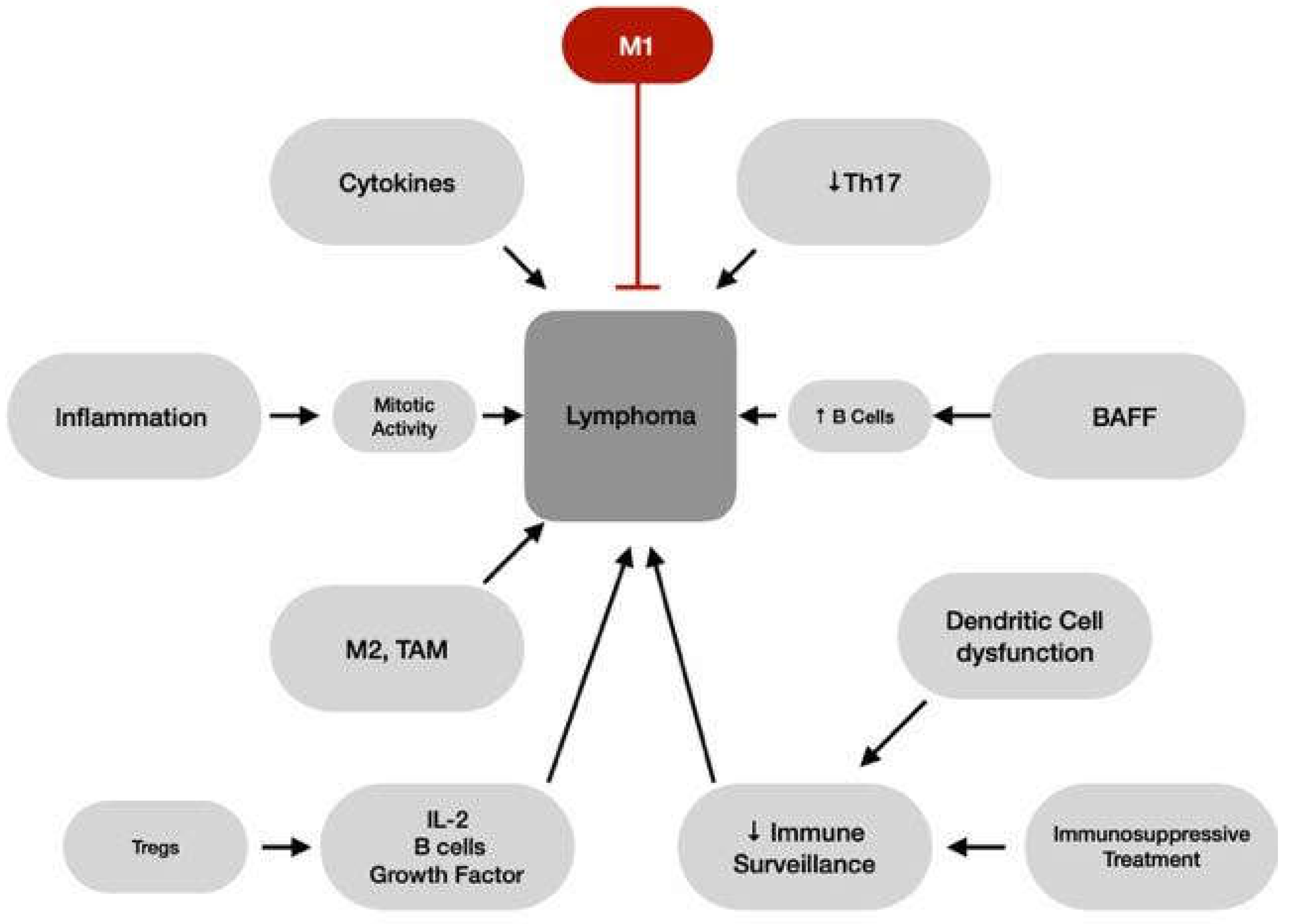
Figure 2.
Genetic and molecular mechanisms that connect sarcoidosis to lymphoma.
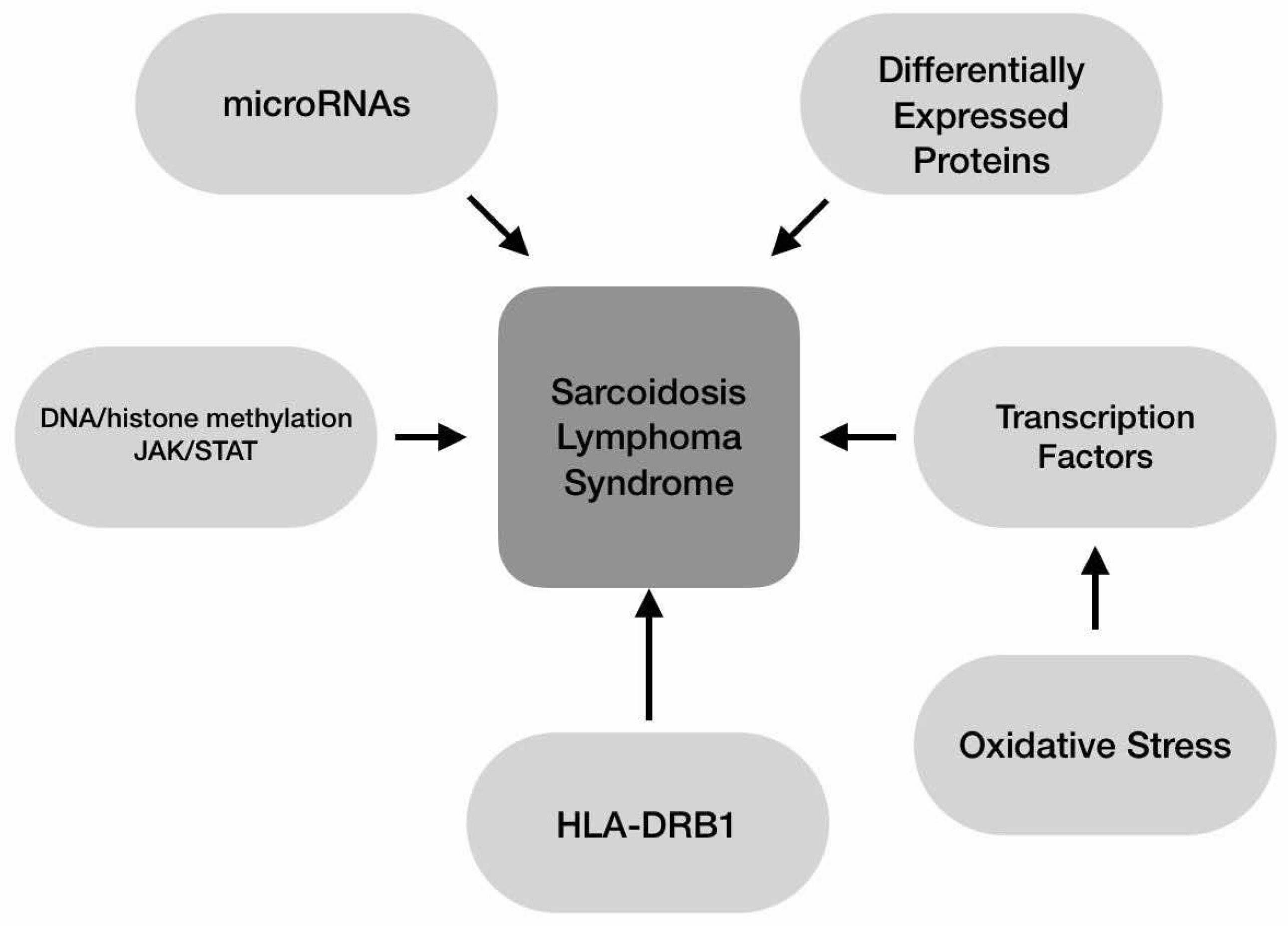
Figure 3.
Summary of the immunopathogenic mechanisms that cause Sarcoid-Like Reaction formation.
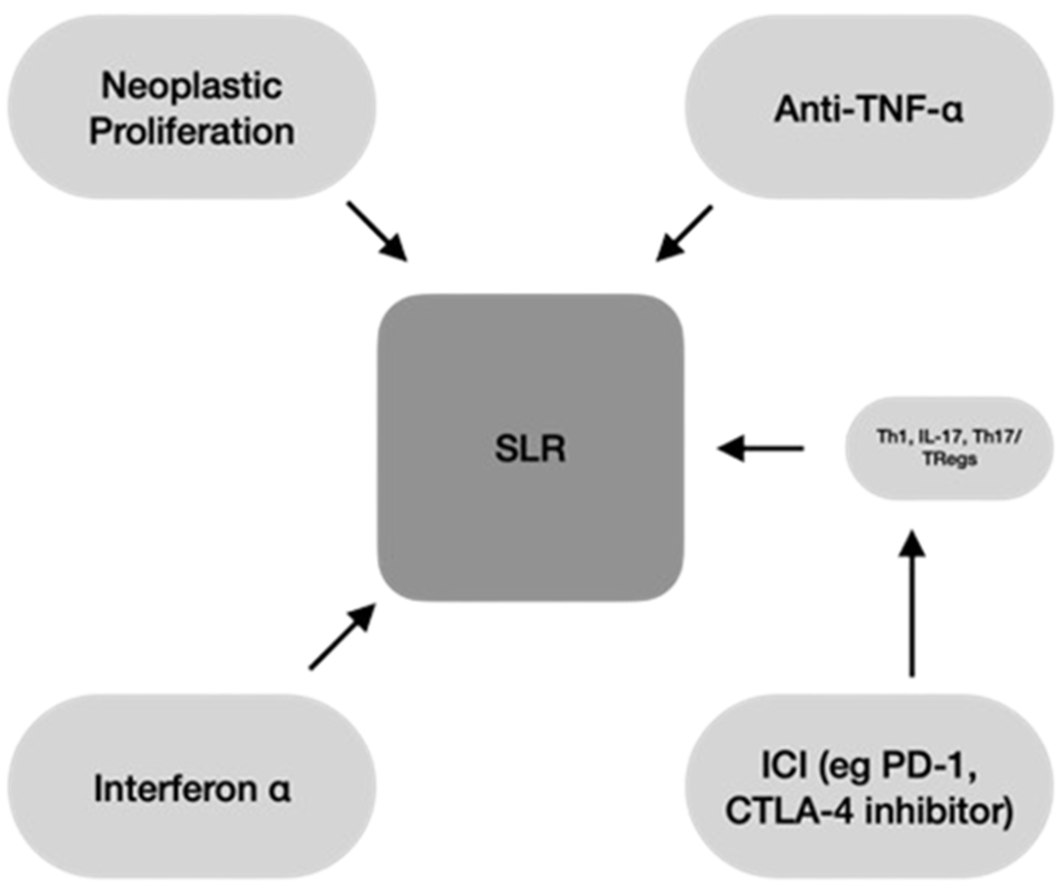

Table 1.
In this table the different risk factors of NHL, HL and Sarcoidosis are listed. For certain risk factors associated with lymphoma, the specific type of malignancy they predispose to is indicated in parentheses. Abbreviations used: NHL: Non-Hodgkin Lymphoma, HL: Hodgkin Lymphoma, FL: Follicular Lymphoma, CLL: Chronic Lymphocytic Leukemia, TCE: trichloroethylene, UV: Ultraviolet, BMI: Body Mass Index. According to references: [57,58,59,60,61].
Table 1.
In this table the different risk factors of NHL, HL and Sarcoidosis are listed. For certain risk factors associated with lymphoma, the specific type of malignancy they predispose to is indicated in parentheses. Abbreviations used: NHL: Non-Hodgkin Lymphoma, HL: Hodgkin Lymphoma, FL: Follicular Lymphoma, CLL: Chronic Lymphocytic Leukemia, TCE: trichloroethylene, UV: Ultraviolet, BMI: Body Mass Index. According to references: [57,58,59,60,61].
| NHL | HL | Sarcoidosis |
|---|---|---|
| Smoking (FL) | Smoking | Decreased sun exposure |
| Hair Dye 1 (FL, CLL) | Eczema | Inhalation of organic bioaerosols (musty odors, industrial organic dusts) |
| UV Radiation | Ionizing Radiation (especially Uranium) | Inhalation of inorganic aerosol exposures (several metal dusts)3 |
| Dietary Fat | Wood stove, Fireplace use | |
| Dessert Foods | Exposure to photocopier tone | |
| Carbohydrates (B-cell lymphoma) | Silica exposure | |
| Broiled meat | Man-mademineral fiber exposure | |
| Solvents (especially benzene and TCE) | Silicate exposure | |
| Some pesticides | Working with vegetable dust | |
| Higher BMI | Higher BMI | Higher BMI |
| Farmers2 | Living/Working in a Farm2 | |
| Blood Transfusion (nodal B-CLL, high-grade extranodal lymphomas) | Working with high humidity |
1 Frequent use before 1980 for over 25 years and especially permanent, dark dye. 2 In relation to occupation, farmers and agricultural workers have the strongest evidence for elevated NHL risk, suggesting that chemical exposure or viral agents are to blame. 3 beryllium, zirconium, titanium, nickel, chromium, cobalt, silicon, earth elements, and aluminum.
Table 2.
In this table the different protective factors of NHL, HL and Sarcoidosis are listed. For certain lymphoma-related factors, the specific type of malignancy they are known to prevent is noted in parentheses. Abbreviations used: DLBCL: Diffuse Large B-cell Lymphoma, FL: Follicular Lymphoma, EBV: Epstein-Barr Virus, BMI: Body Mass Index. According to references: [59,60,61,62].
Table 2.
In this table the different protective factors of NHL, HL and Sarcoidosis are listed. For certain lymphoma-related factors, the specific type of malignancy they are known to prevent is noted in parentheses. Abbreviations used: DLBCL: Diffuse Large B-cell Lymphoma, FL: Follicular Lymphoma, EBV: Epstein-Barr Virus, BMI: Body Mass Index. According to references: [59,60,61,62].
| NHL | HL | Sarcoidosis |
|---|---|---|
| UV Radiation (B-cell subtypes, DLBCL) | UV Radiation (EBV-positive HL) | Smoking |
| Vitamin C (FL) | Physical activity (younger women) | |
| Dietary B12 | Higher BMI (Older Women) | |
| Vitamin B6, Methionine, Folate | Low-dose aspirin use1 | |
| Other Antioxidants: Dietary Manganese, Proanthocyanidins, alpha-carotene | ||
| High intake of some vegetables (cruciferous vegetables) and fruits | ||
| Sun Exposure | ||
| Atopic Diseases | ||
| Blood Transfusion | ||
| Alcohol |
1 Protective factor only for never/rare users of nonaspirin Nonsteroidal Anti-Inflammatory Drugs (NSAIDs).
Disclaimer/Publisher’s Note: The statements, opinions and data contained in all publications are solely those of the individual author(s) and contributor(s) and not of MDPI and/or the editor(s). MDPI and/or the editor(s) disclaim responsibility for any injury to people or property resulting from any ideas, methods, instructions or products referred to in the content. |
© 2024 by the authors. Licensee MDPI, Basel, Switzerland. This article is an open access article distributed under the terms and conditions of the Creative Commons Attribution (CC BY) license (http://creativecommons.org/licenses/by/4.0/).
Copyright: This open access article is published under a Creative Commons CC BY 4.0 license, which permit the free download, distribution, and reuse, provided that the author and preprint are cited in any reuse.


