

Submitted:
11 December 2024
Posted:
12 December 2024
You are already at the latest version
Abstract
Background and Aims: This study aims to evaluate the quantitative changes in retinal nerve fiber layer (RNFL) thickness and radial peripapillary capillary (RPC) vessel density in patients with chronic central serous chorioretinopathy (CSCR), specifically excluding the peripapillary region. Methods: A prospective case-control study was conducted at the Antalya Training and Research Hospital, Health Sciences University, involving 65 patients with chronic CSCR. Participants were categorized into two groups based on the presence or regression of subretinal fluid (SRF). A control group of age- and sex-matched healthy individuals was also included. Optical coherence tomography angiography (OCTA) was used to assess RNFL thickness and RPC vessel density. Statistical analyses were conducted using SPSS, with non-parametric tests employed for between-group comparisons. Results: Patients with persistent SRF exhibited significant increases in RNFL thickness in the inferior and nasal quadrants compared to healthy controls (p = 0.003 and p = 0.014, respectively). Additionally, RPC vessel density in the small vessel disc area (%) was significantly lower in the persistent SRF group compared to controls (p = 0.021). A significant negative correlation was found between nasal quadrant RNFL thickness and small vessel disc area (p = 0.014, r = -0.306). Conclusions: Chronic SRF in CSCR patients, even when not involving the peripapillary region, leads to significant structural changes in both the neural and vascular components of the retina. These findings suggest that SRF contributes to broader retinal alterations and supports the need for early detection and management of CSCR to prevent long-term visual impairment.
Keywords:
1. Introduction
2. Materials and Methods
2.1. Assessment of Retinal Nerve Fiber Layer Thickness and Radial Peripapillary Capillary Density
2.2. Statistical Analyses
3. Results
3.1. Demographic and Clinical Characteristics of Participants
3.2. Radial Peripapillary Capillary Vessel Density (RPC VD)
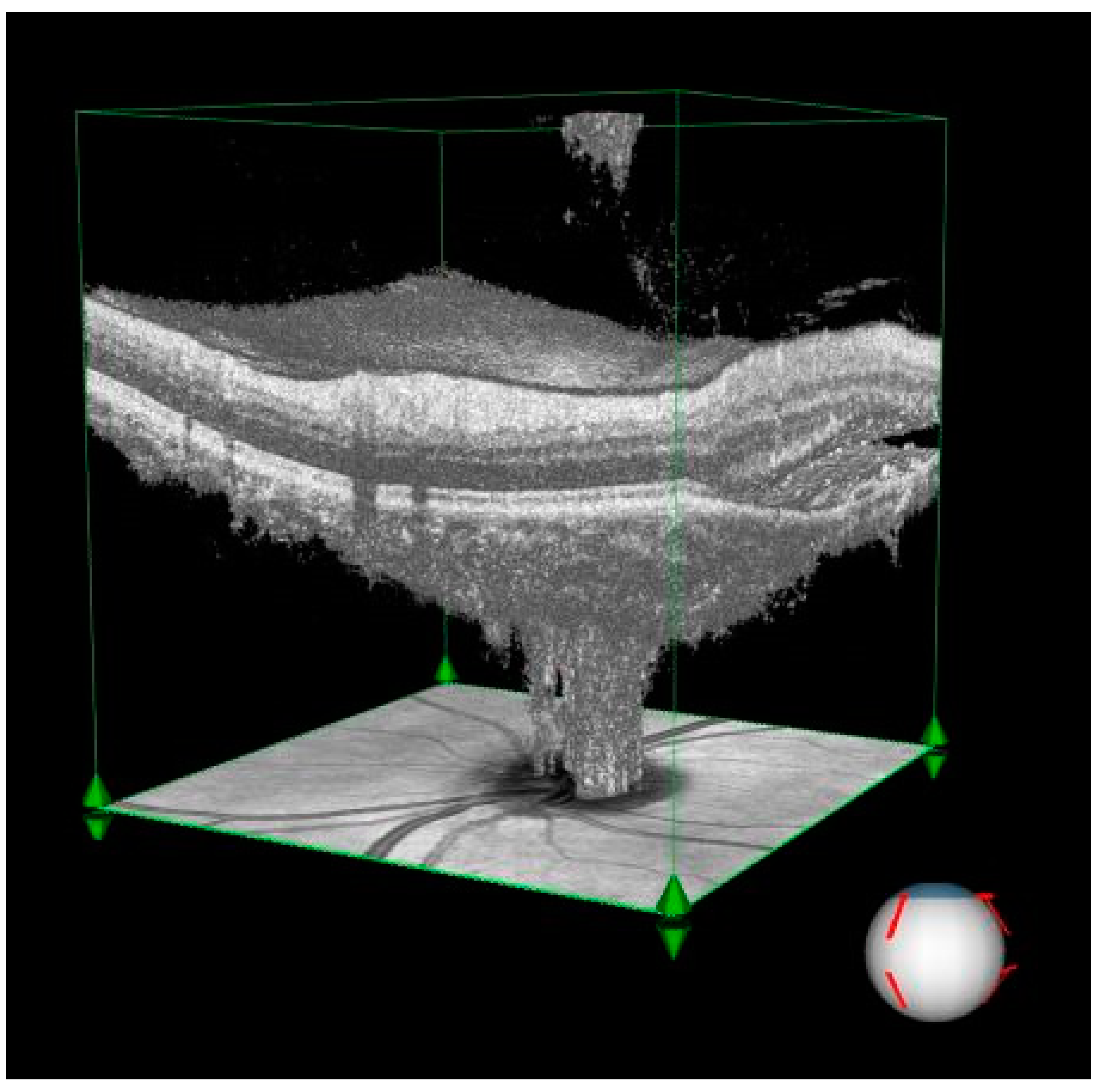
- Subretinal fluid: The visualization demonstrates an extensive fluid pocket, which extends into the peripapillary area, directly impacting the optic nerve head.
- Structural changes: The image highlights how the presence of fluid distorts the normal retinal architecture, including potential thinning of the surrounding neural layers and vascular structures.
- Clinical relevance: This degree of peripapillary involvement emphasizes the importance of early detection and management in CSCR to prevent long-term complications such as optic nerve compression or irreversible damage.
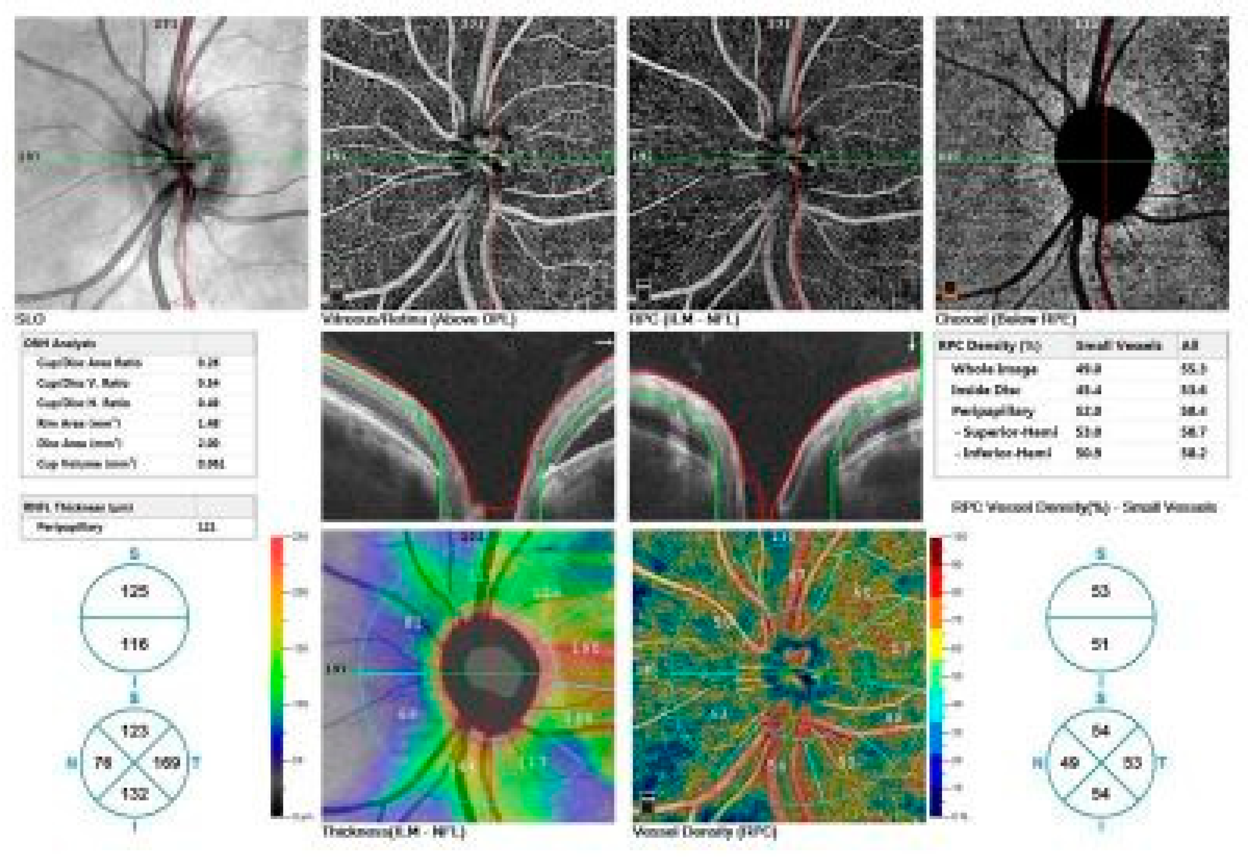
- Top row: Enface views highlight the vascular structure in different retinal layers, including the vitreous/retina (above OPL), radial peripapillary capillaries (ILM-NFL), and choroid (below RPE).
- Middle row: ONH analysis provides detailed measurements of cup/disc area ratio, rim area, disc area, and cup volume. Retinal nerve fiber layer (RNFL) thickness and vessel density maps are shown, illustrating structural and vascular integrity.
- Bottom row: RNFL thickness map (left) and vessel density map (right) offer quantitative insights into the peripapillary region's structural and vascular changes.
3.3. Radial Nerve Fiber Layer Thickness (RNFL)
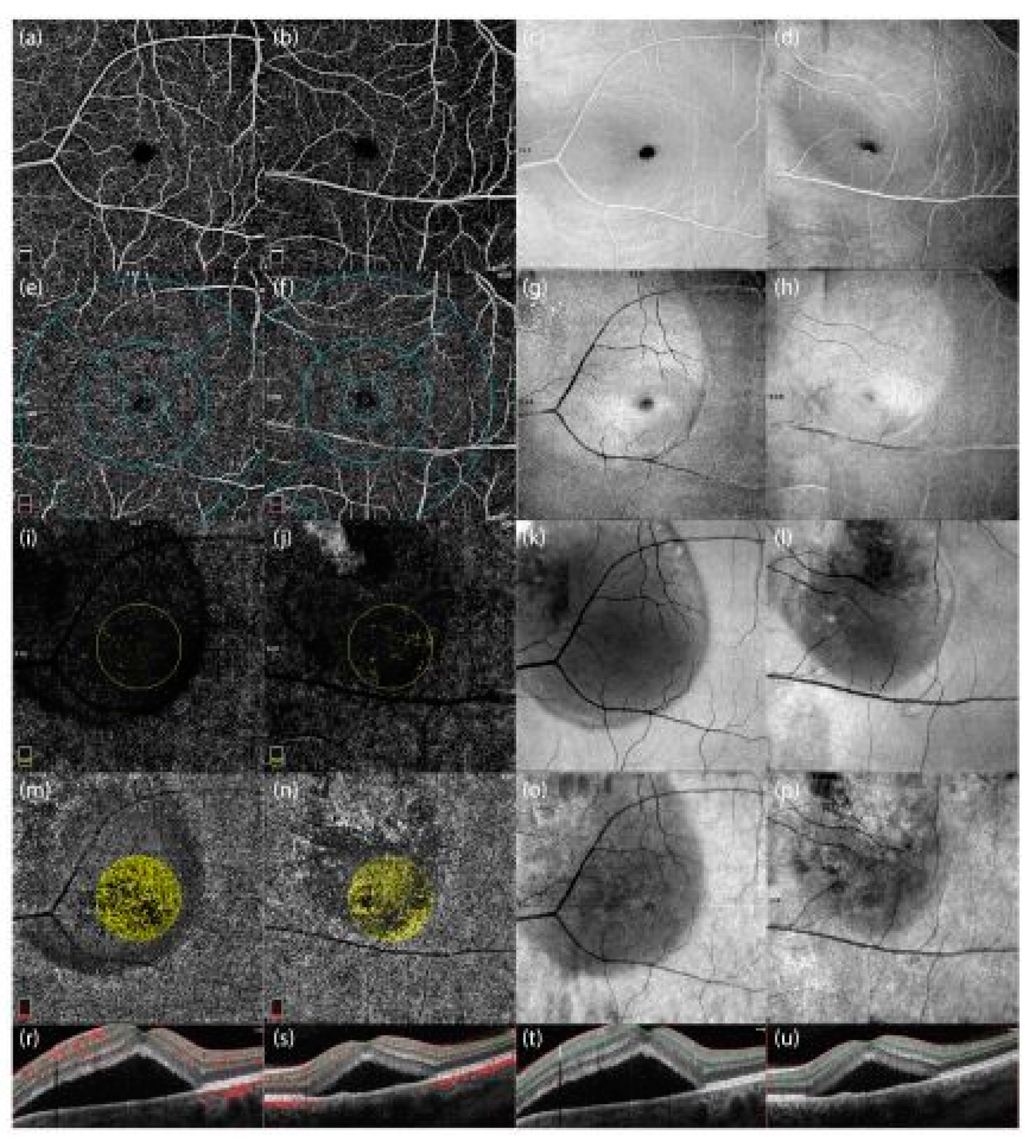
- Enface images: Panels c, g, k, o correspond to the 48-year-old patient, and panels d, h, l, p to the 46-year-old patient.
- OCT cross-sectional scans: Panels t and u display the structural characteristics of the retina in both patients.
- OCTA with angiographic overlays: Panels r and s demonstrate vascular alterations associated with subretinal fluid accumulation.
3.4. Correlation Analyses
4. Discussion
5. Study Limitations and the Need for Functional Correlates
6. Recommendations for Future Research Directions
7. Conclusions
Author Contributions
Funding
Informed Consent Statement
Data Availability Statement
Conflicts of Interest
References
- Daruich, A., Matet, A., Dirani, A., Bousquet, E., Zhao, M., Farman, N., Jaisser, F., &Behar-Cohen, F. (2015). Central serouschorioretinopathy: Recentfindingsandnewphysiopathologyhypothesis. Progress in RetinalandEyeResearch, 48, 82–118. [CrossRef]
- Liew, G., Quin, G., Gillies, M., &Fraser-Bell, S. (2013). Central serouschorioretinopathy: A review of epidemiologyandpathophysiology. ClinicalandExperimentalOphthalmology, 41(2), 201–214. [CrossRef]
- Gemenetzi, M., De Salvo, G., &Lotery, A. J. (2010). Central serouschorioretinopathy: An update on pathogenesisandtreatment. Eye, 24(11), 1743–1756 . [CrossRef]
- Nicholson, B., Noble, J., Forooghian, F., & Meyerle, C. (2013). Central serous chorioretinopathy: Update on pathophysiology and treatment. Survey of Ophthalmology, 58(2), 103-126. [CrossRef]
- Goto, K., Miki, A., Yamashita, T., Araki, S., Takizawa, G., Ieki, Y., &Kiryu, J. (2019). Retinalnerve fiber layerandperipapillarycapillarydensityreductiondetectedusingopticalcoherencetomographyenfaceimagesandangiography in optictractsyndrome. Journal of Neuro-Ophthalmology, 39(2), 253–256. [CrossRef]
- Cunha-Vaz, J. (2017). Mechanisms of retinalfluidaccumulationandblood-retinalbarrierbreakdown. Ophthalmologica, 238(1-2), 11–20. [CrossRef]
- Wang, M., Munch, I. C., Hasler, P. W., Prünte, C., &Larsen, M. (2008). Central serouschorioretinopathy. ActaOphthalmologica, 86(2), 126–145. [CrossRef]
- Imai, A., Toriyama, Y., Iesato, Y., Hirano, T., & Murata, T. (2014). En face swept-source optical coherence tomography detecting thinning of inner retinal layers as an indicator of capillary nonperfusion. European Journal of Ophthalmology, 25(2), 153-158. [CrossRef]
- Jirarattanasopa, P., Ooto, S., Tsujikawa, A., Yamashiro, K., Hangai, M., Hirata, M., Matsumote, A., &Yoshimura, N. (2012). Assessment of macularchoroidalthicknessbyopticalcoherencetomographyandangiographicchanges in centralserouschorioretinopathy. Ophthalmology, 119(8), 1666–1678. [CrossRef]
- Jain, M., Mohan, S., &vanDijk, E. H. C. (2022). Central serouschorioretinopathy: Pathophysiology, systemicassociations, and a noveletiologicalclassification. TaiwanJournal of Ophthalmology, 12, 381–393. [CrossRef]
- Cunha-Vaz, J. (2017). Mechanisms of retinalfluidaccumulationandblood-retinalbarrierbreakdown. Ophthalmologica, 238(1-2), 11–20. [CrossRef]
- Zhang, X., Lim, C. Z. F., Chhablani, J., & Wong, Y. M. (2023). Central serous chorioretinopathy: Updates in the pathogenesis, diagnosis and therapeutic strategies. BMC Ophthalmology, 10(1). [CrossRef]
- Semeraro, F., Morescalchi, F., Russo, A., Gambicorti, E., Pilotto, A., Parmeggiani, F., Bartollino, S., &Costagliola, C. (2019). Central serouschorioretinopathy: Pathogenesisandmanagement. DoveMedicalPress. https://www.dovepress.com/getfile.php?fileID=54405.
- Pujari, A., Surve, A., Azad, S. V., Beniwal, A., SJ, V., Chawla, R., Sachan, A., Kumar, A., & Kumar, A. (2021). Optical coherencetomographyangiography in centralserouschorioretinopathy: Thecurrentclinical role andfutureperspectives. Survey of Ophthalmology. [CrossRef]
- Regatieri, C. V., Novais, E. A., Branchini, L., Adhi, M., Cole, E. D., Louzada, R. N., Lane, M., Reichel, E., &Duker, J. S. (2016). Choroidalthickness in olderpatientswithcentralserouschorioretinopathy. International Journal of Retina andVitreous, 2(1). [CrossRef]
- Montero, J. A., &Ruiz-Moreno, J. M. (2005). Optical coherencetomographycharacterisation of idiopathiccentralserouschorioretinopathy. British Journal of Ophthalmology, 89(5), 562–564. [CrossRef]
- Sikorski, B. L., Bukowska, D., Kałuzný, J., Kowalczyk, A., & Wojtkowski, M. (2012). Restoration of photoreceptor structure and function in nonischaemic central retinal vein occlusion. Acta Ophthalmologica, 91(2). [CrossRef]
- Reich, M., Boehringer, D., Rothaus, K., Çakır, B., Bucher, F., Daniel, M. C., Láng, S., Lagrèze, W. A., Agostini, H., & Lange, C. (2020). Swept-source optical coherence tomography angiography alleviates shadowing artifacts caused by subretinal fluid. International Ophthalmology, 40(8), 2007-2016. [CrossRef]
- Breukink, M. B., Dingemans, A. J., Hollander, A. I. D., Keunen, J. E., MacLaren, R. E., Fauser, S., Querques, G., Hoyng, C. B., Downes, S. M., &Boon, C. J. F. (2016). Chroniccentralserouschorioretinopathy: Long-termfollow-upandvision-relatedquality of life. DoveMedicalPress, 11, 39–46. [CrossRef]
- Shimada, Y., Imai, D., Ota, Y., Kanai, K., Mori, K., Murayama, K., & Yoneya, S. (2010). Retinal adaptability loss in serous retinal detachment with central serous chorioretinopathy. Investigative Ophthalmology & Visual Science, 51(6), 3210-3210. [CrossRef]
- Kim, K. T., Chae, J. B., Lee, S., Seo, E. J., & Kim, D. Y. (2021). Analyses of the effects of persistent subretinal fluid on visual/anatomic outcomes according to the type of macular neovascularization during the relaxed treat-and-extend protocol in age-related macular degeneration patients. BMC Ophthalmology, 21, Article 294. [CrossRef]
- Berni, A., Oakley, J. D., Dolz-Marco, R., Reibaldi, M., Bandello, F., & Borrelli, E. (2021). Topographical quantification of retinal fluid in type 3 MNV and associations with short-term visual outcomes. Scientific Reports, 11, Article 13845. [CrossRef]
- Ferrara, D., Calucci, D., Oréfice, J. L., Magalhães, É. P., Oréfice, F., &Costa, R. (2008). Proposedphysiopathologicalmechanismsandpotentialtherapeutictargetsforcentralserouschorioretinopathy. ExpertReview of Ophthalmology, 3(5), 553–565. [CrossRef]
- Lim, J. S., Moon, C., & Lee, J. (2022). Subretinalfluiddisturbstheretinalvenousbloodflow in centralserouschorioretinopathy. ScientificReports, 12(1), 8865. [CrossRef]
- Mohabati, D., vanRijssen, T. J., vanDijk, E. H. C., Luyten, G. P. M., Missotten, T., Hoyng, C. C. B., Yzer, S., &Boon, C. J. F. (2018). Clinicalcharacteristicsandlong-termvisualoutcome of severe phenotypes of chroniccentralserouschorioretinopathy. DoveMedicalPress, 12, 1061–1070. [CrossRef]
- Gawęcki, M., Jaszczuk-Maciejewska, A., Jurska-Jaśko, A., Kneba, M., &Grzybowski, A. (2019). Impairment of visualacuityandretinalmorphologyfollowingresolvedchroniccentralserouschorioretinopathy. BMC Ophthalmology, 19(1), 1171. [CrossRef]
- Steinberg, J. S., Saßmannshausen, M., Fleckenstein, M., Fimmers, R., Ishibashi, T., Holz, F. G., &Schmitz-Valckenberg, S. (2016). Correlation of partialouterretinalthicknesswithscotopicandmesopicfundus-controlledperimetry in patientswithreticulardrusen. AmericanJournal of Ophthalmology, 168, 52–61. [CrossRef]
- Choi, S. S., Zawadzki, R. J., Keltner, J. L., &Werner, J. S. (2008). Changes in cellularstructuresrevealedby ultra-high-resolutionretinalimaging in opticneuropathies. InvestigativeOphthalmology& Visual Science, 49(5), 2103. [CrossRef]
- Diagnosing and managing central serous chorio retinopathy. (2023, February 6). American Academy of Ophthalmology. https://www.aao.org/eyenet/article/diagnosing-managing-central-serous-chorioretinopat.
- Han, K. J., Kim, H. J., Woo, J. M., &Min, J. K. (2020). Comparison of retinallayerthicknessandcapillaryvesseldensity in thepatientswithspontaneouslyresolvedacutecentralserouschorioretinopathy. Journal of ClinicalMedicine, 10(1), 45. [CrossRef]
- Abouammoh, M. A. (2015). Advances in thetreatment of centralserouschorioretinopathy. SaudiJournal of Ophthalmology, 29(4), 278–286. [CrossRef]
- Yavuz, S., Balsak, S., Karahan, M., & Dursun, B. (2021). Investigating the efficacy and safety of oral spironolactone in patients with central serous chorioretinopathy. Journal Français d'Ophtalmologie, 44(1), 13-23. [CrossRef]

| CSCR patients with regressed* SRF Median (min-max) n: 27 |
CSCR patients with SRF Median (min-max) n: 38 |
Pvalue | |
| Age (year) | 47.00 (31.00-78.00) | 45.00 (32.00-78.00) | 0.457 |
| Vision (LogMar) | 0.045 (0.154-0.00) | 1.301 (1.00-0.096) | <0.001 |
| IOP (mmHg) | 14.00 (11.00-20.00) | 13.00 (9.00-20.00) | 0.469 |
| CSCR patients with regressed SRF Median (min-max) n: 27 |
CSCR patients with SRF Median (min-max) n: 38 |
Control Median (min-max) n: 65 |
Pvalue | ||
| RNFL (µ) | Peripappillary | 112.00 (97.00-145.00) | 117.00 (92.00-151.00) | 113.00 (98.00-140.00) | 0.242 |
| Superior | 136.00 (114.00-239.00) | 132.00 (101.00-175.00) | 130.00 (105.00-165.00) | 0.489 | |
| Inferior | 50.20 (48.00-72.00) | 51.90 (45.00-88.00) | 50.30 (47.00-55.00) | 0.003 | |
| Temporal | 148.00 (119.00-206.00) | 146.00 (123.00-196.00) | 142.00 (114.00-190.00) | 0.609 | |
| Nasal | 74.00 (50.00-89.00) | 78.00 (60.00-115.00) | 75.00 (58.00-113.00) | 0.014 | |
| RPC VDs (%) | Small VesselWholearea | 106.00 (48.30-162.00) | 102.00 (46.80-164.00) | 100.00 (71.00-152.00) | 0.599 |
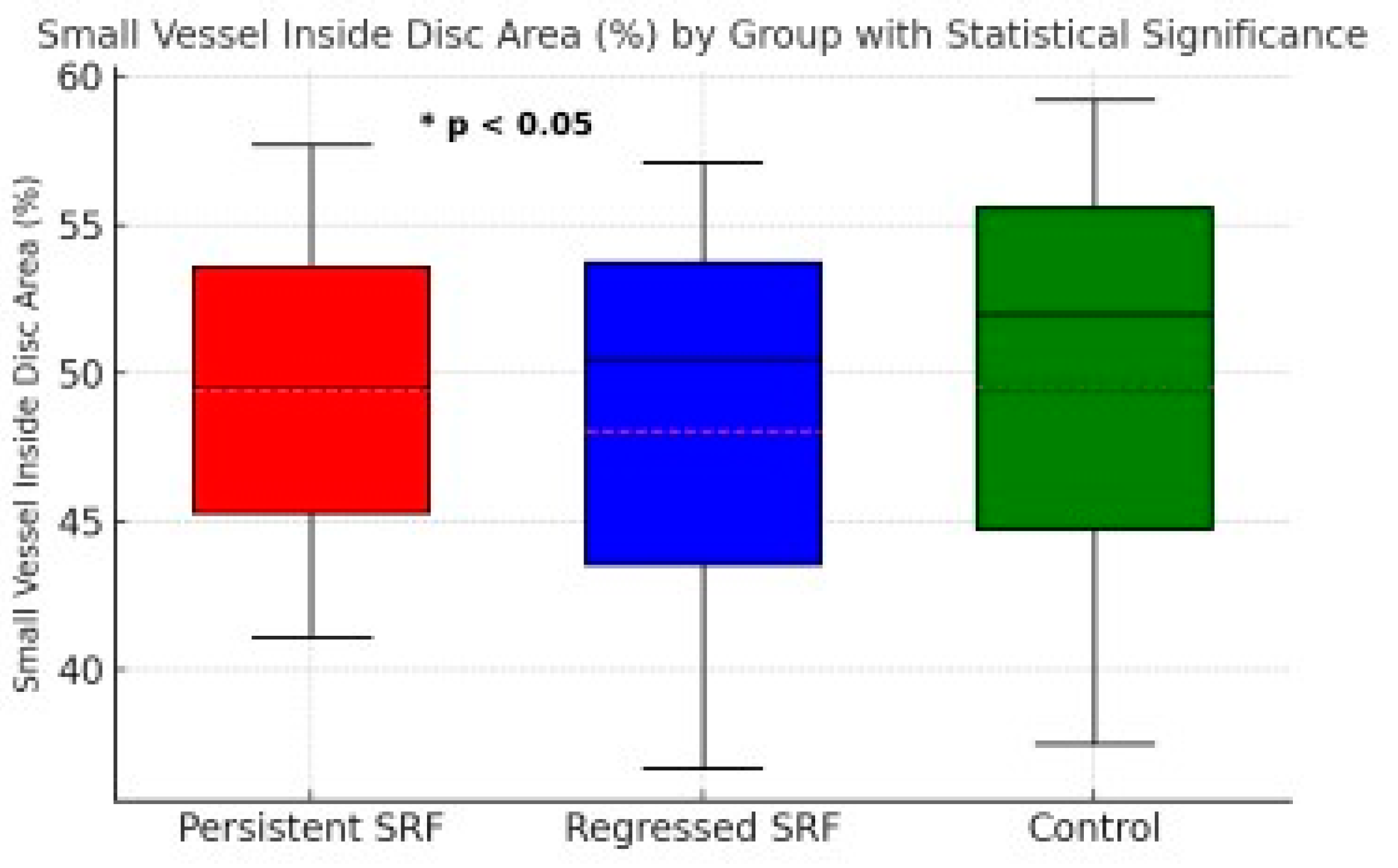
| Small Vessel Inside discarea | 50.40 (36.70-57.10) | 49.50 (41.10-57.70) | 52.00 (37.50-59.20) | 0.021 | |
| Small VesselPeripapillaryarea | 53.10 (48.40-58.60) | 52.55 (45.30-57.50) | 52.90 (49.40-57.80) | 0.331 | |
| AllVesselWholearea | 56.90 (54.10-61.00) | 57.05 (51.50-60.90) | 57.30 (52.70-61.00) | 0.987 | |
| AllVessel Inside discarea | 61.30 (50.40-67.40) | 59.70 (47.90-66.80) | 61.40 (47.10-67.90) | 0.090 | |
| AllVesselPeripapillaryarea | 59.80 (55.80-64.10) | 59.25 (45.80-63.50) | 59.20 (56.20-63.80) | 0.439 | |
| P | P1* | P2* | P3* | |
| RNFL Inferiorthickness (µ) | 0,003 | 1 | 0,003 | 0,054 |
| RNFL Nasalthickness (µ) | 0,014 | 1 | 0,023 | 0,053 |
| RPC Small VesselInsidediscareaVDs (%) | 0,021 | 0,538 | 0,019 | 0,940 |
| RNFL Inferiorthickness (µ) | RNFL Nasalthickness (µ) | ||||
| P | R† | P | R | ||
| RPC VDs (%) | Small VesselWholearea | 0.206 | -0.160 | 0.048* | -0.249 |
| Small Vessel Inside discarea | 0.393 | -0.109 | 0.014* | -0.306 | |
| Small VesselPeripapillaryarea | 0.015* | 0.300 | 0.434 | -0.099 | |
| AllVesselWholearea | 0.003** | 0.358 | 0.498 | -0.085 | |
| AllVessel Inside discarea | 0.431 | -0.099 | 0.061 | -0.233 | |
| AllVesselPeripapillaryarea | 0.006** | 0.335 | 0.835 | -0.026 | |
Disclaimer/Publisher’s Note: The statements, opinions and data contained in all publications are solely those of the individual author(s) and contributor(s) and not of MDPI and/or the editor(s). MDPI and/or the editor(s) disclaim responsibility for any injury to people or property resulting from any ideas, methods, instructions or products referred to in the content. |
© 2024 by the authors. Licensee MDPI, Basel, Switzerland. This article is an open access article distributed under the terms and conditions of the Creative Commons Attribution (CC BY) license (http://creativecommons.org/licenses/by/4.0/).