

Submitted:
01 November 2024
Posted:
05 November 2024
You are already at the latest version
Abstract
ChatGPT, a state-of-the-art AI model created by OpenAI, is renowned for its ability to produce text responses that closely mimic human communication. Recently, it has broadened its scope to encompass multimodal interactions, rendering it particularly advantageous for medical applications. In this paper, we explore into the potential of ChatGPT in the realm of Ultrasound (US) imaging, Our investigation encompasses a series of case studies and examples, each tailored to demonstrate ChatGPT’s application in various aspects of US imaging. These include emphasizing its capacity for intricate analyses, including grayscale texture examination and perfusion metrics in contrast-enhanced ultrasound (CEUS). Moreover, its involvement in real-time imaging guidance, its role in streamlining workflow efficiencies, and its utility in the simplification of medical report generation to facilitate better patient communication. The case studies, while successful, unveil certain limitations and challenges, prominently featuring concerns regarding privacy and technical constraints. Despite these challenges, the evidence gathered from our examples indicates that ChatGPT's innovative features and its ability to operate across multiple modes significantly enhance the efficiency of quantitative image analysis in US imaging. By improving diagnostic precision and potentially elevating patient care outcomes, ChatGPT marks a significant stride forward in healthcare technology, though it necessitates careful consideration of its associated ethical and technical challenges.
Keywords:
Introduction
Technical Overview of ChatGPT
ChatGPT Example Applications in Ultrasound Imaging
Examples for Using GPT-4 for Quantitative Image Analysis
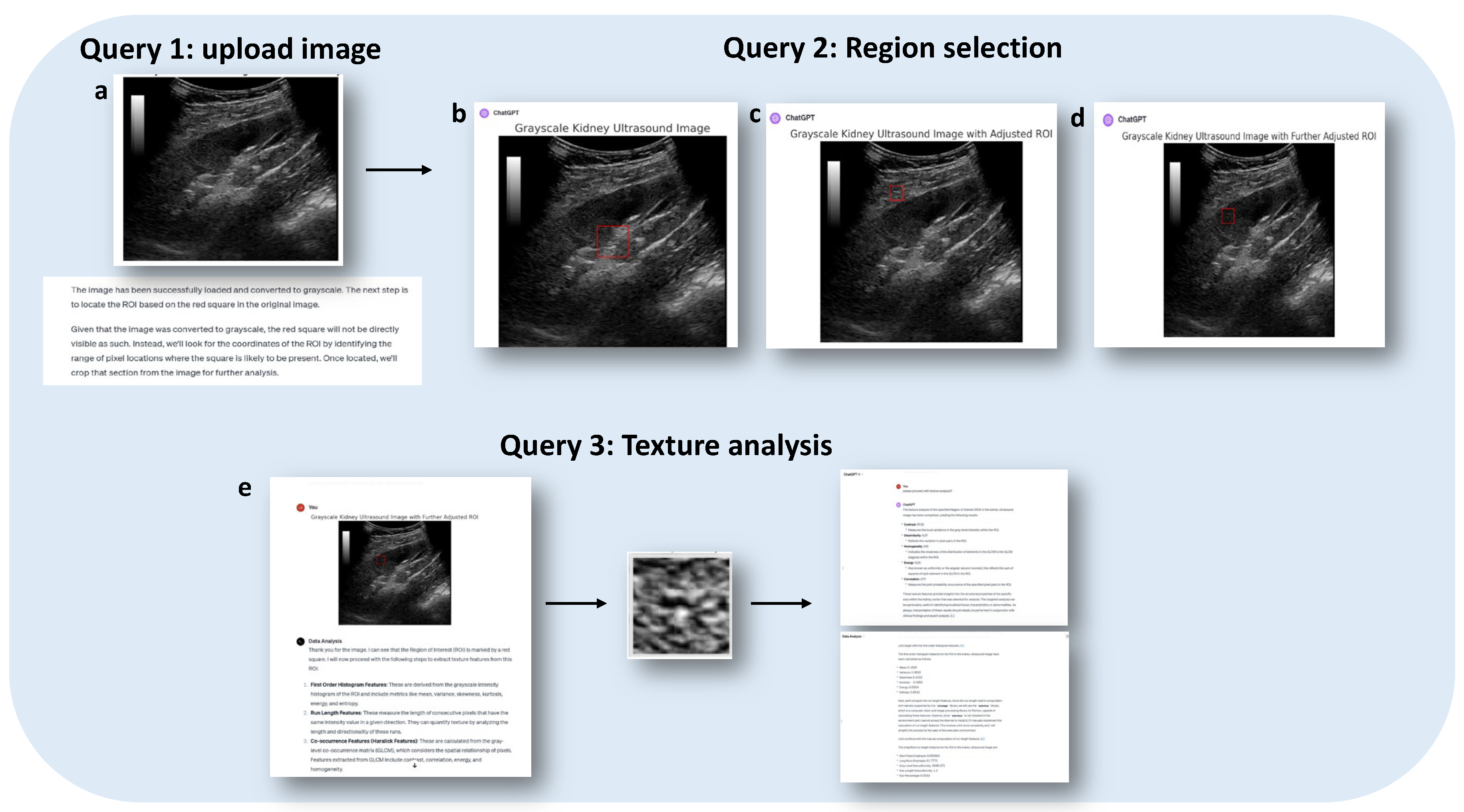
Example 1: Grayscale Texture Analysis Based on B-Mode Ultrasound Images
Example 2: Time Intensity Curve Analysis for Contrast Enhanced Ultrasound
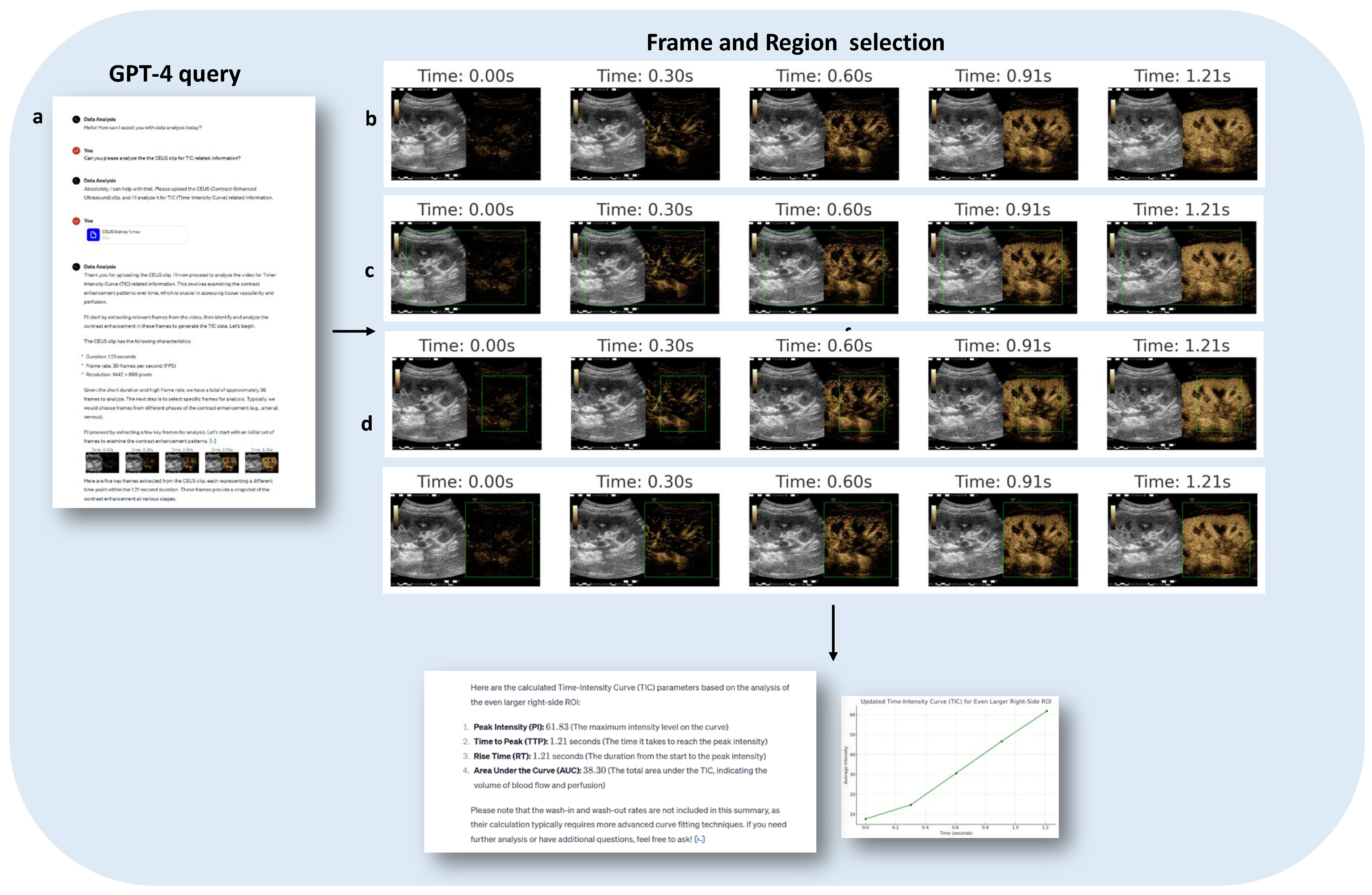
Example 3: Generating Super Resolution Images Based on Vascular Enhancement of CEUS
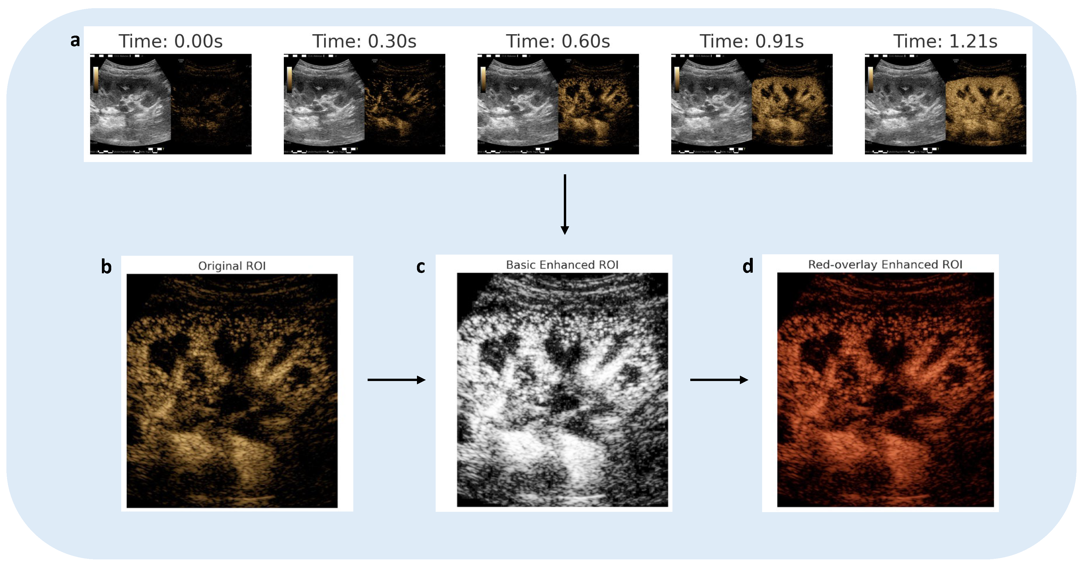
Examples for Using ChatGPT for Improving Workflow
Example 1: Simplifying Report for Better Patient Communication
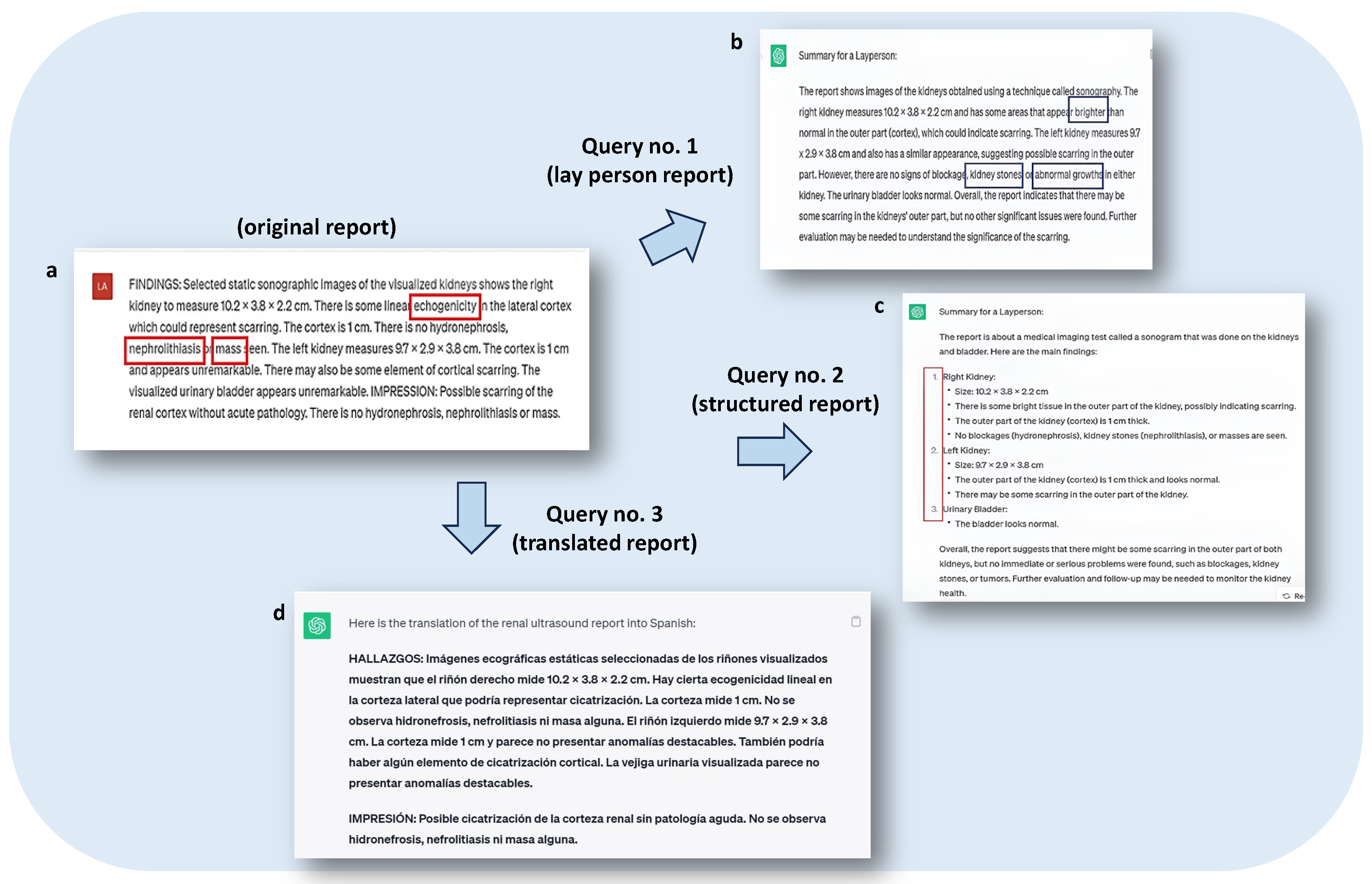
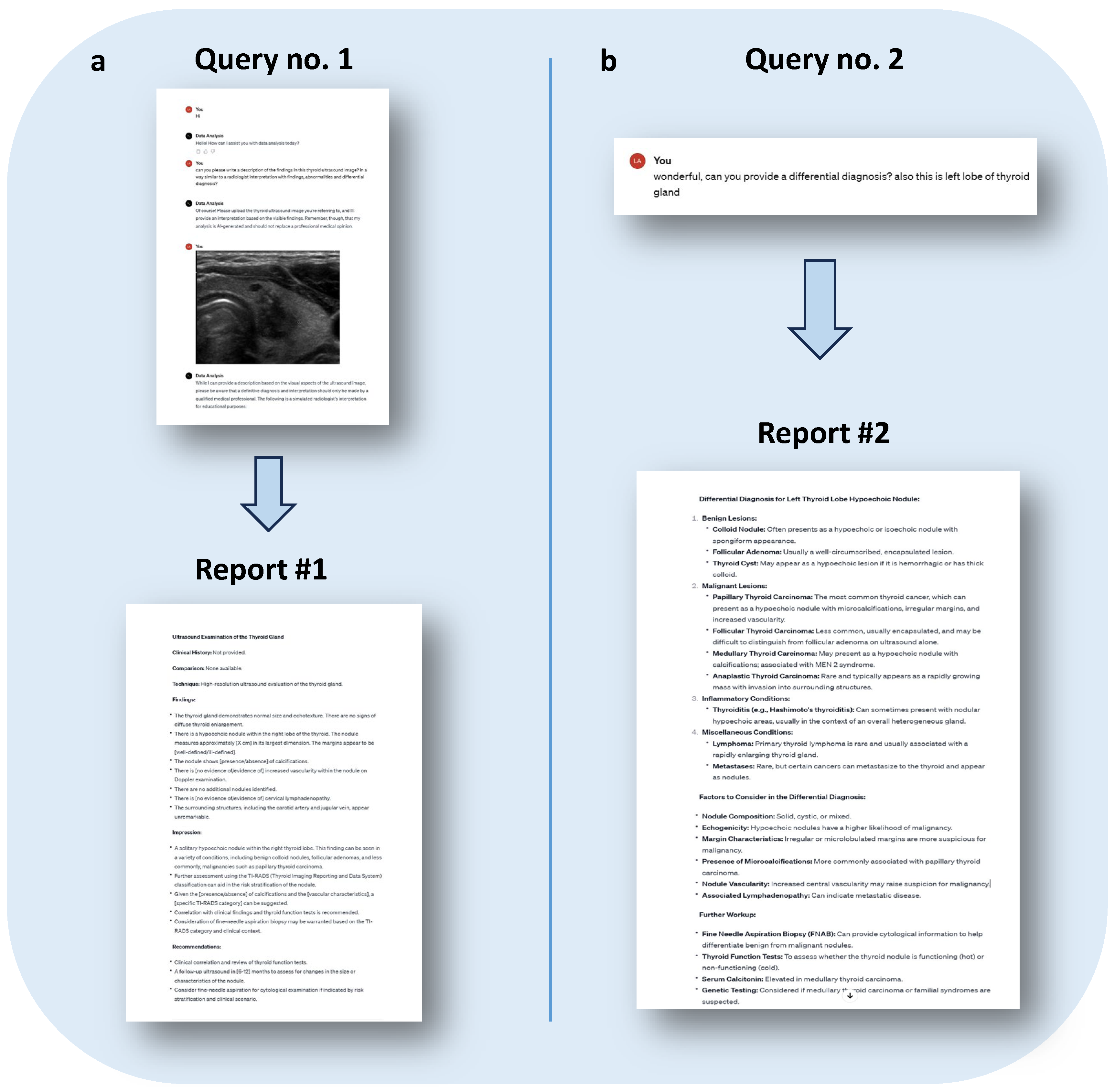
Example 2: Image Interpretation and Diagnosis Report Generation
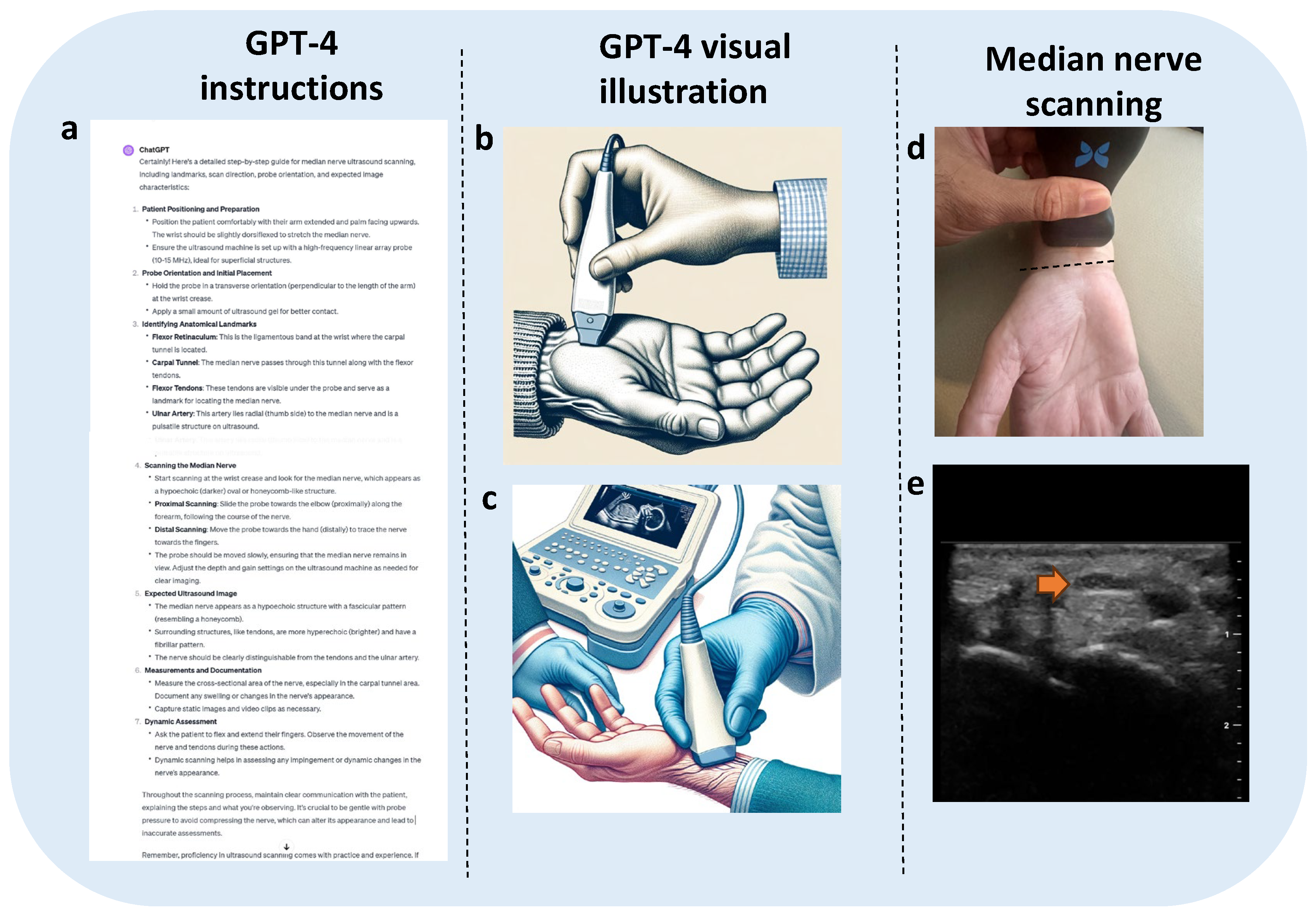
Example 3: ChatGPT Provides Real-Time US Imaging Guidance
Challenges and Considerations for Implementing ChatGPT in Practice
Conclusions
Author Contributions:
Funding
Institutional Review Board Statement
Informed Consent Statement
Data Availability Statement
Conflicts of Interest
References
- Oren, O.; Gersh, B.J.; Bhatt, D.L. Artificial intelligence in medical imaging: switching from radiographic pathological data to clinically meaningful endpoints. Lancet Digital Health. 2020, 2, e486–e488. [Google Scholar] [CrossRef] [PubMed]
- Kim, H.E.; Kim, H.H.; Han, B.K.; Ghafoorian, M.; Tan, T.; Andriessen, T.; Vyvere, T.V.; Hauwe, L.v.D.; Romeny, B.t.H.; Goraj, B.; et al. Changes in cancer detection and false-positive recall in mammography using artificial intelligence: a retrospective, multireader study. Lancet Digital Health 2020, 2, e138–48. [Google Scholar] [CrossRef]
- van den Heuvel, T.L.; van der Eerden, A.W.; Manniesing, R.; et al. Automated detection of cerebral microbleeds in patients with traumatic brain injury. Neuroimage Clin. 2016, 12, 241–251. [Google Scholar] [CrossRef]
- Becker, A.S.; Marcon, M.; Ghafoor, S.; Wurnig, M.C.; Frauenfelder, T.; Boss, A. Deep learning in mammography: diagnostic accuracy of a multipurpose image analysis software in the detection of breast cancer. Invest. Radiol. 2017, 52, 434–440. [Google Scholar] [CrossRef] [PubMed]
- Liu, X.; Faes, L.; Kale, A.U.; et al. A comparison of deep learning performance against health-care professionals in detecting diseases from medical imaging: a systematic review and meta-analysis. Lancet Digital Health 2019, 1, e271–97. [Google Scholar] [CrossRef]
- Bello, G.A.; Dawes, T.J.W.; Duan, J.; Wagner, S.K.; Fu, D.J.; Bruynseels, A.; Mahendiran, T.; Moraes, G.; Shamdas, M.; Kern, C.; et al. Deep learning cardiac motion analysis for human survival prediction. Nat. Mach. Intell. 2019, 1, 95–104. [Google Scholar] [CrossRef] [PubMed]
- Hosny, A.; Parmar, C.; Quackenbush, J.; Schwartz, L.H.; Aerts, H.J.W.L. Artificial intelligence in radiology. Nat. Rev. Cancer 2018, 18, 500–510. [Google Scholar] [CrossRef]
- Lauritzen, A.D.; Rodríguez-Ruiz, A.; von Euler-Chelpin, M.C.; Lynge, E.; Vejborg, I.; Nielsen, M.; Karssemeijer, N.; Lillholm, M. An Artificial Intelligence-based Mammography Screening Protocol for Breast Cancer: Outcome and Radiologist Workload. Radiology. 2022, 304, 41–49. [Google Scholar] [CrossRef] [PubMed]
- Subhan, S.; Malik, J.; Haq, A.U.; Qadeer, M.S.; Zaidi, S.M.J.; Orooj, F.; Zaman, H.; Mehmoodi, A.; Majeedi, U. Role of Artificial Intelligence and Machine Learning in Interventional Cardiology. Curr. Probl. Cardiol. 2023, 48, 101698. [Google Scholar] [CrossRef] [PubMed]
- Di Serafino, M.; Iacobellis, F.; Schillirò, M.L.; D’auria, D.; Verde, F.; Grimaldi, D.; Dell’Aversano Orabona, G.; Caruso, M.; Sabatino, V.; Rinaldo, C.; et al. Common and Uncommon Errors in Emergency Ultrasound. Diagnostics 2022, 12, 631. [Google Scholar] [CrossRef]
- Pinto, A.; Pinto, F.; Faggian, A.; Rubini, G.; Caranci, F.; Macarini, L.; Genovese, E.A.; Brunese, L.; et al. Sources of error in emergency ultrasonography. Crit. Ultrasound J. 2013, 5 (Suppl. S1), S1. [Google Scholar] [CrossRef] [PubMed]
- Le, M.P.T.; Voigt, L.; Nathanson, R.; Maw, A.M.; Johnson, G.; Dancel, R.; Mathews, B.; Moreira, A.; Sauthoff, H.; Gelabert, C.; et al. Comparison of four handheld point-of-care ultrasound devices by expert users. Ultrasound J. 2022, 14, 27. [Google Scholar] [CrossRef] [PubMed]
- Malik, A.N.; Rowland, J.; Haber, B.D.; Thom, S.; Jackson, B.; Volk, B.; Ehrman, R.R. The Use of Handheld Ultrasound Devices in Emergency Medicine. Curr. Emerg. Hosp. Med. Rep. 2021, 9, 73–81. [Google Scholar] [CrossRef]
- Kim, Y.H. Artificial intelligence in medical ultrasonography: driving on an unpaved road. Ultrasonography. 2021, 40, 313–317. [Google Scholar] [CrossRef] [PubMed]
- Komatsu, M.; Sakai, A.; Dozen, A.; Shozu, K.; Yasutomi, S.; Machino, H.; Asada, K.; Kaneko, S.; Hamamoto, R. Towards Clinical Application of Artificial Intelligence in Ultrasound Imaging. Biomedicines. 2021, 9, 720. [Google Scholar] [CrossRef]
- Shao, D.; Yuan, Y.; Xiang, Y.; Yu, Z.; Liu, P.; Liu, D.C. Artifacts detection-based adaptive filtering to noise reduction of strain imaging. Ultrasonics. 2019, 98, 99–107. [Google Scholar] [CrossRef]
- Wen, T.; Gu, J.; Li, L.; Qin, W.; Wang, L.; Xie, Y. Nonlocal Total-Variation-Based Speckle Filtering for Ultrasound Images. Ultrason. Imaging. 2016, 38, 254–275. [Google Scholar] [CrossRef]
- Zhang, L.; Zhang, J. Ultrasound image denoising using generative adversarial networks with residual dense connectivity and weighted joint loss. PeerJ Comput. Sci. 2022, 8, e873. [Google Scholar] [CrossRef] [PubMed]
- Else, H. Abstracts written by ChatGPT fool scientists. Nature 2023. [CrossRef]
- Dwivedi, Y.K.; Kshetri, N.; Hughes, L.; Slade, E.L.; Jeyaraj, A.; Kar, A.K.; Baabdullah, A.M.; Koohang, A.; Raghavan, V.; Ahuja, M.; et al. Opinion Paper: “So what if ChatGPT wrote it?” Multidisciplinary perspectives on opportunities, challenges and implications of generative conversational AI for research, practice, and policy. International Journal of Information Management. 2023, 71, 102642. [Google Scholar] [CrossRef]
- Kitamura, F.C. ChatGPT Is Shaping the Future of Medical Writing but Still Requires Human Judgment. Radiology; 0:230171.
- Jeblick, K.; Johnson, L.; Arora, S.; et al. ChatGPT makes medicine easy to swallow: an exploratory case study on simplified radiology reports. arXiv preprint arXiv:, arXiv:2212.14882. [CrossRef]
- Alhasan, K.; Lee, K.; Kumar, A.; Jamal, A.; Al-Eyadhy, A.; Temsah, M.-H.; Al-Tawfiq, J. Aet al. Mitigating the burden of severe pediatric respiratory viruses in the post-COVID-19 era: ChatGPT insights and recommendations. Cureus, 2023; 15. [Google Scholar] [CrossRef]
- Ali, S.R.; Dobbs, T.D.; Hutchings, H.A.; Whitaker, I.S. Using ChatGPT to write patient clinic letters. Lancet Digit. Health 2023, 5, e179–e181. [Google Scholar] [CrossRef] [PubMed]
- Santandreu-Calonge D, Ortiz-Martinez Y, Agusti-Toro A; et al. Can ChatGPT improve communication in hospitals? Prof. De La Inf. 2023, 32. [Google Scholar] [CrossRef]
- Biswas, S.S. Role of ChatGPT in radiology with a focus on pediatric radiology: proof by examples. Pediatr. Radiol. 2023, 53, 818–822. [Google Scholar] [CrossRef] [PubMed]
- Arya S Rao, John Kim, Meghana Kamineni, Michael Pang, Winston Lie, and Marc Succi. Evaluating chatgpt as an adjunct for radiologic decision-making. medRxiv, pages 2023–02, 2023.
- Wiggers, Kyle. OpenAI releases GPT-4, a multimodal AI that it claims is state-of-the-art”. TechCrunch. Archived from the original on March 15, 2023. Retrieved March 15, 2023.
- ubeck, Sébastien; Chandrasekaran, Varun; Eldan, Ronen; Gehrke, Johannes; Horvitz, Eric; Kamar, Ece; Lee, Peter; Lee, Yin Tat; Li, Yuanzhi; Lundberg, Scott; Nori, Harsha; Palangi, Hamid; Ribeiro, Marco Tulio; Zhang, Yi (March 22, 2023). “Sparks of Artificial General Intelligence: Early experiments with GPT-4”. arXiv:2303.12712 [cs.CL].
- ChatGPT – Release Notes”. Archived from the original on August 4, 2023. Retrieved August 4, 2023.
- Ortiz, Sabrina (February 2, 2023). “What is ChatGPT and why does it matter? Here’s what you need to know”. ZDNET. Archived from the original on January 18, 2023. Retrieved December 18, 2022.
- “Artificial intelligence technology behind ChatGPT was built in Iowa — with a lot of water”. AP News. September 9, 2023. Retrieved September 10, 2023.
- “What’s the next word in large language models?”. Nature Machine Intelligence. 5 (4): 331–332. April 2023. https://doi.org/10.1038/s42256-023-00655-z. ISSN 2522-5839. S2CID 258302563. Archived from the original on June 11, 2023. Retrieved June 10, 2023.
- “What is ChatGPT and why does it matter? Here’s what you need to know”. ZDNET. May 30, 2023. Retrieved June 22, 2023.
- Varghese, B.A.; Cen, S.Y.; Hwang, D.H.; Duddalwar, V.A. ChatGPT-4: a breakthrough in ultrasound image analysis, Radiology Advances 2024, 1, umae006,Varghese BA, Cen SY, Hwang DH, Duddalwar VA. Texture Analysis of Imaging: What Radiologists Need to Know. AJR Am. J. Roentgenol. 2019, 212, 520–528. [Google Scholar] [CrossRef] [PubMed]
- Gabrielsen, D.A.; Carney, M.J.; Weissler, J.M.; Lanni, M.A.; Hernandez, J.; Sultan, L.R.; Enriquez, F.; Sehgal, C.M.; Fischer, J.P.; Chauhan, A. Application of ARFI-SWV in Stiffness Measurement of the Abdominal Wall Musculature: A Pilot Feasibility Study. Ultrasound Med. Biol. 2018, 44, 1978–1985. [Google Scholar] [CrossRef]
- Habibollahi, P.; Sultan, L.R.; Bialo, D.; Nazif, A.; Faizi, N.A.; Sehgal, C.M.; Chauhan, A. Hyperechoic Renal Masses: Differentiation of Angiomyolipomas from Renal Cell Carcinomas using Tumor Size and Ultrasound Radiomics. Ultrasound Med. Biol. 2022, 48, 887–894. [Google Scholar] [CrossRef]
- Wu, M.; Hu, Y.; Hang, J.; Peng, X.; Mao, C.; Ye, X.; Li, A. Qualitative and Quantitative Contrast-Enhanced Ultrasound Combined with Conventional Ultrasound for Predicting the Malignancy of Soft Tissue Tumors. Ultrasound Med. Biol. 2022, 48, 237–247. [Google Scholar] [CrossRef]
- Sultan, L.R.; Karmacharya, M.B.; Al-Hasani, M.; Cary, T.W.; Sehgal, C.M. Hydralazine-augmented contrast ultrasound imaging improves the detection of hepatocellular carcinoma. Med. Phys. 2023, 50, 1728–1735. [Google Scholar] [CrossRef]
- Zhang, Z.; Hwang, M.; Kilbaugh, T.J.; Sridharan, A.; Katz, J. Cerebral microcirculation mapped by echo particle tracking velocimetry quantifies the intracranial pressure and detects ischemia. Nat. Commun. 2022, 13, 666. [Google Scholar] [CrossRef]
- Frosch, D.L.; Elwyn, G. Don’t blame patients, engage them: transforming health systems to address health literacy. J. Health Commun. 2014, 19 Suppl. s2, 10–14. [Google Scholar] [CrossRef] [PubMed]
- CDC Patient Engagement | Health Literacy | CDC.
- Zarcadoolas, C.; Vaughon, W.L.; Czaja, S.J.; Levy, J.; Rockoff, M.L. Consumers’ perceptions of patient-accessible electronic medical records. J. Med. Internet Res. 2013, 15, e168. [Google Scholar] [CrossRef] [PubMed]
- NDX-Imaging. https://www.ndximaging.com/radiology-reads/sample-reports/.
- “OpenAI API”. platform.openai.com. Archived from the original on March 3, 2023. Retrieved March 3, 2023.
Disclaimer/Publisher’s Note: The statements, opinions and data contained in all publications are solely those of the individual author(s) and contributor(s) and not of MDPI and/or the editor(s). MDPI and/or the editor(s) disclaim responsibility for any injury to people or property resulting from any ideas, methods, instructions or products referred to in the content. |
© 2024 by the authors. Licensee MDPI, Basel, Switzerland. This article is an open access article distributed under the terms and conditions of the Creative Commons Attribution (CC BY) license (http://creativecommons.org/licenses/by/4.0/).