Submitted:
02 November 2024
Posted:
04 November 2024
You are already at the latest version
Abstract
Background/Objectives: Recent advancements in artificial intelligence (AI) have spurred interest in developing computer-assisted analysis for imaging examinations. However, the lack of high-quality datasets remains a significant bottleneck. Labeling instructions are critical to improving dataset quality but are often lacking. This study aimed to establish a liver MRI segmentation protocol and assess its impact on annotation quality and inter-reader agreement; Methods: This retrospective study included 20 patients with chronic liver disease. Manual liver segmentations were performed by a radiologist in training and a radiology technician on T2 weighted imaging (wi) and T1wi at the portal venous phase. Based on the inter-reader discrepancies identified after the first segmentation round, a segmentation protocol was established, guiding the second round of segmentation, resulting in a total of 160 segmentations. Dice Similarity Coefficient (DSC) assessed inter-reader agreement pre- and post-protocol with a Wilcoxon signed-rank test. Slice selection at extreme cranial or caudal liver positions was evaluated using the McNemar test; Results: The per-volume DSC significantly increased after protocol implementation for both T2wi (p<0.001) and T1wi (p=0.03). Per-slice DSC also improved significantly for both T2wi and T1wi (p<0.001). The protocol reduced the number of liver segmentations with a non-annotated slice on T1wi (p=0.04), but the change was not significant on T2wi (p=0.16); Conclusions: Establishing a liver MRI segmentation protocol improves annotation robustness and reproducibility, paving the way for advanced computer-assisted analysis. Moreover, segmentation protocols could be extended to other organs and lesions and incorporated into guidelines, thereby expanding the potential applications of AI in daily clinical practice.
Keywords:
1. Introduction
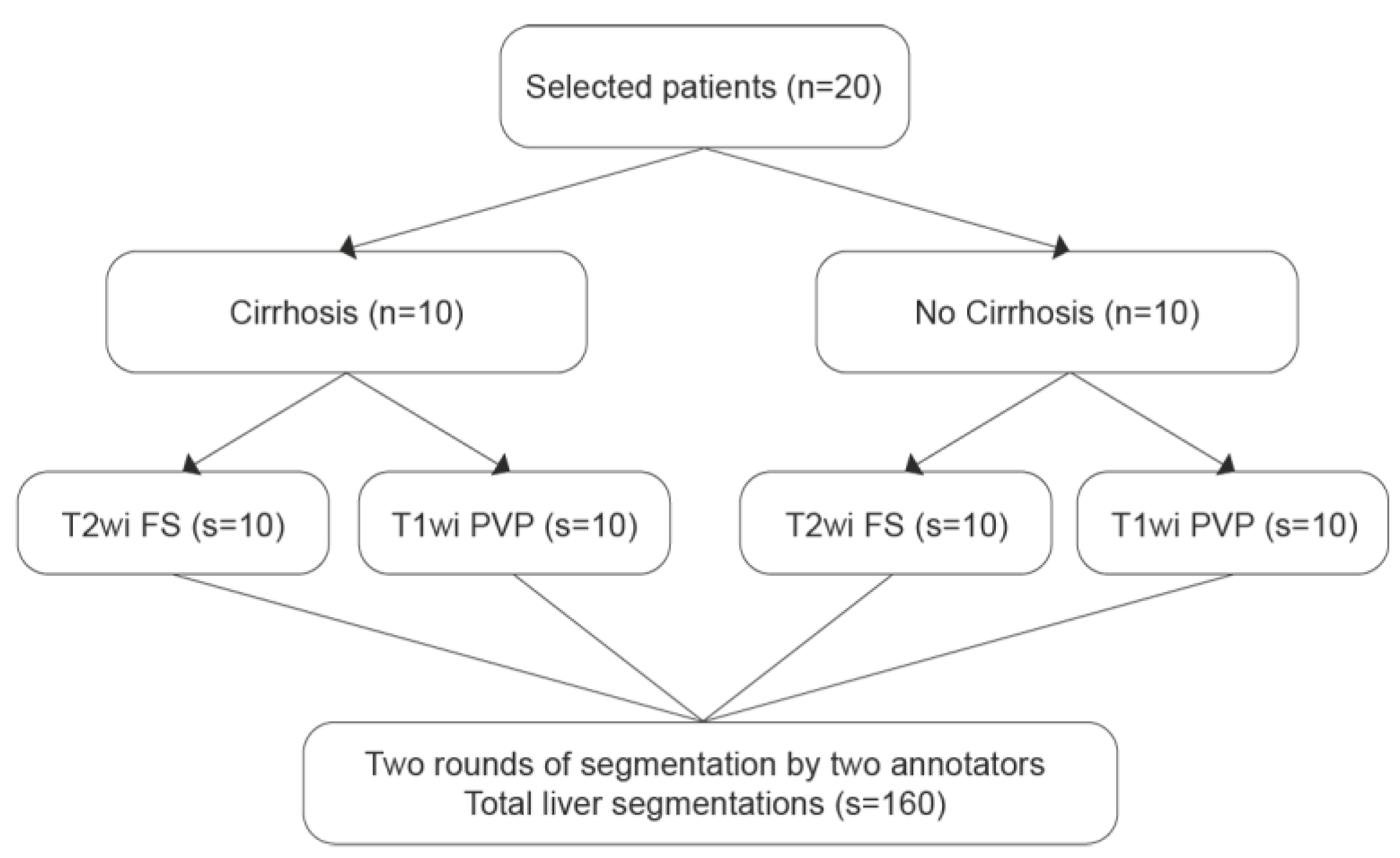
2. Materials and Methods
2.1. Patient Selection
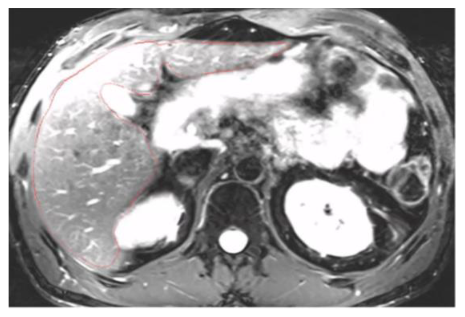
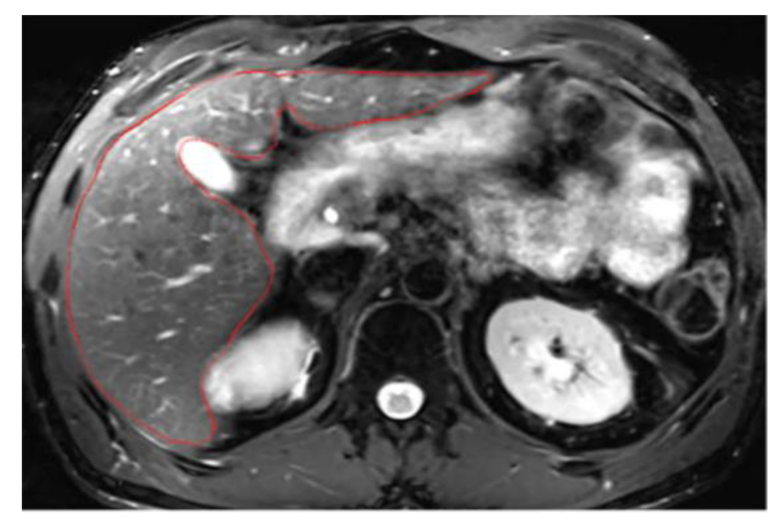
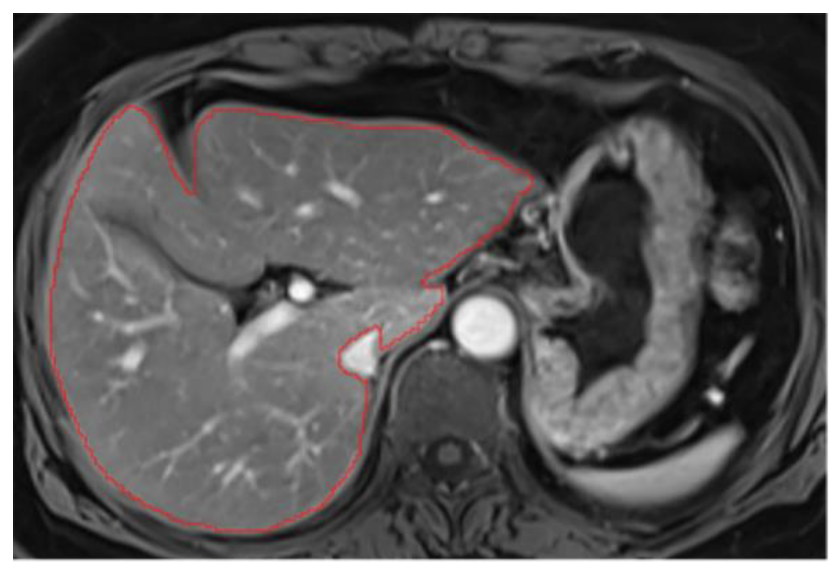
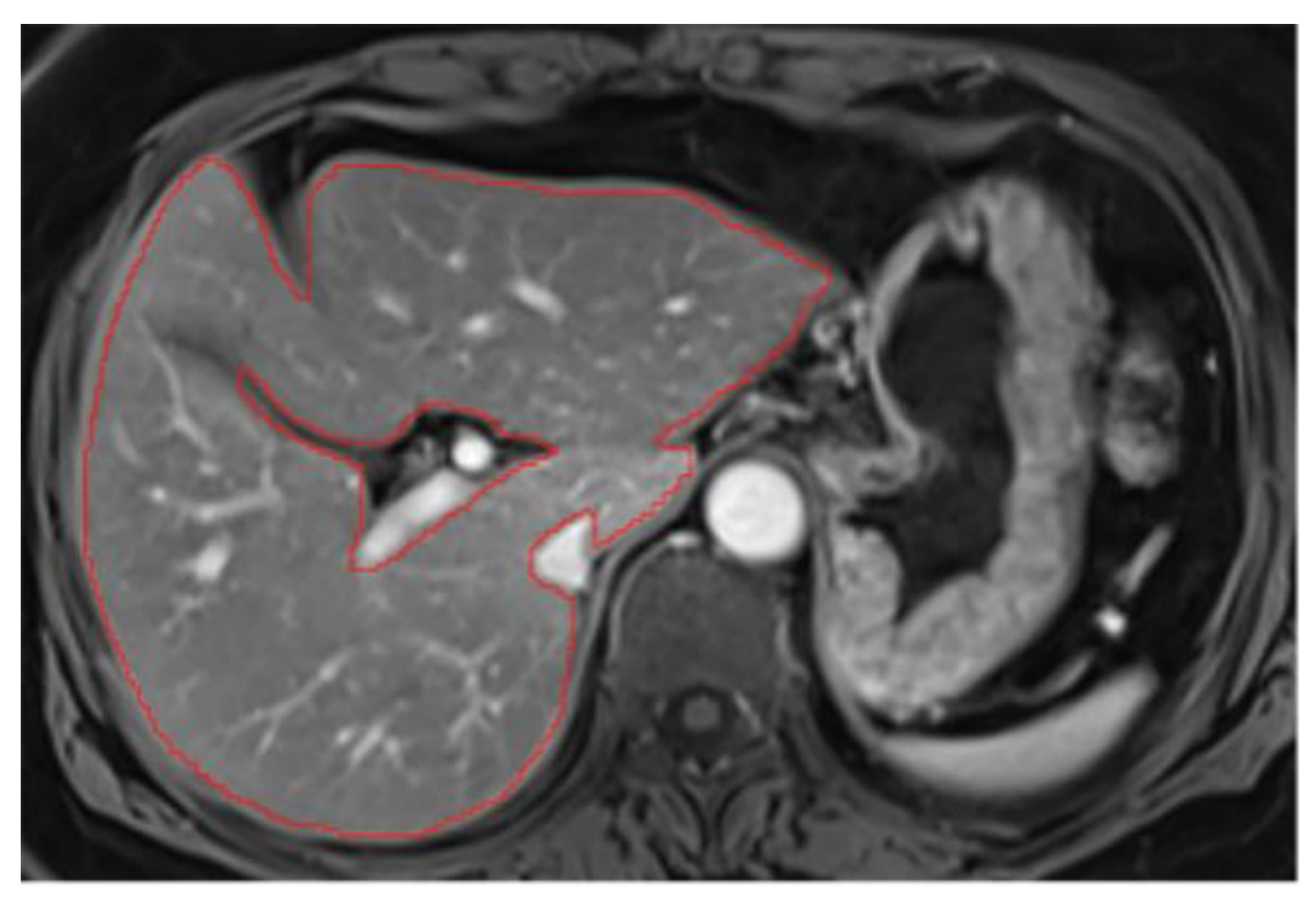
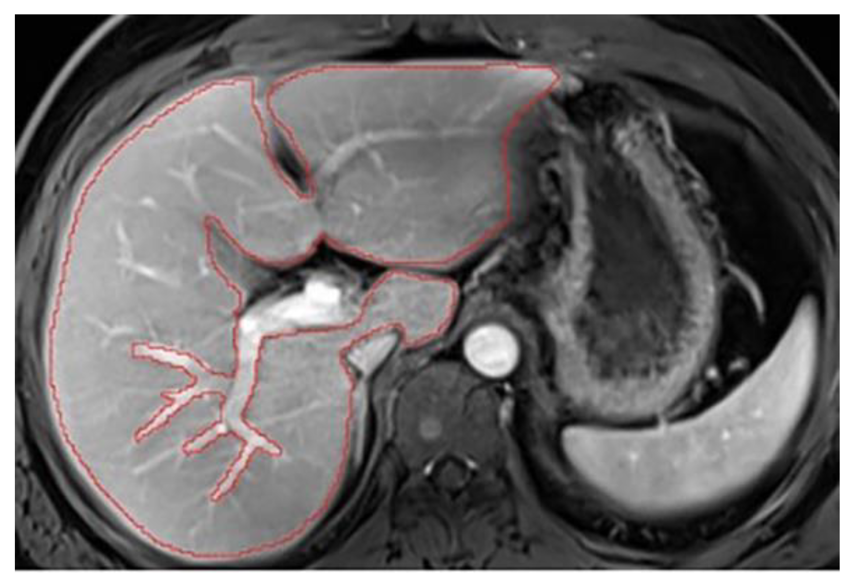
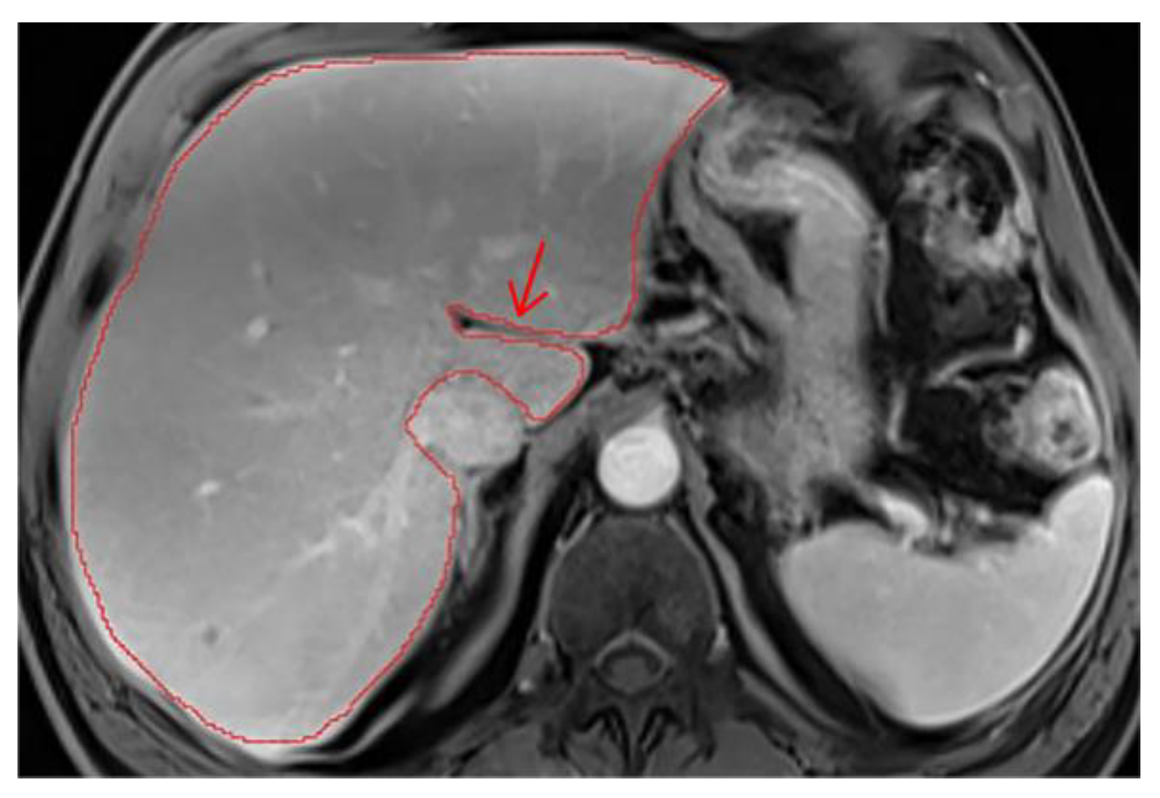
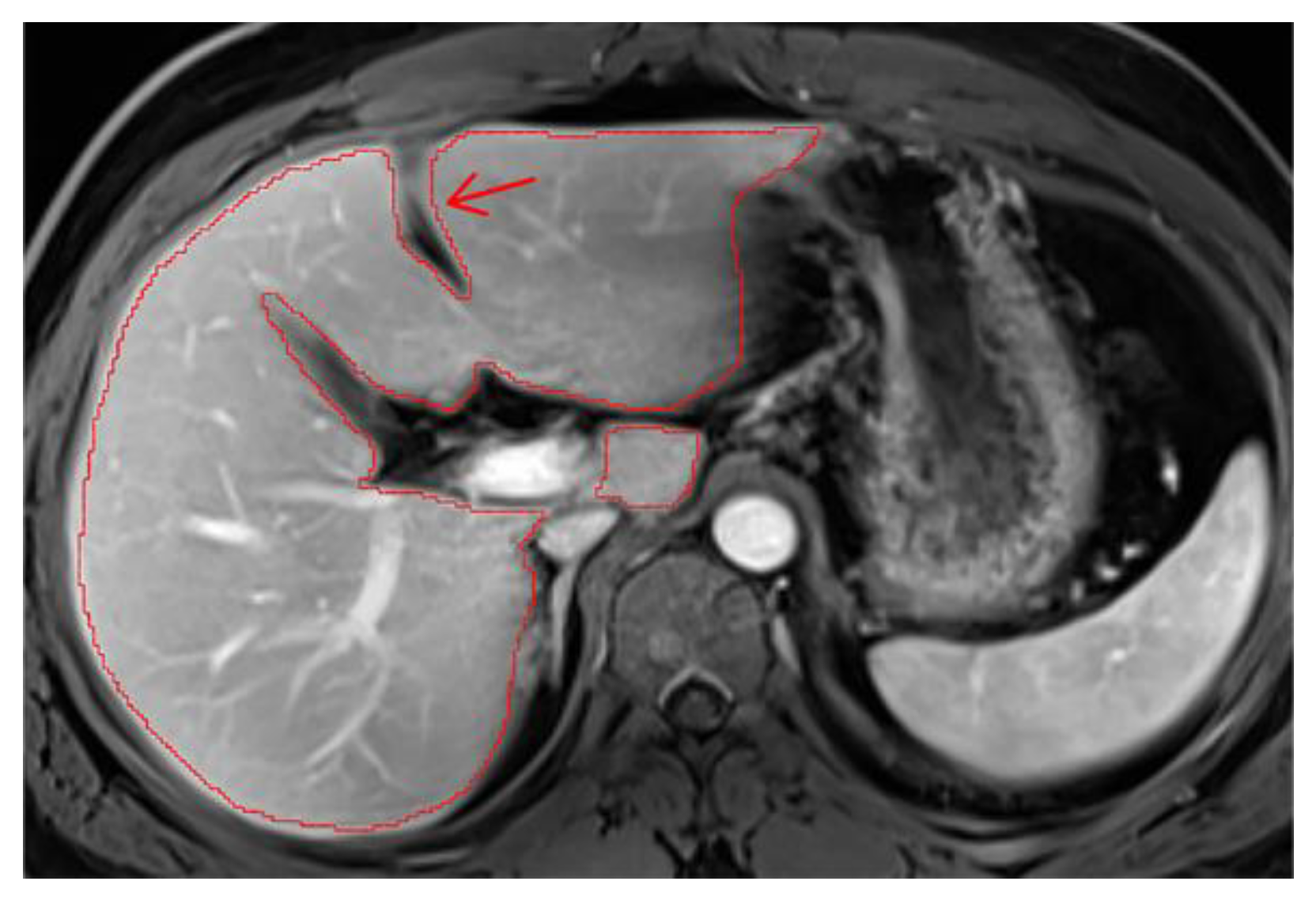
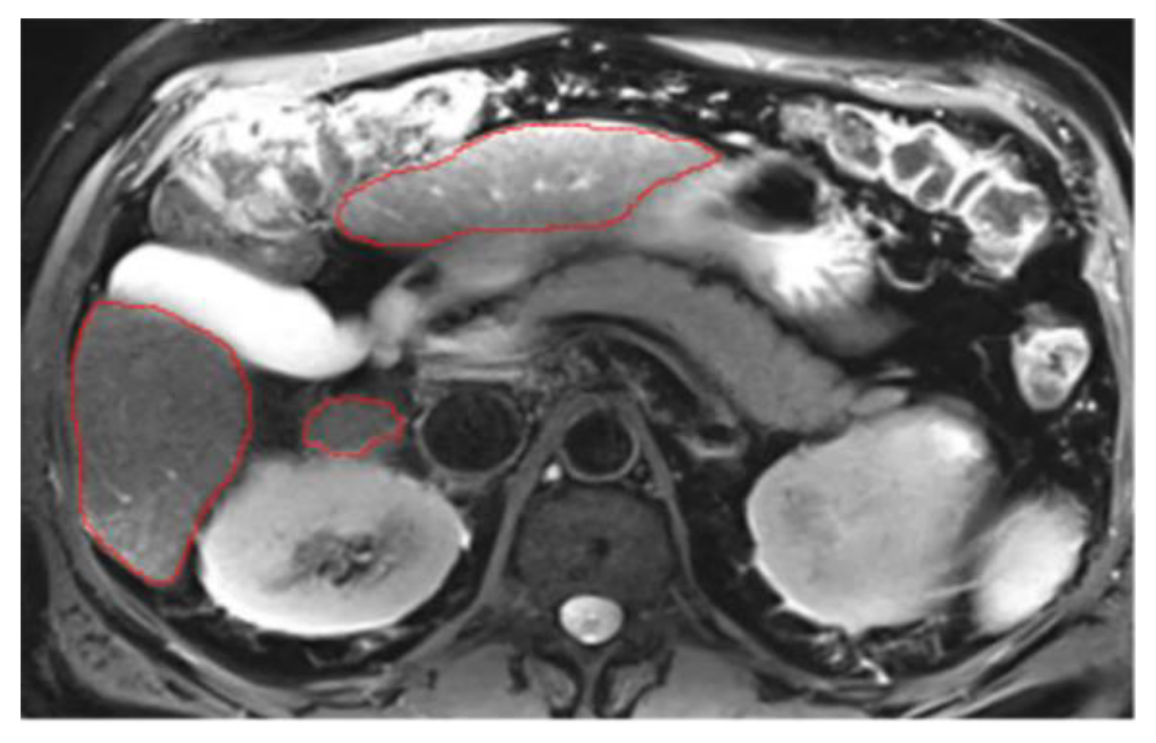
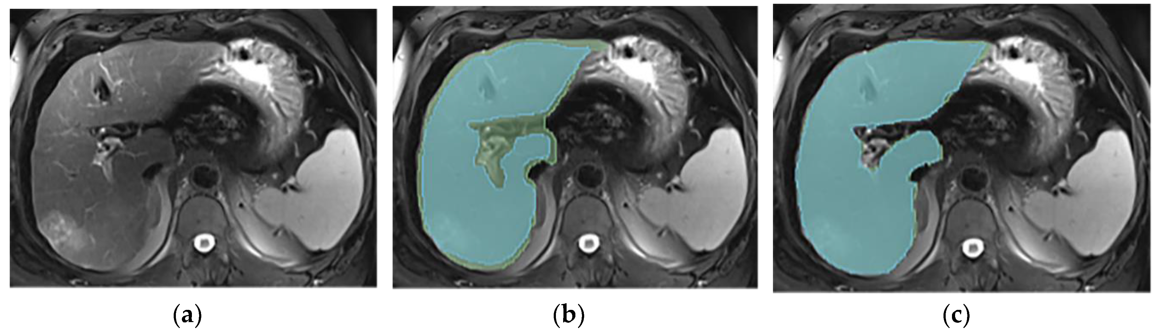
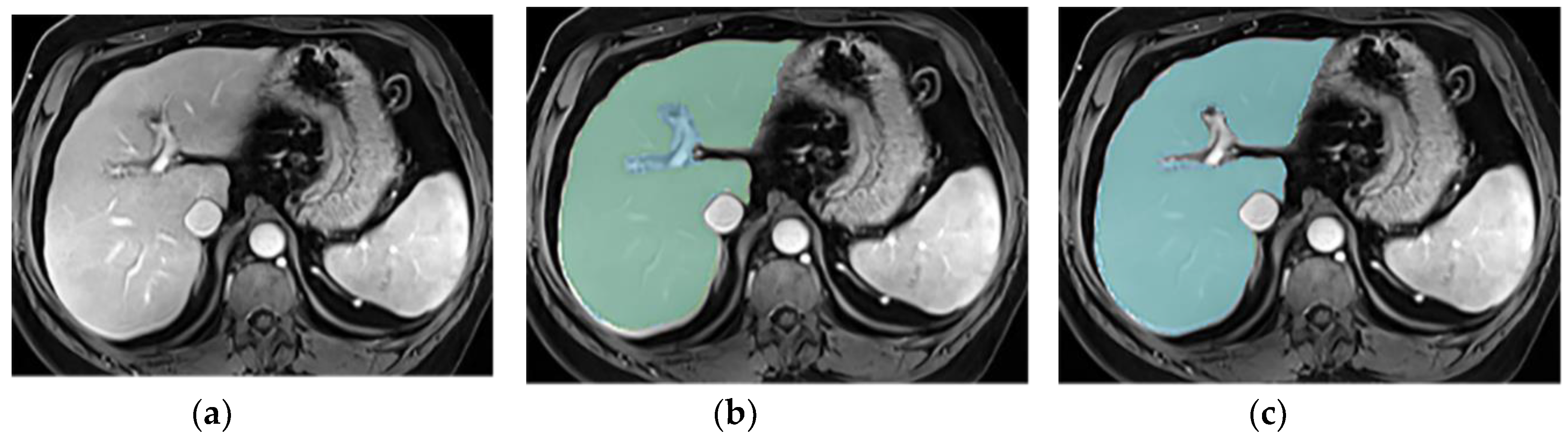
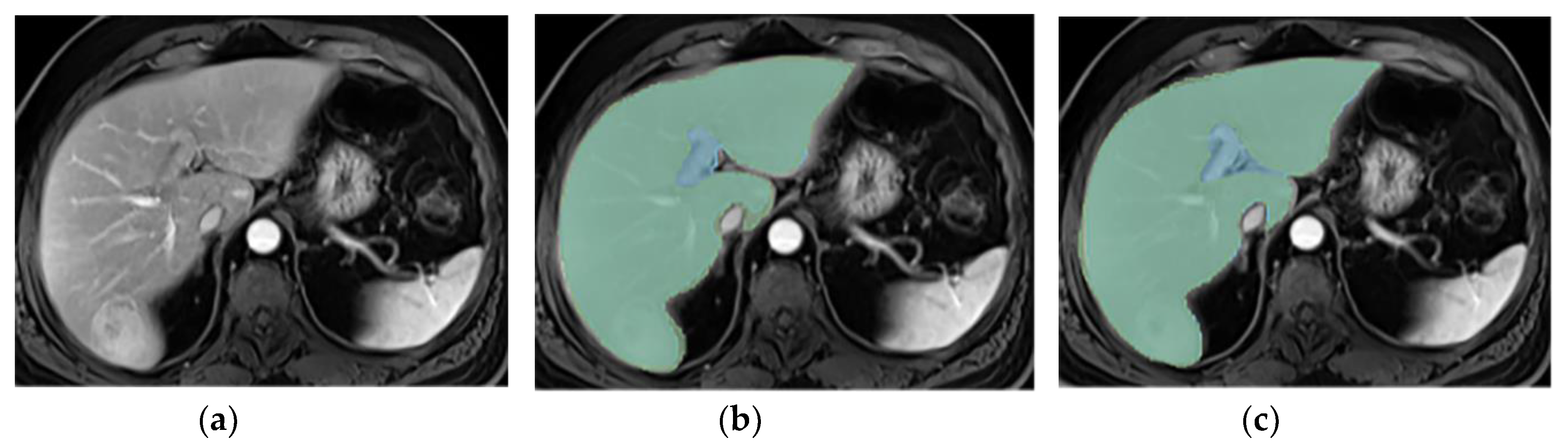
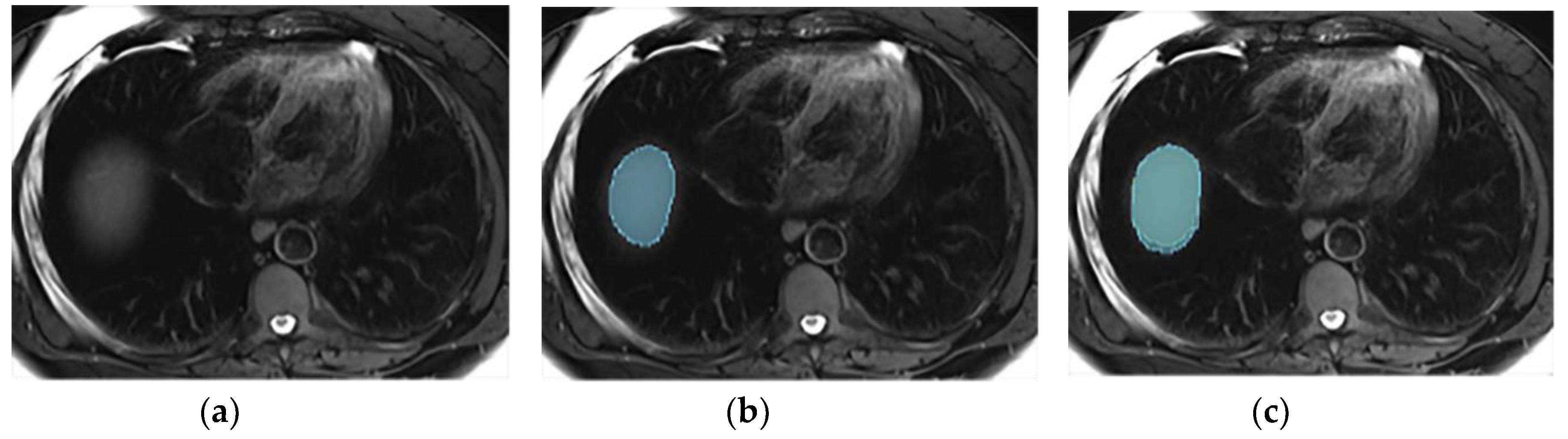
2.2. Liver Segmentations and Protocol
2.3. Statistical Analysis
3. Results
3.1. Patient Population
3.2. Liver Segmentation Correlation
3.3. Annotations Comparison
4. Discussion
5. Conclusions
Author Contributions
Funding
Institutional Review Board Statement
Informed Consent Statement
Conflicts of Interest
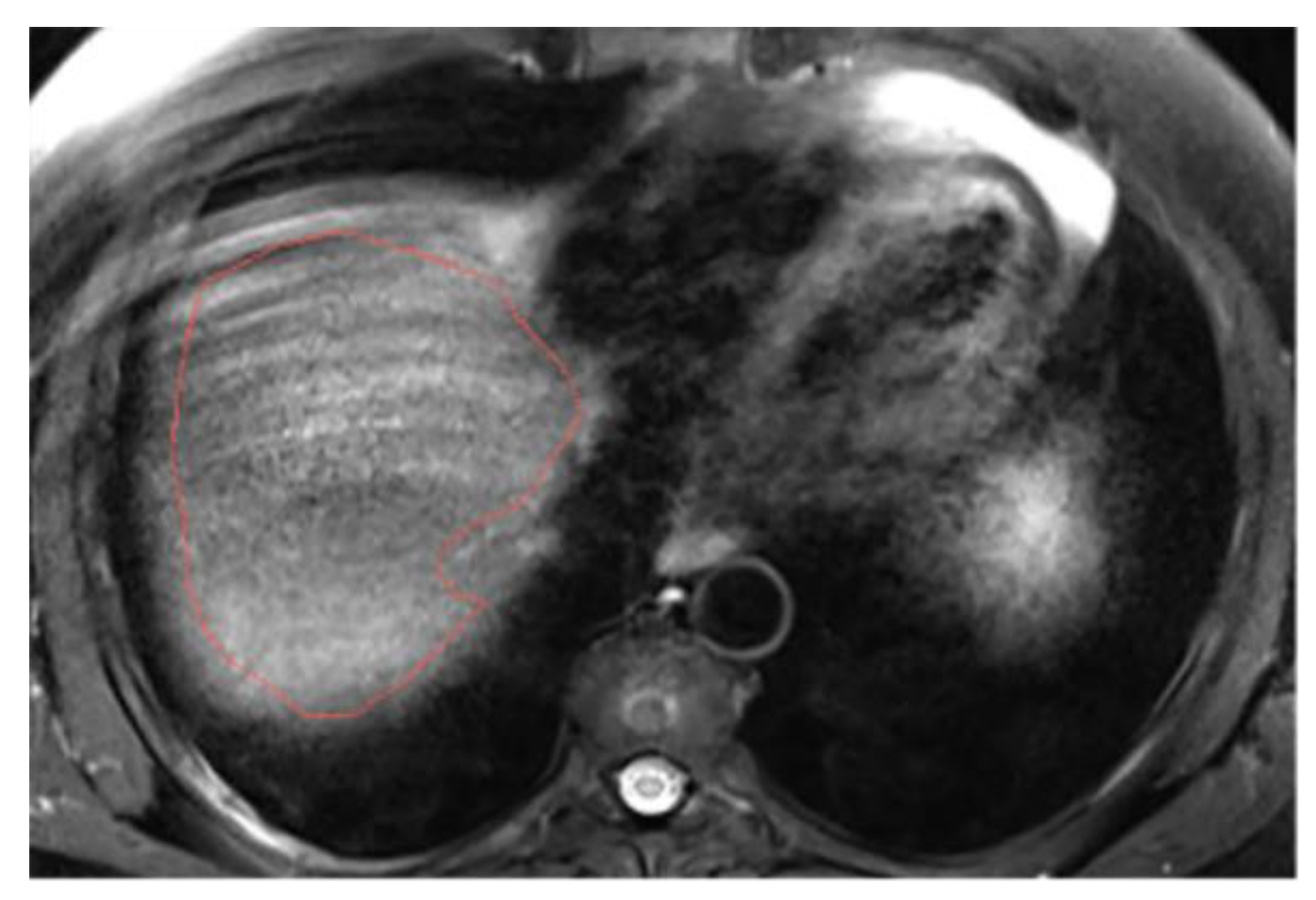
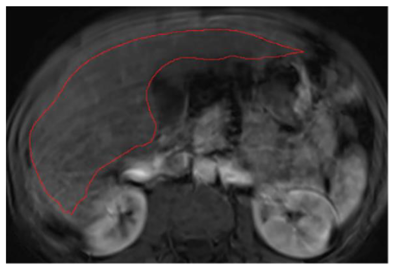
Appendix A. MRI Liver Segmentation Protocol
1. Select appropriate windowing and magnification
2. Hepatic hilum segmentation
3. Vascular segmentation

4. Ligaments segmentation
5. Multi-part liver parenchyma
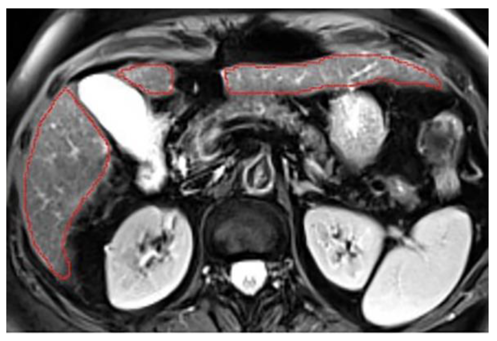
6. Presence of respiratory artifacts
7. Ascites

References
- B. Ryerson et al., « Annual Report to the Nation on the Status of Cancer, 1975-2012, featuring the increasing inci-dence of liver cancer: Report on Status of Cancer, 1975-2012 », Cancer, vol. 122, no 9, p. 1312-1337, mai 2016. [CrossRef]
- L. A. Torre, F. Bray, R. L. Siegel, J. Ferlay, J. Lortet-Tieulent, et A. Jemal, « Global cancer statistics, 2012: Global Can-cer Statistics, 2012 », CA: A Cancer Journal for Clinicians, vol. 65, no 2, p. 87-108, mars 2015. [CrossRef]
- J. Ferlay et al., « Cancer incidence and mortality patterns in Europe: Estimates for 40 countries and 25 major cancers in 2018 », European Journal of Cancer, vol. 103, p. 356-387, nov. 2018. [CrossRef]
- N. S. Parra et al., « Advancements in the Diagnosis of Hepatocellular Carcinoma », IJTM, vol. 3, no 1, p. 51-65, janv. 2023. [CrossRef]
- V. Chernyak et al., « Liver Imaging Reporting and Data System (LI-RADS) Version 2018: Imaging of Hepatocellular Carcinoma in At-Risk Patients », Radiology, vol. 289, no 3, p. 816-830, déc. 2018. [CrossRef]
- D. A. Bluemke et al., « Assessing Radiology Research on Artificial Intelligence: A Brief Guide for Authors, Review-ers, and Readers—From the Radiology Editorial Board », Radiology, vol. 294, no 3, p. 487-489, mars 2020. [CrossRef]
- H. J. Park, B. Park, et S. S. Lee, « Radiomics and Deep Learning: Hepatic Applications », Korean J Radiol, vol. 21, no 4, p. 387, 2020. [CrossRef]
- S. Yao, Z. Ye, Y. Wei, H.-Y. Jiang, et B. Song, « Radiomics in hepatocellular carcinoma: A state-of-the-art review », WJGO, vol. 13, no 11, p. 1599-1615, nov. 2021. [CrossRef]
- Y. LeCun, Y. Bengio, et G. Hinton, « Deep learning », Nature, vol. 521, no 7553, p. 436-444, mai 2015. [CrossRef]
- M. Gross et al., « Automated MRI liver segmentation for anatomical segmentation, liver volumetry, and the extrac-tion of radiomics », Eur Radiol, vol. 34, no 8, p. 5056-5065, janv. 2024. [CrossRef]
- L. C. Chu, S. Park, S. Kawamoto, A. L. Yuille, R. H. Hruban, et E. K. Fishman, « Current Status of Radiomics and Deep Learning in Liver Imaging », J Comput Assist Tomogr, vol. 45, no 3, p. 343-351, mai 2021. [CrossRef]
- Ibrahim et al., « Radiomics for precision medicine: Current challenges, future prospects, and the proposal of a new framework », Methods, vol. 188, p. 20-29, avr. 2021. [CrossRef]
- M. Pavic et al., « Influence of inter-observer delineation variability on radiomics stability in different tumor sites », Acta Oncologica, vol. 57, no 8, p. 1070-1074, août 2018. [CrossRef]
- T. Rädsch et al., « Labelling instructions matter in biomedical image analysis », Nat Mach Intell, vol. 5, no 3, p. 273-283, mars 2023. [CrossRef]
- Gotra et al., « Liver segmentation: indications, techniques and future directions », Insights Imaging, vol. 8, no 4, p. 377-392, août 2017. [CrossRef]
- X.-Q. Gong et al., « Progress of MRI Radiomics in Hepatocellular Carcinoma », Front. Oncol., vol. 11, p. 698373, sept. 2021. [CrossRef]
- J. Dockès, G. Varoquaux, et J.-B. Poline, « Preventing dataset shift from breaking machine-learning biomarkers », GigaScience, vol. 10, no 9, p. giab055, sept. 2021. [CrossRef]
- C. Yu, B. Mohajer, et J. Eng, « External Validation of Deep Learning Algorithms for Radiologic Diagnosis: A Sys-tematic Review », Radiology: Artificial Intelligence, vol. 4, no 3, p. e210064, mai 2022. [CrossRef]
- P. Xu et al., « Efficient knowledge distillation for liver CT segmentation using growing assistant network », Phys. Med. Biol., vol. 66, no 23, p. 235005, déc. 2021. [CrossRef]
- R. Jin, M. Wang, L. Xu, J. Lu, E. Song, et G. Ma, « Automatic 3D CT liver segmentation based on fast global minimi-zation of probabilistic active contour », Medical Physics, vol. 50, no 4, p. 2100-2120, avr. 2023. [CrossRef]
- J. Senthilvelan et N. Jamshidi, « A pipeline for automated deep learning liver segmentation (PADLLS) from contrast enhanced CT exams », Sci Rep, vol. 12, no 1, p. 15794, sept. 2022. [CrossRef]
- K. Wang et al., « Automated CT and MRI Liver Segmentation and Biometry Using a Generalized Convolutional Neural Network », Radiology: Artificial Intelligence, vol. 1, no 2, p. 180022, mars 2019. [CrossRef]
- F. López-Mir, V. Naranjo, J. Angulo, M. Alcañiz, et L. Luna, « Liver segmentation in MRI: A fully automatic method based on stochastic partitions », Computer Methods and Programs in Biomedicine, vol. 114, no 1, p. 11-28, avr. 2014. [CrossRef]
- F. Quinton et al., « A Tumour and Liver Automatic Segmentation (ATLAS) Dataset on Contrast-Enhanced Magnetic Resonance Imaging for Hepatocellular Carcinoma », Data, vol. 8, no 5, p. 79, avr. 2023. [CrossRef]
- Stanzione et al., « Oncologic Imaging and Radiomics: A Walkthrough Review of Methodological Challenges », Cancers, vol. 14, no 19, p. 4871, oct. 2022. [CrossRef]
- M. Y. Ansari et al., « Practical utility of liver segmentation methods in clinical surgeries and interventions », BMC Med Imaging, vol. 22, no 1, p. 97, mai 2022. [CrossRef]
- E. Zuñiga-Aguilar et O. Ramírez-Fernández, « Fibrosis and hepatic regeneration mechanism », Transl Gastroenterol Hepatol, vol. 7, p. 9-9, janv. 2022. [CrossRef]
- L. Maier-Hein et al., « Why rankings of biomedical image analysis competitions should be interpreted with care », Nat Commun, vol. 9, no 1, p. 5217, déc. 2018. [CrossRef]
- R. Girardet et al., « The combination of non-contrast abbreviated MRI and alpha foetoprotein has high performance for hepatocellular carcinoma screening », Eur Radiol, juill. 2023. [CrossRef]
- J. Jia, Jingnan-Jia/segmentation_metrics: v1.1.3. (30 octobre 2022). Zenodo. [CrossRef]
- P. Virtanen et al., « SciPy 1.0: fundamental algorithms for scientific computing in Python », Nat Methods, vol. 17, no 3, p. 261-272, mars 2020. [CrossRef]
- « Introduction — statsmodels ». Consulté le: 1 avril 2024. [En ligne]. Disponible sur: https://www.statsmodels.org/v0.13.5/.
- M. C. Lim, C. H. Tan, J. Cai, J. Zheng, et A. W. C. Kow, « CT volumetry of the liver: Where does it stand in clinical practice? », Clinical Radiology, vol. 69, no 9, p. 887-895, sept. 2014. [CrossRef]
- M. D’Onofrio, « Liver volumetry: Is imaging reliable? Personal experience and review of the literature », WJR, vol. 6, no 4, p. 62, 2014. [CrossRef]
- Y. Nakayama et al., « Automated Hepatic Volumetry for Living Related Liver Transplantation At Multisection CT », Radiology, vol. 240, no 3, p. 743-748, sept. 2006. [CrossRef]
- M. G. Poirot et al., « Robustness of radiomics to variations in segmentation methods in multimodal brain MRI », Sci Rep, vol. 12, no 1, p. 16712, oct. 2022. [CrossRef]
- Q. Qiu et al., « Reproducibility and non-redundancy of radiomic features extracted from arterial phase CT scans in hepatocellular carcinoma patients: impact of tumor segmentation variability », Quant. Imaging Med. Surg, vol. 9, no 3, p. 453-464, mars 2019. [CrossRef]
- J. Duan et al., « Reproducibility for Hepatocellular Carcinoma CT Radiomic Features: Influence of Delineation Var-iability Based on 3D-CT, 4D-CT and Multiple-Parameter MR Images », Front. Oncol., vol. 12, p. 881931, avr. 2022. [CrossRef]
- M. Gross, S. Arora, S. Huber, A. S. Kücükkaya, et J. A. Onofrey, « LiverHccSeg: A publicly available multiphasic MRI dataset with liver and HCC tumor segmentations and inter-rater agreement analysis », Data in Brief, vol. 51, p. 109662, déc. 2023. [CrossRef]
- G. Suman et al., « Development of a volumetric pancreas segmentation CT dataset for AI applications through trained technologists: a study during the COVID 19 containment phase », Abdom Radiol, vol. 45, no 12, p. 4302-4310, déc. 2020. [CrossRef]
- X. Zhang et al., « The effects of volume of interest delineation on MRI-based radiomics analysis: evaluation with two disease groups », Cancer Imaging, vol. 19, no 1, p. 89, déc. 2019. [CrossRef]
- E. Gundersen et S. Kjensmo, « State of the Art: Reproducibility in Artificial Intelligence », AAAI, vol. 32, no 1, avr. 2018. [CrossRef]
- Kocak et al., « CheckList for EvaluAtion of Radiomics research (CLEAR): a step-by-step reporting guideline for authors and reviewers endorsed by ESR and EuSoMII », Insights Imaging, vol. 14, no 1, p. 75, mai 2023. [CrossRef]
- J. A. Macdonald, Z. Zhu, B. Konkel, M. A. Mazurowski, W. F. Wiggins, et M. R. Bashir, « Duke Liver Dataset: A Pub-licly Available Liver MRI Dataset with Liver Segmentation Masks and Series Labels », Radiology: Artificial Intelli-gence, vol. 5, no 5, p. e220275, sept. 2023. [CrossRef]

| Characteristic | Study Cohort (n=20) |
|---|---|
| Sex (M/F) | 15/5 |
| Age (median, range) | 54, 29-79 |
| Ethnicity | |
| Caucasian | 13 (65%) |
| Asian | 4 (20%) |
| African | 3 (15%) |
| Liver Disease Etiology | |
| HBV | 7 (35%) |
| Alcohol consumption | 6 (30%) |
| HCV | 4 (20%) |
| NASH | 3 (15%) |
| Cirrhosis | |
| Yes | 10 (50%) |
| No | 10 (50%) |
| Child-Pugh class (if cirrhosis) | |
| A | 9 (90%) |
| B | 1 (10%) |
| Before protocol (± SD) | After protocol (± SD) | Wilcoxon signed-rank test p-value | |
|---|---|---|---|
| Per-volume analysis | |||
| T2wi | DSC = 0.944 ±0.013 | DSC = 0.957 ±0.008 | <0.001 |
| HD = 24.47 ±13.01 | HD = 19.94 ±5.38 | 0.216 | |
| T1wi | DSC = 0.953 ±0.011 | DSC = 0.957 ±0.009 | 0.03 |
| HD = 21.85 ±11.15 | HD = 16.40 ±5.68 | 0.048 | |
| Per-slice analysis | |||
| T2wi | DSC = 0.885 ±0.208 | DSC = 0.924 ±0.134 | <0.001 |
| HD = 8.73 ±15.81 | HD = 7.22 ±13.45 | <0.001 | |
| T1wi | DSC = 0.918 ±0.14 | DSC = 0.925 ±0.12 | <0.001 |
| HD = 6.26 ±11.48 | HD = 5.70 ±9.80 | 0.035 | |
| Weights | Per-volume analysis (p-value) |
Per-slice analysis (p-value) |
DSC improvement |
|
|---|---|---|---|---|
| Cirrhosis | T2wi | 0.002 | <0.001 | Yes |
| Without cirrhosis | T2wi | 0.002 | <0.001 | Yes |
| Cirrhosis | T1wi | 0.012 | <0.001 | Yes |
| Without cirrhosis | T1wi | 0.556 | 0.82 | No |
Disclaimer/Publisher’s Note: The statements, opinions and data contained in all publications are solely those of the individual author(s) and contributor(s) and not of MDPI and/or the editor(s). MDPI and/or the editor(s) disclaim responsibility for any injury to people or property resulting from any ideas, methods, instructions or products referred to in the content. |
© 2024 by the authors. Licensee MDPI, Basel, Switzerland. This article is an open access article distributed under the terms and conditions of the Creative Commons Attribution (CC BY) license (http://creativecommons.org/licenses/by/4.0/).


