Preprint
Review

This version is not peer-reviewed.

Research Progress of Lactate and Its Regulated Small Molecule Drugs

A peer-reviewed article of this preprint also exists.

Submitted:

18 October 2024

Posted:

21 October 2024

You are already at the latest version

Abstract

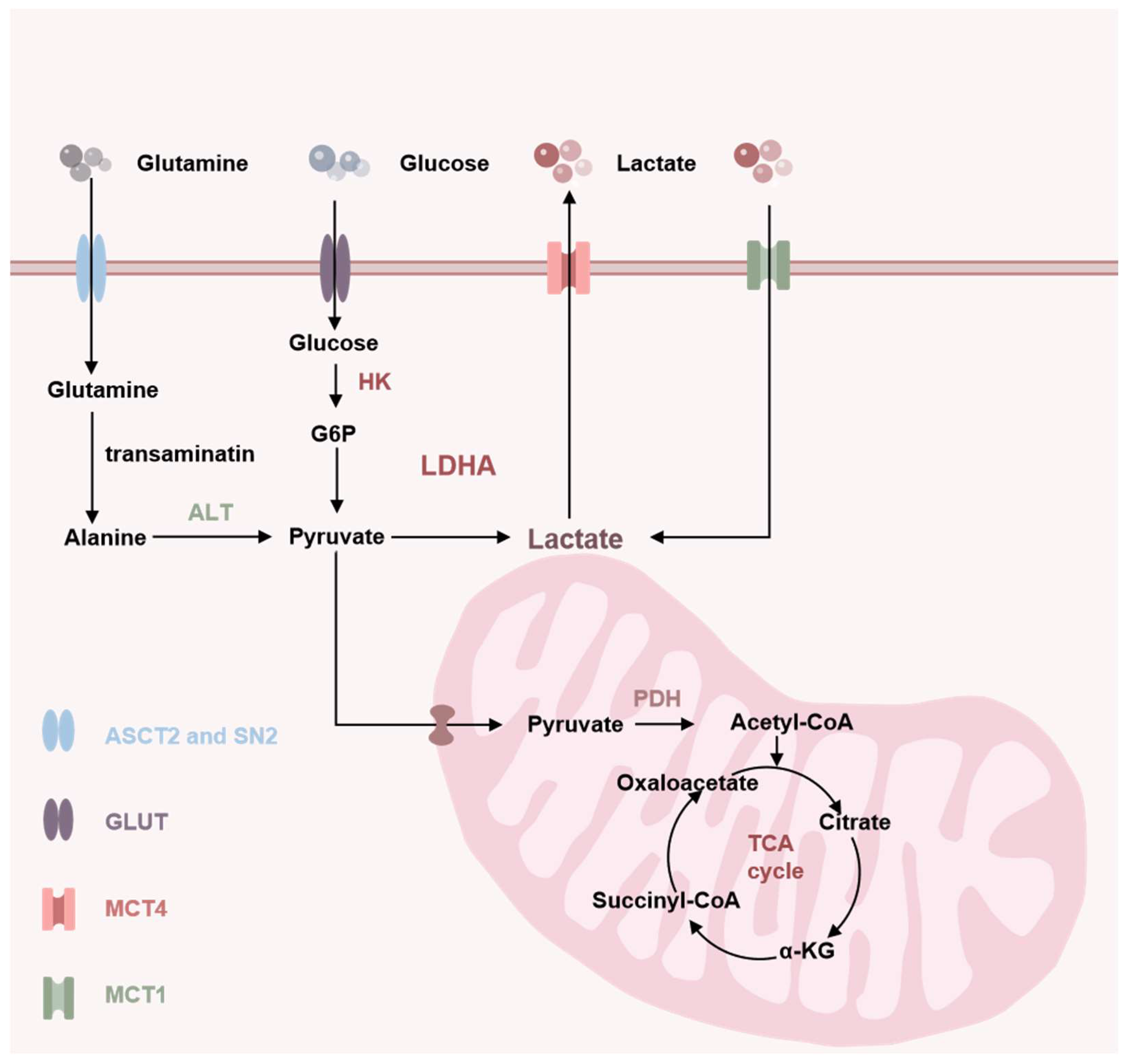
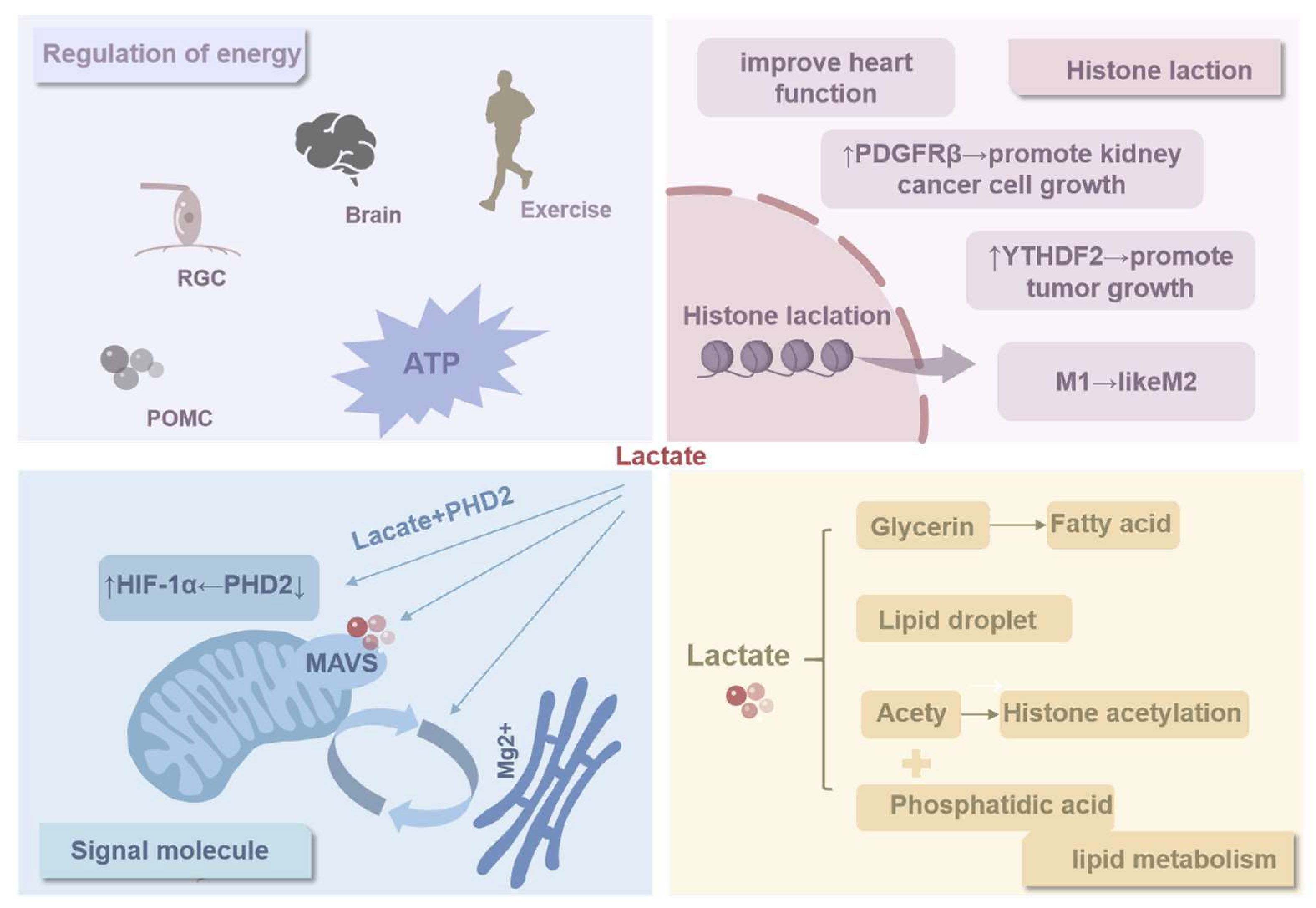
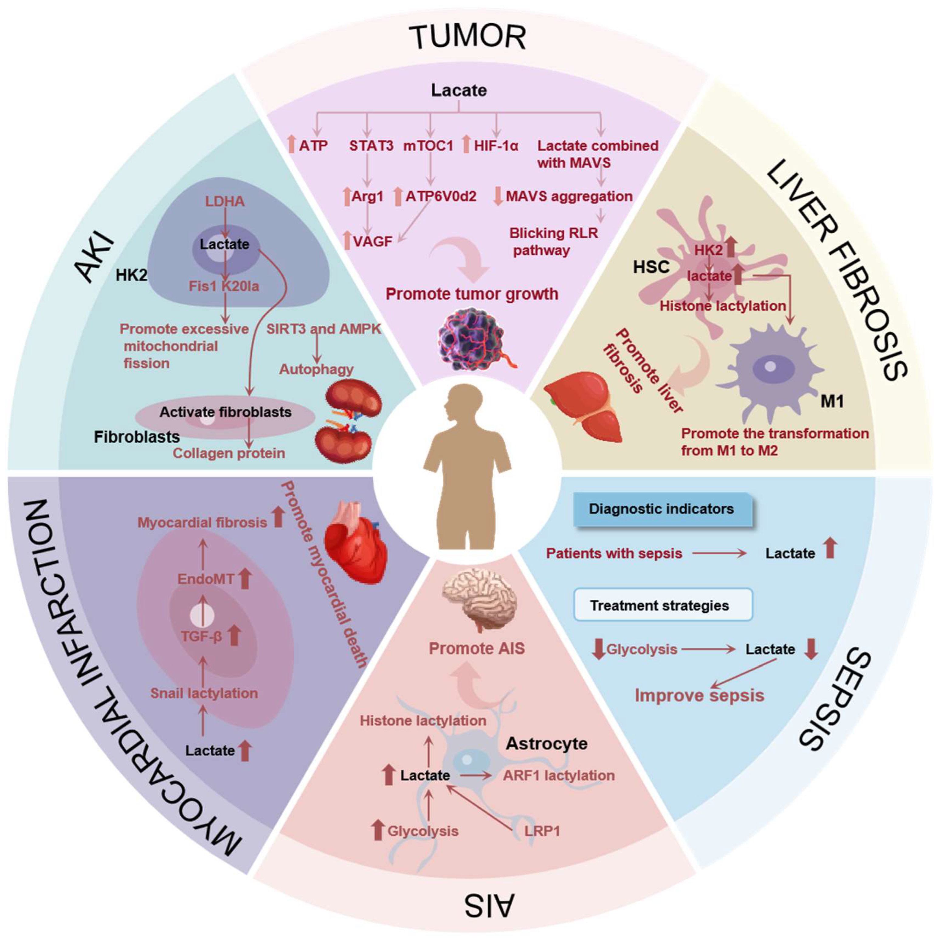
Lactate plays a critical role in cell metabolism and disease development. Under conditions of hypoxia or high-intensity exercise, cells use the glycolytic pathway to convert glucose into pyruvate, which is then reduced to lactate to quickly obtain energy. The traditional role of lactate is being redefined, as it is not only a provider of energy but also an important signaling molecule that regulates cell physiological functions, including histone lactylation modification, which affects gene expression. However, excessive accumulation of lactate is associated with the development of various diseases, such as liver disease, cancer, and cardiovascular disease. Research has also found that small-molecule drugs can regulate lactate levels, providing new possibilities for treating related diseases.

Keywords: 
;  ;  ;  

1. Introduction

In conditions of hypoxia or high-intensity exercise, cells undergo a metabolic process wherein glucose is converted to pyruvate, which is then reduced to lactate through the glycolytic pathway. lactate production represents an adaptive cellular response, enabling the rapid acquisition of energy. It is frequently a byproduct of glycolysis and has historically been regarded as a metabolic “waste product.” Nevertheless, as scientific research has advanced, it has become evident that lactate plays a far more significant role in living organisms than was previously assumed. Lactate serves not only as a source of cellular energy but also as a signaling molecule capable of regulating cellular physiological functions [1]. For instance, lactate may participate in the lactylation modification of histones, which could potentially influence gene expression [2]. The discovery of lactylation modification offers a novel perspective on the function of lactate in cellular regulation. However, the excessive accumulation of lactate is also closely associated with the development of several pathological conditions. An elevated lactate level may be indicative of underlying pathology in the liver, kidneys, heart muscle, and other organs. In such instances, the generation of lactate in substantial quantities may result in poisoning and impairment of the organism. It is encouraging to note that some small-molecule drugs are capable of modulating lactate levels. These small-molecules regulate lactate production and metabolism through a variety of mechanisms and signaling pathways, thereby providing new avenues for the treatment of diseases related to lactate. lactate plays a pivotal role in numerous physiological processes and pathological responses in living organisms. To enhance the utilisation of lactate as a biomolecule, it is essential to conduct comprehensive research and gain a deeper understanding of its sources, clearance, physiological functions, and its relationship to disease. In the meantime, the discovery of small molecule drugs that modulate lactate has yielded novel therapeutic strategies and concepts.

2. Soure of Lactate

In accordance with the prevailing physiological conditions, the foodstuffs consumed by the body are subjected to a series of intricate biochemical processes, ultimately resulting in the conversion of these substances into glucose. The glucose molecules are transported into the cell via the glucose transporter protein (GLUT) on the cell membrane [3]. Subsequently, glucose is converted to glucose-6-phosphate (G6P) by hexokinase (HK) [4]. G6P is converted to pyruvate (PA) via a series of enzymatic reactions. In the presence of adequate oxygen, pyruvate continues its journey into the mitochondria. In the presence of adequate oxygen, pyruvate is transported into the mitochondria by the mitochondrial pyruvate carrier (MPC) [5], where it is converted to acetyl-CoA by pyruvate dehydrogenase (PDH). This process generates adenosine triphosphate (ATP), providing the energy necessary for cellular functions [6]. However, under hypoxic conditions, pyruvate within the cell is unable to produce energy through the mitochondrial pathway. Instead, it is converted to lactate, which generates ATP through the action of lactate dehydrogenase (LDH) [7]. It is noteworthy that cancer cells, even when exposed to an oxygen-rich environment, tend to adopt the aerobic glycolytic pathway, also known as the Warburg effect, in order to obtain energy through the production of lactate [8]. This aberrant metabolic pattern is attributable to the inhibition of PDH activity by a multitude of tumor-inducing factors, which in turn activates the glycolytic pathway and precipitates a reliance on lactate as a source of energy for cancer cells [9]. This metabolic shift ultimately results in the accumulation of lactate in cancer cells, which in turn facilitates tumor growth and metastasis. Another source of lactate production is glutamine and amino acid catabolism. Glutamylamine and amino acid catabolism represent an additional source of lactate production. When cells utilize glutamyl ammonia and other amino acids, they typically undergo decomposition and transamination reactions, which are accompanied by the production of substantial quantities of alanine, a proportion of alanine is transaminated to pyruvate as a result of the action of alanine aminotransferase (ALT), pyruvate is catalyzed by LDHA to produce lactate [10].
Figure 1. The source of transport of lactate.
Preprints 121612 g001

3. Clearance of Lactate

Lactate accumulation occurs when intracellular lactate is not excreted in a timely manner, subsequently, the aforementioned lactate has the potential to cause harm to the human body. For instance, a substantial accumulation of lactate in the serum may precipitate acidosis [11], the accumulation of lactate in tumor cells has been demonstrated to promote their growth and facilitate immune escape [12]. It is therefore evident that the timely removal of lactate plays a crucial role in maintaining the homeostasis of the human microenvironment. Lactate clearance is primarily accomplished through a multitude of metabolic pathways. On the one hand, lactate can undergo an irreversible clearance process in the human body by the metabolic enzyme PDH [13], on the other hand, cells depend on monocarboxylate transporters (MCTs) and exosomes (Exos) to facilitate the efficient removal of lactate. The MCT family comprises 14 members, collectively constituting the solute carrier family 16 (SLC16), these transporters play an integral role in the transport of cellular nutrients and in cellular metabolism [14]. To illustrate, MCT1 is principally accountable for the conveyance of lactate from glycolytic cells, such as tumor cells, to oxidative cells, such as cardiomyocytes and slow muscle fibers, for subsequent oxidative utilization, a process designated as the “lactate shuttle”, MCT4 is a high-affinity lactate transporter that exports lactate in a high-lactate microenvironment, thereby maintaining equilibrium between intracellular and extracellular lactate levels [15]. In tumor cells, the expression levels of MCT1 and MCT4 are found to be abnormally increased [16]. In solid tumors, MCT facilitates the exchange of lactate, and cancer cells exhibit a preference for oxidizing lactate as an oxidative fuel, thereby supplying energy to the cancer cells [17]. Accordingly, the modulation of lactate equilibrium through the modulation of these transporters represents a promising avenue for the treatment of neoplastic growth. Exosomes are a type of cystic vesicle secreted by active cells. They are capable of transporting macromolecular signals, including lipids, proteins, and genetic materials, from the interior of the cell to the extracellular space [18]. Recent studies have revealed that lactate can act not only as a metabolite but also as a signaling molecule to stimulate cells to produce exosomes. For example, lactate promotes the lactation of HMGB1 through a p300 / CBP-dependent mechanism, and the lactation of HMGB1 can then be transported outside the cell via exosomes produced by lactate-stimulated cells [19], consequently, the clearance of lactate is accomplished indirectly. Lactate clearance has the potential to restore the normal physiological function of cells and maintain the normal metabolic processes and oxidation of cells.4. Physiological Roles of Lactate

3.1. Regulation of Energy

Lactate plays a significant role in the physiological processes of the human body, both under normal physiological conditions and in the context of pathological processes. In normal physiological activities, the concentration of lactate in the human bloodstream increases in response to physical exertion [20]. The necessity for rapid energy acquisition during exercise is a primary factor in this phenomenon. While the tricarboxylic acid cycle is capable of providing a substantial amount of energy, its intricate and time-consuming process renders it an inadequate source for the demands of exercise. In contrast, the process of glycolysis is more straightforward and occurs more rapidly. The production of lactate is a crucial aspect of the glycolysis pathway, enabling the body to meet its energy demands during exercise [21]. Moreover, lactate has been identified as the primary energy source for specific cells, including retinal ganglion cells (RGCs) [22], The data indicated that these cells were more likely to utilize lactate as an energy source than glucose, suggesting that lactate plays a unique role in energy metabolism in specific cellular contexts. It has been evidenced that lactate enhances cerebral energy provision and facilitates ATP accumulation under normoglycemic circumstances [23]. This finding offers new insights into our understanding of energy metabolism in the brain. The observed dysregulation of energy metabolism in the precursor melanocortin (POMC) cells, induced by MCT inhibitors, suggests that these cells may rely primarily on lactate as an energy source [24]. Furthermore, the study of live guinea pig ventricular cardiomyocytes has confirmed that lactate activates cardiac ATP-sensitive potassium channels, thereby affecting the electrophysiological characteristics of cardiomyocytes [25]. The aforementioned studies indicate that certain cells and tissues may utilize lactate as an alternative source of energy instead of glucose. In cancer cells, lactate is the primary source of energy in both aerobic and anaerobic environments. In human non-small cell lung cancer, the role of lactate is even more complex and diverse, it is not only the carbon source of the tricarboxylic acid cycle, which provides the necessary material basis for the growth and metabolism of tumor cells, but also can be used as an energy source to support the rapid proliferation and spread of tumor cells [26]. Lactate is essential in human physiological activities. In addition to serving as an energy source for cells during hypoxia, it fulfills distinct functions in various cells and tissues. It serves as the primary source of rapid energy during exercise and is the preferred energy source for select cells, such as retinal ganglion cells. It has been demonstrated to enhance the energy supply of the brain and to exert an influence on the electrophysiological characteristics of cardiomyocytes, while also supporting the growth and metabolic processes of tumor cells.

3.2. Histone Lactylation

Protein modification, also referred to as protein post-translational modification(PTM), entails a series of biochemical reactions that alter the chemical structure of the protein molecule subsequent to mRNA translation [27], such alterations have the potential to impact a number of protein characteristics, including its structure, stability, and activity [27]. The function of proteins is regulated by cells through a number of different processes, including phosphorylation [28], ubiquitination [29], methylation [30], and acetylation [31]. A novel protein modification, histone lactation, has recently been identified. In 2019, Zhang D [2] et al. employed high-performance liquid chromatography (HPLC) coupled with tandem mass spectrometry (MS/MS) to demonstrate that lactate can be incorporated at histone lysine residues, thereby conferring M2-like functions in M1-type macrophages at later stages. These findings indicate that histone lactylation may serve as a lactate-based temporal regulator, orchestrating the transition of macrophages from an inflammatory to a homeostatic phenotype. Furthermore, PI3K B cell adapters (BCAP) have been demonstrated to facilitate the transition of macrophages from a pro-inflammatory phenotype to a homeostatic phenotype [32]. It has been shown in studies that a loss of BCAP results in a reduction in aerobic glycolysis, a decrease in lactate production, and an impairment of histone lactylation, which in turn impairs the repair expression of tissue genes [33]. These findings indicate that BCAP, when modified by histone lactylation, may facilitate the transition of pro-inflammatory macrophages to reparative macrophages [33]. Subsequent to histone lactylation, it may facilitate the expression of m6A-modified protein YTHDF2, thus contributing to the promotion of tumorigenesis [34]. Concurrently, histone lactylation is concentrated in the promoter regions of glycolytic genes, which activates transcription and enhances glycolytic activity [35]. In renal cell carcinoma, histone lactylation levels are elevated, which facilitates the transcription of platelet-derived growth factor receptor β (PDGFRβ) and stimulates the proliferation of renal cell carcinoma [36]. In studies examining sepsis, a positive correlation has been observed between the level of histone lactylation and the expression of arginase-1 (Arg1) [37]. The literature indicates that histone lactylation may facilitate the transcription of repair genes, regulate the anti-inflammatory and pro-angiogenic effects of monocytes and macrophages, and thereby contribute to the repair of the microenvironment and the improvement of cardiac function following myocardial infarction [38].
Protein modification refers to the process of chemical alterations to proteins that occur as a result of biochemical reactions subsequent to mRNA translation. These modifications impact the properties of the proteins in question. The recently identified protein lactate modification has been demonstrated to play a pivotal role in a multitude of biological processes, including the regulation of macrophage function, the modulation of glycolytic activity, and the promotion of tumorigenesis. Furthermore, histone lactylation has been linked to the transcription of repair genes and enhanced cardiac function, underscoring the intricate regulatory processes involved.

3.3. Signal Molecule

As research progresses, it becomes increasingly evident that lactate plays a multifaceted and pivotal role in biological systems. In addition to its role as a pivotal enzyme in cellular energy metabolism, it also functions as a signaling molecule, regulating various cellular physiological functions. The study by Xu Aiming et al. [39] revealed the mechanism of lactate accumulation in obese mice and its effect on the inflammatory response of macrophages in adipose tissue. They found that the expression of glycolytic genes was increased in obese mice, while the expression of tricarboxylic acid cycle (TCA) genes was reduced, leading to lactate accumulation. Macrophages have a strong lactate uptake capacity, which allows lactate from fat to enter adipose tissue macrophages (ATM), thereby promoting ATM polarization and the production of pro-inflammatory factors. Furthermore, lactate can directly bind to prolyl hydroxylase 2 (PHD2) and inhibit its activity, while promoting the protein stability of hypoxia-inducible factor 1α (HIF-1α), thereby aggravating inflammation. This suggests that lactate secreted by adipocytes can act as a “metabolic signaling molecule” to regulate the inflammatory response of macrophages in adipose tissue. Besides, Lactate can bind to the transmembrane domain of mitochondrial antiviral signaling proteins (MAVS) and prevent MAVS aggregation, thereby inhibiting pattern recognition receptor (RLR) signaling mediated by glycolysis [40]. The results of this study confirm that lactate directly inhibits RLR signaling and identifies MAVS as a sensor of lactate. In addition, lactate is also involved in the regulation of intracellular Mg2+ transport. Mg2+ transport plays an important role in the regulation of life energy. Lactate can regulate the transfer of Mg2+ between the endoplasmic reticulum and mitochondria, act as an intracellular Mg2+ kinetic agonist activation signal, and link the mitochondrial Mg2+ transport with the main metabolic feedback loop and mitochondrial bioenergy metabolism [41]. The aforementioned studies suggest that lactate, serving as a crucial signaling molecule, exerts a significant influence on the regulation of diverse physiological and pathological processes. Lipids, as a class of highly complex biomolecules within organisms, serve a function that extends far beyond the fundamental structure of biological membranes [42].

3.4. Biomimetic Principles of Biomembranes

Lipids are integral to several fundamental biological processes, including cell signaling, energy metabolism, and a variety of other biological functions [43]. In recent years, as research has progressed, the relationship between lactate and lipid metabolism has been elucidated in greater detail. A series of biochemical reactions has been observed in rat muscle, whereby lactate is converted to glycerol. The subsequent production of fatty acids is promoted by glycerol, thereby regulating lipid metabolism [44]. Furthermore, lactate secreted by prostate cancer fibroblasts (CAFs) has been demonstrated to regulate intracellular lipid accumulation in lipid droplets (LDs), lactate regulates the expression of lipid metabolism genes in prostate cancer cells by providing acetyl groups for histone acetylation [45]. This mechanism elucidates the pivotal function of lactate in cancer cell metabolism. In neurons, lactate also fulfills an essential regulatory function. Neurons with higher levels of reactive oxygen species (ROS) showed increased lactate production, lactate is transported from glial cells to neurons by MCT, where it is converted to phosphatidic acid (PA) and acetyl-CoA [46]. These substances provide the required substrates for fatty acid synthesis, which in turn regulates the adipogenic process. The is a close relationship between lactate and lipid metabolism. Lactate is not only involved in the production of fatty acids and the regulation of fat metabolism but also affects lipid metabolism by affecting the accumulation of lipid droplets and regulating gene expression.
Figure 2. The physiological functions involving lactate.
Preprints 121612 g002

4. Lactate’s Association with Diseases

4.1. Lactate’s Role in Tumor

In 2020, it was estimated that there were 19.3 million new cases of cancer and nearly 10 million cancer-related deaths worldwide [47]. The microenvironment of a tumor is a complex and distinctive ecosystem, markedly distinguished by hypoxia. The substantial reproduction and expansion of tumor cells frequently exceed the capacity of the oxygen supply to meet their metabolic demands, resulting in the formation of a hypoxic microenvironment [48]. This hypoxia state will result in further disruption of the tumor microenvironment, which will in turn affect tumor growth, metastasis, and invasion. In the context of hypoxic conditions, there is the potential for significant alterations to occur in the metabolic pathways of tumor cells [49]. Normally, cells undergo aerobic oxidation through the tricarboxylic acid (TCA) cycle in the presence of sufficient oxygen, thereby generating a substantial amount of energy [9]. However, in an environment with low oxygen levels, the TCA cycle is suppressed, prompting tumor cells to rely on glycolysis for energy production [50]. This metabolic shift can result in the production of substantial quantities of lactate, which serves to further exacerbate the acidification of the tumor microenvironment. Lactate plays a significant role in the tumor microenvironment. In addition to providing energy for tumor cells, lactate plays a role in regulating tumor cell growth and proliferation. For instance, in many cancers, including lung, liver, and prostate cancers, the level of lactate is significantly correlated with tumor growth and prognosis [51,52]. In addition, lactate can also affect tumor growth by affecting signaling pathways and immune cell activity in the tumor microenvironment. For example, tumor-derived lactate induces macrophages to convert to M2 by stimulating STAT3 to activate endothelial growth factor (VEGF) and arginine (Arg-1), which further promotes tumor development [53]. HIF-1α is a further crucial regulator of the tumor hypoxic microenvironment [54]. In hypoxic conditions, the activation of HIF-α subsequently activates a series of HIF target genes, thereby promoting tumor growth and metastasis [55,56]. Lactate has been demonstrated to reduce intracellular levels of cyclic adenosine monophosphate (cAMP) and the activation of protein kinase A (PKA) through the action of the G protein-coupled receptor 81 (GPR81). Subsequently, PKA-mediated ubiquitination of HIF-1α results in the inhibition of degradation, thereby causing HIF-1α protein accumulation. Subsequently, HIF-1α induces transcription of Rab27a, which promotes the release of sEVs, in turn, it promotes tumor growth [57]. Moreover, lactate can also affect the immune surveillance of tumor cells by interacting with MAVS adaptor proteins [58]. Lactate can directly bind to the transmembrane domain of MAVS, thereby inhibiting the aggregation of MAVS and blocking RLR signaling. This enables tumor cells to evade the monitoring and clearance of the immune system [59]. In an anaerobic environment, lactate produced by tumor cells inhibits the expression of the macrophage-specific vacuolar ATPase subunit ATP6V0d2 by stimulating the microtubule-targeting chimera 1 (mTOC1) pathway, this results in elevated HIF-2α-mediated production of VEGF in macrophages, which subsequently display tumor-cell growth-promoting characteristics [60].
The tumor microenvironment is a complex and distinctive entity, distinguished by hypoxia, which instigates alterations in the metabolic pathways of tumor cells, predominantly glycolysis, resulting in the generation of a substantial quantity of lactate and a further exacerbation of the acidification of the microenvironment. Lactate serves not only as an energy source for tumor cells but also as a regulator of their growth and proliferation. Furthermore, it plays a role in tumor development by affecting signaling pathways and immune cell activity, including the induction of macrophages to transform into the M2 type. HIF is also a crucial regulator, influencing metabolic processes, oxygen delivery, and other biological functions by regulating the expression of hypoxia response genes. Moreover, lactate can interact with MAVS adaptor proteins, thereby affecting tumor cell immune surveillance and enabling tumor cells to evade immune system clearance. In an anaerobic environment, lactate also inhibits the expression of specific genes in macrophages by stimulating specific pathways, thereby promoting the growth of tumor cells.

4.2. Lactate’s Role in Liver Fibrosis

The occurrence of liver fibrosis can be regarded as an excessive repair response of the liver to injury. This is manifested as abnormal proliferation and deposition of extracellular matrix in the liver tissue, the formation of scar tissue, and ultimately, abnormal liver structure or function [61]. This pathological process involves multiple cell types and complex molecular mechanisms [62], among the cells involved in the process of liver fibrosis, hepatic stellate cells (HSCs) are of particular importance [63]. In the resting state, HSCs undergo activation and transformation into cells with anti-inflammatory and pro-fibrotic characteristics when subjected to disturbances caused by pathogenic factors, such as damaged epithelial cells, and immune or metabolic dysregulation [64]. The activation process is accompanied by a significant expenditure of energy, primarily provided by aerobic glycolysis, which results in an increased production of lactate [65,66]. In inactivated HSC, HK2 will undergo lactate modification, and in HK2 knockout HSC, the activation of HSC will be inhibited [67], the impact of lactate on liver fibrosis was further substantiated. Moreover, macrophages within the liver contribute significantly to the development of liver fibrosis. Macrophages are capable of undergoing polarization into either the M1 or M2 phenotype in response to disparate internal environmental stimuli, upon stimulation by lipopolysaccharide or interferon-γ, M1 macrophages release a substantial number of inflammatory factors, thereby contributing to the immune response against pathogens and facilitating the transition of metabolism to the aerobic glycolysis pathway, which results in the production of lactate [68,69]. In contrast, M2 macrophages produce anti-inflammatory factors and promote the fibrotic process [70]. Studies have shown that the accumulation of lactate in activated HSCs can promote the transformation of macrophages from M1 to M2, thereby further promoting the progression of liver fibrosis [71].
Liver fibrosis is defined as an excessive repair response of the liver to injury, which is characterized by abnormal proliferation and deposition of extracellular matrix. A multitude of cell types and molecular mechanisms are implicated in this process, with HSCs and macrophages occupying a pivotal position. HSCs are activated when exposed to pathogenic factors and undergo a transformation into cells with anti-inflammatory and pro-fibrotic characteristics, which is accompanied by increased lactate production. Macrophages can be classified into two distinct polarization states: M1 and M2. The M1 phenotype is anti-inflammatory, whereas the M2 phenotype is pro-fibrotic. The accumulation of lactate has been demonstrated to facilitate the transformation of macrophages from the M1 to the M2 phenotype, which in turn serves to accelerate the progression of liver fibrosis. Therefore, coordinating the interactions between these cells helps to reverse the process of liver fibrosis and restore the normal structure and function of the liver.

4.3. Lactate’s Role in Sepsis

The mechanism of sepsis is a complex and multifaceted process, which involves a variety of factors such as immune response triggered by infectious agents, immunosuppression, coagulation dysfunction, and organ dysfunction [72]. The interaction of these factors leads to the appearance of systemic inflammatory response syndrome, which in turn may trigger sepsis. In the context of sepsis development, alterations in lactate levels possess diagnostic value. The level of lactate is closely related to the prognosis of patients with sepsis [73]. The higher the lactate level, the more severe the tissue hypoperfusion and the worse the patient’s prognosis [74]. Accordingly, monitoring lactate levels can be employed to ascertain the severity and prognosis of sepsis patients. Moreover, lactate modification has emerged as a promising marker for the treatment and diagnosis of sepsis in recent years [75]. Inhibition of the isoenzyme PFKFB3, which is involved in the glycolytic pathway, has been demonstrated to reduce lactate production, thereby improving the symptoms of sepsis [76]. These findings indicate that the regulation of the glycolytic pathway may represent a novel strategy for the treatment of sepsis.

4.4. Lactate’s Role in Ischemic Stroke

Stroke represents the second leading cause of disability and mortality on a global scale, with ischemic stroke (AIS) accounting for approximately 87% of cases [77]. AIS is typified by high rates of recurrence, mortality, and disability [78]. LDH, functioning as a lactate convertase, has been employed as a biomarker for a number of pathological conditions [79]. Some scholars have identified the presence of LDH in the serum of AIS patients, noting an elevated LDH content in these individuals. This observation may serve as a potential biomarker for the diagnosis of AIS in the future [80]. In the context of cerebral ischemia, astrocytes rely on the glycolytic process as their primary source of energy. This is accompanied by the inhibition of OXPHOS and disruption of mitochondrial function, which collectively result in an elevated lactate content within the cerebrospinal fluid [81], an elevated lactate level will serve to exacerbate the effects of an ischemic stroke [82]. Previous studies have shown that reducing lactate content by inhibiting LDHA or glycolysis can significantly improve brain damage in mice with ischemic stroke, The study used a pan-antibody for lactacitization to detect protein lactacitization in astrocytes and found that protein lactacitization would be upregulated in an ischemic stroke model [82], but the study did not further indicate which protein was lactatated. Low-density lipoprotein receptor (LDLR)-related protein-1 (LRP1) is a multifunctional transmembrane protein [83], LRP1 has been reported to be associated with AIS [84]. In a mouse model of ischemic stroke, the inhibition of LRP1 in astrocytes would impede the transfer of mitochondria to neurons, thereby exacerbating ischemic stroke [85]. To provide further clarification regarding the inhibitory effect of LRP1 in astrocytes on lactate production and the reduction of ADP-ribosylation factor 1 (ARF1) lactylation, as well as the promotion of astrocyte mitochondrial transfer to neurons and the alleviation of ischemic stroke [85]. In SAI, there is an increase in lactate content in both serum and cerebrospinal fluid. These elevated lactate levels lead to protein undergoing protein lactate, which in turn contributes to ischemic stroke.

4.5. Lactate’s Role in Myocardial Infarction

Myocardial infarction (MI) is a pathological condition resulting from prolonged ischemia of the heart or coronary arteries [86]. This is typically accompanied by hypoxia and an imbalance in energy metabolism within cardiomyocytes [87]. In a hypoxic microenvironment, mitochondrial oxidative phosphorylation of cardiomyocytes is reduced, while glycolysis is enhanced, which ultimately results in elevated lactate levels [88]. Clinical studies have corroborated the notion that the concentration of lactate in patients who have experienced an MI will increase. The measurement of serum lactate levels is of paramount importance for the prognostic evaluation of patients who have suffered a myocardial infarction [89,90]. As early as 1991, it was proposed that peripheral blood lactate levels could serve as a diagnostic indicator for myocardial infarction [91]. In addition, patients with myocardial infarction are often accompanied by cardiac fibrosis [92], although early cardiac fibrosis has an inhibitory effect on the rupture of infarcted cardiac tissue at the time of myocardial infarction, persistent cardiac fibrosis is a significant contributing factor to the development of heart failure [93]. Endothelial-to-mesenchymal transition (EndoMT) plays an important role in cardiac fibrosis. MI results in myocardial cell necrosis and interstitial fibrosis, whereas EndoMT can exacerbate cardiac fibroblast generation and collagen deposition, thereby further aggravating cardiac fibrosis [94]. Transforming growth factor beta (TGF-β) plays a pivotal role in the activation of EndoMT. It stimulates the phosphorylation of Smad2/3, thereby promoting the expression of EndoMT transcription factors, including Snail1 [95]. Some researchers have discovered that the inhibition of lactate production with 2-DG can enhance the mitigation of myocardial infarction-induced EndoMT and cardiac fibrosis, thereby alleviating the symptoms of myocardial infarction. However, the addition of lactate has been observed to exacerbate both EndoMT and myocardial infarction. Lactate has been demonstrated to upregulate TGF-β expression by promoting the lactoacylation of Snail1, a transcription factor of TGF-β, and enhancing its nuclear translocation and binding to the TGF-β promoter [96]. In conclusion, lactate level is increased in patients with myocardial infarction, and elevated lactate up-regulates TGF-β by promoting the lactoacylation of Snail1, inducing EndoMT and cardiac fibrosis, thereby exacerbating myocardial infarction.

4.6. Lactate’s Role in Acute Kidney Injury

Acute kidney injury (AKI) is a clinical syndrome characterized by a rapid decline in kidney function, which may be caused by a variety of factors [97]. It is estimated to cause a mortality rate as high as 20-50% worldwide [98]. Clinical studies have shown that the serum lactate content of AKI patients is significantly increased, and the increase of lactate will further aggravate the course of the disease [99]. In a mouse model of AKI, the addition of exogenous lactate aggravated the histopathological damage and apoptosis of the kidney [100]. Mitochondrial fission 1 protein (Fis1), which serves as an adaptor protein on the mitochondrial outer membrane, interacts with the fission execution gene dynamin-related protein 1 (DRP1) to facilitate mitochondrial fission [101]. In vitro and in vivo experiments have evidenced that the incorporation of lactate can facilitate the lactoacylation of Fis1 lysine 20 (Fis1 K20la), which in turn induces mitochondrial excessive fission and dysfunction, thereby exacerbating kidney injury [100]. In addition, autophagy plays a protective role in AKI. When AKI occurs, autophagy in renal tubular epithelial cells is activated. Blocking autophagy can further aggravate kidney injury, and inducing autophagy can help alleviate kidney injury [102]. The mRNA levels of lactate and glycolysis-related proteins, including PKM2 and LDHA, were elevated in HK2 cells that had been induced by LPS. Conversely, the autophagy process was enhanced when the glycolytic inhibitor 2-DG was employed, subsequent research demonstrated that lactate impeded the induction of autophagy by suppressing the autophagy regulators SIRT3 and AMPK [103]. Additionally, fibroblasts are a significant contributor to the pathogenesis of AKI. The proliferation of fibroblasts is increased in the renal interstitium of numerous AKI models, and the activation of fibroblasts plays a pivotal role in the process of renal tubular injury, repair, and recovery following AKI [104,105,106]. In AKI, glucose metabolism in renal tubular epithelial cells is dominated by glycolysis, producing large amounts of lactate. Lactate-rich microenvironment can activate fibroblasts and promote collagen deposition, which has a repair effect in the early stage of AKI, but excessive collagen deposition can lead to renal fibrosis [107]. Thus, inhibition of lactate production inhibits fibroblast activation and collagen deposition in a model of AKI, thus potentially contributing to the reduction of kidney injury.
Figure 3. The impact of lactate on disease.
Preprints 121612 g003

5. Small Molecule Drugs for Regulating Lactate Levels

The first rate-limiting enzyme in glycolysis, HK-2, has been demonstrated to significantly promote lactate production [4] and is highly expressed in a variety of diseases [108]. Since its initial design in 1950, 2-deoxy-D-glucose (2DG), a competitive inhibitor of glucose [109], has demonstrated anti-tumor effects in a multitude of cancer types, including breast cancer, prostate cancer, ovarian cancer, lung cancer, and glioma [110,111,112,113,114]. Its mechanism involves inhibition of glycolysis, ATP synthesis, disruption of N-glycosylation of proteins, reduction of energy metabolism and NADPH levels, and interference with cellular thiol metabolism [115]. Nevertheless, the 2DG clinical trial was terminated due to the emergence of adverse effects [116]. In recent years, new inhibitors targeting HK-2 have been identified. For example, benzenserazide, or Benz for short, can bind specifically to HK-2 and inhibit its activity. This results in a reduction in glucose uptake, lactate production, intracellular ATP levels, and, in certain cases, the potential induction of apoptosis. This effect is particularly noticeable in rectal cancer cells [117]. As an analog of pyruvate, 3-bromopropanoic acid (3-BP) can inhibit HK-2 and inhibit the proliferation of colorectal cancer cells by inducing ferroptosis, autophagy, and apoptosis [118]. Metformin and Pachymic acid inhibited the expression of HK-2 by mimicking the physiological effects of glucose-6-phosphate (G6P). Metformin can inhibit the expression of HK2, and reduce glucose intake and lactate production, thereby inhibiting the proliferation of hepatocellular carcinoma cells [119]. Pachymic acid was observed to exert a specific inhibitory effect on HK-2 expression and to reduce lactate production in breast cancer cells [120]. Similarly, Ikarugamycin displays a similar physiological effect to that of G6P, namely the inhibition of HK-2. Consequently, glycolysis and lactate production in pancreatic cancer cells are also inhibited, which in turn leads to a reduction in the proliferation of these cells [121]. The voltage-dependent anion channel (VDAC) plays a pivotal role in the transportation of mitochondrial metabolites. VDAC1 represents the predominant anion channel form on the mitochondrial outer membrane (OMM) [122]. The binding of HK2 to VDAC1 at the outer mitochondrial membrane (OMM) results in the preferential transport of adenosine triphosphate (ATP) from the mitochondria to the cytoplasm, which is utilized for the promotion of glycolysis [123]. Consequently, the inhibition of HK2 binding to VDAC1 results in the suppression of glycolysis, which ultimately leads to a reduction in lactate production. Chrysin [124] and piperlongumine [125] are two naturally occurring compounds that have been demonstrated to inhibit this binding, thereby preventing glycolysis and lactate production. In vitro studies have shown that these compounds exert anti-tumor effects in liver cancer cells and non-small cell lung cancer cells, respectively.
In light of the distinctive attributes of HK-2, its inhibitors can be classified into three principal categories: The inhibitors can be classified into three main categories: 1) glucose-competitive inhibitors, 2) G6P analog inhibitors, and 3) HK2-VDAC1 binding inhibitors. The combined action of these inhibitors results in a reduction of lactate production by inhibiting the activity of HK-2, thereby exerting a disease-modifying effect.

5.1. Targeted Small Molecule Inhibitors for LDHA

As a catalytic enzyme involved in the production of lactate, LDHA is highly expressed in a variety of pathological conditions, including tumors [126], liver fibrosis [127], pulmonary fibrosis [128], sepsis [129], and hypertension [130]. The development of effective and selective LDH inhibitors represents a significant area of interest for the treatment of these diseases. Oxamate, which is chemically analogous to pyruvate, competes with pyruvate to inhibit LDHA expression [131]. Preclinical studies have indicated that Oxamate can impede the growth of invasive pituitary adenomas, medulloblastoma, and glioblastoma by specifically inhibiting LDHA, thereby regulating the glycolytic pathway [132]. Another small molecule inhibitor has also been developed, Hydroxyisoxazole-4-carboxylic acid (HICA) has been demonstrated to compete with pyruvate, thereby inhibiting the enzyme LDHA. This inhibition of LDHA has been shown to disrupt the integrity of the mitochondrial membrane, which in turn inhibits the proliferation of Human colon cancer cells [133]. Similarly, 1-(phenylseleno)-4-(trifluoromethyl) benzene (PSTMB) is another pyruvate competitive inhibitor. Molecular docking showed that PSTMB binds well to LDHA and inhibits glycolysis by specifically inhibiting LDHA activity. Disruption of mitochondrial membrane integrity can inhibit the apoptosis of human colon cancer cells [134]. The equilibrium between NAD+ and NDHA is of paramount importance in the synthesis of lactate [135]. In the final stage of the glycolytic pathway, pyruvate is reduced to lactate via the action of LDHA, while NAD+ is reduced to NADH. In this process, NAD+ is involved in the reaction as an oxidant, and the formation of NADH marks the transfer of electrons [136].Gossypol, a polyphenolic compound that inhibits LDHA by competing with NDHA, exerts anti-pulmonary fibrosis effects by inhibiting LDHA in a mouse model of radiation-induced pulmonary fibrosis [137]. In addition, as a novel LDHA inhibitor competing with NDHA, FX-11 is chemically similar to Gossypol in structure, and treatment of lymphoma cells with FX-11 promotes their death [138]. Galloflavin, as A competitive LDHA inhibitor of NDHA, can inhibit the A and B isoforms of LDH. GF has been shown to effectively disrupt aerobic glycolysis and reduce the cell viability of many cancer cell types, including breast cancer, colon cancer, and liver cancer [139]. In addition to the above two modes to inhibit LDHA, there are other inhibitors with no clear mode, for example: LDHA-IN3, as a selenobenzene compound, inhibits the proliferation of melanoma cells by inhibiting LDHA [140]. Azm-33, another novel LDHA inhibitor, has been shown to promote cell death in both MCF-7 and HCT116 [141]. In the context of pulmonary fibrosis, the administration of a small molecule inhibitor, designated GPEG-140, has been observed to exert anti-pulmonary fibrosis effects. This is achieved by inhibiting LDHA, which in turn reduces the production of lactate and inhibits histone lactylation [142]. Currently, LDHA inhibitors can be classified into three categories: 1) pyruvate competitive inhibitors; 2) coenzyme (NADH) competitive inhibitors; And 3) other model inhibitors. Together, these inhibitors effectively inhibit the activity of LDHA, which in turn reduces lactate production. This mechanism has shown therapeutic potential for a variety of diseases, especially in the field of tumor treatment, and LDHA inhibitors are expected to become a new therapeutic target.

5.2. Targeted Small Molecule Inhibitors for HIF-1α

In the hypoxic microenvironment, HIF-1α enters the nucleus and activates the expression of a series of genes related to glycolysis, such as HK-2, thereby promoting the glycolysis process and promoting the production of lactate [143]. HIF-1α is overexpressed in cancer cells to accelerate the progression of cancer [144]. Targeted inhibition of HIF-1α activity to inhibit glycolysis and thus reduce lactate production has been a strategy for the treatment of tumors. PX-478 is currently undergoing phase I clinical trials. It was demonstrated that PX-478 enhanced mitochondrial functionality by targeting HIF-1α, impeded glycolysis and diminished lactate synthesis, and impeded the formation of gastric mucosal lesions [145]. In addition, PX-478 could significantly improve the function and activity of pancreatic β cells in diabetic mice by inhibiting HIF-1α to inhibit the key proteins of glycolysis, such as HK-2, to inhibit the overloading of β cells and reduce blood glucose [146]. Oligomycin has the capacity to specifically inhibit the transport of hydrogen ions by inhibiting the enzyme H+-ATP synthase, thereby inhibiting the synthesis and decomposition of ATP. By inhibiting ATP synthesis, oligomycin blocks the energy metabolism pathway in cells, which indirectly inhibits the stability and activity of HIF-1α [147]. Oligomycin exerts an inhibitory effect on senescent cells by downregulating the expression of HIF-1α in these cells, thereby inhibiting glycolysis and reducing the production of lactate [148]. Steppogenin extracted from mulberry root bark can inhibit the transcriptional activity of HIF-1a and vascular endothelial growth factor in HEK293T cells, thereby exerting anti-tumor properties [149]. It has been reported that albendazole possesses anti-tumor properties. In non-small cell lung cancer (NSCLC) cells, albendazole has been observed to down-regulate HIF-1α, thereby inhibiting the glycolysis of HK-2, PK, and LDHA, which are key enzymes in the process. This ultimately results in the inhibition of lactate formation and, consequently, the proliferation of NSCLC [150]. Furthermore, CRLX101, a novel nanoparticle-drug conjugate, demonstrated the capacity to selectively deliver camptothecin to cancer cells, thereby sustaining the inhibition of HIF-1α. In preclinical models of rectal cancer, the combination of CRLX101 and anti-angiogenesis drugs such as bevacizumab (Avastin) has been observed to exhibit a notable synergistic effect [151]. SENP-1 is a SUMOylation-specific protease that has the capacity to remove SUMO modifications from substrate proteins in an overly specific manner [152]. In the cell, HIF-1α is stabilized and accumulated under hypoxic conditions, in part, due to the removal of SUMO modification by SENP-1, which avoids degradation by the ubiquitin-proteasome pathway. Consequently, SENP-1 exerts a positive feedback regulatory effect on HIF-1α stability and activity through desumoylation modification [153]. Chloramphenicol, isolated from Streptomyces Venezuela, has a broad spectrum of antibacterial effects [154]. Recently, chloramphenicol has been reported to promote autophagy in non-small cell lung cancer cells and inhibit HIF-1α/SENP-1 protein interaction, thereby disrupting the stability of HIF-1α. Finally, inhibition of glycolysis can inhibit the proliferation of non-small cell lung cancer [155]. The current research on inhibitors of HIF-1α can be broadly classified into two categories. The first category of inhibitors directly inhibits the activity of HIF-1α, while the second category acts through indirect mechanisms. These include the modulation of the function of H+-ATP synthase and the interference with the interaction between HIF-1α and SENP-1 protein. This ultimately results in the indirect inhibition of HIF-1α activity. A more profound comprehension of these mechanisms will furnish a crucial foundation for the development of innovative and efficacious HIF-1α inhibitors.

5.3. Targeted Small Molecule Inhibitors for MCT1 or MCT4

Monocarboxylate transporters MCT1 and MCT4 play a significant role in the metabolism of lactate. MCT1 and MCT4 have been proposed as potential therapeutic targets in a variety of disease treatment strategies [156,157,158]. As a pyrimidine derivative, AR-C155858 exhibits high specificity for MCT1.In a mouse breast cancer model, AR-C155858 demonstrated efficacy in inhibiting extracellular lactate uptake and exerting an inhibitory effect on tumor growth [159]. AZD3965, a derivative of AR-C155858, is currently undergoing phase I clinical trials. Azd3965 is a selective MCT1 inhibitor with a Ki value of 1.6 nM. Studies have shown that it can impede the growth and proliferation of breast cancer cells by inhibiting MCT1 activity [160]. AZD0095 is a clinical candidate for MCT4, developed by AstraZeneca. Obtained through a high-throughput phenotypic screening approach, the resulting compound AZD0095 has been optimized through multiple rounds of synthesis and has been shown to be highly active and highly MCT1 selective. Pharmacological studies have demonstrated that the compound affects the transport of lactate in lung cancer cells, ultimately leading to the inhibition of their growth [161].
Table 1. Small molecule drugs, their inhibitory mechanisms, and their roles in diseases or cells: An overview of the most significant lactate drugs.
Small molecules drugs Mechanism Disease or Cell type Refs.
Targeted inhibition of HK-2 to decrease lactate
2-DG Competition with glucose Breast cancer [110]
Prostate cancer [111]
Ovarian cancer [112]
Lung cancer [113]
Glioma [114]
Benz Specific binding to HK2 Rectal cancer cell [117]
3-BP Pyruvate acid analogs Colorectal cancer cell [118]
Metformi mimicking the physiological effects of G6P Hepatocellular carcinoma cell [119]
Pachymic acid Breast cancer cell [120]
Ikarugamycin Pancreatic cancer cell [121]
Chrysin Inhibition of the binding between HK2 and VDAC Liver cancer cell [124]
Piperlongumine Non-small cell lung cancer cell [125]
Targeted inhibition of LDHA to decrease lactate
Oxamate Structurally similar to pyruvate Pituitary adenoma [131]
Medulloblastoma
Glioblastoma
HICA Human colon cancer cell [133]
PSTMB [134]
Gossypol Compete with NADH Pulmonary fibrosis [137]
FX-11 Lymphoma cell [138]
Galloflavin Breast cancer [139]
Colon cancer
Liver cancer
LDHA-IN3 Unknown mechanism of action Melanoma cell [140]
Az-33 MCF-7 and HCT116 [141]
GPEG-140 Pulmonary fibrosis [142]
Targeted inhibition of HIF-1α to decrease lactate
PX-478 Directly inhibiting HIF-1α Gastric mucosal lesion [145]
Diabetic [146]
Oligomycin Inhibiting the enzyme H+-ATP synthase Senescent cell [147]
Steppogenin Directly inhibiting HIF-1α HEK293T cell [149]
Albendazole Directly inhibiting HIF-1α NSCLC [150]
CRLX101 Directly inhibiting HIF-1α rectal cancer [151]
Chloramphenicol Inhibition of the HIF-1α/SENP-1 protein interaction Non-small cell lung cancer [155]
Targeted inhibition of MCT1 or MCT4 to decrease lactate
AR-C155858 Inhibition of MCT1 Breast cancer [159]
AZD3965 Breast cancer cell [160]
AZD0095 Inhibition of MCT4 lung cancer cell [161]

6. Conclusion and Perspectives

In vivo, cells consume energy to decompose and synthesize molecules. Glucose, a key molecule for energy supply, is converted to pyruvate by the glycolytic process, and pyruvate produced by this process is often regarded as the core of cellular energy metabolism. In healthy cells, pyruvate is transported into the mitochondria, where it undergoes oxidative phosphorylation to release energy. Conversely, under conditions of hypoxia or during high-intensity exercise, cells rely on glycolysis to produce lactate as a rapid source of energy [20,21]. Notably, the excessive accumulation of lactate has been closely associated with the pathogenesis and progression of various diseases, including liver disorders, tumors, and cardiovascular diseases [50,71,82,96]. Therefore, a comprehensive understanding of lactate metabolism and its implications in disease processes is crucial for developing novel therapeutic strategies. lactate was once regarded as a simple by-product of glycolysis. With the deepening of research, its important physiological and pathological functions in organisms have been gradually revealed. Histone lactation modification proposed in 2019 further amplified the importance of lactate [2], and its role in cellular energy supply [21], signal pathway regulation [39], epigenetic modification [2], and fat metabolism regulation [44] has been gradually revealed. Lactate is not only an energy provider for cells under hypoxic conditions, but also a signaling molecule that can regulate the physiological functions of cells. When oxygen is in short supply, cells produce lactate through glycolysis as an alternative energy source to power the cells [7]. At the same time, lactate can also participate in the regulation of cell metabolic activities by affecting the pH value inside and outside the cell [165]. Even more exciting is the new role of lactate in epigenetics. The discovery of histone lactation modification has opened a new perspective for us to understand the role of lactate in the regulation of gene expression [2]. This modification may have similar functions as acetylation, methylation, and acetic acid modification, and affect gene transcription and expression by changing the structure and stability of histones [27]. Although the study of gene expression changes after histone lactation modification is still in its infancy, this field undoubtedly has great research potential and application value. However, lactate accumulation is a problem that cannot be ignored in many diseases. In tumors, the accumulation of lactate not only provides energy for tumor cells but also participates in the regulation of immune responses and angiogenesis in the tumor microenvironment, promoting tumor growth and metastasis [57]. In liver fibrosis [71], sepsis [74], ischemic stroke [82], myocardial infarction [96], and acute kidney injury [100], the change of lactate level is also closely related to the severity and prognosis of the disease. These findings suggest that lactate may become an important target for disease diagnosis and treatment. Targeting the regulation of lactate, several small molecule inhibitors have shown therapeutic potential. Lactate production and transport are affected by key enzymes such as HK-2 [4] and LDHA [126], as well as the MCT1 and MCT4 transporters [156,157,158]. By regulating these key proteins, the production and transport of lactate can be regulated, and then the process of the disease can be alleviated. Inhibitors of HK-2 are broadly divided intoglucose-competitive inhibitors (e.g., 2-DG) [110], direct inhibitors (e.g., Benz [117], 3-BP [118]), and inhibitors that mimic the physiological effects of glucose-6-phosphate (G6P) (e.g., Metformin [119] and Pachymic acid [120]). In addition, both Chrysin [124] and piperlongumine [125] inhibited glycolysis by inhibiting the binding of HK2 to VDAC1, which in turn reduced lactate production. LDHA inhibitors are mainly divided into pyruvate competitive inhibitors, coenzyme (NADH) competitive inhibitors, and other mode inhibitors. For example, Oxamate [131], as a pyruvate competitive inhibitor, has been shown in preclinical studies to regulate the glycolytic pathway and block tumor growth by specifically inhibiting LDHA. Gossypol [137] and FX-11 [138], as coenzyme competitive inhibitors, also showed anti-disease effects. In addition, there are other modalities of inhibitors such as LDHA-IN3 [140] and AZ-33 [141], which inhibit cell proliferation by inhibiting LDHA in melanoma and breast cancer cells. Inhibitors targeting HIF-1α, such as PX-478 [145], have shown therapeutic potential by reducing the activity of HIF-1α to reduce lactate production. In addition, Oligomycin [147], Steppogenin [149] and Albendazole [150] also reduce lactate production by inhibiting HIF-1α. CRLX101 [151], as a novel nanomedicine, shows synergistic effects with antiangiogenic drugs by directly inhibiting HIF-1α. These findings suggest that glycolysis and lactate production can be effectively inhibited by targeting HIF-1α and its related pathways, providing new strategies for the treatment of cancer and other diseases. By regulating the MCT1 and MCT4 transporters, the transport of lactate between different cells can be further regulated, thus having a regulatory effect on the disease. AR-C155858 [159] and AZD3965 [160], as a pyrimidine derivative, have been shown to inhibit MCT1 with high specificity. In addition, AZD0095 [161], as an MCT4 inhibitor, also affected lactate transport. Future research may focus on the following aspects: the discovery of novel therapeutic targets, the search, and validation of new therapeutic targets based on new findings in lactate metabolism and signaling; the Development of clinical treatment strategies, including drug development, gene therapy, immunotherapy, and other novel treatment strategies. Future studies also need to focus on the interaction of lactate with other metabolites such as ketone bodies, and fatty acids, and their common mechanisms of action in disease. The biological functions of lactate are far more complex than previously appreciated, and a deep understanding of the metabolic and signaling roles of lactate will provide new strategies and targets for the diagnosis and treatment of diseases. Future studies will further reveal the full role of lactate in health and disease and provide more options for clinical treatment.

Author Contributions

Jin Liu and Feng Zhou drafted the manuscript. Yang Tan, Yang Tang, and Lihui Li collected important background information and translated manuscripts. Ling Li and Gang Pei reviewed and revised manuscripts. All authors contributed to data analysis, drafting or revising the article, have agreed on the journal to which the article will be submitted, gave final approval of the version to be published, and agree to be accountable for all aspects of the work.

Funding

This research was supported by the National Natural Science Foundation of China (82174271).

Data Availability Statement

All data are contained within the article.

Conflicts of Interest

All authors of this paper declare that they have no conflicts of interest related to the publication of this research.

References

  1. Cai, X.; Ng, C.P.; Jones, O.; Fung, T.S.; Ryu, K.W.; Li, D.; Thompson, C.B. Lactate activates the mitochondrial electron transport chain independently of its metabolism. Mol. Cell 2023, 83, 3904–3920. [Google Scholar] [CrossRef] [PubMed]
  2. Zhang, D.; Tang, Z.; Huang, H.; Zhou, G.; Cui, C.; Weng, Y.; Liu, W.; Kim, S.; Lee, S.; Perez-Neut, M.; et al. Metabolic regulation of gene expression by histone lactylation. Nature 2019, 574, 575–580. [Google Scholar] [CrossRef] [PubMed]
  3. Zhang, D.; Tang, Z.; Huang, H.; Zhou, G.; Cui, C.; Weng, Y.; Liu, W.; Kim, S.; Lee, S.; Perez-Neut, M.; et al. Metabolic regulation of gene expression by histone lactylation. Nature 2019, 574, 575–580. [Google Scholar] [CrossRef] [PubMed]
  4. Tan VP, Miyamoto S. HK2/hexokinase-II integrates glycolysis and autophagy to confer cellular protection. Autophagy. 2015;11(6):963-4.
  5. Tavoulari, S.; Sichrovsky, M.; Kunji, E.R.S. Fifty years of the mitochondrial pyruvate carrier: New insights into its structure, function, and inhibition. Acta Physiol. 2023, 238, e14016. [Google Scholar] [CrossRef] [PubMed]
  6. Bonora, M.; Patergnani, S.; Rimessi, A.; De Marchi, E.; Suski, J.M.; Bononi, A.; Giorgi, C.; Marchi, S.; Missiroli, S.; Poletti, F.; et al. ATP synthesis and storage. Purinergic Signal. 2012, 8, 343–357. [Google Scholar] [CrossRef]
  7. Fantin VR, St-Pierre J, Leder P. Attenuation of LDH-A expression uncovers a link between glycolysis, mitochondrial physiology, and tumor maintenance. Cancer Cell. 2006 Jun;9(6):425-34.
  8. Liberti MV, Locasale JW. The Warburg Effect: How Does it Benefit Cancer Cells? Trends Biochem Sci. 2016 Mar;41(3):211-218.
  9. Zhang, S.-L.; Hu, X.; Zhang, W.; Yao, H.; Tam, K.Y. Development of pyruvate dehydrogenase kinase inhibitors in medicinal chemistry with particular emphasis as anticancer agents. Drug Discov. Today 2015, 20, 1112–1119. [Google Scholar] [CrossRef]
  10. Adeva-Andany, M.; López-Ojén, M.; Funcasta-Calderón, R.; Ameneiros-Rodríguez, E.; Donapetry-García, C.; Vila-Altesor, M.; Rodríguez-Seijas, J. Comprehensive review on lactate metabolism in human health. Mitochondrion 2014, 17, 76–100. [Google Scholar] [CrossRef]
  11. Bennis, Y.; Bodeau, S.; Batteux, B.; Gras-Champel, V.; Masmoudi, K.; Maizel, J.; De Broe, M.E.; Lalau, J.-D.; Lemaire-Hurtel, A.-S. A Study of Associations Between Plasma Metformin Concentration, Lactic Acidosis, and Mortality in an Emergency Hospitalization Context. Crit. Care Med. 2020, 48, e1194–e1202. [Google Scholar] [CrossRef]
  12. Bennis, Y.; Bodeau, S.; Batteux, B.; Gras-Champel, V.; Masmoudi, K.; Maizel, J.; De Broe, M.E.; Lalau, J.-D.; Lemaire-Hurtel, A.-S. A Study of Associations Between Plasma Metformin Concentration, Lactic Acidosis, and Mortality in an Emergency Hospitalization Context. Crit. Care Med. 2020, 48, e1194–e1202. [Google Scholar] [CrossRef]
  13. Jha MK, Lee IK, Suk K. Metabolic reprogramming by the pyruvate dehydrogenase kinase-lactate axis: Linking metabolism and diverse neuropathophysiologies. Neurosci Biobehav Rev. 2016 Sep;68:1-19.
  14. Halestrap, A.P. The SLC16 gene family – Structure, role and regulation in health and disease. Mol. Asp. Med. 2013, 34, 337–349. [Google Scholar] [CrossRef]
  15. Halestrap, A.P. The SLC16 gene family – Structure, role and regulation in health and disease. Mol. Asp. Med. 2013, 34, 337–349. [Google Scholar] [CrossRef] [PubMed]
  16. Felmlee, M.A.; Jones, R.S.; Rodriguez-Cruz, V.; Follman, K.E.; Morris, M.E. Monocarboxylate Transporters (SLC16): Function, Regulation, and Role in Health and Disease. Pharmacol. Rev. 2020, 72, 466–485. [Google Scholar] [CrossRef] [PubMed]
  17. Felmlee, M.A.; Jones, R.S.; Rodriguez-Cruz, V.; Follman, K.E.; Morris, M.E. Monocarboxylate Transporters (SLC16): Function, Regulation, and Role in Health and Disease. Pharmacol. Rev. 2020, 72, 466–485. [Google Scholar] [CrossRef] [PubMed]
  18. Zhang, Y.; Bi, J.; Huang, J.; Tang, Y.; Du, S.; Li, P. Exosome: A Review of Its Classification, Isolation Techniques, Storage, Diagnostic and Targeted Therapy Applications. Int. J. Nanomed. 2020, 15, 6917–6934. [Google Scholar] [CrossRef] [PubMed]
  19. Yang, K.; Fan, M.; Wang, X.; Xu, J.; Wang, Y.; Tu, F.; Gill, P.S.; Ha, T.; Liu, L.; Williams, D.L.; et al. Lactate promotes macrophage HMGB1 lactylation, acetylation, and exosomal release in polymicrobial sepsis. Cell Death Differ. 2022, 29, 133–146. [Google Scholar] [CrossRef]
  20. Snarr, R.L.; Esco, M.R.; Tolusso, D.V.; Hallmark, A.V.; Earley, R.L.; Higginbotham, J.C.; Fedewa, M.V.; Bishop, P. Comparison of Lactate and Electromyographical Thresholds After an Exercise Bout. J. Strength Cond. Res. 2019, 33, 3322–3331. [Google Scholar] [CrossRef]
  21. Ganapathy-Kanniappan, S.; Geschwind, J.-F.H. Tumor glycolysis as a target for cancer therapy: progress and prospects. Mol. Cancer 2013, 12, 1–11. [Google Scholar] [CrossRef]
  22. Vohra, R.; Aldana, B.I.; Bulli, G.; Skytt, D.M.; Waagepetersen, H.; Bergersen, L.H.; Kolko, M. Lactate-Mediated Protection of Retinal Ganglion Cells. J. Mol. Biol. 2019, 431, 1878–1888. [Google Scholar] [CrossRef]
  23. Richter, J.; Rabe, D.; Duysen, K.; Melchert, U.H.; Oltmanns, K.M. Lactate infusion increases brain energy content during euglycemia but not hypoglycemia in healthy men. NMR Biomed. 2019, 32, e4167. [Google Scholar] [CrossRef]
  24. Lhomme, T.; Clasadonte, J.; Imbernon, M.; Fernandois, D.; Sauve, F.; Caron, E.; Lima, N.d.S.; Heras, V.; Martinez-Corral, I.; Mueller-Fielitz, H.; et al. Tanycytic networks mediate energy balance by feeding lactate to glucose-insensitive POMC neurons. J. Clin. Investig. 2021, 131. [Google Scholar] [CrossRef]
  25. Keung, E.C.; Li, Q. Lactate activates ATP-sensitive potassium channels in guinea pig ventricular myocytes. J. Clin. Investig. 1991, 88, 1772–1777. [Google Scholar] [CrossRef] [PubMed]
  26. Faubert, B.; Li, K.Y.; Cai, L.; Hensley, C.T.; Kim, J.; Zacharias, L.G.; Yang, C.; Do, Q.N.; Doucette, S.; Burguete, D.; et al. Lactate Metabolism in Human Lung Tumors. Cell 2017, 171, 358–371. [Google Scholar] [CrossRef] [PubMed]
  27. Wang R, Wang G. Protein Modification and Autophagy Activation. Adv Exp Med Biol. 2019;1206:237-259.
  28. Bickel, D.; Vranken, W. Effects of Phosphorylation on Protein Backbone Dynamics and Conformational Preferences. J. Chem. Theory Comput. 2024, 20, 4998–5011. [Google Scholar] [CrossRef] [PubMed]
  29. Akhter, D.; Zhang, Y.; Hu, J.; Pan, R. Protein ubiquitination in plant peroxisomes. J. Integr. Plant Biol. 2023, 65, 371–380. [Google Scholar] [CrossRef] [PubMed]
  30. Akhter, D.; Zhang, Y.; Hu, J.; Pan, R. Protein ubiquitination in plant peroxisomes. J. Integr. Plant Biol. 2023, 65, 371–380. [Google Scholar] [CrossRef]
  31. Lin, Y.; Lin, A.; Cai, L.; Huang, W.; Yan, S.; Wei, Y.; Ruan, X.; Fang, W.; Dai, X.; Cheng, J.; et al. ACSS2-dependent histone acetylation improves cognition in mouse model of Alzheimer’s disease. Mol. Neurodegener. 2023, 18, 1–21. [Google Scholar] [CrossRef]
  32. Troutman, T.D.; Hu, W.; Fulenchek, S.; Yamazaki, T.; Kurosaki, T.; Bazan, J.F.; Pasare, C. Role for B-cell adapter for PI3K (BCAP) as a signaling adapter linking Toll-like receptors (TLRs) to serine/threonine kinases PI3K/Akt. Proc. Natl. Acad. Sci. USA 2011, 109, 273–278. [Google Scholar] [CrossRef]
  33. Irizarry-Caro, R.A.; McDaniel, M.M.; Overcast, G.R.; Jain, V.G.; Troutman, T.D.; Pasare, C. TLR signaling adapter BCAP regulates inflammatory to reparatory macrophage transition by promoting histone lactylation. Proc. Natl. Acad. Sci. USA 2020, 117, 30628–30638. [Google Scholar] [CrossRef]
  34. Yu, J.; Chai, P.; Xie, M.; Ge, S.; Ruan, J.; Fan, X.; Jia, R. Histone lactylation drives oncogenesis by facilitating m6A reader protein YTHDF2 expression in ocular melanoma. Genome Biol. 2021, 22, 1–21. [Google Scholar] [CrossRef]
  35. Pan, R.-Y.; He, L.; Zhang, J.; Liu, X.; Liao, Y.; Gao, J.; Liao, Y.; Yan, Y.; Li, Q.; Zhou, X.; et al. Positive feedback regulation of microglial glucose metabolism by histone H4 lysine 12 lactylation in Alzheimer’s disease. Cell Metab. 2022, 34, 634–648. [Google Scholar] [CrossRef]
  36. Pan, R.-Y.; He, L.; Zhang, J.; Liu, X.; Liao, Y.; Gao, J.; Liao, Y.; Yan, Y.; Li, Q.; Zhou, X.; et al. Positive feedback regulation of microglial glucose metabolism by histone H4 lysine 12 lactylation in Alzheimer’s disease. Cell Metab. 2022, 34, 634–648. [Google Scholar] [CrossRef] [PubMed]
  37. Chu, X.; Di, C.; Chang, P.; Li, L.; Feng, Z.; Xiao, S.; Yan, X.; Xu, X.; Li, H.; Qi, R.; et al. Lactylated Histone H3K18 as a Potential Biomarker for the Diagnosis and Predicting the Severity of Septic Shock. Front. Immunol. 2022, 12, 786666. [Google Scholar] [CrossRef] [PubMed]
  38. Wang, N.; Wang, W.; Wang, X.; Mang, G.; Chen, J.; Yan, X.; Tong, Z.; Yang, Q.; Wang, M.; Chen, L.; et al. Histone Lactylation Boosts Reparative Gene Activation Post–Myocardial Infarction. Circ. Res. 2022, 131, 893–908. [Google Scholar] [CrossRef] [PubMed]
  39. Feng, T.; Zhao, X.; Gu, P.; Yang, W.; Wang, C.; Guo, Q.; Long, Q.; Liu, Q.; Cheng, Y.; Li, J.; et al. Adipocyte-derived lactate is a signalling metabolite that potentiates adipose macrophage inflammation via targeting PHD2. Nat. Commun. 2022, 13, 1–14. [Google Scholar] [CrossRef] [PubMed]
  40. Zhang, W.; Wang, G.; Xu, Z.-G.; Tu, H.; Hu, F.; Dai, J.; Chang, Y.; Chen, Y.; Lu, Y.; Zeng, H.; et al. Lactate Is a Natural Suppressor of RLR Signaling by Targeting MAVS. Cell 2019, 178, 176–189. [Google Scholar] [CrossRef]
  41. Daw, C.C.; Ramachandran, K.; Enslow, B.T.; Maity, S.; Bursic, B.; Novello, M.J.; Rubannelsonkumar, C.S.; Mashal, A.H.; Ravichandran, J.; Bakewell, T.M.; et al. Lactate Elicits ER-Mitochondrial Mg2+ Dynamics to Integrate Cellular Metabolism. Cell 2020, 183, 474–489. [Google Scholar] [CrossRef]
  42. Parton RG, Simons K. The Biology of Lipids. Cold Spring Harb Perspect Biol. 2024 Aug 1;16(8):a041713.
  43. Snaebjornsson, M.T.; Janaki-Raman, S.; Schulze, A. Greasing the Wheels of the Cancer Machine: The Role of Lipid Metabolism in Cancer. Cell Metab. 2020, 31, 62–76. [Google Scholar] [CrossRef]
  44. Lund J, Aas V, Tingstad RH, Van Hees A, Nikolić N. Utilization of lactate in human myotubes and interplay with glucose and fatty acid metabolism. Sci Rep. 2018 Jun 29;8(1):9814.
  45. Ippolito L, Comito G, Parri M, et al. Lactate Rewires Lipid Metabolism and Sustains a Metabolic-Epigenetic Axis in Prostate Cancer. Cancer Res. 2022 Apr 1;82(7):1267-1282.
  46. Liu, L.; MacKenzie, K.R.; Putluri, N.; Maletić-Savatić, M.; Bellen, H.J. The Glia-Neuron Lactate Shuttle and Elevated ROS Promote Lipid Synthesis in Neurons and Lipid Droplet Accumulation in Glia via APOE/D. Cell Metab. 2017, 26, 719–737. [Google Scholar] [CrossRef]
  47. Sung, H.; Ferlay, J.; Siegel, R.L.; Laversanne, M.; Soerjomataram, I.; Jemal, A.; Bray, F. Global Cancer Statistics 2020: GLOBOCAN Estimates of Incidence and Mortality Worldwide for 36 Cancers in 185 Countries. CA: A Cancer J. Clin. 2021, 71, 209–249. [Google Scholar] [CrossRef]
  48. Brown, J.M. Evidence for acutely hypoxic cells in mouse tumours, and a possible mechanism of reoxygenation. Br. J. Radiol. 1979, 52, 650–656. [Google Scholar] [CrossRef]
  49. Brown, J.M. Evidence for acutely hypoxic cells in mouse tumours, and a possible mechanism of reoxygenation. Br. J. Radiol. 1979, 52, 650–656. [Google Scholar] [CrossRef] [PubMed]
  50. Inamdar, S.; Suresh, A.P.; Mangal, J.L.; Ng, N.D.; Sundem, A.; Wu, C.; Lintecum, K.; Thumsi, A.; Khodaei, T.; Halim, M.; et al. Rescue of dendritic cells from glycolysis inhibition improves cancer immunotherapy in mice. Nat. Commun. 2023, 14, 1–12. [Google Scholar] [CrossRef] [PubMed]
  51. Wang, C.; Xue, L.; Zhu, W.; Liu, L.; Zhang, S.; Luo, D. Lactate from glycolysis regulates inflammatory macrophage polarization in breast cancer. Cancer Immunol. Immunother. 2023, 72, 1917–1932. [Google Scholar] [CrossRef] [PubMed]
  52. Gong, H.; Xu, H.-M.; Ma, Y.-H.; Zhang, D.-K. Demethylzeylasteral targets lactate to suppress the tumorigenicity of liver cancer stem cells: It is attributed to histone lactylation? Pharmacol. Res. 2023, 194, 106869. [Google Scholar] [CrossRef] [PubMed]
  53. Hen, P. , Zuo H., Xiong H., Kolar M.J., Chu Q., Saghatelian A., Siegwart D.J., Wan Y. Gpr132 sensing of lactate mediates tumor-macrophage in-terplay to promote breast cancer metastasis. Proc. Natl. Acad. Sci. USA. 2017;114:580–585.
  54. Cassavaugh J, Lounsbury KM. Hypoxia-mediated biological control. J Cell Biochem. 2011 Mar;112(3):735-744.
  55. Li, T.; Mao, C.; Wang, X.; Shi, Y.; Tao, Y. Epigenetic crosstalk between hypoxia and tumor driven by HIF regulation. J. Exp. Clin. Cancer Res. 2020, 39, 1–25. [Google Scholar] [CrossRef]
  56. Colegio OR, Chu NQ, Szabo AL, et al. Functional polarization of tumour-associated macrophages by tumour-derived lactate. Nature. 2014 Sep 25;513(7519):559-563.
  57. Luo M, Zhu J, Ren J, Tong Y, Wang L, Ma S, Wang J. Lactate increases tumor malignancy by promoting tumor small extracellular vesicles production via the GPR81-cAMP-PKA-HIF-1α axis. Front Oncol. 2022 Dec 1;12:1036543.
  58. Daw, C.C.; Ramachandran, K.; Enslow, B.T.; Maity, S.; Bursic, B.; Novello, M.J.; Rubannelsonkumar, C.S.; Mashal, A.H.; Ravichandran, J.; Bakewell, T.M.; et al. Lactate Elicits ER-Mitochondrial Mg2+ Dynamics to Integrate Cellular Metabolism. Cell 2020, 183, 474–489. [Google Scholar] [CrossRef]
  59. Zhang, W.; Wang, G.; Xu, Z.-G.; Tu, H.; Hu, F.; Dai, J.; Chang, Y.; Chen, Y.; Lu, Y.; Zeng, H.; et al. Lactate Is a Natural Suppressor of RLR Signaling by Targeting MAVS. Cell 2019, 178, 176–189. [Google Scholar] [CrossRef]
  60. Liu, N.; Luo, J.; Kuang, D.; Xu, S.; Duan, Y.; Xia, Y.; Wei, Z.; Xie, X.; Yin, B.; Chen, F.; et al. Lactate inhibits ATP6V0d2 expression in tumor-associated macrophages to promote HIF-2α–mediated tumor progression. J. Clin. Investig. 2019, 129, 631–646. [Google Scholar] [CrossRef]
  61. Parola, M.; Pinzani, M. Liver fibrosis: Pathophysiology, pathogenetic targets and clinical issues. Mol. Asp. Med. 2018, 65, 37–55. [Google Scholar] [CrossRef]
  62. Kisseleva, T.; Brenner, D. Molecular and cellular mechanisms of liver fibrosis and its regression. Nat. Rev. Gastroenterol. Hepatol. 2020, 18, 151–166. [Google Scholar] [CrossRef]
  63. Zhou WC, Zhang QB, Qiao L. Pathogenesis of liver cirrhosis. World J Gastroenterol. 2014 Jun 21;20(23):7312-7324.
  64. Higashi, T.; Friedman, S.L.; Hoshida, Y. Hepatic stellate cells as key target in liver fibrosis. Adv. Drug Deliv. Rev. 2017, 121, 27–42. [Google Scholar] [CrossRef] [PubMed]
  65. Rho, H.; Terry, A.R.; Chronis, C.; Hay, N. Hexokinase 2-mediated gene expression via histone lactylation is required for hepatic stellate cell activation and liver fibrosis. Cell Metab. 2023, 35, 1406–1423. [Google Scholar] [CrossRef] [PubMed]
  66. Li, F.; Huangyang, P.; Burrows, M.; Guo, K.; Riscal, R.; Godfrey, J.; Lee, K.E.; Lin, N.; Lee, P.; Blair, I.A.; et al. FBP1 loss disrupts liver metabolism and promotes tumorigenesis through a hepatic stellate cell senescence secretome. Nat. Cell Biol. 2020, 22, 728–739. [Google Scholar] [CrossRef]
  67. Rho, H.; Terry, A.R.; Chronis, C.; Hay, N. Hexokinase 2-mediated gene expression via histone lactylation is required for hepatic stellate cell activation and liver fibrosis. Cell Metab. 2023, 35, 1406–1423. [Google Scholar] [CrossRef] [PubMed]
  68. Ubil, E.; Caskey, L.; Holtzhausen, A.; Hunter, D.; Story, C.; Earp, H.S. Tumor-secreted Pros1 inhibits macrophage M1 polarization to reduce antitumor immune response. J. Clin. Investig. 2018, 128, 2356–2369. [Google Scholar] [CrossRef]
  69. Galván-Peña S, O’Neill LA. Metabolic reprograming in macrophage polarization. Front Immunol. 2014 Sep 2;5:420.
  70. Gharib, S.A.; McMahan, R.S.; Eddy, W.E.; Long, M.E.; Parks, W.C.; Aitken, M.L.; Manicone, A.M. Transcriptional and functional diversity of human macrophage repolarization. J. Allergy Clin. Immunol. 2018, 143, 1536–1548. [Google Scholar] [CrossRef]
  71. Chen, J.; Huang, X.; Huang, Z.; Cao, Y. Activated Hepatic Stellate Cells Promote the M1 to M2 Macrophage Transformation and Liver Fibrosis by Elevating the Histone Acetylation Level. Dis. Markers 2022, 2022, 1–8. [Google Scholar] [CrossRef]
  72. Gyawali, B.; Ramakrishna, K.; Dhamoon, A.S. Sepsis: The evolution in definition, pathophysiology, and management. SAGE Open Med. 2019, 7. [Google Scholar] [CrossRef]
  73. Schlapbach, L.J.; Watson, R.S.; Sorce, L.R.; Argent, A.C.; Menon, K.; Hall, M.W.; Akech, S.; Albers, D.J.; Alpern, E.R.; Balamuth, F.; et al. International Consensus Criteria for Pediatric Sepsis and Septic Shock. JAMA 2024, 331, 665–674. [Google Scholar] [CrossRef]
  74. Zhu, B.; Zhou, R.; Qin, J.; Li, Y. Hierarchical Capability in Distinguishing Severities of Sepsis via Serum Lactate: A Network Meta-Analysis. Biomedicines 2024, 12, 447. [Google Scholar] [CrossRef]
  75. Sun, Z.; Song, Y.; Li, J.; Li, Y.; Yu, Y.; Wang, X. Potential biomarker for diagnosis and therapy of sepsis: Lactylation. Immunity, Inflamm. Dis. 2023, 11, e1042. [Google Scholar] [CrossRef] [PubMed]
  76. Wang Z, Kong L, Tan S, et al. Zhx2 Accelerates Sepsis by Promoting Macrophage Glycolysis via Pfkfb3. J Immunol. 2020 Apr 15;204(8):2232-2241.
  77. Feigin VL, Brainin M, Norrving B, et al. World Stroke Organization (WSO): Global Stroke Fact Sheet 2022 [published correction appears in Int J Stroke. 2022 Apr;17(4):478.
  78. Herpich F, Rincon F. Management of Acute Ischemic Stroke. Crit Care Med. 2020;48(11):1654-1663.
  79. Sharma, D.; Singh, M.; Rani, R. Role of LDH in tumor glycolysis: Regulation of LDHA by small molecules for cancer therapeutics. Semin. Cancer Biol. 2022, 87, 184–195. [Google Scholar] [CrossRef] [PubMed]
  80. Jin XX, Fang MD, Hu LL, Yuan Y, Xu JF, Lu GG, Li T. Elevated lactate dehydrogenase predicts poor prognosis of acute ischemic stroke. PLoS One. 2022 Oct 7;17(10):e0275651.
  81. Magistretti, P.J.; Allaman, I. Lactate in the brain: from metabolic end-product to signalling molecule. Nat. Rev. Neurosci. 2018, 19, 235–249. [Google Scholar] [CrossRef] [PubMed]
  82. Xiong, X.-Y.; Pan, X.-R.; Luo, X.-X.; Wang, Y.-F.; Zhang, X.-X.; Yang, S.-H.; Zhong, Z.-Q.; Liu, C.; Chen, Q.; Wang, P.-F.; et al. Astrocyte-derived lactate aggravates brain injury of ischemic stroke in mice by promoting the formation of protein lactylation. Theranostics 2024, 14, 4297–4317. [Google Scholar] [CrossRef] [PubMed]
  83. Oue, H.; Yamazaki, Y.; Qiao, W.; Yuanxin, C.; Ren, Y.; Kurti, A.; Shue, F.; Parsons, T.M.; Perkerson, R.B.; Kawatani, K.; et al. LRP1 in vascular mural cells modulates cerebrovascular integrity and function in the presence of APOE4. J. Clin. Investig. 2023, 8. [Google Scholar] [CrossRef]
  84. Wang, S.; Li, C.; Kang, X.; Su, X.; Liu, Y.; Wang, Y.; Liu, S.; Deng, X.; Huang, H.; Li, T.; et al. Agomelatine promotes differentiation of oligodendrocyte precursor cells and preserves white matter integrity after cerebral ischemic stroke. J. Cereb. Blood Flow Metab. 2024. [Google Scholar] [CrossRef]
  85. Zhou, J.; Zhang, L.; Peng, J.; Zhang, X.; Zhang, F.; Wu, Y.; Huang, A.; Du, F.; Liao, Y.; He, Y.; et al. Astrocytic LRP1 enables mitochondria transfer to neurons and mitigates brain ischemic stroke by suppressing ARF1 lactylation. Cell Metab. 2024, 36, 2054–2068. [Google Scholar] [CrossRef]
  86. Salari, N.; Morddarvanjoghi, F.; Abdolmaleki, A.; Rasoulpoor, S.; Khaleghi, A.A.; Hezarkhani, L.A.; Shohaimi, S.; Mohammadi, M. The global prevalence of myocardial infarction: a systematic review and meta-analysis. BMC Cardiovasc. Disord. 2023, 23, 1–12. [Google Scholar] [CrossRef]
  87. Wal, P.; Aziz, N.; Singh, Y.K.; Wal, A.; Kosey, S.; Rai, A.K. Myocardial Infarction as a Consequence of Mitochondrial Dysfunction. Curr. Cardiol. Rev. 2023, 19, 1–1. [Google Scholar] [CrossRef]
  88. Ouyang, J.; Wang, H.; Huang, J. The role of lactate in cardiovascular diseases. Cell Commun. Signal. 2023, 21, 1–14. [Google Scholar] [CrossRef]
  89. Chen, Y.; Lai, W.; Yang, K.; Wu, B.; Xie, D.; Peng, C. Association between lactate/albumin ratio and prognosis in patients with acute myocardial infarction. Eur. J. Clin. Investig. 2023, 54, e14094. [Google Scholar] [CrossRef] [PubMed]
  90. Wang, D.; Luo, C.; Li, Q.; Zheng, T.; Gao, P.; Wang, B.; Duan, Z. Association between lactate/albumin ratio and all-cause mortality in critical patients with acute myocardial infarction. Sci. Rep. 2023, 13, 1–9. [Google Scholar] [CrossRef] [PubMed]
  91. Mavrić, Z.; Zaputović, L.; Žagar, D.; Matana, A.; Smokvina, D. Usefulness of blood lactate as a predictor of shock development in acute myocardial infarction. Am. J. Cardiol. 1991, 67, 565–568. [Google Scholar] [CrossRef] [PubMed]
  92. Talman, V.; Ruskoaho, H. Cardiac fibrosis in myocardial infarction—from repair and remodeling to regeneration. Cell Tissue Res. 2016, 365, 563–581. [Google Scholar] [CrossRef] [PubMed]
  93. González A, Schelbert EB, Díez J, Butler J. Myocardial Interstitial Fibrosis in Heart Failure: Biological and Translational Perspectives. J Am Coll Cardiol. 2018 Apr 17;71(15):1696-1706.
  94. Zeisberg EM, Tarnavski O, Zeisberg M, Dorfman AL, McMullen JR, Gustafsson E, Chandraker A, Yuan X, Pu WT, Roberts AB, Neilson EG, Sayegh MH, Izumo S, Kalluri R. Endothelial-to-mesenchymal transition contributes to cardiac fibrosis. Nat Med. 2007 Aug;13(8):952-61.
  95. Medici, D.; Potenta, S.; Kalluri, R. Transforming growth factor-β2 promotes Snail-mediated endothelial–mesenchymal transition through convergence of Smad-dependent and Smad-independent signalling. Biochem. J. 2011, 437, 515–520. [Google Scholar] [CrossRef]
  96. Fan M, Yang K, Wang X, Chen L, Gill PS, Ha T, Liu L, Lewis NH, Williams DL, Li C. Lactate promotes endothelial-to-mesenchymal transition via Snail1 lactylation after myocardial infarction. Sci Adv. 2023 Feb 3;9(5):eadc9465.
  97. Levey AS, James MT. Acute Kidney Injury. Ann Intern Med. 2017 Nov 7;167(9):ITC66-ITC80.
  98. Pickering, J.W.; Blunt, I.R.H.; Than, M.P. Acute Kidney Injury and mortality prognosis in Acute Coronary Syndrome patients: A meta-analysis. Nephrology 2018, 23, 237–246. [Google Scholar] [CrossRef]
  99. Cho, H.; Jung, J.-Y.; Yoon, H.-K.; Yang, S.-M.; Lee, H.-J.; Kim, W.H.; Jung, C.-W.; Suh, K.-S. Serum neutrophil gelatinase-associated lipocalin and lactate level during surgery predict acute kidney injury and early allograft dysfunction after liver transplantation. Sci. Rep. 2023, 13, 1–10. [Google Scholar] [CrossRef]
  100. An, S.; Yao, Y.; Hu, H.; Wu, J.; Li, J.; Li, L.; Wu, J.; Sun, M.; Deng, Z.; Zhang, Y.; et al. PDHA1 hyperacetylation-mediated lactate overproduction promotes sepsis-induced acute kidney injury via Fis1 lactylation. Cell Death Dis. 2023, 14, 1–13. [Google Scholar] [CrossRef]
  101. Jin JY, Wei XX, Zhi XL, Wang XH, Meng D. Drp1-dependent mitochondrial fission in cardiovascular disease. Acta Pharmacol Sin. 2021 May;42(5):655-664.
  102. Lin, Q.; Li, S.; Jiang, N.; Jin, H.; Shao, X.; Zhu, X.; Wu, J.; Zhang, M.; Zhang, Z.; Shen, J.; et al. Inhibiting NLRP3 inflammasome attenuates apoptosis in contrast-induced acute kidney injury through the upregulation of HIF1A and BNIP3-mediated mitophagy. Autophagy 2020, 17, 2975–2990. [Google Scholar] [CrossRef]
  103. Tan C, Gu J, Li T, Chen H, Liu K, Liu M, Zhang H, Xiao X. Inhibition of aerobic glycolysis alleviates sepsis induced acute kidney injury by promoting lactate/Sirtuin 3/AMPK regulated autophagy. Int J Mol Med. 2021 Mar;47(3):19.
  104. Livingston, M.J.; Shu, S.; Fan, Y.; Li, Z.; Jiao, Q.; Yin, X.-M.; Venkatachalam, M.A.; Dong, Z. Tubular cells produce FGF2 via autophagy after acute kidney injury leading to fibroblast activation and renal fibrosis. Autophagy 2022, 19, 256–277. [Google Scholar] [CrossRef]
  105. Niculae, A.; Gherghina, M.-E.; Peride, I.; Tiglis, M.; Nechita, A.-M.; Checherita, I.A. Pathway from Acute Kidney Injury to Chronic Kidney Disease: Molecules Involved in Renal Fibrosis. Int. J. Mol. Sci. 2023, 24, 14019. [Google Scholar] [CrossRef] [PubMed]
  106. Li, Y.; Hu, C.; Zhai, P.; Zhang, J.; Jiang, J.; Suo, J.; Hu, B.; Wang, J.; Weng, X.; Zhou, X.; et al. Fibroblastic reticular cell-derived exosomes are a promising therapeutic approach for septic acute kidney injury. Kidney Int. 2023, 105, 508–523. [Google Scholar] [CrossRef] [PubMed]
  107. Shen, Y.; Jiang, L.; Wen, P.; Ye, Y.; Zhang, Y.; Ding, H.; Luo, J.; Xu, L.; Zen, K.; Zhou, Y.; et al. Tubule-derived lactate is required for fibroblast activation in acute kidney injury. Am. J. Physiol. Physiol. 2020, 318, F689–F701. [Google Scholar] [CrossRef] [PubMed]
  108. Ciscato, F.; Ferrone, L.; Masgras, I.; Laquatra, C.; Rasola, A. Hexokinase 2 in Cancer: A Prima Donna Playing Multiple Characters. Int. J. Mol. Sci. 2021, 22, 4716. [Google Scholar] [CrossRef]
  109. Pelicano, H.; Martin, D.S.; Xu, R.-H.; Huang, P. Glycolysis inhibition for anticancer treatment. Oncogene 2006, 25, 4633–4646. [Google Scholar] [CrossRef]
  110. Zhang T, Zhu X, Wu H, Jiang K, Zhao G, Shaukat A, Deng G, Qiu C. Targeting the ROS/PI3K/AKT/HIF-1α/HK2 axis of breast cancer cells: Combined administration of Polydatin and 2-Deoxy-d-glucose. J Cell Mol Med. 2019 May;23(5):3711-3723.
  111. Wanyan Y, Xu X, Liu K, Zhang H, Zhen J, Zhang R, Wen J, Liu P, Chen Y. 2-Deoxy-d-glucose Promotes Buforin IIb-Induced Cytotoxicity in Prostate Cancer DU145 Cells and Xenograft Tumors. Molecules. 2020 Dec 7;25(23):5778.
  112. Zhang, L.; Su, J.; Xie, Q.; Zeng, L.; Wang, Y.; Yi, D.; Yu, Y.; Liu, S.; Li, S.; Xu, Y. 2-Deoxy-d-Glucose Sensitizes Human Ovarian Cancer Cells to Cisplatin by Increasing ER Stress and Decreasing ATP Stores in Acidic Vesicles. J. Biochem. Mol. Toxicol. 2015, 29, 572–578. [Google Scholar] [CrossRef]
  113. Hou, X.-B.; Li, T.-H.; Ren, Z.-P.; Liu, Y. Combination of 2-deoxy d-glucose and metformin for synergistic inhibition of non-small cell lung cancer: A reactive oxygen species and P-p38 mediated mechanism. Biomed. Pharmacother. 2016, 84, 1575–1584. [Google Scholar] [CrossRef]
  114. Sun X, Fan T, Sun G, Zhou Y, Huang Y, Zhang N, Zhao L, Zhong R, Peng Y. 2-Deoxy-D-glucose increases the sensitivity of glioblastoma cells to BCNU through the regulation of glycolysis, ROS and ERS pathways: In vitro and in vivo validation. Biochem Pharmacol. 2022 May;199:115029.Chen M, Wang S, Chen Y, et al. Precision cardiac targeting: empowering curcumin therapy through smart exosome-mediated drug delivery in myocardial infarction. Regen Biomater. 2023;11:rbad108.
  115. Pajak B, Siwiak E, Sołtyka M, et al. 2-Deoxy-d-Glucose and Its Analogs: From Diagnostic to Therapeutic Agents. Int J Mol Sci. 2019;21(1):234. Published 2019 Dec 29.
  116. Raez, L.E.; Papadopoulos, K.; Ricart, A.D.; Chiorean, E.G.; DiPaola, R.S.; Stein, M.N.; Rocha Lima, C.M.; Schlesselman, J.J.; Tolba, K.; Langmuir, V.K.; et al. A phase I dose-escalation trial of 2-deoxy-d-glucose alone or combined with docetaxel in patients with advanced solid tumors. Cancer Chemother. Pharmacol. 2013, 71, 523–530. [Google Scholar] [CrossRef]
  117. Li, W.; Zheng, M.; Wu, S.; Gao, S.; Yang, M.; Li, Z.; Min, Q.; Sun, W.; Chen, L.; Xiang, G.; et al. Benserazide, a dopadecarboxylase inhibitor, suppresses tumor growth by targeting hexokinase 2. J. Exp. Clin. Cancer Res. 2017, 36, 1–12. [Google Scholar] [CrossRef]
  118. Mu, M.; Zhang, Q.; Zhao, C.; Li, X.; Chen, Z.; Sun, X.; Yu, J. 3-Bromopyruvate overcomes cetuximab resistance in human colorectal cancer cells by inducing autophagy-dependent ferroptosis. Cancer Gene Ther. 2023, 30, 1414–1425. [Google Scholar] [CrossRef]
  119. DeWaal D, Nogueira V, Terry AR, et al. Hexokinase-2 depletion inhibits glycolysis and induces oxidative phosphorylation in hepatocellular carcinoma and sensitizes to metformin [published correction appears in Nat Commun. 2018 Jun 26;9(1):2539.
  120. Miao, G.; Han, J.; Zhang, J.; Wu, Y.; Tong, G. Targeting Pyruvate Kinase M2 and Hexokinase II, Pachymic Acid Impairs Glucose Metabolism and Induces Mitochondrial Apoptosis. Biol. Pharm. Bull. 2019, 42, 123–129. [Google Scholar] [CrossRef] [PubMed]
  121. Jiang SH, Dong FY, Da LT, Yang XM, Wang XX, Weng JY, Feng L, Zhu LL, Zhang YL, Zhang ZG, Sun YW, Li J, Xu MJ. Ikarugamycin inhibits pancreatic cancer cell glycolysis by targeting hexokinase 2. FASEB J. 2020 Mar;34(3):3943-3955.
  122. Tian, G.; Zhou, J.; Quan, Y.; Kong, Q.; Li, J.; Xin, Y.; Wu, W.; Tang, X.; Liu, X. Voltage-dependent anion channel 1 (VDAC1) overexpression alleviates cardiac fibroblast activation in cardiac fibrosis via regulating fatty acid metabolism. Redox Biol. 2023, 67, 102907. [Google Scholar] [CrossRef] [PubMed]
  123. Quach CH, Jung KH, Lee JH, Park JW, Moon SH, Cho YS, Choe YS, Lee KH. Mild Alkalization Acutely Triggers the Warburg Effect by Enhancing Hexokinase Activity via Voltage-Dependent Anion Channel Binding. PLoS One. 2016 Aug 1;11(8):e0159529.
  124. Xu, D.; Jin, J.; Yu, H.; Zhao, Z.; Ma, D.; Zhang, C.; Jiang, H. Chrysin inhibited tumor glycolysis and induced apoptosis in hepatocellular carcinoma by targeting hexokinase-2. J. Exp. Clin. Cancer Res. 2017, 36, 1–11. [Google Scholar] [CrossRef] [PubMed]
  125. Zhou, L.; Li, M.; Yu, X.; Gao, F.; Li, W. Repression of Hexokinases II-Mediated Glycolysis Contributes to Piperlongumine-Induced Tumor Suppression in Non-Small Cell Lung Cancer Cells. Int. J. Biol. Sci. 2019, 15, 826–837. [Google Scholar] [CrossRef] [PubMed]
  126. Wang, L.; Liu, Y.; Dai, Y.; Tang, X.; Yin, T.; Wang, C.; Wang, T.; Dong, L.; Shi, M.; Qin, J.; et al. Single-cell RNA-seq analysis reveals BHLHE40-driven pro-tumour neutrophils with hyperactivated glycolysis in pancreatic tumour microenvironment. Gut 2022, 72, 958–971. [Google Scholar] [CrossRef]
  127. Wang F, Chen L, Kong D, Zhang X, Xia S, Liang B, Li Y, Zhou Y, Zhang Z, Shao J, Zheng S, Zhang F. Canonical Wnt signaling promotes HSC glycolysis and liver fibrosis through an LDH-A/HIF-1α transcriptional complex. Hepatology. 2024 Mar 1;79(3):606-623.
  128. Wang, L.; Zhu, M.; Li, Y.; Yan, P.; Li, Z.; Chen, X.; Yang, J.; Pan, X.; Zhao, H.; Wang, S.; et al. Serum Proteomics Identifies Biomarkers Associated With the Pathogenesis of Idiopathic Pulmonary Fibrosis. Mol. Cell. Proteom. 2023, 22, 100524. [Google Scholar] [CrossRef]
  129. Zhang, Q.; Luo, P.; Xia, F.; Tang, H.; Chen, J.; Zhang, J.; Liu, D.; Zhu, Y.; Liu, Y.; Gu, L.; et al. Capsaicin ameliorates inflammation in a TRPV1-independent mechanism by inhibiting PKM2-LDHA-mediated Warburg effect in sepsis. Cell Chem. Biol. 2022, 29, 1248–1259. [Google Scholar] [CrossRef]
  130. Wu, D.; Wang, S.; Wang, F.; Zhang, Q.; Zhang, Z.; Li, X. Lactate dehydrogenase A (LDHA)-mediated lactate generation promotes pulmonary vascular remodeling in pulmonary hypertension. J. Transl. Med. 2024, 22, 738. [Google Scholar] [CrossRef]
  131. Valvona, C.J.; Fillmore, H.L. Oxamate, but Not Selective Targeting of LDH-A, Inhibits Medulloblastoma Cell Glycolysis, Growth and Motility. Brain Sci. 2018, 8, 56. [Google Scholar] [CrossRef]
  132. Altinoz, M.A.; Ozpinar, A. Oxamate targeting aggressive cancers with special emphasis to brain tumors. Biomed. Pharmacother. 2022, 147, 112686. [Google Scholar] [CrossRef]
  133. Kim, E.-Y.; Chung, T.-W.; Han, C.W.; Park, S.Y.; Park, K.H.; Jang, S.B.; Ha, K.-T. A Novel Lactate Dehydrogenase Inhibitor, 1-(Phenylseleno)-4-(Trifluoromethyl) Benzene, Suppresses Tumor Growth through Apoptotic Cell Death. Sci. Rep. 2019, 9, 1–12. [Google Scholar] [CrossRef] [PubMed]
  134. Kim, E.-Y.; Chung, T.-W.; Han, C.W.; Park, S.Y.; Park, K.H.; Jang, S.B.; Ha, K.-T. A Novel Lactate Dehydrogenase Inhibitor, 1-(Phenylseleno)-4-(Trifluoromethyl) Benzene, Suppresses Tumor Growth through Apoptotic Cell Death. Sci. Rep. 2019, 9, 1–12. [Google Scholar] [CrossRef] [PubMed]
  135. Patgiri, A.; Skinner, O.S.; Miyazaki, Y.; Schleifer, G.; Marutani, E.; Shah, H.; Sharma, R.; Goodman, R.P.; To, T.-L.; Bao, X.R.; et al. An engineered enzyme that targets circulating lactate to alleviate intracellular NADH:NAD+ imbalance. Nat. Biotechnol. 2020, 38, 309–313. [Google Scholar] [CrossRef] [PubMed]
  136. Read, J.A.; Winter, V.J.; Eszes, C.M.; Sessions, R.B.; Brady, R.L. Structural basis for altered activity of M- and H-isozyme forms of human lactate dehydrogenase. Proteins: Struct. Funct. Bioinform. 2001, 43, 175–185. [Google Scholar] [CrossRef]
  137. Judge, J.L.; Lacy, S.H.; Ku, W.-Y.; Owens, K.M.; Hernady, E.; Thatcher, T.H.; Williams, J.P.; Phipps, R.P.; Sime, P.J.; Kottmann, R.M. The Lactate Dehydrogenase Inhibitor Gossypol Inhibits Radiation-Induced Pulmonary Fibrosis. Radiat. Res. 2017, 188, 35–43. [Google Scholar] [CrossRef]
  138. Ward, R.A.; Brassington, C.; Breeze, A.L.; Caputo, A.; Critchlow, S.; Davies, G.; Goodwin, L.; Hassall, G.; Greenwood, R.; Holdgate, G.A.; et al. Design and Synthesis of Novel Lactate Dehydrogenase A Inhibitors by Fragment-Based Lead Generation. J. Med. Chem. 2012, 55, 3285–3306. [Google Scholar] [CrossRef]
  139. Han, X.; Sheng, X.; Jones, H.M.; Jackson, A.L.; Kilgore, J.; E Stine, J.; Schointuch, M.N.; Zhou, C.; Bae-Jump, V.L. Evaluation of the anti-tumor effects of lactate dehydrogenase inhibitor galloflavin in endometrial cancer cells. J. Hematol. Oncol. 2015, 8, 2–2. [Google Scholar] [CrossRef]
  140. Peppicelli, S.; Kersikla, T.; Menegazzi, G.; Andreucci, E.; Ruzzolini, J.; Nediani, C.; Bianchini, F.; Calorini, L. The critical role of glutamine and fatty acids in the metabolic reprogramming of anoikis-resistant melanoma cells. Front. Pharmacol. 2024, 15, 1422281. [Google Scholar] [CrossRef]
  141. Alobaidi, B.; Hashimi, S.M.; Alqosaibi, A.I.; Alqurashi, N.; Alhazmi, S. Targeting the monocarboxylate transporter MCT2 and lactate dehydrogenase A LDHA in cancer cells with FX-11 and AR-C155858 inhibitors. 2023, 27, 6605–6617. [CrossRef]
  142. Li, J.; Zeng, G.; Zhang, Z.; Wang, Y.; Shao, M.; Li, C.; Lu, Z.; Zhao, Y.; Zhang, F.; Ding, W. Urban airborne PM2.5 induces pulmonary fibrosis through triggering glycolysis and subsequent modification of histone lactylation in macrophages. Ecotoxicol. Environ. Saf. 2024, 273, 116162. [Google Scholar] [CrossRef]
  143. Cao, L.; Wang, M.; Dong, Y.; Xu, B.; Chen, J.; Ding, Y.; Qiu, S.; Li, L.; Zaharieva, E.K.; Zhou, X.; et al. Circular RNA circRNF20 promotes breast cancer tumorigenesis and Warburg effect through miR-487a/HIF-1α/HK2. Cell Death Dis. 2020, 11, 1–10. [Google Scholar] [CrossRef]
  144. Infantino, V.; Santarsiero, A.; Convertini, P.; Todisco, S.; Iacobazzi, V. Cancer Cell Metabolism in Hypoxia: Role of HIF-1 as Key Regulator and Therapeutic Target. Int. J. Mol. Sci. 2021, 22, 5703. [Google Scholar] [CrossRef] [PubMed]
  145. Ilegems E, Bryzgalova G, Correia J, Yesildag B, Berra E, Ruas JL, Pereira TS, Berggren PO. HIF-1α inhibitor PX-478 preserves pancreatic β cell function in diabetes. Sci Transl Med. 2022 Mar 30;14(638):eaba9112.
  146. Ilegems E, Bryzgalova G, Correia J, Yesildag B, Berra E, Ruas JL, Pereira TS, Berggren PO. HIF-1α inhibitor PX-478 preserves pancreatic β cell function in diabetes. Sci Transl Med. 2022 Mar 30;14(638):eaba9112.
  147. Hearne, A.; Chen, H.; Monarchino, A.; Wiseman, J.S. Oligomycin-induced proton uncoupling. Toxicol. Vitr. 2020, 67, 104907. [Google Scholar] [CrossRef] [PubMed]
  148. Leontieva, O.V.; Blagosklonny, M.V. M(o)TOR of pseudo-hypoxic state in aging: Rapamycin to the rescue. Cell Cycle 2014, 13, 509–515. [Google Scholar] [CrossRef] [PubMed]
  149. Cha, S.; Kim, H.-G.; Jang, H.; Lee, J.; Chao, T.; Baek, N.-I.; Song, I.-S.; Lee, Y.M. Steppogenin suppresses tumor growth and sprouting angiogenesis through inhibition of HIF-1α in tumors and DLL4 activity in the endothelium. Phytomedicine 2022, 108, 154513. [Google Scholar] [CrossRef] [PubMed]
  150. Zhou, F.; Du, J.; Wang, J. Albendazole inhibits HIF-1α-dependent glycolysis and VEGF expression in non-small cell lung cancer cells. Mol. Cell. Biochem. 2017, 428, 171–178. [Google Scholar] [CrossRef]
  151. Tian X, Nguyen M, Foote HP, Caster JM, Roche KC, Peters CG, Wu P, Jayaraman L, Garmey EG, Tepper JE, Eliasof S, Wang AZ. CRLX101, a Nanoparticle-Drug Conjugate Containing Camptothecin, Improves Rectal Cancer Chemoradiotherapy by Inhibiting DNA Repair and HIF1α. Cancer Res. 2017 Jan 1;77(1):112-122.
  152. Wada, H.; Maruyama, T.; Niikura, T. SUMO1 modification of 0N4R-tau is regulated by PIASx, SENP1, SENP2, and TRIM11. Biochem. Biophys. Rep. 2024, 39, 101800. [Google Scholar] [CrossRef]
  153. Zhou, F.; Dai, A.; Jiang, Y.; Tan, X.; Zhang, X. SENP-1 enhances hypoxia-induced proliferation of rat pulmonary artery smooth muscle cells by regulating hypoxia-inducible factor-1α. Mol. Med. Rep. 2016, 13, 3482–3490. [Google Scholar] [CrossRef]
  154. Nakano, H.; Matsuhashi, Y.; Takeuchi, T.; Umezawa, H. Distribution of chloramphenicol acetyltransferase and chloramphenicol-3-acetate esterase among Streptomyces and Corynebacterium. J. Antibiot. 1977, 30, 76–82. [Google Scholar] [CrossRef]
  155. Hsu, H.-L.; Liao, P.-L.; Cheng, Y.-W.; Huang, S.-H.; Wu, C.-H.; Li, C.-H.; Kang, J.-J. Chloramphenicol Induces Autophagy and Inhibits the Hypoxia Inducible Factor-1 Alpha Pathway in Non-Small Cell Lung Cancer Cells. Int. J. Mol. Sci. 2019, 20, 157. [Google Scholar] [CrossRef]
  156. Benjamin, D.; Robay, D.; Hindupur, S.K.; Pohlmann, J.; Colombi, M.; El-Shemerly, M.Y.; Maira, S.-M.; Moroni, C.; Lane, H.A.; Hall, M.N. Dual Inhibition of the Lactate Transporters MCT1 and MCT4 Is Synthetic Lethal with Metformin due to NAD+ Depletion in Cancer Cells. Cell Rep. 2018, 25, 3047–3058. [Google Scholar] [CrossRef]
  157. Bonen, A. The expression of lactate transporters (MCT1 and MCT4) in heart and muscle. Eur. J. Appl. Physiol. 2001, 86, 6–11. [Google Scholar] [CrossRef] [PubMed]
  158. Droździk, M.; Szeląg-Pieniek, S.; Grzegółkowska, J.; Łapczuk-Romańska, J.; Post, M.; Domagała, P.; Miętkiewski, J.; Oswald, S.; Kurzawski, M. Monocarboxylate Transporter 1 (MCT1) in Liver Pathology. Int. J. Mol. Sci. 2020, 21, 1606. [Google Scholar] [CrossRef] [PubMed]
  159. Alobaidi, B.; Hashimi, S.M.; Alqosaibi, A.I.; Alqurashi, N.; Alhazmi, S. Targeting the monocarboxylate transporter MCT2 and lactate dehydrogenase A LDHA in cancer cells with FX-11 and AR-C155858 inhibitors. 2023, 27, 6605–6617. [CrossRef]
  160. Guan, X.; Rodriguez-Cruz, V.; Morris, M.E. Cellular Uptake of MCT1 Inhibitors AR-C155858 and AZD3965 and Their Effects on MCT-Mediated Transport of L-Lactate in Murine 4T1 Breast Tumor Cancer Cells. AAPS J. 2019, 21, 13. [Google Scholar] [CrossRef] [PubMed]
  161. Goldberg, F.W.; Kettle, J.G.; Lamont, G.M.; Buttar, D.; Ting, A.K.T.; McGuire, T.M.; Cook, C.R.; Beattie, D.; Gutierrez, P.M.; Kavanagh, S.L.; et al. Discovery of Clinical Candidate AZD0095, a Selective Inhibitor of Monocarboxylate Transporter 4 (MCT4) for Oncology. J. Med. Chem. 2022, 66, 384–397. [Google Scholar] [CrossRef]
Disclaimer/Publisher’s Note: The statements, opinions and data contained in all publications are solely those of the individual author(s) and contributor(s) and not of MDPI and/or the editor(s). MDPI and/or the editor(s) disclaim responsibility for any injury to people or property resulting from any ideas, methods, instructions or products referred to in the content.
Copyright: This open access article is published under a Creative Commons CC BY 4.0 license, which permit the free download, distribution, and reuse, provided that the author and preprint are cited in any reuse.
Prerpints.org logo

Preprints.org is a free preprint server supported by MDPI in Basel, Switzerland.

Subscribe

Disclaimer

Terms of Use

Privacy Policy

Privacy Settings

© 2025 MDPI (Basel, Switzerland) unless otherwise stated