
Submitted:
24 August 2024
Posted:
26 August 2024
You are already at the latest version
Abstract
Urethral catheterization, a common procedure in canine patients, often compromises urinary tract defenses, leading to bacterial colonization and potential systemic infection. Despite posi-tive urine cultures, clinical signs of urinary tract infection (UTI) may be absent in catheterized dogs. This study evaluated the correlation between urinalysis results and catheter-associated UTIs in hospitalized dogs, considering prior antibiotic treatment and antibiotic resistance. Twen-ty-eight dogs at Kasetsart University Veterinary Teaching Hospital were included. Urine cul-tures and antibiotic sensitivity tests were performed on days 0, 3, and 7 and prior to catheter re-moval, with a positive urine culture defined at ≥104 CFU/mL. Instances of pyuria (>5 WBCs/HPF), hematuria (>5 RBCs/HPF), and bacteriuria were recorded. A statistical analysis showed no significant association between the urine culture results and urinalysis parameters, catheterization duration, breed, sex, neutering status, or age. Dogs with prior antibiotic treat-ments exhibited UTI-free periods after catheter placement that were longer than those previous-ly reported. A Kaplan–Meier analysis showed a probability of being UTI-free of 92.8% at 3 days, declining to 60.7% by 7 days and 53.6% by 10 days. Alarmingly, eighty percent of the isolates (12/15) were multidrug-resistant organisms (MDROs), resistant to ≥3 antimicrobials. Routine urinalysis cannot reliably predict catheter-associated UTI, and the high rate of MDROs under-scores the need for judicious antibiotic use and improved diagnostic tools.
Keywords:
urinary catheter
; urinary tract infection
; urinalysis
; MDROs
1. Introduction
Urethral catheterization is a common procedure in canine patients. Several natural defense mechanisms prevent urinary tract infection (UTI), such as normal micturition, anatomical structures, mucosal defense barriers, the antimicrobial properties of urine, and systemic immunocompetence [13]. However, urinary catheter placements frequently compromise these defense mechanisms, allowing the introduction of bacteria into the urinary tract during the procedure. This situation could lead to bacterial colonization and systemic infection caused by infection ascending to the kidneys [2,8]. The prevalence of bacteriuria in catheterized dogs and cats is high [2,8], but clinical signs of UTI are often absent in catheterized patients, even when a urine bacterial culture yields positive results [9]. Urinalysis is routinely employed to assess for catheter-associated urinary tract inflammation. However, its ability to predict UTI remains contentious [9].
The duration of catheterization is the most significant risk factor for the development of infection. Therefore, catheterization should be as brief as possible [8]. Each additional day of catheterization increases the incidence of UTI by 27% [12]. One study discovered that the placement of an indwelling urinary catheter in dogs is associated with a low risk of catheter-associated UTI during the first 3 days after catheter placement [11].
Prophylactic antimicrobial therapy is not indicated for the prevention of catheter-associated bacterial cystitis [8,12]. One study showed that administering antimicrobials increased the likelihood of UTI by 454% in catheterized dogs [12]. Additionally, other studies have found that administering antimicrobials during catheterization leads to the occurrence of more antimicrobial-resistant bacteria [26,27]. The rise in antimicrobial resistance is a growing concern in both small animal medicine and human health. Increasing resistance among canine pathogens complicates treatment and poses a public health risk, especially when the pathogens are zoonotic or when resistance genes can transfer between bacteria of animal and human origin [21,22,23,24]. Conversely, there is debate over whether a single dose of antibiotics can delay biofilm development for up to 4 days [10]. A retrospective study analyzed 278 human patients with community-acquired multidrug-resistant organism (MDRO)-associated urinary tract infections (UTIs) [25]. The study found that MDRO-associated UTIs primarily occurred in elderly, frail patients with a history of invasive urinary tract procedures, imposing a more significant economic burden compared to non-MDRO UTIs.
This study aimed to determine the occurrence of catheter-associated urinary tract infections among hospitalized dogs, assess the ability of urinalysis parameters to predict them, identify possible risk factors, and identify causative bacteria and patterns of antimicrobial resistance.
2. Materials and Methods
This prospective study was conducted at the Kasetsart University Veterinary Teaching Hospital in Bangkok, Thailand. Dogs that were hospitalized in the Intensive Care Unit or Critical Care Unit between March 2023 and March 2024 and underwent urinary catheter placement either upon admission or during hospitalization were included in the study. Approval was obtained from the Kasetsart University Institutional Animal Care and Use Committee (ACKU66-VET-016), and owner consent was obtained. The information obtained for each dog enrolled in the study included the dog’s signalment, disease status, the antimicrobials administered, and the duration of urinary catheterization.
For the placement of a urinary catheter, the area around the vulva or preputial opening was clipped of hair and then prepared with a chlorhexidine scrub. Subsequently, an appropriately sized silicone-coated latex Foley catheter for female dogs or a silicone Foley catheter for male dogs was inserted into the bladder using sterile gloves and lubricant. Immediately following placement, baseline urine samples (Day 0) were collected via the urinary catheter for urinalysis, aerobic bacterial culture, and drug sensitivity testing. A sterile closed collection system was promptly connected to the catheter following placement. Urinary catheters were regularly inspected to detect any issues that could increase the risk of infection, such as breaks or gross fecal contamination. The exposed portion of the catheter was then cleaned with a chlorhexidine solution, and the vulvar or preputial area was also cleaned and flushed with chlorhexidine solution. Urine was aseptically collected from the drainage port of the urinary catheter on days 3 and 7 post-placement and prior to removal, then aseptically transferred via syringe into a sterile tube containing no preservative. Subsequently, the urine was subjected to urinalysis, aerobic bacterial culture, and drug sensitivity testing within one hour of collection.
Urinalysis was performed by means of dipstick analysis, urine sediment evaluation, and measurement of the urinary specific gravity (USG) using a refractometer; instances of pyuria (white blood cell count of >5 cells/HPF), hematuria (red blood cell count of >5 cells/HPF), proteinuria, and bacteriuria were recorded. In the bacterial culture techniques, the urine samples were plated on MacConkey and blood agar plates, followed by incubation at 37°C for 72-96 hours. Colony counts were conducted on all urine samples displaying growth. A positive urine culture was defined as a culture from the urinary catheter from which >104 CFU/ml of an identified pathogen was isolated [14]. Bacterial isolates were tested for their susceptibility to antimicrobials depending on the bacterial genus and species. Microbial isolates were identified using routine biochemical and automated systems (VI-TEK 2 COMPACT; Biomerieux). Susceptibility testing for antibacterial agents was performed on Mueller–Hinton agar using antimicrobial discs (MASTDISC® AST; England). The antibiotics included in the testing were amikacin, amoxicillin, amoxicillin/clavulanic acid, azithromycin, ceftriaxone, cephalexin, ciprofloxacin, enrofloxacin, gentamicin, imipenem, marbofloxacin, meropenem, norfloxacin, nitrofurantoin, sulfamethoxazole/trimethoprim, and vancomycin. A bacterial isolate was classified as multidrug-resistant (MDR) if it demonstrated intermediate susceptibility or resistance to three or more antimicrobial classes [15]. Dogs with a positive urine culture, pyuria, or bacteriuria prior to catheter placement were excluded from the study.
Data analyses were conducted using GraphPad Prism version 10.0.2 software (GraphPad Software, Inc.; La Jolla, CA, USA). A Kaplan–Meier survival analysis was used to assess the probability of a dog being free from UTIs over time. A dog was considered free from UTIs until the day when the first urine sample yielded positive bacterial culture results. The Cox proportional hazard test and Fisher’s exact test were utilized to examine associations for the individual breed (versus all breeds), sex, neutering status, age, duration of catheterization, and urinalysis parameters with the rate of UTI. Univariate Cox regression analyses were considered at a P-value of ≤0.2. The final model was analyzed using a Backward stepwise selection. The significance level was set at 0.05.
3. Results
Thirty-two dogs were initially included in this study. Two dogs were excluded due to positive urine cultures, while another two exhibited pyuria or bacteriuria (one each). Subsequently, 28 dogs were enrolled in the study. There were 3 males (2 sexually intact and 1 neutered) and 25 females (6 sexually intact and 19 spayed). The median age was 5 years (range, 2 to 17 years), and the median weight was 12.05 kg (range, 3.25 to 36.3 kg). The most common breeds were crossbreed (9/28 [32.1%]), French Bulldog (4/28 [14.3%]), and Thai Bangkaew (3/28 [10.7%]). The other represented breeds included Pomeranian, Poodle, Shih Tzu, Siberian Husky, and Welsh Corgi, with two dogs each, and Chihuahua and Golden Retriever, with one dog each.
Regarding the dogs’ disease history, the following conditions were noted: closed pyometra, vector-borne diseases, pneumonia, pancreatitis, hepatic mass, acute kidney injury, canine distemper virus infection, encephalitis, diabetes mellitus, post-operative cases, lymphoma, cholangiohepatitis, and hemorrhagic gastroenteritis.
In their medical histories of administered antimicrobials, eleven dogs had received a single antibiotic: amoxicillin/clavulanic acid (eight dogs) or imipenem (one dog). Twenty-five dogs had received combinations of two antibiotics: imipenem and metronidazole (four dogs), amoxicillin/clavulanic acid and metronidazole (three dogs), amoxicillin/clavulanic acid and azithromycin (two dogs), amoxicillin/clavulanic acid and doxycycline (two dogs), imipenem and doxycycline (two dogs), imipenem and sulfamethoxazole/trimethoprim (one dog), imipenem and azithromycin (one dogs), doxycycline and clindamycin (one dog), doxycycline and marbofloxacin (one dog), or metronidazole and enrofloxacin (one dog). Additionally, one dog had received three antibiotics: imipenem, metronidazole, and tylosin.
The mean duration of urinary catheterization for the twenty-eight dogs was 5.5 days, with a median value of 6 days (range, 2 to 16 days). The bacterial cultures of urine samples resulted in bacterial growth of at least one species in 13 out of 28 cases (46.4%). Among these, two dogs showed a positive urine culture on day 3 (2/13; 15.4%), three dogs on day 4 (3/13; 23%), one dog on day 6 (1/13; 7.7%), five dogs on day 7 (5/13; 38.5%), and two dogs on day 10 (2/13; 15.4%). The mean duration of catheterization in the urine culture-positive group was 7.5 days, and the median was 7 days (4 to 16 days). Fifteen dogs were urine culture-negative until the catheter was removed. The mean duration of catheterization in the urine culture-negative group was 4.2 days, and the median was 4 days (2 to 12 days). According to the Kaplan–Meier survival analysis, the probability of a dog being free from UTIs after three days was 92.8%; it decreased to 60.7% after seven days and declined further to 53.6% after ten days (Figure 1).
Twelve dogs had a single bacterial growth: six had Enterococcus faecalis, two had Escherichia coli, two had Acinetobacter spp., and one each had Klebsiella pneumonia and Pseudomonas aeruginosa. Additionally, one dog showed the growth of three bacterial species: Escherichia coli, Klebsiella pneumoniae, and Pseudomonas aeruginosa. The drug sensitivity test results for all samples are shown in Table 1. Fifteen bacterial isolates were obtained from twelve dogs. Eighty percent of the isolates were classified as MDROs, resistant to ≥3 antimicrobials. The distribution of the multidrug-resistant (MDR) pathogens is shown in Table 2. Enterococcus faecalis had the highest proportion of MDR isolates.
No statistical association was found between positive or negative urine culture results and the breed, sex, neutering status, age, duration of catheterization, or any urinalysis parameter, as determined via the Cox proportional hazard test and Fisher’s exact test.
In the univariate Cox proportional hazard test, a significant association was found between UTI and bacteriuria and between UTI and age greater than seven years. However, the multivariate analysis found no significant association between UTI and bacteriuria or age greater than seven years (P-values of 0.087 and 0.093, respectively; 95% confidence interval).
4. Discussion
The incidence rate of catheter-associated urinary tract infection in dogs in this study was 46.4% (13 out of 28 cases), a figure that demands our immediate attention. This high incidence rate, significantly higher than those in other studies [11,16], is likely due to the longer mean and median durations of catheterization, which were 5.5 and 6 days, respectively (range, 2 to 16 days). Previous studies reported that the duration of catheterization is a significant risk factor for catheter-associated UTI, the likelihood of which increases by 27% for each additional day of catheterization [12]. A retrospective study in human patients with MDRO-associated urinary tract infections found that the independent risk factors for these infections include a high white blood cell count, multiple urinary tract obstructive diseases, the use of third-generation cephalosporins, and a history of invasive urological procedures. The MDRO-positive group of patients also had longer hospital stays, greater antibiotic requirements, and increased rates of bladder catheter use [25].
In this study, the probability of a dog remaining free from UTIs after three days was 92.8%; it decreased to 60.7% after seven days and further declined to 53.6% after ten days. A longer duration was observed compared to another study where the probability of a dog remaining free from UTIs decreased to 63.3% by day 4 [11], possibly due to the use of antibiotics for every dog in our study. Another study suggested that a single dose of antibiotic, especially a later-generation antibiotic [10], can delay UTI onset and reduce the likelihood of UTI development in dogs, as compared to those that do not receive antimicrobials [11]. However, it cannot prevent UTIs and may lead to antimicrobial resistance. Additionally, this study found a delay in UTI onset but identified pathogens with multidrug resistance in 13 out of 15 isolates (80%), confirming the recommendations in guidelines for diagnosing and managing bacterial urinary tract infections in dogs and cats [8]. These guidelines state that prophylactic antimicrobial therapy is not indicated for the prevention of cystitis in catheterized animals, and the duration of catheterization should be as short as possible.
One possible reason for the failure of systemic antimicrobial administration to prevent catheter-associated UTI is biofilm formation. After a catheter is placed and comes into contact with urine, a conditioning film forms due to the deposition of host-derived factors. This surface provides binding sites for bacterial colonization, leading to biofilm formation and maturation. Bacteria within the biofilm display decreased susceptibility to antimicrobials [2,3,10,17].
Widespread antimicrobial resistance is an emerging problem in both small animal medicine and human health. The close contact between pets and humans creates favorable conditions for the transmission of bacteria, either through direct contact or via the domestic environment. The transmission of antimicrobial-resistant bacteria from pets to humans is particularly concerning when the strains carry resistance genes that are relevant to human medicine. There is a risk that resistant bacteria and/or resistance genes could transfer from pets to humans, including bacterial species and resistance genotypes of clinical significance [21,22,23,24].
Microscopic examinations of urine sediment can generally assist in diagnosing urinary tract infections, but no urinalysis parameters in this study were able to predict catheter-associated UTI. One study found that light microscopic examinations of specimens stained with modified Wright’s stain are more sensitive and specific than examinations of routine unstained preparations. The sensitivity values were 93.2% and 82.4% and the specificity values were 99.0% and 76.4% for samples with and without staining, respectively, when compared to urine culture results [19]. The prevalence of positive aerobic bacterial urine cultures in dogs with inactive urine sediment is low (3.4%) [18]. Therefore, urine sediment examination could help identify specimens likely to yield positive culture results. While bacteriuria was likely associated with UTIs in our study, it was not statistically significant. Therefore, using modified Wright’s stain may provide a more sensitive and specific prediction of catheter-associated UTI and should be considered.
Age may be a risk factor for developing catheter-associated UTIs, though it was not found to be significant in this study. A previous study reported that each additional year of age increases the risk of developing a UTI by 20% [12]. This increased risk may be attributed to age-related changes in the immune system [20]. In older dogs, urinary catheterization compromises the normal defense mechanisms, and the altered immune response associated with aging may contribute to the development of UTIs.
To the best of the authors’ knowledge, there is currently no effective prevention method for catheter-associated UTIs. However, the potential of future research to find a solution is promising. While urinary catheters coated with antibacterial substances, such as silver or chlorhexidine, have shown potential in reducing bacterial colonization and biofilm formation [1,2,3,4,5,6,7], further research is needed to confirm their efficacy. These coatings may offer a promising alternative for prevention in the future.
Our study had several limitations, which we acknowledge to maintain transparency and honesty. First, the small sample size may have affected the power and reliability of the study results. Increasing the sample size could potentially reveal more significant findings. Second, there was a lower number of male dogs compared to female dogs in our study. Additionally, differences in the types of urinary catheters used for male and female dogs may have limited the ability to draw comparisons between these groups. Finally, this study did not have specific criteria for antibiotic use, as all dogs received antibiotics for their underlying conditions. This lack of standardized antibiotic criteria could have influenced the urine culture results. However, our results demonstrated the trend of a higher risk associated with antimicrobial pre-treatment that created unexpected MDROs in the catheterized dogs.
5. Conclusion
Routine urinalysis is unreliable for predicting catheter-associated UTIs. In this study, dogs that received prior antibiotic treatments exhibited UTI-free periods following catheter placement that were longer than those reported in previous studies. However, a concerning finding emerged from the drug sensitivity tests, which revealed a high prevalence of pathogens with multidrug resistance. These MDROs created by antimicrobial pre-treatment highlight a growing issue of antibiotic resistance in veterinary medicine, which could complicate treatment and management strategies. Given these concerns, it may be advisable to reconsider prophylactic antibiotics, even for dogs at high risk of developing UTIs, such as older dogs undergoing short-term urinary catheterization. Implementing serial bacterial cultures, drug sensitivity tests, and urine sediment examinations using modified Wright’s stain could become the new standard routine. This proposed approach aims to prevent or delay the development of further antibiotic resistance in catheter-associated urinary tract infections. Future research should focus on validating this approach and exploring additional strategies to mitigate antibiotic resistance in veterinary practice.
Supplementary Materials
The following supporting information can be downloaded at the website of this paper posted on Preprints.org, Table S1: Signalment and history, Table S2: Urinalysis, Table S3: Bacterial culture, Table S4: Drug sensitivity test
Author Contributions
Conceptualization, G.K.; methodology, P.A., and S.K.; software, P.A., and S.K.; validation, S.K., and G.K.; formal analysis, P.A., S.K., and G.K.; investigation, P.A.; clinical case handling, N.P., N.E., and P.A.; resources, O.D.; data curation, S.K.; writing—original draft preparation, P.A., and G.K..; writing—review and editing, G.K.; visualization, S.K., and O.D.; supervision, G.K.; project administration, S.K. All authors have read and agreed to the published version of the manuscript.
Funding
This research was funded by Research grants for graduate development, Faculty of Veterinary Medicine, Kasetsart University (VET.KU2023-06).
Institutional Review Board Statement
All animal studies were reviewed, approved, and carried out following the guidelines and regulations of the Ethics of Animal Experimentation of the National Research Council of Thailand. The procedures in this study received approval from the Kasetsart University Institutional Animal Care and Use Committee (KUIACUC) (ACKU66-VET-061, approval date: 7 April 2023).
Informed Consent Statement
Informed consent was obtained from the owners of the animals.
Data Availability Statement
The datasets generated and/or analyzed during the current study are available from the corresponding author upon reasonable request.
Acknowledgments
The authors extend their sincere gratitude to the dog owners and the staff at the staff at the Veterinary Diagnostic Center, Critical Care Unit, and Intensive Care Unit, Kasetsart University Veterinary Teaching Hospital for their contributions to the successful completion of this study.
Conflicts of Interest
The authors declare no conflict of interest.
References
- Gefter Shenderovich J, Zaks B, Kirmayer D, Lavy E, Steinberg D, Friedman M. Chlorhexidine sustained-release varnishes for catheter coating - Dissolution kinetics and antibiofilm properties. Eur J Pharm Sci. 2018;112:1-7.
- Segev G, Bankirer T, Steinberg D, Duvdevani M, Shapur NK, Friedman M, et al. Evaluation of urinary catheters coated with sustained-release varnish of chlorhexidine in mitigating biofilm formation on urinary catheters in dogs. J Vet Intern Med. 2013;27(1):39-46. [CrossRef]
- Shapur NK, Duvdevani M, Friedman M, Zaks B, Gati I, Lavy E, et al. Sustained release varnish containing chlorhexidine for prevention of biofilm formation on urinary catheter surface: in vitro study. J Endourol. 2012;26(1):26-31. [CrossRef]
- Srisang S, Boongird A, Ungsurungsie M, Wanasawas P, Nasongkla N. In vivo catheterization study of chlorhexidine-loaded nanoparticle coated Foley urinary catheters in male New Zealand white rabbits. J Biomed Mater Res B Appl Biomater. 2021;109(11):1836-43. [CrossRef]
- Srisang S, Nasongkla N. Layer-by-layer dip coating of Foley urinary catheters by chlorhexidine-loaded micelles. Journal of Drug Delivery Science and Technology. 2019;49:235-42. [CrossRef]
- Srisang S, Nasongkla N. Spray coating of foley urinary catheter by chlorhexidine-loadedpoly(epsilon-caprolactone) nanospheres: effect of lyoprotectants, characteristics, and antibacterial activity evaluation. Pharm Dev Technol. 2019;24(4):402-9.
- Srisang S, Wongsuwan N, Boongird A, Ungsurungsie M, Wanasawas P, Nasongkla N. Multilayer nanocoating of Foley urinary catheter by chlorhexidine-loaded nanoparticles for prolonged release and anti-infection of urinary tract. International Journal of Polymeric Materials and Polymeric Biomaterials. 2019;69(17):1081-9. [CrossRef]
- Weese JS, Blondeau J, Boothe D, Guardabassi LG, Gumley N, Papich M, et al. International Society for Companion Animal Infectious Diseases (ISCAID) guidelines for the diagnosis and management of bacterial urinary tract infections in dogs and cats. Vet J. 2019;247:8-25. [CrossRef]
- Schwartz DS, Barone JE. Correlation of urinalysis and dipstick results with catheter-associated urinary tract infections in surgical ICU patients. Intensive Care Med. 2006;32(11):1797-801. [CrossRef]
- Koseoglu H, Aslan G, Esen N, Sen BH, Coban H. Ultrastructural stages of biofilm development of Escherichia coli on urethral catheters and effects of antibiotics on biofilm formation. Urology. 2006;68(5):942-6. [CrossRef]
- Smarick SD, Haskins SC, Aldrich J, Foley JE, Kass PH, Fudge M, et al. Incidence of catheter-associated urinary tract infection among dogs in a small Animal Intensive Care Unit. Journal of the American Veterinary Medical Association. 2004;224:1936–40. [CrossRef]
- Bubenik LJ, Hosgood GL, Waldron DR, Snow LA. Frequency of urinary tract infection in catheterized dogs and comparison of bacterial culture and susceptibility testing results for catheterized and noncatheterized dogs with urinary tract infections. Journal of the American Veterinary Medical Association. 2007;231:893–9. [CrossRef]
- Ettinger, S.J.; Feldman, E.C.; Cóteé, E. Textbook of Veterinary Internal Medicine: Diseases of the Dog and the Cat, 8th ed.; Elsevier: St. Louis, MI, USA, 2017.
- Elliott J, Grauer GF, Westropp JL. BSAVA Manual of Canine and Feline Nephrology and Urology. Gloucester: British Small Animal Veterinary Association; 2017.
- Magiorakos AP, Srinivasan A, Carey RB, Carmeli Y, Falagas ME, Giske CG, et al. Multidrug-resistant, extensively drug-resistant and pandrug-resistant bacteria: an international expert proposal for interim standard definitions for acquired resistance. Clin Microbiol Infect. 2012;18(3):268-81. [CrossRef]
- Ogeer-Gyles J, Mathews K, Weese JS, Prescott JF, Boerlin P. Evaluation of catheter-associated urinary tract infections and multi–drug-resistant escherichia coli isolates from the urine of dogs with indwelling urinary catheters. Journal of the American Veterinary Medical Association. 2006;229:1584–90. [CrossRef]
- Trautner BW, Darouiche RO. Catheter-associated infections: pathogenesis affects prevention. Arch Intern Med. 2004;164(8):842-50.
- Magiorakos AP, Srinivasan A, Carey RB, Carmeli Y, Falagas ME, Giske CG, et al. Multidrug-resistant, extensively drug-resistant and pandrug-resistant bacteria: an international expert proposal for interim standard definitions for acquired resistance. Clin Microbiol Infect. 2012;18(3):268-81. [CrossRef]
- Swenson CL, Boisvert AM, Kruger JM, Gibbons-Burgener SN. Evaluation of modified Wright-staining of urine sediment as a method for accurate detection of Bacteriuria in dogs. Journal of the American Veterinary Medical Association. 2004;224:1282–9. [CrossRef]
- Graham JE, Christian LM, Kiecolt-Glaser JK. Stress, age, and immune function: toward a lifespan approach. J Behav Med. 2006;29(4):389-400. [CrossRef]
- Cooke CL, Singer RS, Jang SS, Hirsh DC. Enrofloxacin resistance in escherichia coli isolated from dogs with urinary tract infections. Journal of the American Veterinary Medical Association. 2002;220:190–2. [CrossRef]
- Wong C, Epstein SE, Westropp JL. Antimicrobial Susceptibility Patterns in Urinary Tract Infections in Dogs (2010-2013). J Vet Intern Med. 2015;29(4):1045-52. [CrossRef]
- Windahl U, Holst BS, Nyman A, Grönlund U, Bengtsson B. Characterisation of bacterial growth and antimicrobial susceptibility patterns in canine urinary tract infections. BMC Veterinary Research. 2014;10. [CrossRef]
- Guardabassi L, Schwarz S, Lloyd DH. Pet animals as reservoirs of antimicrobial-resistant bacteria. J Antimicrob Chemother. 2004;54(2):321-32. [CrossRef]
- Bian C, Zhu Y, Fang X, Ding R, Hu X, Lu J, et al. Risk factors and economic burden for community-acquired multidrug-resistant organism-associated urinary tract infections: A retrospective analysis. Medicine (Baltimore). 2024;103(21):e38248. [CrossRef]
- Barsanti JA, Blue J, Edmunds J. Urinary tract infection due to indwelling bladder catheters in dogs and cats. J Am Vet Med Assoc. 1985;187(4):384-8.
- Lees GE, Osborne CA. Urinary tract infections associated with the use and misuse of urinary catheters. Vet Clin North Am Small Anim Pract. 1980;9(4):713-27. [CrossRef]
Figure 1.
Kaplan–Meier plot of the probability of a dog being free from catheter-associated urinary tract infection over time.
Figure 1.
Kaplan–Meier plot of the probability of a dog being free from catheter-associated urinary tract infection over time.
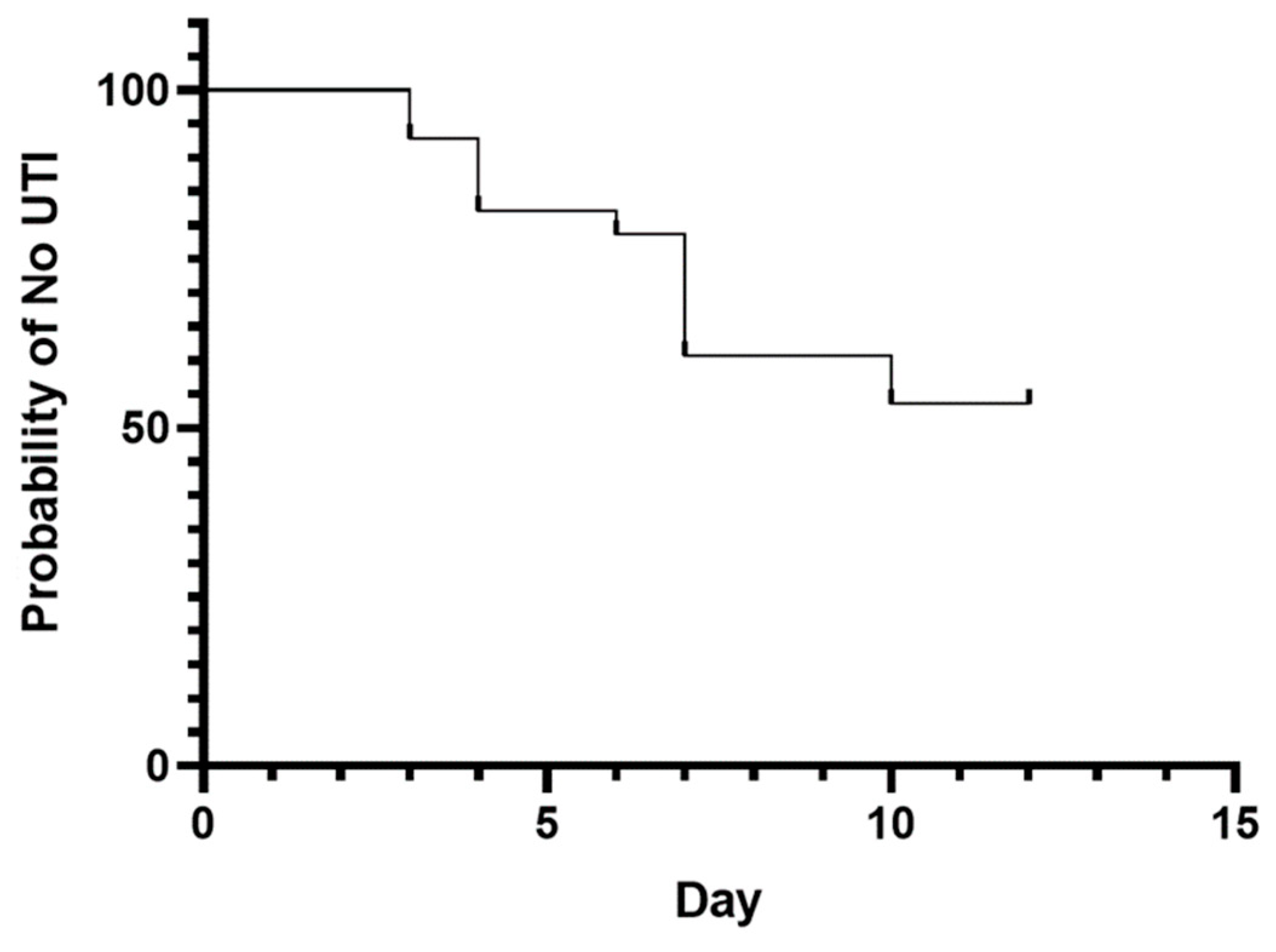

Table 1.
Antimicrobial susceptibility (%) of all positive urine culture samples.
| Antimicrobial | Acinetobacter spp. (n=2) | Enterococcus faecalis (n=6) | Escherichia coli (n=3) | Klebsiella pneumoniae (n=2) | Pseudomonas aeruginosa (n=2) |
|---|---|---|---|---|---|
| Amikacin | 50 | 0 | 67 | 50 | 100 |
| Amoxicillin | 0 | 0 | 33 | 0 | NA |
| Amoxicillin/Clavulanic Acid | 0 | 0 | 33 | 0 | NA |
| Azithromycin | 0 | 0 | 33 | 0 | NA |
| Ceftriaxone | 0 | 0 | 33 | 0 | NA |
| Cephalexin | 0 | 0 | 33 | 0 | NA |
| Ciprofloxacin | 0 | 0 | 33 | 0 | 100 |
| Enrofloxacin | 0 | 0 | 0 | 0 | 50 |
| Gentamicin | 0 | 0 | 33 | 0 | 100 |
| Imipenem | 0 | 0 | 100 | 50 | 100 |
| Marbofloxacin | 0 | 0 | 33 | 0 | 100 |
| Meropenem | 0 | 0 | 100 | 50 | 100 |
| Norfloxacin | 0 | 0 | 33 | 0 | 100 |
| Nitrofurantoin | NA | 0 | NA | 0 | NA |
| Sulfamethoxazole/Trimethoprim | 0 | 0 | 67 | 0 | NA |
| Vancomycin | NA | 100 | NA | 0 | NA |
Table 2.
Distribution of pathogens with percentages of multidrug-resistant (MDR) and non-MDR isolates.
Table 2.
Distribution of pathogens with percentages of multidrug-resistant (MDR) and non-MDR isolates.
| Bacterial isolates (15) | % MDR | % Non-MDR |
|---|---|---|
| Acinetobacter spp. (n=2) | 2 | 0 |
| Enterococcus faecalis (n=6) | 6 | 0 |
| Escherichia coli (n=3) | 2 | 1 |
| Klebsiella pneumoniae (n=2) | 2 | 0 |
| Pseudomonas aeruginosa (n=2) | 0 | 2 |
| 12 (80%) | 3 (20%) |
Disclaimer/Publisher’s Note: The statements, opinions and data contained in all publications are solely those of the individual author(s) and contributor(s) and not of MDPI and/or the editor(s). MDPI and/or the editor(s) disclaim responsibility for any injury to people or property resulting from any ideas, methods, instructions or products referred to in the content. |
© 2024 by the authors. Licensee MDPI, Basel, Switzerland. This article is an open access article distributed under the terms and conditions of the Creative Commons Attribution (CC BY) license (http://creativecommons.org/licenses/by/4.0/).
Copyright: This open access article is published under a Creative Commons CC BY 4.0 license, which permit the free download, distribution, and reuse, provided that the author and preprint are cited in any reuse.


