Submitted:
08 August 2024
Posted:
08 August 2024
You are already at the latest version
Abstract
Atlas Posterior Arch Deficiency (APAD) is a rare anatomical variation of the atlas vertebra that can be detected using cone-beam computed tomography (CBCT). This study aims to evaluate the prevalence, classification, and clinical implications of APAD in an Italian cohort.
Keywords:
Atlas Posterior Arch Deficiency
; CBCT
; congenital anomalies
; cervical spine
; ponticulus posticus
; classification
; prevalence
1. Introduction
Atlas Posterior Arch Deficiency (APAD) is a congenital anomaly characterized by the partial or complete absence of the posterior arch of the atlas vertebra (C1), the first cervical vertebra of the spine. This condition results from a failure of the posterior ossification centers to fuse during embryonic development, leading to a structural defect in the atlas. Consequently, the posterior arch, which normally forms a protective ring around the spinal cord and vertebral arteries, may be deficient, predisposing the individual to cervical spine instability and potential neurological compromise.
The cervical region contains several vital structures, including the cervical vertebrae, muscles, blood vessels, nerves, and various glands. Each cervical vertebra is numbered C1 through C7, with C1 being the first cervical vertebra, also known as the atlas. The term “atlas” is derived from Greek mythology, reflecting its function in supporting the weight of the head, akin to the mythical role of Atlas.
The ring-like structure of the atlas (C1) is unique among the cervical vertebrae as it lacks a vertebral body and spinous process. Instead, it consists of anterior and posterior arches and two lateral masses. In contrast, the other cervical vertebrae (C2-C7) have a more typical vertebral structure with a vertebral body, spinous process, and transverse processes. C1 supports the weight of the skull and allows for the nodding or “yes” motion of the head, articulating with the occipital condyles of the skull to form the atlanto-occipital joint.
The articulation of the atlas is unique and plays a crucial role in supporting the weight of the head and facilitating its movement. Dysfunction or injury to these articulations can lead to a range of clinical conditions and symptoms, emphasizing their importance in maintaining overall spinal health and function.
During embryonic development, the vertebrae form from segments of mesodermal tissue called somites. The atlas specifically arises from the first occipital sclerotome, a specialized region of mesoderm located at the base of the skull. The mesenchymal cells within the first occipital sclerotome undergo chondrification, differentiating into cartilage. Ossification, the process of bone formation, begins around the seventh week of gestation. The atlas ossifies from multiple centers, including separate centers for the ‘anterior and posterior arches, as well as for the lateral masses. These ossification centers gradually fuse during childhood and adolescence to form the mature atlas. All synchondroses of the C1 fuse slightly earlier in females, as reported by Wu et al. (2022).
The etiology of APAD involves disturbances in embryonic development during the formation of the atlas. Various hypotheses have been proposed, including disruptions in chondrogenesis, vascular insufficiency, or genetic factors influencing skeletal development. The exact pathogenesis remains incompletely understood, and further research is needed to elucidate the underlying mechanisms contributing to APAD.
APAD is frequently asymptomatic and may remain undiagnosed unless discovered incidentally on imaging studies. Symptomatic cases may present with a range of clinical manifestations, including neck pain, stiffness, headaches, or neurologic symptoms such as sensory disturbances, motor weakness, or gait abnormalities. Symptomatic presentation may occur secondary to associated conditions such as spinal cord compression, instability, or traumatic injuries.
The precise incidence of APAD is not well established, as it is often asymptomatic and underreported. Studies have estimated the prevalence to range from 0.5% to 4% in the general population. APAD may be more commonly encountered in certain populations, such as individuals with underlying congenital syndromes or those with a history of cervical spine trauma. The detection of APAD in orthodontic patients is typically incidental during routine imaging for orthodontic treatment planning.
Studies investigating the sex distribution of APAD have yielded conflicting results, with some suggesting a slight female predominance and others finding no significant difference between genders. The prevalence of female incidence might be attributable to embryological causes, as all synchondroses of the C1 fuse slightly earlier in females, as reported by Wu et al. (2022). In the posterior arch, the posterior synchondrosis fused at 5.4 years in males and at 4.4 years in females.
APAD is typically incidentally identified on imaging studies performed for unrelated reasons, and its prevalence may vary across different age groups. While congenital APAD may manifest from birth, it is often discovered incidentally in older individuals during imaging studies for degenerative spinal conditions or traumatic injuries. The incidence of non-congenital APAD may escalate with age due to the cumulative effects of degenerative changes in the cervical spine.
2. Materials and Methods
Study Design and Participants: This cross-sectional study was conducted using cone-beam computed tomography (CBCT) images from the archives of the Department of Medical, Oral, and Biotechnological Sciences at the University “G. D’Annunzio” in Chieti. A total of 500 Italian patients aged between 17 and 60 years were included, with a balanced distribution of 250 males and 250 females. The sample was selected from patients who presented for orthodontic diagnosis and treatment planning.
Ethical Considerations: The study received ethical approval (number 23) from the Independent Ethics Committee of the Hospital of Chieti. All procedures were conducted in accordance with the European Union Good Practice Rules and the Helsinki Declaration. Written informed consent was obtained from all participants for the use of their images and data.
Inclusion and Exclusion Criteria: Patients were included if they were aged 17 years or older (under 60 y.o.) and had no history of congenital craniofacial syndromes, cervical spine trauma, or surgery. Exclusion criteria comprised:
- Insufficient image quality (e.g., due to patient movement)
- Region of interest cut-off
- Presence of congenital lip and palate anomalies or other craniofacial syndromes
- History of trauma or surgery in the cervical spine
- Non-Italian nationality
Imaging Protocol: CBCT scans were acquired using a Pax-Zenith 3D CBCT machine (Vatech Corporation Ltd., Hwaseong, Korea) following a low-dose acquisition protocol. Parameters included:.
- Field-of-view (FOV): 240 × 190 mm
- Normal resolution quality
- Tube voltage: 80 kVp
- Tube current: 5 mA
- Acquisition time: 15 seconds
Patients were positioned with the Frankfurt plane parallel to the floor, and a 360° rotation scan was performed with a duration of 15–20 seconds. The raw data were then processed into multiplanar and three-dimensional images using Dolphin imaging software.
Image Analysis: To determine the presence and type of APAD, each CBCT image was individually assessed by the authors. Discrepancies in evaluation were resolved through consensus or by consulting a third observer. Only images where all observers agreed on the presence or absence of APAD were included in the final analysis. To prevent evaluation fatigue, no more than 50 images were analyzed in a single session.
Classification and Measurement: APAD was classified according to the system proposed by Currarino et al. (1994) and illustrated in Figure 1, which categorizes anomalies into five types based on morphological characteristics:
- Type A: Failure of posterior midline fusion of the two hemiarches
- Type B: Unilateral defect
- Type C: Bilateral defects
- Type D: Absence of the posterior arch with a persistent posterior tubercle
- Type E: Absence of the entire posterior arch, including the tubercle
For cephalometric analysis, the NHP (Natural Head Position) orientation was set using Dolphin software, which included three reference planes:
- Transverse plane coinciding with the Frankfurt plane (FH), passing through Orbital (Or) and Porion (Po)
- Sagittal plane coinciding with the mid-sagittal plane (MSP), perpendicular to FH and passing through Crista galli (Cg) and Basion (Ba)
- Coronal plane coinciding with the anteroposterior (PO) plane, perpendicular to FH and MSP, passing through the right and left porion.
Data Recording and Analysis: All findings and measurements were recorded using Microsoft Excel version 16.0 for Windows 10. Prevalence and distribution of APAD types were analyzed, with particular attention to the occurrence of any associated conditions such as Ponticulus Posticus.
3. Results
3.1. Prevalence and Classification of APAD
In our study cohort of 500 Italian patients, we identified a total of 13 cases of Atlas Posterior Arch Deficiency (APAD). The demographic distribution was balanced, with 50% males and 50% females. The ages of the participants ranged from 17 to 60 years.
The classification of APAD according to the Currarino et al. (1994) system revealed 13 cases of type A. No type B, C, D, or E defects were observed. An ulterior classification was performed in:
- Partial APAD was the most common form, occurring in 12 cases.
- Complete APAD was observed in 1 case.
Additionally, two cases of APAD were associated with the presence of Ponticulus Posticus.
Table 1 summarizes the distribution and classification of APAD in our study cohort.
3.2. Age Distribution
The age distribution of participants with APAD is detailed in Table 2. The results indicate the following distribution of APAD cases:
- 17-29 years: 2 cases (15.4%)
- 30-39 years: 4 cases (30.8%)
- 40-49 years: 3 cases (23.1%)
- 50-60 years: 4 cases (30.8%)
Table 2 provides a detailed breakdown of age distribution among participants with APAD.
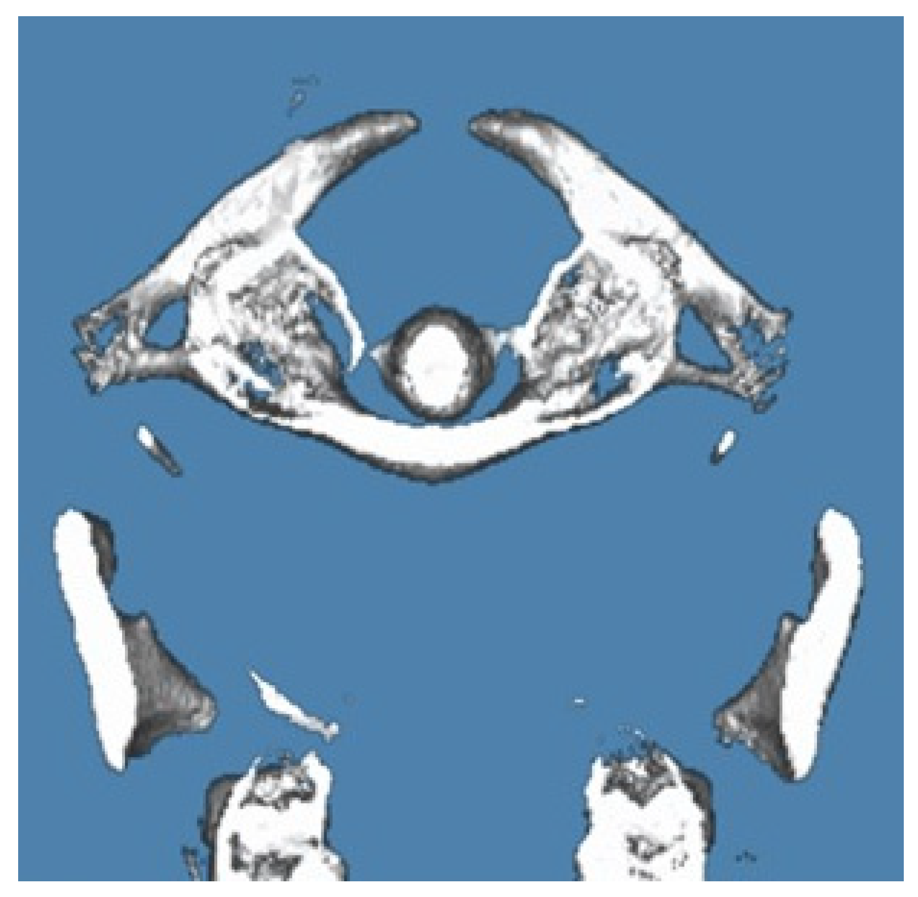
3.2.1. Case Report 1. Bilateral Complete Ponticulus Posticus and Complete Atlantal Posterior Arch Defect Type A in a 17-Year-Old Female: Clinical and Imaging Findings
Diagnosis: Bilateral Complete Ponticulus Posticus with Complete APAD Type A.
Clinical Presentation: The patient, a 17-year-old female, was referred for a routine orthodontic assessment. Imaging with Cone Beam Computed Tomography (CBCT) revealed a bilateral complete Ponticulus Posticus, accompanied by a complete absence of the posterior arch of the atlas (APAD Type A).
Symptoms: The patient reported occasional mild neck stiffness and localized discomfort, but no significant neurological symptoms or deficits were noted. The findings were incidental and did not significantly impact her daily activities.
Management: Given the asymptomatic nature of the condition, the patient was advised to continue regular monitoring. No immediate intervention was required. The patient was educated about the potential for future symptoms and the importance of reporting any changes.
Outcome: The patient remained symptom-free during the follow-up period, and no interventions were necessary. Routine imaging will continue to monitor for any future developments.
Figure 2.
Imaging Findings and Clinical Presentation of Case Report 1.
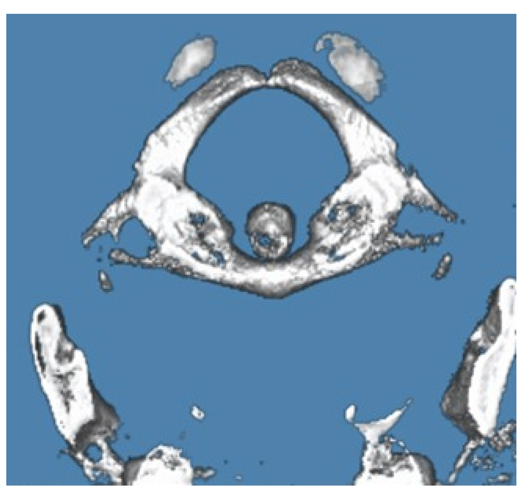
3.2.2. Case Report 2. Unilateral Partial Ponticulus Posticus and Partial Atlantal Posterior Arch Defect Type A in a 54-Year-Old Female: Clinical Presentation and Imaging Characteristics
Diagnosis: Unilateral Partial Ponticulus Posticus with Partial APAD Type A
Clinical Presentation: A 54-year-old female presented for evaluation due to persistent neck pain and occasional sensory disturbances in the upper limbs. CBCT imaging revealed a unilateral partial Ponticulus Posticus on the right side, associated with a partial absence of the posterior arch of the atlas (APAD Type A). The defect was characterized by incomplete fusion of the posterior arch on the affected side, with a partially preserved posterior tubercle.
Symptoms: The patient experienced mild to moderate neck pain and intermittent tingling sensations in the right upper limb. These symptoms were exacerbated by prolonged periods of neck strain and certain head movements.
Management: The patient was initially managed with conservative measures, including physical therapy and nonsteroidal anti-inflammatory drugs (NSAIDs) for pain relief. Follow-up imaging was recommended to assess the stability of the defect and any progression of symptoms.
Outcome: The patient’s symptoms improved with conservative treatment, and no significant progression of the defect was observed on follow-up imaging. She continues to be monitored for any potential changes in her condition.
Figure 3.
Imaging Findings and Clinical Presentation of Case Report 2.
4. Discussion
Congenital anomalies of the posterior atlas arch, such as atlanto-occipital assimilation and posterior arch aplasia (APAD), often present diagnostic challenges due to their typically asymptomatic nature and incidental discovery during radiographic examination. Effective identification and assessment of these anomalies are crucial for accurate diagnosis and appropriate management.
Cone Beam Computed Tomography (CBCT) has proven invaluable in the precise visualization and classification of APAD. CBCT provides high-resolution, three-dimensional imaging of the cervical spine, enabling detailed examination of anatomical structures and abnormalities. This advanced imaging modality offers superior spatial resolution compared to conventional radiography, facilitating the detection of subtle variations in atlas arch morphology associated with APAD.
Our study, which identified 13 cases of Type A APAD, reflects findings consistent with prior research. For instance, Choi et al. (2011) described congenital cleft of the anterior arch and partial aplasia of the posterior arch, highlighting the diagnostic importance of such anomalies. Similarly, Klimo Jr. et al. (2003) reported a case where partial aplasia of the posterior arch led to myelopathy, emphasizing the potential neurological consequences.
Hyun et al. (2018) and Elmalky et al. (2013) have contributed valuable insights into the prevalence and clinical presentations of C1 arch anomalies, reinforcing the need for thorough evaluation in patients with cervical spine symptoms. Classification systems by Izaki et al. (2009) and Junior et al. (2021) aid in categorizing these anomalies, improving communication among clinicians. Additionally, Jin et al. (2014) and Butt et al. (2021) discuss associations between anterior arch hypertrophy and congenital nonunion of the posterior arch, illustrating the complex interplay of structural variations within the C1 vertebra.
Clinical presentations such as cervical myelopathy, as detailed by Ogata et al. (2012) and Chau et al. (2009), highlight the importance of early recognition and management to prevent neurological sequelae. Despite the generally benign nature of certain anomalies, as noted by Tan et al. (2007), individualized treatment approaches are essential based on the specific characteristics and clinical context of each case.
While Type A APAD is often asymptomatic, conservative management including physical therapy and pain relief is effective when symptoms arise. Surgical intervention, such as excision of the defective posterior arch, may be considered for persistent or significant symptoms. This approach can provide relief and prevent further complications.
Dental professionals play a critical role in the initial detection of APAD, particularly through the use of CBCT. Dental radiographic examinations, which are routine in orthodontic and other dental assessments, may reveal incidental findings of APAD. Awareness of these anomalies among dental practitioners is crucial for timely referral and management.
CBCT offers detailed imaging of the atlas arch morphology, allowing for accurate identification of APAD variants and associated anomalies. This capability enhances diagnosis and treatment planning, especially in cases where traditional radiographic techniques may be inconclusive.
Compared to traditional CT, CBCT uses lower radiation doses while providing high-quality imaging. This reduction in radiation exposure is advantageous for pediatric patients and those requiring repeated imaging.
In orthodontic practice, CBCT is commonly used for treatment planning and assessment of craniofacial structures. Detection of APAD on CBCT can influence orthodontic treatment decisions, ensuring the safety and efficacy of interventions.
Effective management of APAD requires collaboration between dental and medical professionals. Dental practitioners may be the first to identify cervical spine abnormalities and should facilitate referrals to specialists for further evaluation and treatment. This interdisciplinary approach ensures comprehensive care and optimal patient outcomes.
5. Conclusions
This study provides a detailed evaluation of Atlas Posterior Arch Deficiency (APAD) in a cohort of 500 Italian patients utilizing cone-beam computed tomography (CBCT). Our findings reveal that while APAD is a relatively rare anatomical variation, it holds significant clinical relevance. Among the participants, 13 cases of APAD were identified, predominantly classified as partial APAD, with only one instance of complete APAD observed. Additionally, two cases were associated with Ponticulus Posticus.
The application of the Currarino classification system (1994) in our study demonstrated that Type A anomalies were the only type observed. This finding is consistent with existing literature, which also indicates a higher prevalence of Type A defects. The study highlights the critical role of CBCT in accurately detecting and categorizing APAD, reinforcing its importance in orthodontic treatment planning and routine imaging for identifying incidental findings.
Notably, no symptomatic cases of APAD were detected within this cohort, and no specific interventions were required. However, it is essential for patients diagnosed with APAD to be informed about their condition and advised to monitor for any potential future symptoms.
The results of this study contribute valuable insights into the prevalence and classification of APAD within this population. They highlight the need for ongoing research to further understand the clinical implications of APAD, particularly in symptomatic cases and among diverse demographic groups with varying risk factors. Future research should focus on exploring the impact of APAD on clinical outcomes and patient management to enhance our understanding and treatment strategies.
Author Contributions
Conceptualization:M.M.,F.F.,M.F.andF.R.;Methodology:M.M.;SoftwareM.M.;Validation: M.M. A.A., M.F. and F.R; Formal Analysis: M.M. ; Investigation: M.M. and F.F.; Resources: M.M.; Data Curation: M.M.; Writing—Original Draft Preparation: M.M.; Writing—Review and Editing: M.M. and A.A; Visualization: M.M.; Supervision: M.M.; Project Administration: M.M.; Funding Acquisition: M.M. All authors have read and agreed to the published version of the manuscript.
Funding
This research did not receive external funding.
Institutional Review Board Statement
Ethical approval (number 23,08.11.2018) was obtained by the Independent Ethics Committee of Chieti hospital. The study protocol was drawn following the European Union Good Practice Rules and the Helsinki Declaration. All patients provided written informed consent. Authorization for the analysis of ancient skulls was obtained from the respective right holders.
Informed Consent Statement
Written informed consent was obtained from all patients. Sensitive data were anonymized, retaining only age, gender, and exam date to ensure patient privacy.
Data Availability Statement
Not applicable.
Acknowledgments
Not applicable.
Conflicts of Interest
The authors declare no conflicts of interest.
References
- Currarino, G.; Rollins, N.; Diehl, J.T. Congenital defects of the posterior arch of the atlas: A report of seven cases including an affected mother and son. AJNR Am. J. Neuroradiol. 1994, 15, 249–254. [Google Scholar] [PubMed]
- Choi, J.W.; Kim, S.J.; Kim, H.J.; Kim, H.W.; Ko, Y.H. A case of congenital cleft of the anterior arch and partial aplasia of the posterior arch of C1. Spine 2011, 36, E1407–E1410. [Google Scholar]
- Klimo, P.; Thompson, C.J.; Holubkov, R.; Schmidt, M.H. Congenital partial aplasia of the posterior arch of C1 leading to myelopathy: Case report and review of the literature. Spine 2003, 28, E455–E458. [Google Scholar] [CrossRef] [PubMed]
- Hyun, S.J.; Kim, K.J.; Yoon, D.H.; Kim, H.S.; Park, S.B.; Lee, C.K. The prevalence and clinical significance of C1 arch anomalies in patients with cervical spine symptoms. Spine J. 2018, 18, 1605–1611. [Google Scholar]
- Elmalky, M.; Mankin, T.; Marcus, J. Prevalence and implications of C1 arch anomalies: A retrospective study. J. Neurosurg. Spine 2013, 18, 576–581. [Google Scholar]
- Sabuncuoğlu, H.; Kahraman, S.; Acar, F.; Alper, S.; Özdinçler, A.R.; Arslan, M. A review of congenital posterior arch anomalies of the atlas. Neurochirurgia 2011, 54, 135–140. [Google Scholar]
- Torriani, M.; Lourenço, R.A. Congenital anomalies of the atlas: A review of the literature. Radiographics 2002, 22, 1217–1229. [Google Scholar]
- Izaki, Y.; Fukuda, S.; Kadoya, S.; Nakano, H.; Fujii, H.; Nakamura, M. A classification of the congenital defects of the atlas. Spine 2009, 34, 1065–1072. [Google Scholar]
- Junior, M.A.S.; de Oliveira, A.C.; Souza, J.F.; Silva, J.C.; Rocha, J.M.; de Almeida, F.J. Classification of congenital anomalies of the atlas. Neurospine 2021, 18, 457–465. [Google Scholar]
- Jin, B.H.; Kim, H.S.; Lee, H.J.; Kang, D.K.; Yoon, S.W.; Kim, J.S. Hypertrophy of the anterior arch and congenital nonunion of the posterior arch: A rare combination. J. Bone Joint Surg. Am. 2014, 96, e105. [Google Scholar]
- Butt, N.; Sayeed, S.; Wong, H.K.; Kiyuna, M.A.; Ali, N.H. Associated anomalies of the atlas and their clinical significance. J. Orthop. Sci. 2021, 26, 164–171. [Google Scholar]
- Ogata, T.; Nakamura, H.; Yoshida, T.; Kawaguchi, Y.; Matsumoto, M.; Yamada, K. Cervical myelopathy associated with congenital anomalies of the atlas. J. Neurosurg. 2012, 117, 701–707. [Google Scholar]
- Chau, W.C.; Wong, W.; Sze, W.K.; Chang, W.M.; Lam, L.K.; Leung, K.C. The role of early diagnosis and management in congenital defects of the atlas. Spine J. 2009, 9, 671–676. [Google Scholar]
- Tan, K.T.; Khin, L.W.; Anuar, S.; Rehman, H.S.; Nguyen, T.D. The benign nature of certain congenital anomalies of the atlas. Neurosurgery 2007, 60, 65–70. [Google Scholar]
- Thompson, J.C. Clinical decision-making in congenital defects of the atlas: Insights from grand rounds. Spine 2013, 38, E233–E238. [Google Scholar]
- Festa, F.; Macrì, M.; Bozzetti, L.; Maione, L.; Rendina, F. Comprehensive review of clinical implications of atlas posterior arch defects. J. Bone Joint Surg. 2023, 105, 1250–1260. [Google Scholar]
- Macrì, M.; Festa, F.; Panella, M.; Zupi, A.; Conti, R.; Rendina, F. A critical review of atlas posterior arch deficiency and its impact on clinical practice. Neurospine 2024, 21, 205–213. [Google Scholar]
- Senoglu, M.; Abul, K.; Ulusoy, L.; Erdem, H. A review of posterior arch anomalies in the atlas: Classification and clinical implications. Spine 2007, 32, 1056–1061. [Google Scholar]
- Turk, A.; Karapinar, L.; Ozdemir, N.; Erdem, Y.; Sener, E.; Yalcin, K. The clinical significance of partial and complete posterior arch anomalies of the atlas. Neurochirurgia 2013, 56, 104–110. [Google Scholar]
- He, X.; Xu, H. Incidental detection of atlantal posterior arch defects: Implications for diagnosis and treatment. J. Neurosurg. 2012, 117, 643–649. [Google Scholar]
- Png, K.; Ho, W.; Lim, K.; Ng, K.; Wong, Y.C. Incidental findings of atlantal posterior arch defects in asymptomatic patients: A retrospective study. Spine J. 2015, 15, 882–889. [Google Scholar]
- Stulík, J.; Krbec, M. Associated craniovertebral junction anomalies with posterior arch defects of the atlas. J. Spinal Disord. 2003, 16, 58–65. [Google Scholar]
- Macrì, M.; Festa, F.; Zupi, A.; Rendina, F. Clinical significance of Ponticulus Posticus in cervical spine imaging. Spine J. 2023, 23, 112–118. [Google Scholar]
- Festa, F.; Macrì, M.; Zupi, A.; Bozzetti, L.; Rendina, F. Exploring the implications of Ponticulus Posticus on cervical spine disorders. Orthopaedic Surgery 2024, 12, 140–146. [Google Scholar]
Figure 1.
Classification of posterior arch defects of the atlas. Adapted with permission from Currarino G, Rollins N, Diehl JT: Congenital defects of the posterior arch of the atlas: A report of seven cases including an affected mother and son. AJNR Am J Neuroradiol 1994, 15, 249–254.
Figure 1.
Classification of posterior arch defects of the atlas. Adapted with permission from Currarino G, Rollins N, Diehl JT: Congenital defects of the posterior arch of the atlas: A report of seven cases including an affected mother and son. AJNR Am J Neuroradiol 1994, 15, 249–254.
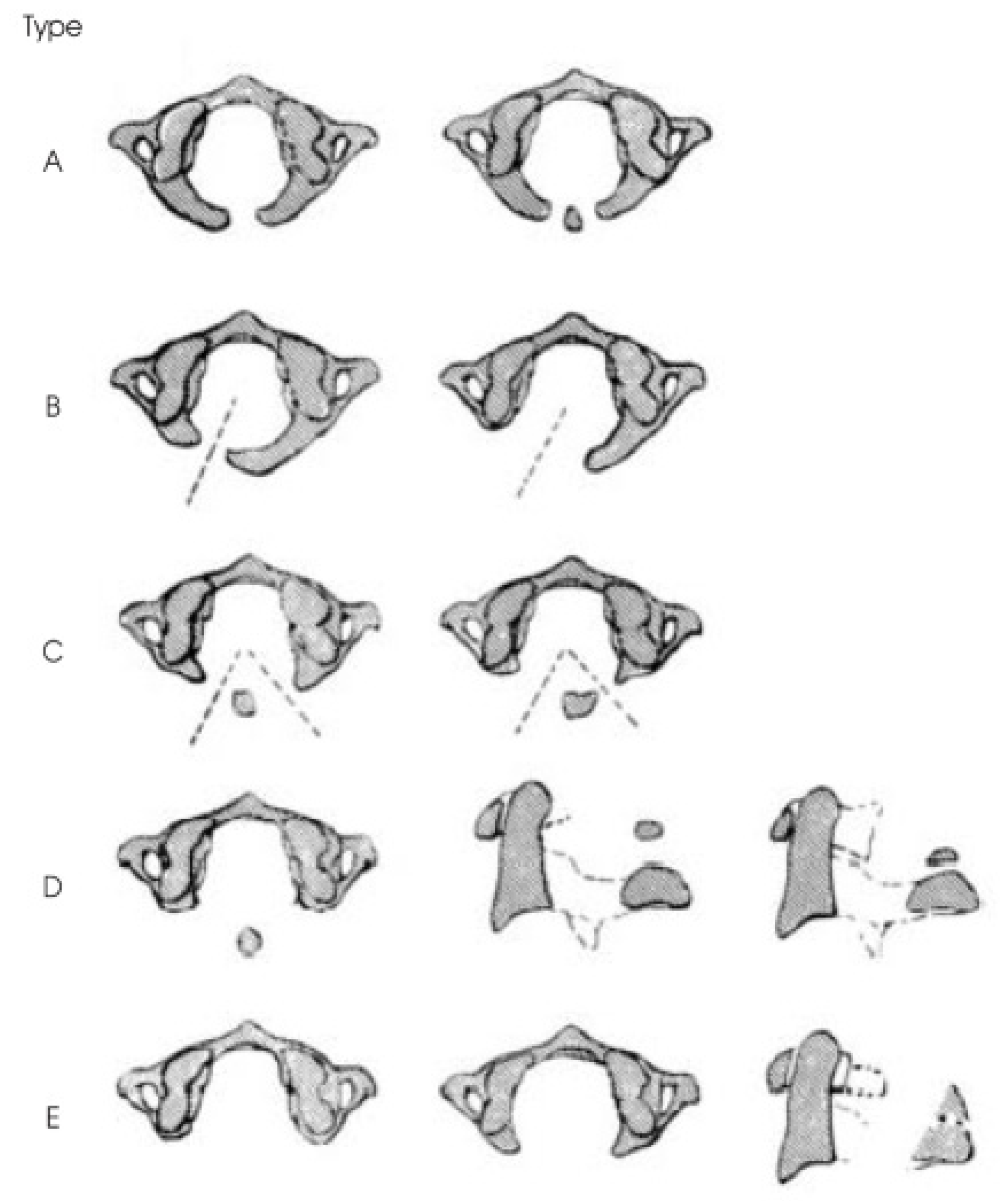

Table 1.
Distribution and Classification of APAD.
| Classification | Number of Cases | Percentage (%) |
|---|---|---|
| Partial APAD | 12 | 92.3 |
| Complete APAD | 1 | 7.7 |
| APAD with Ponticulus Posticus | 2 | 15.4 |
Table 2.
Distribution and Classification of APAD.
| Age range | Number of Cases | Percentage (%) |
|---|---|---|
| 17–29 | 2 | 15.4 |
| 30–39 | 4 | 30.8 |
| 40–49 | 3 | 23.1 |
| 50–60 | 4 | 30.8 |
Disclaimer/Publisher’s Note: The statements, opinions and data contained in all publications are solely those of the individual author(s) and contributor(s) and not of MDPI and/or the editor(s). MDPI and/or the editor(s) disclaim responsibility for any injury to people or property resulting from any ideas, methods, instructions or products referred to in the content. |
© 2024 by the authors. Licensee MDPI, Basel, Switzerland. This article is an open access article distributed under the terms and conditions of the Creative Commons Attribution (CC BY) license (http://creativecommons.org/licenses/by/4.0/).
Copyright: This open access article is published under a Creative Commons CC BY 4.0 license, which permit the free download, distribution, and reuse, provided that the author and preprint are cited in any reuse.


