Submitted:
18 June 2024
Posted:
19 June 2024
You are already at the latest version
Abstract
The leaves of Nicotiana glauca (Solanaceae) plant, are known for its major cause of health concern. This study investigated the antioxidant activity and polyphenols composition of aerial parts of N.glauca collected from its wild habitat in Jordan, using Methanol-Conventional (MC) and deep eutectic solvents (DESs) extraction methods in addition to nicotine content determination using UHPLC. The results have shown that the MC extract contains fewer total phenols and flavonoid content than the DES extract at ratio of 90% (0.1194 ±0.009 and 0.311± 0.020 mg/mL equivalent to gallic acid) and (0.01084±0.005 and 0.928 ± 0.09 mg/mL equivalent to rutin), respectively. Moreover, the study showed that the MC extract contains 635.07 ppm nicotine compared to the DES extraction method, which contains 1194.91 ppm. Both the MC and the DES extracts exhibited weak antioxidant activities with the highest was 33%inhibition equivalent to ascorbic acid was found for DES at (90%) ratio. The UHPLC-MS/MS analysis revealed the presence of variations in the detected compounds between the two extraction methods used. This study found that en-vironmentally friendly DES extraction of N. glauca produced higher phenol and flavonoid content than MC method, highlighting green chemistry methods' superior efficiency and environmental benefits for extracting valuable phytoconstituents.
Keywords:
Nicotiana glauca
; Chromatography
; Mass Spectrometry
; Antioxidants
; deep eutectic solvents
; sustainable extraction.
1. Introduction
Tobacco, which is derived from the leaves of the Nicotiana plant, is known for its abuse and is recognized as a major global health concern. However, it has been used in traditional medicine by Native Americans, to treat respiratory, parasitic, and mental problems [1]. Later in Europe, the tobacco plant was enlisted in various pharmacopeias, with therapeutic applications in treating catarrh, colds, and fevers, as well as being used as a digestion aid, a purgative, and a narcotic. Later in the 20th century, some reports suggested that tobacco might lower the risk of Alzheimer’s disease, Parkinson’s and Tourette’s syndrome [2].
Several compounds, like alkaloids, steroids, tannins, and flavonoids, were isolated from Nicotiana species. Many of these metabolites are bioactive with reported anti-inflammatory, antitumor, antibacterial, and antioxidant activities [3]. For instance, Nicotiana glauca Graham (Solanaceae) was shown to contain anabasine as the major alkaloid in the methanolic extract of their leaves [4], which is known to possess antiparasitic activity [5]. A study by Ameya et al (2017), revealed that N. tabacum L. contains pyridine alkaloids with antibacterial activity against biofilm-forming pathogens [6]. These alkaloids were used to treat strep throat caused by Streptococcus pyogenes [7] and showed activity against Staphylococcus aureus, Enterococcus faecalis, Escherichia coli, and Pseudomonas aeruginosa [8].
Moreover, several reports have highlighted the antioxidant activity of tobacco plant extracts, suggesting potential applications for various purposes. According to a recent study the methanol extract of N. glauca contains high levels of phenolic compounds, such as Chlorogenic acid and rutin [9]. These compounds were found to contribute to anti-inflammatory, anti-aging, and anticancer effects [3]. Another study in Saudi Arabia has demonstrated the antimicrobial effects of N. glauca against E.coli and S. aureus. The study concluded that the extracts from the leaves and flower had the highest amounts of phytochemicals [10].
Conventional methods for extracting natural alkaloids and flavonoids, such as Soxhlet, maceration, percolation, and organic solvent extraction, are well-studied but have significant drawbacks. These techniques are time-consuming, inefficient, and often require large quantities of toxic, flammable, and non-biodegradable solvents, making them non-specific and not cost-effective. To address these issues, innovative solvents like deep eutectic solvents (DES) and natural deep eutectic solvents (NaDES) have been recently utilized [11,12,13,14]. As a subclass of ionic liquids (ILs), NaDES are considered less toxic, lower-cost, greener, and more efficient alternatives to both conventional organic solvents and ILs [15]. NaDES are usually prepared from a hydrogen bond donor and a hydrogen bond acceptor, which, when mixed in certain ratios, form a liquid at room temperature. Overall, the versatility, low toxicity, and environmentally friendly nature of NaDES make them attractive for a broad spectrum of industrial and research applications as solubilizers [16], drug delivery vehicles [17,18,19,20], stability enhancers [21], extraction and purification [22,23]. Additionally, NaDES themselves have been reported to possess antimicrobial [24], antioxidant [25], antibiofilm agents [26], and wound healing activity [17] among other beneficial effects. Choline chloride (a hydrogen bond acceptor) and malonic acid (a hydrogen bond donor) are natural compounds and are among the most commonly used substances for preparing NaDESs [27].
In the context of NaDESs application, this method was utilized for the extraction of polyphenols from Citrus aurantium L. peel [28]. The results showed enhanced recovery efficiency of polyphenols in the obtained extracts. Regarding tobacco plants, Hong et al., (2022) proposed the DES method for the extraction of solanesol from waste tobacco leaves [29]. Recently, cembranoid-type diterpenes compounds, known for their anticancer and antimicrobial effects, were extracted from tobacco flower waste using DESs. Findings revealed the importance of green technologies in waste management and the extraction of bioactive natural compounds [30].
Hassan et al., (2014) conducted a phytochemical analysis of the N. glauca growing in Egypt [31]. Findings showed that the content of flavonoids in N. glauca was influenced by its habitat’s different conditions, which also affected the antioxidant activities. Therefore, research should take into consideration the use of medicinal plants relative to their composition of active and/or toxic metabolites collected from different regional areas.
This study aims to investigate the antioxidant activity and polyphenols composition of aerial parts of N. glauca species collected from its wild habitat in Jordan, using Methanol-Conventional (MC) and deep eutectic solvents (DESs) extraction methods. This may provide information relevant to phenols content and antioxidant effect of the prepared extracts, revealing novel proposed uses with economic values ensuring the sustainable use of this plant species.
2. Materials and Methods
2.1. Plant Material
Fresh leaves of N. glauca were collected from widely grown plants in North Jordan, during the spring of 2022. The plant material was authenticated by an expert botanist in the Royal Botanical Gardens, Jordan. The voucher sample was deposited in the laboratory of Al-Ahliyya Amman University (Amman, Jordan). N. glauca leaves were dried under shade before the reduction in size using a conventional grinder and kept in a dark dry place at room temperature until used.
2.2. Methanol Conventional Extraction (MC)
An extract of the study plants was prepared using 50 g of dry plant material in 500 mL of methanol using the soaking method for 72 hrs. at room temperature. This process was repeated twice, then the suspension was filtered and concentrated until a fine powder was obtained using Benchtop Manifold Freeze Dryer from Millrock Technology®.
2.3. Deep Eutectic Solvents (DESs) Extraction
The DES was prepared from malonic acid and choline chloride in a 1:1 w/w molar ratio by physically mixing the two components gently on a hotplate to around 50-80 ˚C until a clear, homogeneous liquid was formed. The prepared DES was mixed with deionized water to prepare three different extraction mixtures namely 30%, 70%, and 90% v/v. The cold extraction method was utilized by adding 5g of the dry powder plant material in 25 mL of the DES extraction media. The plant material was soaked in the solvent for 72 hours and then filtered to complete the extraction process.
2.4. Determination of Total Phenolic
The total phenolic content was measured using the Folin-Ciocalteu method as described by Alnsour et al., 2022 [32]. The phenolic content was determined calorimetrically at 765 nm. The total phenolic content (mg/mL) was determined as gallic acid equivalent. A stock solution of the plant extract was prepared at a concentration of 5 mg/mL. Serial dilutions were made, and an aliquot of each sample concentration (80 μL) was added to Folin–Ciocalteu (400 μL) reagent in a test tube, mixed with 7.5% sodium carbonate solution (320 μL). The solution was incubated in a dark place at 45◦C water bath for 30 mins. Total phenolic content was expressed as gallic acid equivalent (mg/g), using the standard curve (Equation 1):
y=0.0049x+0.0426, R 2=0.9991
y = absorbance at 765 nm and x = concentration of total phenolic content gallic acid equivalent mg/mL.
2.5. Determination of Total Flavonoids
The determination of total flavonoids was performed using a colorimetric method based on the formation of a complex flavonoid–aluminum, measured at a wavelength of 510 nm using a UV-spectrophotometer as described by Ubaydee, et al., (2022) [33]. The results were expressed as (mg/mL) equivalents to quercetin. Briefly, a stock solution of the plant extract at a concentration of 5 mg/mL was prepared. Serial dilutions were made, 1 mL of each concentration was added into (0.5 mL) AlCl3, (0.5 mL) NaNO2, (2 mL) NaOH, and (4 mL) distilled water. The mixture was incubated at room temperature for 15 mins.
Total Flavonoid content was expressed as rutin equivalent (mg/mL), using the standard curve (Equation 2):
y=0.0009x+0.613 , R2=0.994
y = absorbance at 510 nm and x = concentration of total phenolic content rutin equivalent mg/mL.
2.6. In-Vitro Antioxidant Activity
The 2,2-diphenyl-1-picrylhydrazyl (DPPH) scavenging activity was used as described by Al-Bayati et al., (2023) [34]. For the reaction reagent, DPPH was dissolved in methanol at a concentration of (0.04 g/mL). The reaction was performed by dissolving plant extract in methanol at a concentration of (0.01g/mL). An aliquot of 1 mL of the plant extract solution was mixed with 3 mL of DPPH and completed to a final volume of 10 mL using methanol, then allowed to stand in darkness for 30 minutes. Absorbance was measured at a wavelength of 517 nm. Ascorbic acid was used as a reference for comparison (Sigma Aldrich, Germany). A calibration curve of ascorbic acid was used for the calculation of the effective concentration required for scavenging DPPH free radicals (%inhibition Equation 3).
% inhibition= [ (A control - A sample)/ A control ⌉x100
Where: A_control = absorbance of the control sample, and A_(sample ) = absorbance of the sample.
2.7. UHPLC-MS/MS Methodology
Instrumentation and MS parameters
The UHPLC coupled with Impact II QTOFMS Bruker Daltonik (Bremen, Germany) was used for screening the compounds of interest using the same method previously described by Al-Bayati, et al., (2023) [34]. The instrument operation conditions were as follows: Apollo II ion funnel electrospray source, capillary voltage (2500 V), nebulizer gas (2 bar), and nitrogen dry gas at a flow rate of 8 L/min (200 ºC). The mass accuracy was ˂ 1 ppm; with Full Sensitivity Resolution (50000 FSR) and the TOF repetition rate of 20 kHz.
Bruker Solo 2.0_C-18 UHPLC column (100 mm x 2.1 mm x 2.0 μm) was used for chromatographic separation at a flow rate of 0.51 mL/min (40 ℃). The mobile phase consists of (A: 0.05% formic acid in water), and (B: acetonitrile). Gradient elution was used as follows: 0 – 27 mins linear gradient from 5% - 80% B; 27-29 min 95% B; 29.1 min 5% B. The total analysis time was 35 mins in positive mode and 35 mins in negative mode, with an injection volume of 3 µL.
MC and DES samples stock solutions were prepared by dissolving an appropriate amount of the plant extract in dimethyl sulfoxide-DMSO (analytical grade), then diluted with acetonitrile to complete 50 mL, then centrifugation was performed at 4000 rpm was applied for 2 mins. All the other reagents, Acetonitrile, methanol, water, and formic acid used were LC-MS grade.
Sample preparation: 100 µL of each sample has been dissolved in 900 µL of MeOH. A 1.0 mL was transferred to an autosampler and injected. Identification of phenols and flavonoid compounds was based on the retention time (Rt), mass spectrum (m/z), and molecular formula, compared to a previously developed integrated library of natural compounds.
2.8. Nicotine Content Determination
Nicotine content determination was performed as described by Kheawfu et al., 2021 [35]. Briefly, each obtained extract was analyzed by UHPLC coupled with Impact II QTOFMS Bruker Daltonik (Bremen, Germany). Bruker Solo 2.0_C-18 UHPLC column (100 mm x 2.1 mm x 2.0 μm). A linear elution mobile phase composed of Sodium acetate, methanol, and trimethylamine, (88:12:0.5 v/v) (pH = 4.2) was used. The mobile phase elution was adjusted to a flow rate of 1 mL/min and measured at UV =259 nm. The total analysis time was 20 minutes in positive mode.
Nicotine standard (AccuStandard®, Inc.) was used for establishing the calibration curve at concentrations ranging of (0.10 – 2.00 µg/mL) in water and was used for the calculation of nicotine content in plant extracts as ppm values.
Sample preparation for UHPLC-MS/MS analysis: (A) 500 µL from MC or DES extract samples were diluted with 500 µL methanol, then the solution was centrifuged at 4000 rpm for 2.0 min. Next, 1.0 mL was transferred to the autosampler and 3.0 µL was injected into the system (D.f.=2). (B) Then, 50 µL was taken from sample (A), and diluted with 1950 µL of methanol. Next, 1.0 mL was transferred to the autosampler and 3.0 µL was injected into the system (D.f.=40). (C) Then, 100 µL was taken from sample (B), and diluted with 1900 µL of methanol. Next, 1.0 mL was transferred to the autosampler and 3.0 µL was injected into the system (D.f.=20), (D.f.total = 2*40*20).
3. Results
3.1. Total Phenol Content
The MC extract was shown to contain less total phenolic compounds (0.1194 ±0.009 mg/mL equivalent to gallic acid), compared to the DES extracts which showed a similar total phenols content for the three prepared extracts ratios with almost no significant difference between three tested DES ratios (30,70, and 90%) corresponding to an average of 0.312± 0.13 mg/ mL (equivalent to gallic acid) (Table 1).
3.2. Total Flavonoid Content
The MC extract was shown to contain less total flavonoid content (0.01084 ±0.005 mg (equivalent to rutin)/ mL compared to the DES extracts, which showed the highest total flavonoid content for the extract at 90% ratios (Table 2).
3.3. Antioxidant Activity (DPPH Assay) for N. Glauca Leaf Extracts
The results indicated that both the MC and DES extracts showed weak antioxidant activities. The most concentrated DES extract (90%) exhibited the highest %Inhibition of free radical activity at 33%, equivalent to ascorbic acid. However, all other extract samples did not change the color of the DPPH reagent from dark purple to pale yellow, rendering the antioxidant test ineffective for these samples.
3.4. Identification of Phenols Using UHPLC-MS/MS Analysis
3.4.1. Methanol Conventional Extraction (MC)
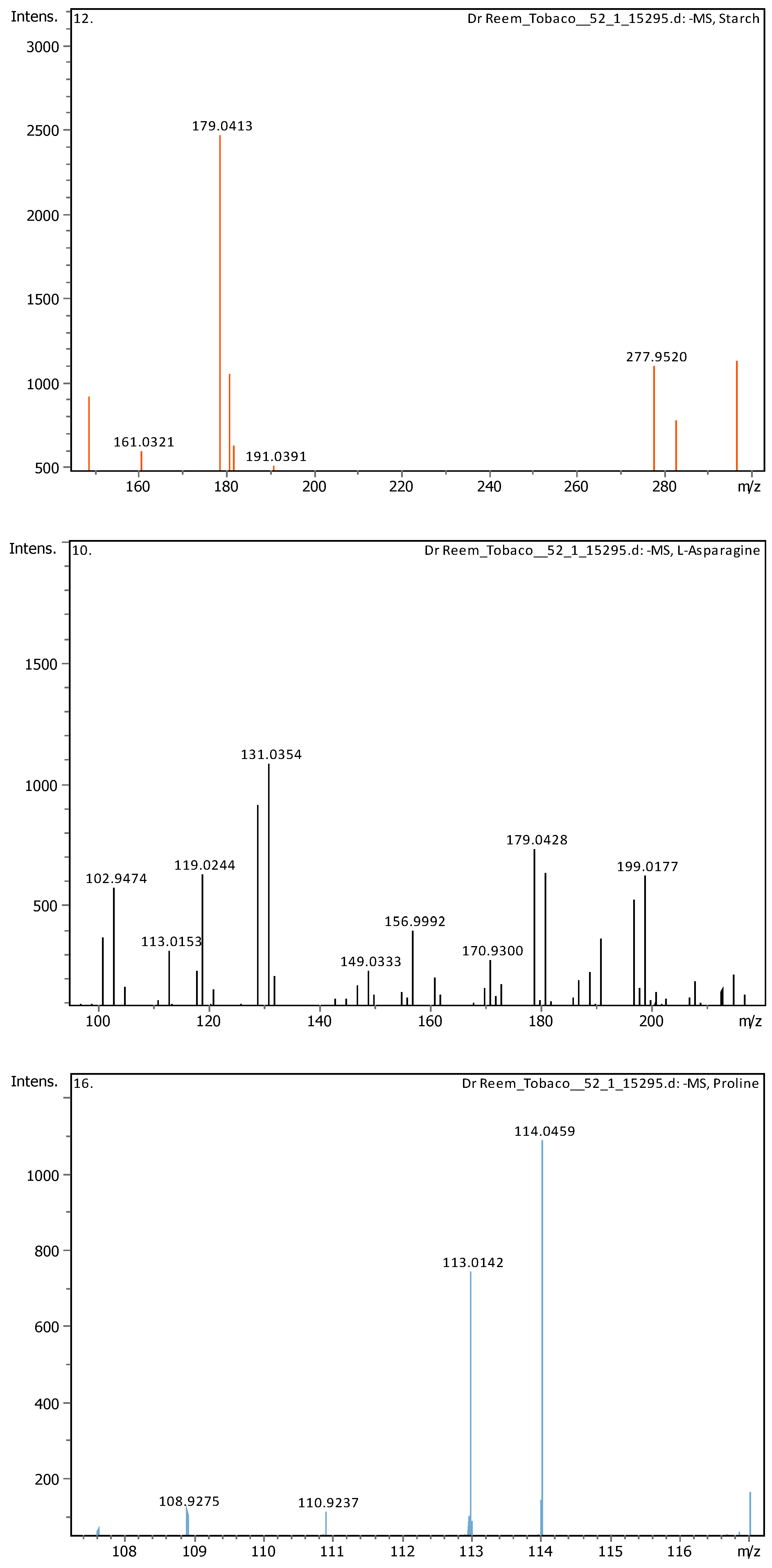
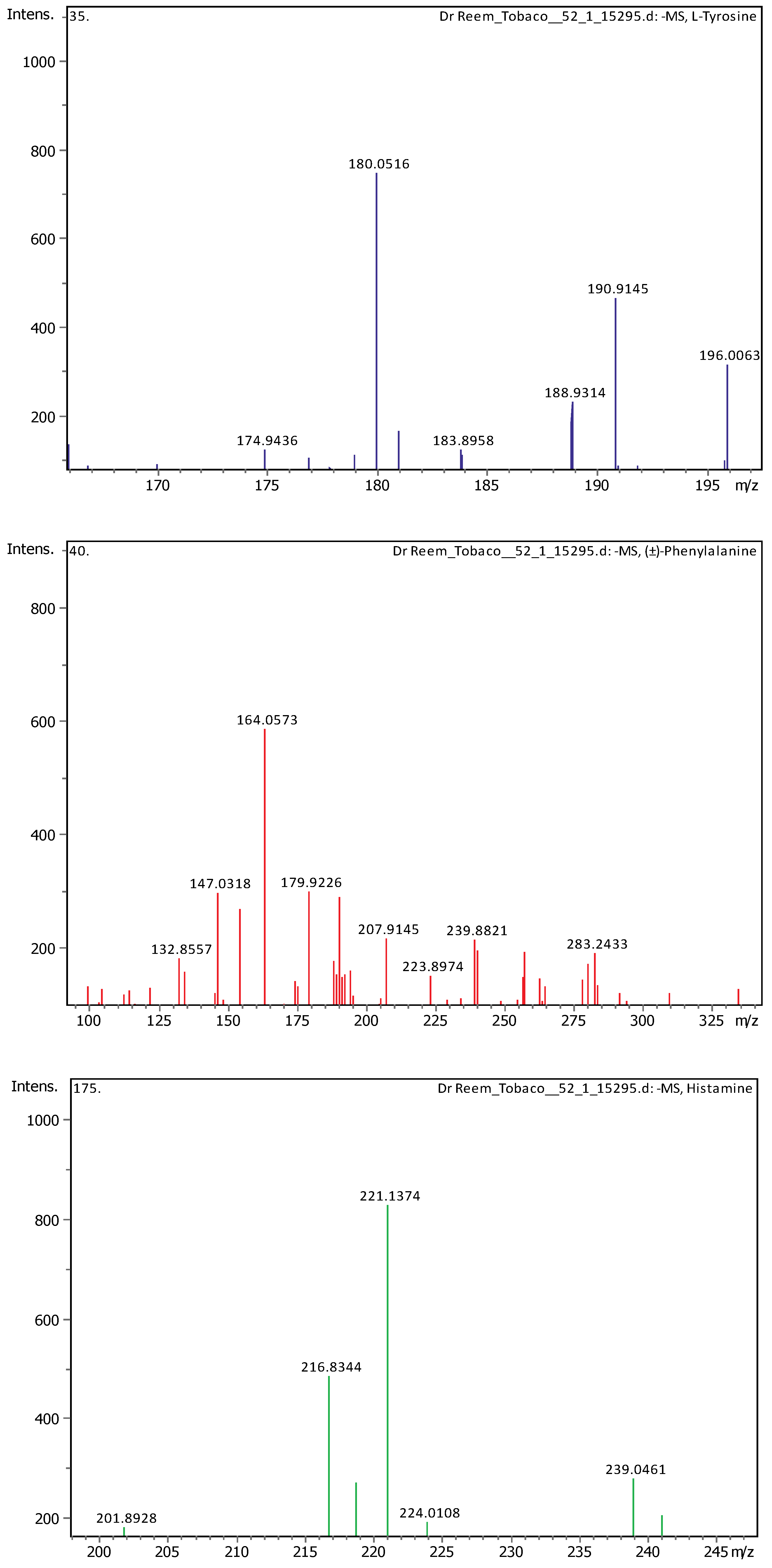
Twenty-three different phenolic components have been detected in the MC extract using the LC-MS/MS analysis, and the integrated natural compounds library. The retention time (Rt)mass-to-charge ratio (m/z), and molecular formula for the detected compounds (positive and negative ion modes) are listed in Table 3. Fourteen compounds were detected- in the negative mode, and nine were detected in the positive mode.
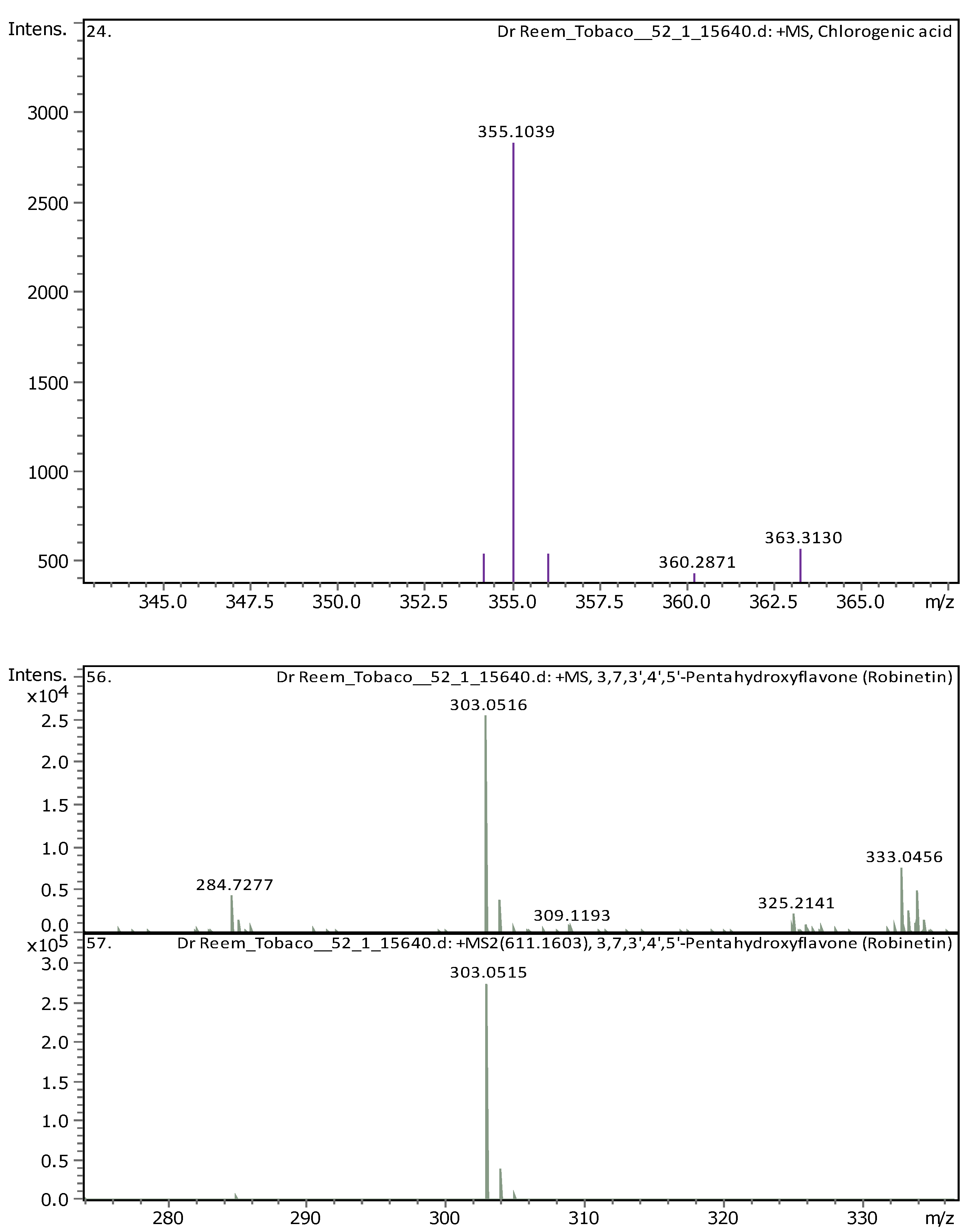
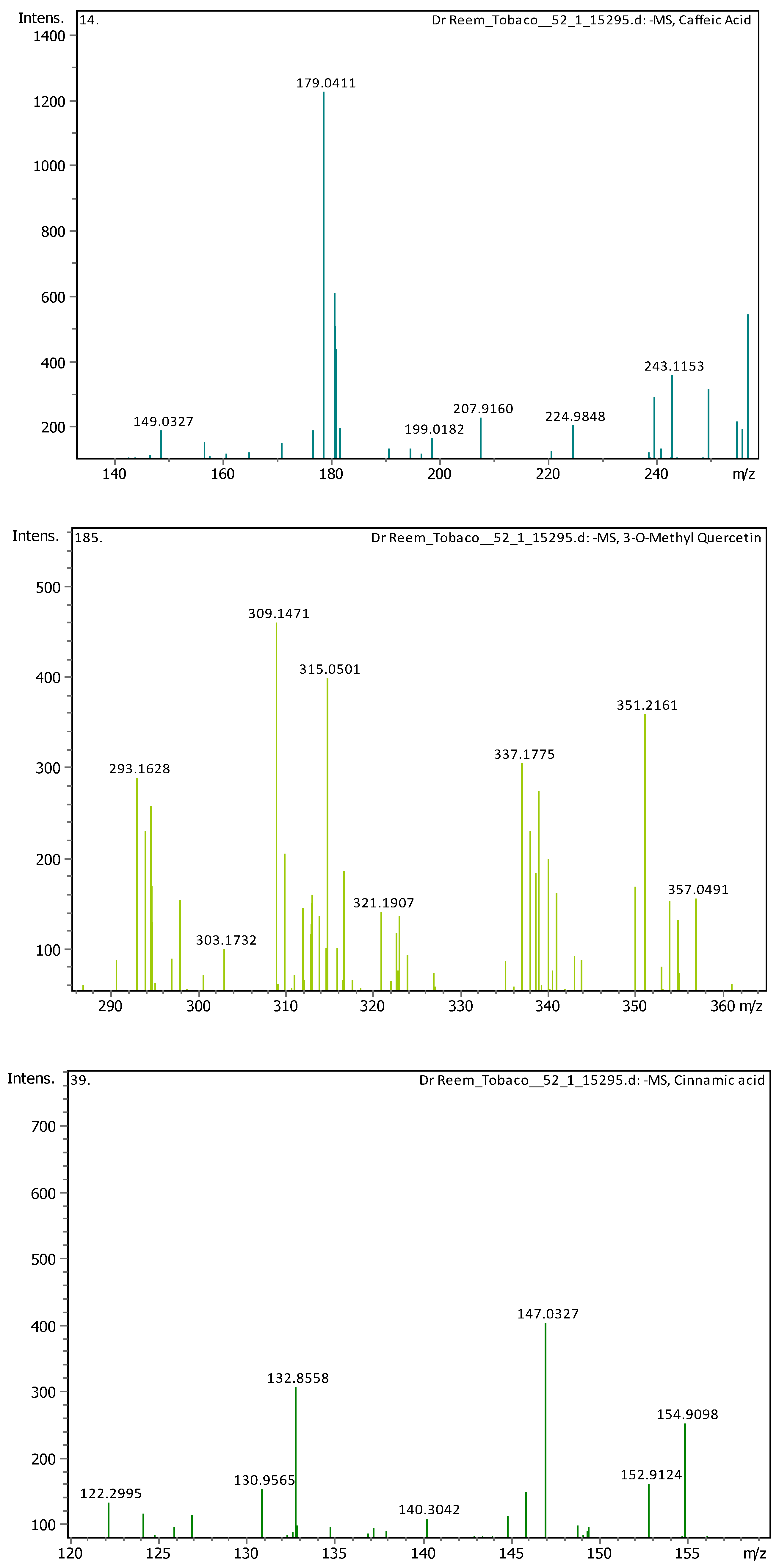
Figure 1 shows the total ion chromatogram for all compounds detected in the MC leaves extracts. The UHPLC-chromatograms which display the peaks and retention time of each compound detected in the extract are shown in Figure 2. The mass spectrum (m/z) and fragments for each compound detected in the MC extract are presented in Appendix A.
3.4.2. Deep Eutectic Extraction (DESs)
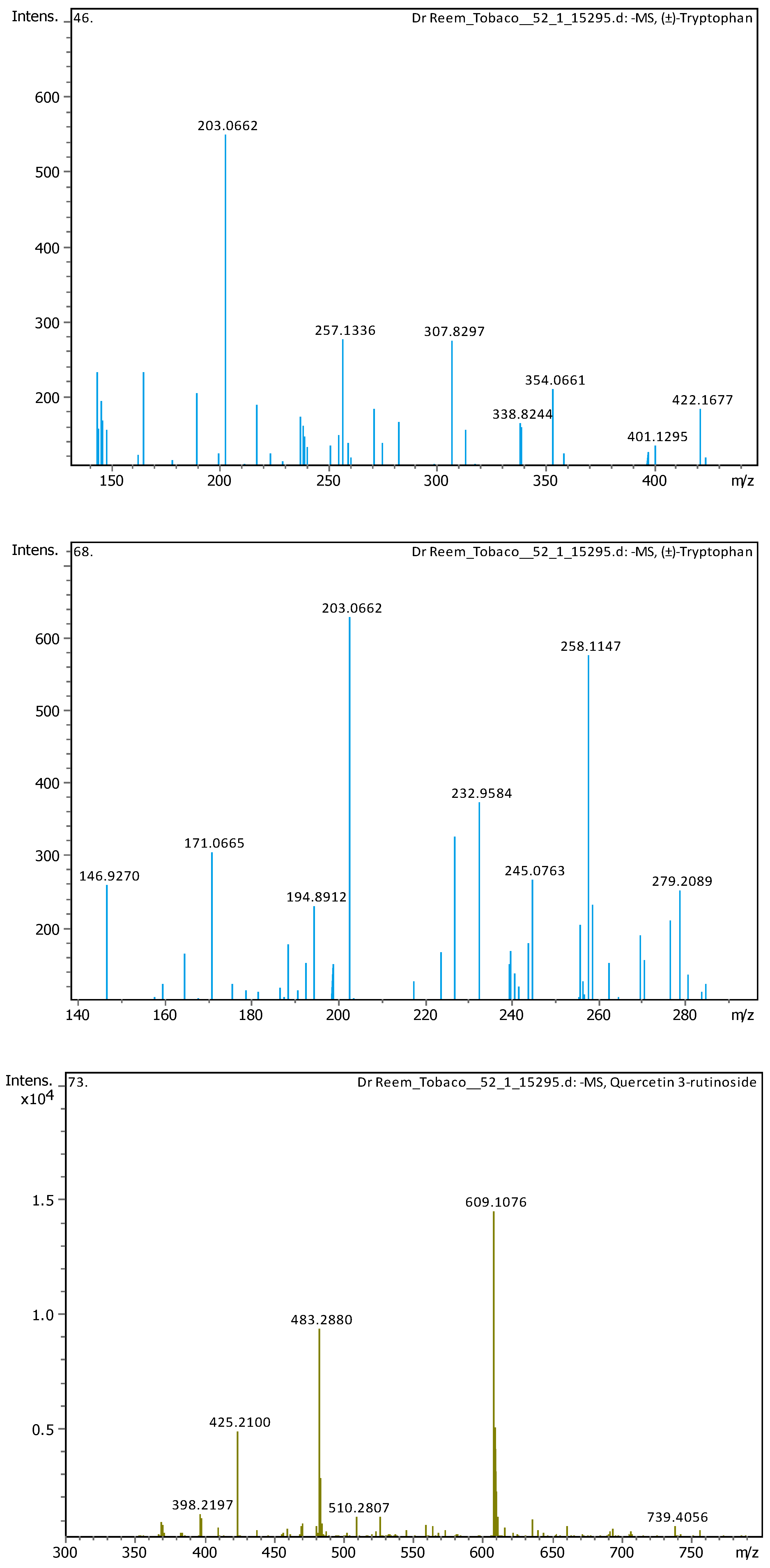
Twenty-three different phenolic components have been detected in the DES extract using the UHPLC-MS/MS analysis, and the integrated natural compounds library. The Rt, m/z, and molecular formula for the detected compounds (positive and negative ion modes) are listed in Table 4. Ten compounds were detected in the negative mode, and thirteen were detected in the positive mode.
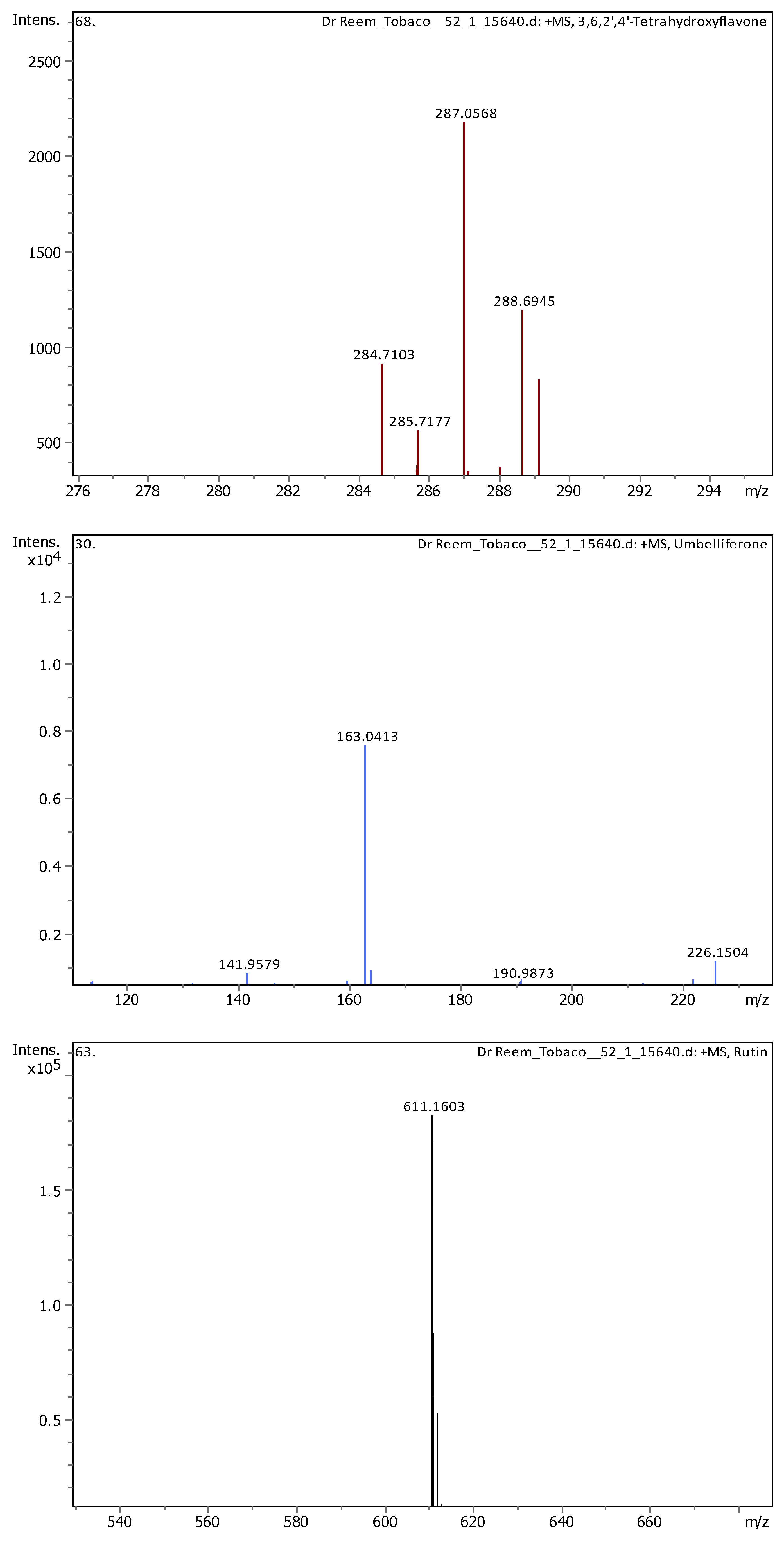
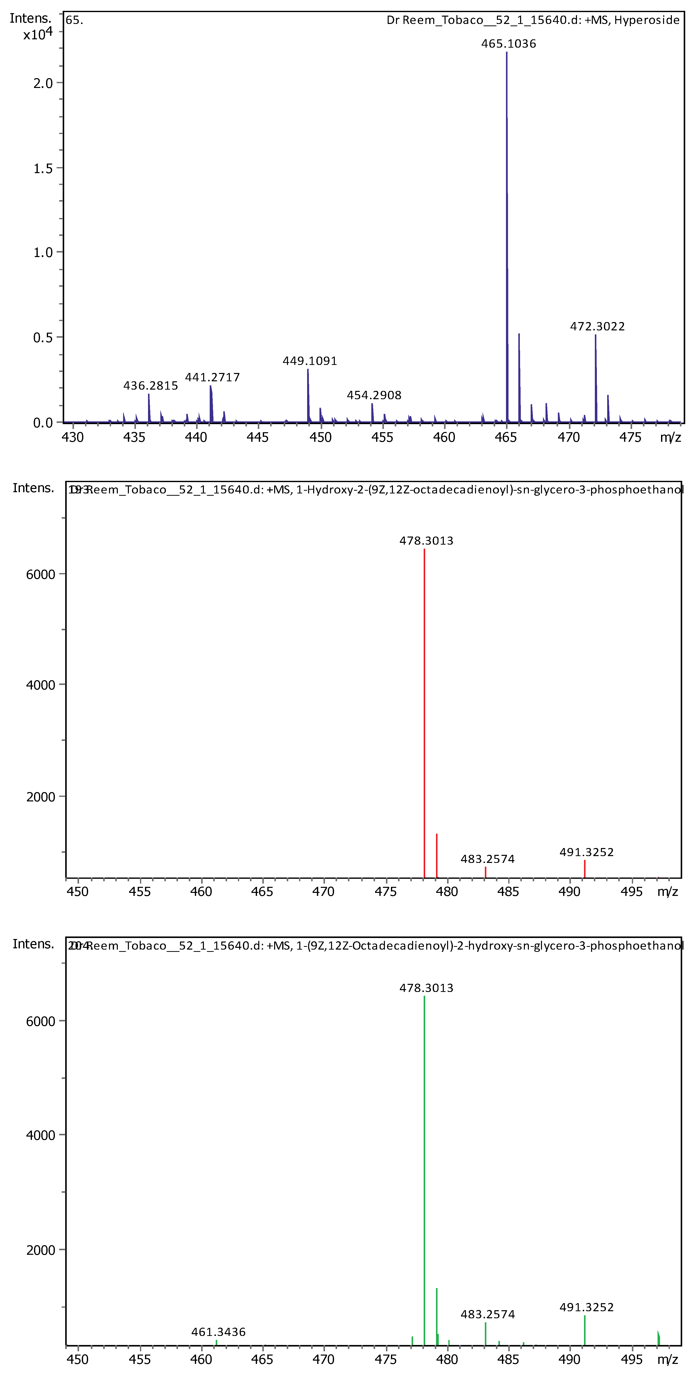
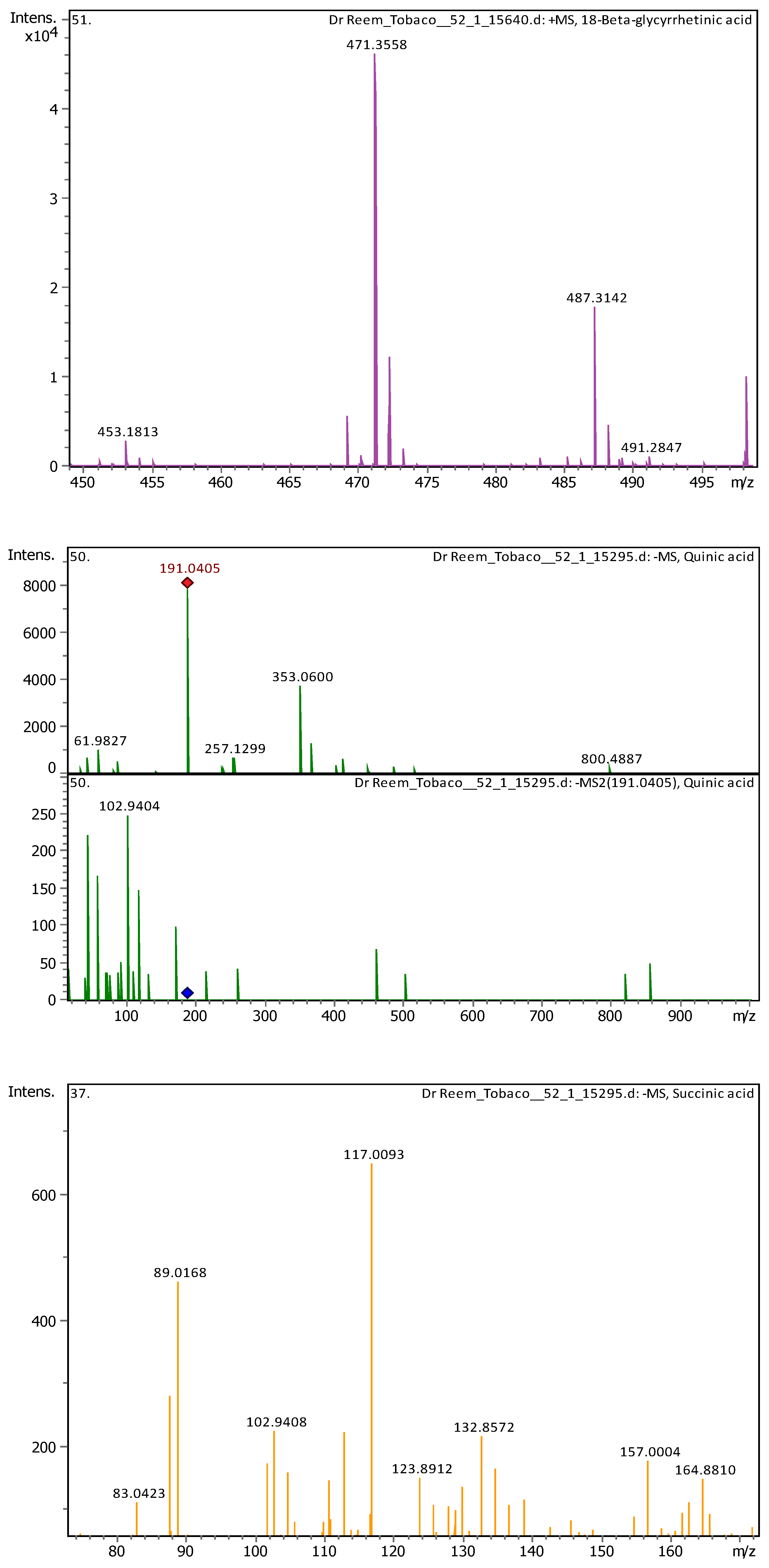
Figure 3 shows the total ion chromatogram for all compounds detected in the DES leaf extracts. The UHPLC-chromatograms showing peaks and retention time of each compound detected in the extract are shown in Figure 4. The Mass spectrum (m/z) and fragments for each compound detected in the MC extract are available in Appendix B.
3.4.3. Identification and Quantification of Nicotine
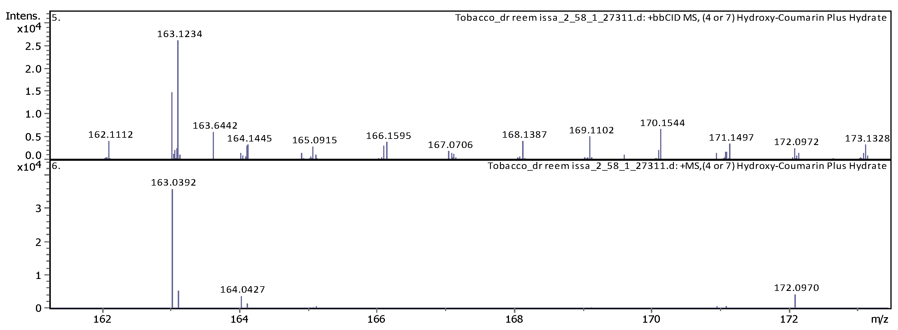
The identification of nicotine in the extracts samples was performed using a multiple external standards method using UHPLC-MS/MS system, based on the retention time, m/z and molecular formula as shown in Table 5 and Figure 5.
The results indicated that the concentration of nicotine in the MC extract was 635.07 ppm (µg/mL) while the concentration in the DES extract was 1194.91 ppm (µg/mL) (i.e. almost twice). This suggests that the DES extraction method yielded a higher concentration of nicotine compared with the MC method, proving that the DES method is more effective for the extraction of this alkaloid. The levels of nicotine extracted using the two techniques are illustrated in Table 6.
4. Discussion
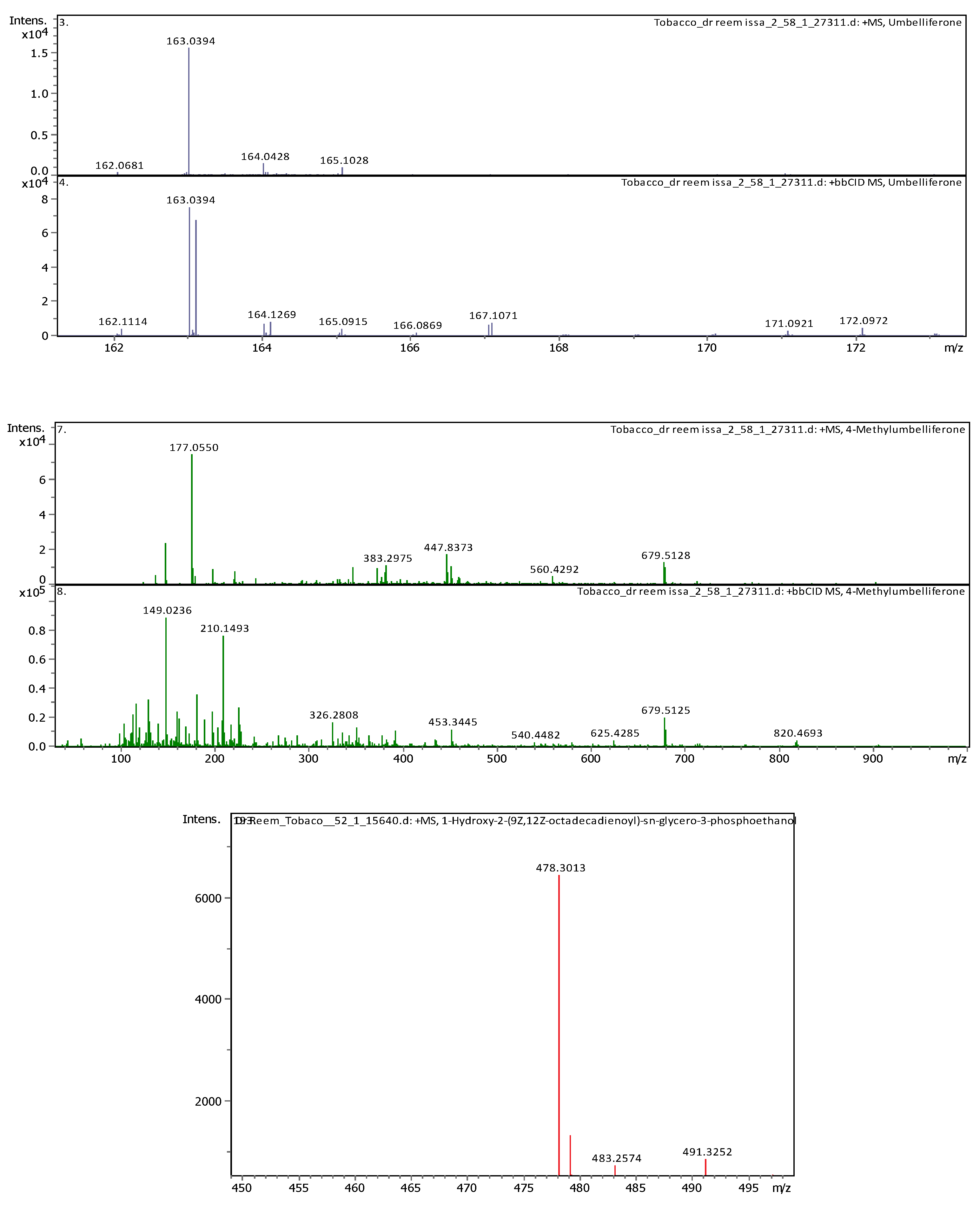
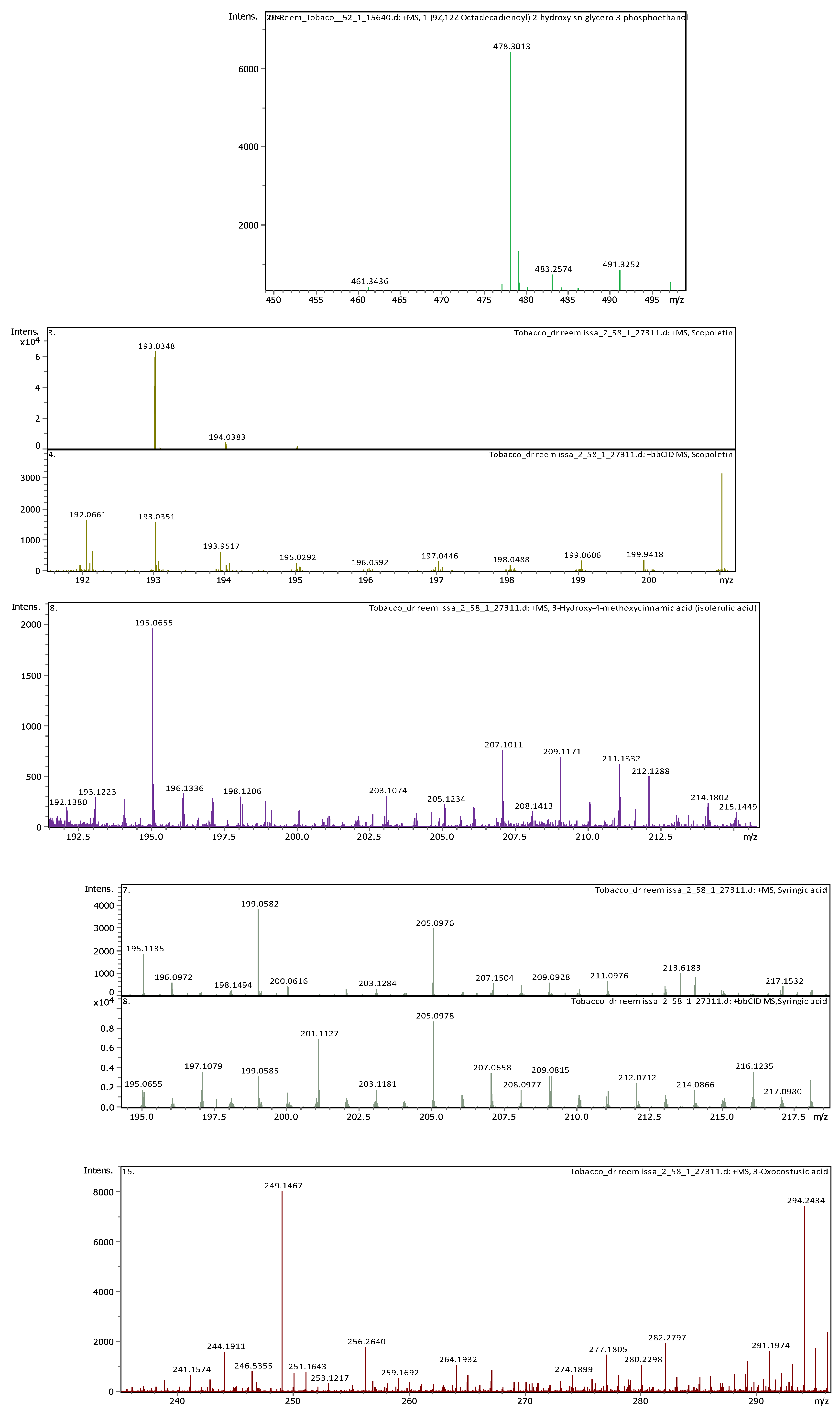
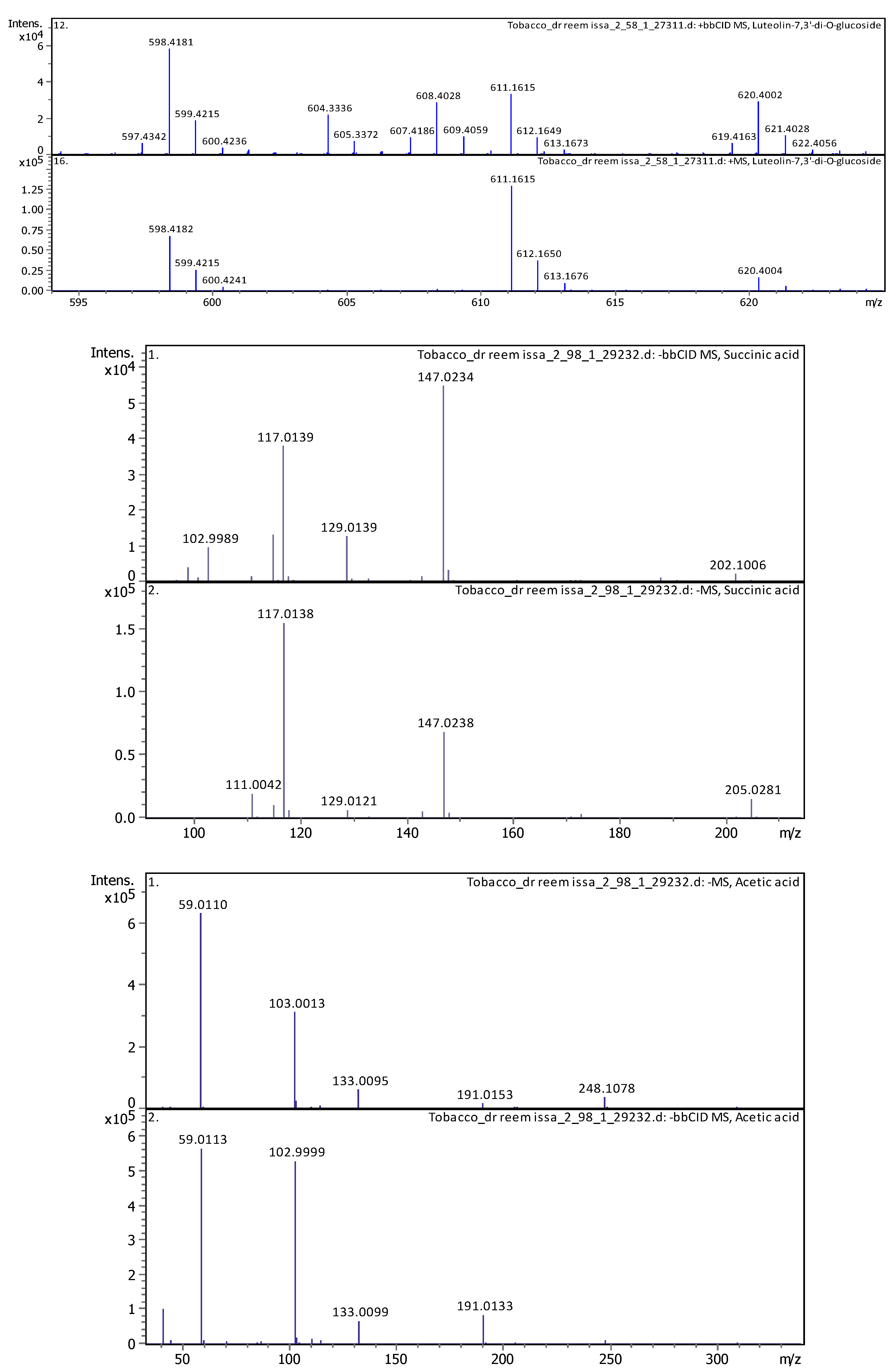
Several studies reported high toxicity of the plant N. glauca caused by its alkaloid content, namely nicotine and other derivatives [36,37]. The use of N. glauca as an anti-jaundice plant among herbalists and traditional healers was reported in Jordanian folk medicine [38]. Nevertheless, few reports investigated the phytochemical composition of the wild species grown in Jordan. Researchers detected the presence of different phenolic compounds in tobacco plants such as kaempferol-3-O-rutinoside, quercetin-3-O-rutinoside, in addition to the main components of tobacco polyphenols are chlorogenic acid and rutin [4,39,40]. In this study, the UHPLC-MS/MS analysis revealed the presence of variations in the detected compounds between the two extraction methods used. Mainly, 3,7,3',4',5'-Pentahydroxyflavone (Robinetin), Chlorogenic acid, Hyperoside, Rutin, and Umbelliferone, were detected in both extracts. Whereas (4 or 7) Hydroxy-Coumarin Plus Hydrate, 3-Hydroxy-4-methoxycinnamic acid (isoferulic acid), 3-Oxocostusic acid, 4-Methylumbelliferone, Luteolin-7,3'-di-O-glucoside, Salvianolic acid A, Saponarin, Scopoletin, and Syringic acid, were detected in the DES extract only. On the other side, 1-(9Z,12Z-Octadecadienoyl)-2-hydroxy-sn-glycero-3-phosphoethanolamine,18-Beta glycyrrhetinic acid, 1-Hydroxy-2-(9Z,12Z-octadecadienoyl)-sn-glycero-3-phosphoethanolamine, and 3,6,2',4'-Tetra hydroxy flavone, were detected in the MC extract only.
Our work on the total content of phenolic phytocomponents showed the MC extract contained fewer total phenols and flavonoid content compared to the DES extract. These findings were expected, as previous studies highlighted the advantages of using NaDES as green solvent in the extraction of phytochemicals, including enhanced extraction yield, and additional environmental benefits [28,29]. The results of the antioxidant DPPH test revealed that both extraction methods have weak antioxidant activities, except for the concentrated (90%) DES extract which showed a moderated antioxidant activity compared to ascorbic acid. The (90%) DES extract was also found to contain the highest total flavonoid content among the other extract samples. These findings are in correlation with phenols and flavonoid content, which are the most contributing natural compounds for antioxidant activity. Our findings agree with a previous study by Trifa, et al (2020), investigating N. glauca extract collected from central Algeria, which showed good antioxidant activity in the ethyl acetate and n-butanol fractions, which was related to the content of polyphenols [41]. Similarly, Sumengen et al., (2023) conducted a study to investigate the phytochemical composition of N. glauca methanol leaf extract collected from Northern Cyprus [42]. Findings showed that N. glauca methanolic extract had the highest antioxidant activity determined using the DPPH test and were correlated to their content of phenols and flavonoids. The latter compared his findings with others’ previous work, showing that variations in the antioxidant effects are expected, due to variations in the phytochemical composition. These variations may occur due to several factors, such as growing conditions, environmental variations, stage of plant development, season of collection, and geographical origin, in addition to methods of extraction and solvent used.
It was previously found that the acid-base extraction method contains the highest nicotine content, as this method aids in solubilizing the alkaloids by converting them to the salt form, which enhances their solubility in polar solvents [35]. It was concluded that methods of extraction and solvent used, plant part, and many other variables would largely be affecting nicotine content, in addition to other phytocomponents obtained in the plant extract.
The findings of the presented study showed that the MC extract contains 635.07 ppm of nicotine compared to 1194.91 ppm of nicotine was found according to the DES extraction method. In agreement with other published work, Kheawfu et al. (2021) found that extraction with water for 24 hrs gave the highest amount of nicotine [35]. Whereas Puripattanavong et al. (2013) suggested that using methanol and ethanol gave the highest yield percentage of nicotine extraction from N. tabacum leaves compared with other solvents and extraction media [43]. A similar work by Massadeh, et al (2022) screening the phytochemical constituent in the leaves of N. glauca collected from the north region in Jordan [4]. Using the UPLC-MS and GC-MS analysis, anabasine was detected as the major alkaloid, while nicotine was not detected in their studied extract.
5. Conclusions
This work analyzed the phenol, flavonoid, antioxidant, and nicotine content in N. glauca extracts using both methanol and environmentally friendly DESs extraction methods. The study found significant differences in phytochemical compositions, with the conventional methanol method yielding lower total phenols and flavonoid content, with also less nicotine content. These results emphasize the critical importance of adopting green chemistry techniques, not only for their environmental benefits, but also for their superior efficiency in producing higher yields of valuable phytoconstituents.
Author Contributions
Conceptualization, R.I. and F.A.; methodology, R.I., L.A., and T.A.; validation, S.A and K.O.; formal analysis, R.I; data curation, F.A.; writing—original draft preparation, R.I, F.A and S.A.; writing—review and editing, S.A and K.O. All authors have read and agreed to the published version of the manuscript.
Funding
This research received no external funding.
Conflicts of Interest
The authors declare no conflicts of interest.
Appendix A
Appendix B
References
- Berlowitz, I.; Torres, E.G.; Walt, H.; Wolf, U.; Maake, C.; Martin-Soelch, C. “Tobacco Is the Chief Medicinal Plant in My Work”: Therapeutic Uses of Tobacco in Peruvian Amazonian Medicine Exemplified by the Work of a Maestro Tabaquero. Frontiers in Pharmacology 2020, 11. [Google Scholar] [CrossRef]
- Charlton, A. Medicinal Uses of Tobacco in History. J R Soc Med 2004, 97, 292–296. [Google Scholar] [CrossRef] [PubMed]
- Zou, X.; Bk, A.; Abu-Izneid, T.; Aziz, A.; Devnath, P.; Rauf, A.; Mitra, S.; Emran, T.B.; Mujawah, A.A.H.; Lorenzo, J.M.; et al. Current Advances of Functional Phytochemicals in Nicotiana Plant and Related Potential Value of Tobacco Processing Waste: A Review. Biomedicine & Pharmacotherapy 2021, 143, 112191. [Google Scholar] [CrossRef]
- Massadeh, R.; El-Elimat, T.; Al-Gharaibeh, M.; Tawaha, K.; Alali, F. UPLC-HRESI-MS and GC-MS Analysis of the Leaves of Nicotiana Glauca. Acta Pharmaceutica 2022, 72, 97–108. [Google Scholar] [CrossRef]
- Silva, F.D.S.; Albuquerque, U.P.; Costa Júnior, L.M.; Lima, A.D.S.; Nascimento, A.L.B.D.; Monteiro, J.M. An Ethnopharmacological Assessment of the Use of Plants against Parasitic Diseases in Humans and Animals. Journal of Ethnopharmacology 2014, 155, 1332–1341. [Google Scholar] [CrossRef]
- Ameya, G.; Manilal, A.; Merdekios, B. In Vitro Antibacterial Activity and Phytochemical Analysis of Nicotiana Tabacum L. Extracted in Different Organic Solvents. Open Microbiol J 2017, 11, 352–359. [Google Scholar] [CrossRef]
- Anumudu, C.; Mi, N.; Cc, O.; Io, N.; Ihenetu, F. Antimicrobial Activities of Extracts of Tobacco Leaf (Nicotiana Tabacum) and Its Grounded Snuff (Utaba) on Candida Albicans and Streptococcus Pyogenes. PLoS Neglected Tropical Diseases 2019, 7, 000300. [Google Scholar] [CrossRef]
- Fernanda, S.A.; Amru, B.A.; Rahmani, H.A.; Gozan, M.; Irsyad, N.S.; Bahar, M.; Puspita, O.S.; Zulfa, F.; Pramono, A. Antibacterial Potential of Nicotiana Tabacum L. Var Virginia Pyrolysis Extract Against Staphylococcus Aureus, Enterococcus Faecalis, Escherichia Coli, and Pseudomonas Aeruginosa. IOP Conf. Ser.: Earth Environ. Sci. 2021, 755, 012013. [Google Scholar] [CrossRef]
- Gutiérrez A, D.M.; Bah, M.; Garduño R, M.L.; Mendoza D, S.O.; Serrano C, V. Anti-Inflammatory and Antioxidant Activities of Methanol Extracts and Alkaloid Fractions of Four Mexican Medicinal Plants of Solanaceae. African journal of traditional, complementary, and alternative medicines : AJTCAM / African Networks on Ethnomedicines 2014, 11, 259–267. [Google Scholar] [CrossRef]
- Ali Alghamdi, A. Phytoconstituents Screening and Antimicrobial Activity of the Invasive Species Nicotiana Glauca Collected from Al-Baha Region of Saudi Arabia. Saudi J Biol Sci 2021, 28, 1544–1547. [Google Scholar] [CrossRef]
- Al-Akayleh, F.; Ali, H.H.M.; Ghareeb, M.M.; Al-Remawi, M. Therapeutic deep eutectic system of capric acid and menthol: Characterization and pharmaceutical application. Journal of Drug Delivery Science and Technology 2019, 53, 101159. [Google Scholar] [CrossRef]
- Al-Mawla, L.; Al-Akayleh, F.; Daadoue, S.; Mahyoob, W.; Al-Tameemi, B.; Al-Remawi, M.; …; Agha, A.S.A. Development, characterization, and ex vivo permeation assessment of diclofenac diethylamine deep eutectic systems across human skin. Journal of Pharmaceutical Innovation 2023, 18, 2196–2209. [Google Scholar] [CrossRef]
- Alkhawaja, B.; Al-Akayleh, F.; Nasereddin, J.; Kamran, M.; Woodman, T.; Al-Rubaye, Z.; …; Olaimat, A.R. Structural insights into novel therapeutic deep eutectic systems with capric acid using 1D, 2D NMR and DSC techniques with superior gut permeability. RSC advances 2024, 14, 14793–14806. [Google Scholar] [CrossRef]
- Nakaweh, A.; Al-Akayleh, F.; Al-Remawi, M.; Abdallah, Q.; Agha, A.S. Deep Eutectic System-Based Liquisolid Nanoparticles as Drug Delivery System of Curcumin for In-Vitro Colon Cancer Cells. Journal of Pharmaceutical Innovation 2024, 19, 1–11. [Google Scholar] [CrossRef]
- Al-Akayleh, F.; Al-Remawi, M.; Agha, A.; Abu-Nameh, E. Applications and Risk Assessments of Ionic Liquids in Chemical and Pharmaceutical Domains: An Updated Overview. Jordan Journal of Chemistry (JJC) 2023, 18, 53–76. [Google Scholar]
- Alkhawaja, B.; Al-Akayleh, F.; Nasereddin, J.; Malek, S.A.; Alkhawaja, N.; Kamran, M.; …; Aburayyan, W.S. Levofloxacin–Fatty Acid Systems: Dual Enhancement Through Deep Eutectic Formation and Solubilization for Pharmaceutical Potential and Antibacterial Activity. AAPS PharmSciTech 2023, 24, 244.–content content. [Google Scholar] [CrossRef] [PubMed]
- Alkhawaja, B.; Al-Akayleh, F.; Al-Khateeb, A.; Nasereddin, J.; Ghanim, B.Y.; Bolhuis, A.; …; Qinna, N.A. Deep eutectic liquids as a topical vehicle for tadalafil: characterisation and potential wound healing and antimicrobial activity. Molecules 2023, 28, 2402. [Google Scholar] [CrossRef]
- Luhaibi, D.K.; Ali, H.H.M.; Al-Ani, I.; Shalan, N.; Al-Akayleh, F.; Al-Remawi, M.; …; Khanfar, M. The formulation and evaluation of deep eutectic vehicles for the topical delivery of azelaic acid for acne treatment. Molecules 2023, 28, 6927. [Google Scholar] [CrossRef]
- Daadoue, S.; Al-Remawi, M.; Al-Mawla, L.; Idkaidek, N.; Khalid, R.M.; Al-Akayleh, F. Deep eutectic liquid as transdermal delivery vehicle of Risperidone. Journal of Molecular Liquids 2022, 345, 117347. [Google Scholar] [CrossRef]
- Al-Akayleh, F.; Adwan, S.; Khanfar, M.; Idkaidek, N.; Al-Remawi, M. A novel eutectic-based transdermal delivery system for risperidone. AAPS PharmSciTech 2021, 22, 1–11. [Google Scholar] [CrossRef] [PubMed]
- Alkhawaja, B.; Al-Akayleh, F.; Al-Rubaye, Z.; Bustami, M.; Smairat, M.A.; Agha, A.S.; …; Watts, A.G. Dissecting the stability of Atezolizumab with renewable amino acid-based ionic liquids: Colloidal stability and anticancer activity under thermal stress. International Journal of Biological Macromolecules 2024, 270, 132208. [Google Scholar] [CrossRef]
- Airouyuwa, J.O.; Sivapragasam, N.; Redha, A.A.; Maqsood, S. Sustainable green extraction of anthocyanins and carotenoids using natural deep eutectic solvents (NaDES): A review of recent developments. Food Chemistry 2024, 139061. [Google Scholar] [CrossRef]
- Wang, Z.; Wang, D.; Fang, J.; Song, Z.; Geng, J.; Zhao, J.; …; Li, M. Green and efficient extraction of flavonoids from Perilla frutescens (L.) Britt. leaves based on natural deep eutectic solvents: Process optimization, component identification, and biological activity. Food Chemistry 2024, 452, 139508. [Google Scholar] [CrossRef]
- Al-Akayleh, F.; Khalid, R.M.; Hawash, D.; Al-Kaissi, E.; Al-Adham, I.S.I.; Al-Muhtaseb, N.; …; Collier, P.J. Antimicrobial potential of natural deep eutectic solvents. Letters in Applied Microbiology 2022, 75, 607–615. [Google Scholar] [CrossRef]
- Yu, J.; Xu, S.; Goksen, G.; Yi, C.; Shao, P. Chitosan films plasticized with choline-based deep eutectic solvents: UV shielding, antioxidant, and antibacterial properties. Food Hydrocolloids 2023, 135, 108196. [Google Scholar] [CrossRef]
- Nystedt, H.L.; Grønlien, K.G.; Rolfsnes, R.R.; Winther-Larsen, H.C.; Økstad, O.A.L.; Tønnesen, H.H. Neutral natural deep eutectic solvents as anti-biofilm agents. Biofilm 2023, 5, 100114. [Google Scholar] [CrossRef]
- Tsvetov, N.; Pasichnik, E.; Korovkina, A.; Gosteva, A. Extraction of bioactive components from Chamaenerion angustifolium (L. ) Scop. with choline chloride and organic acids natural deep eutectic solvents. Molecules 2022, 27, 4216. [Google Scholar]
- Edrisi, S.; Bakhshi, H. Separation of Polyphenolic Compounds from Citrus Aurantium L. Peel by Deep Eutectic Solvents and Their Recovery Using a New DES-Based Aqueous Two-Phase System. Journal of Molecular Liquids 2024, 402, 124790. [Google Scholar] [CrossRef]
- Hong, J.; Deng, M.; Zhao, L. Natural Deep Eutectic Solvent Combined with Ultrasonic Enhancement: A Green Extraction Strategy for Solanesol in Tobacco Leaves. Industrial Crops and Products 2022, 187, 115355. [Google Scholar] [CrossRef]
- Yu, T.; Yang, L.; Shang, X.; Bian, S. Recovery of Cembratrien-Diols from Waste Tobacco (Nicotiana Tabacum L.) Flowers by Microwave-Assisted Deep Eutectic Solvent Extraction: Optimization, Separation, and In Vitro Bioactivity. Molecules 2024, 29, 1563. [Google Scholar] [CrossRef] [PubMed]
- Nasr, H. ECOLOGICAL AND PHYTOCHEMICAL STUDIES ON NICOTIANA GLAUCA FROM EGYPT. THE EGYPTIAN JOURNAL OF EXPERIMENTAL BIOLOGY 2014. [Google Scholar]
- Alnsour, L.; Issa, R.; Awwad, S.; Albals, D.; Al-Momani, I. Quantification of Total Phenols and Antioxidants in Coffee Samples of Different Origins and Evaluation of the Effect of Degree of Roasting on Their Levels. Molecules 2022, 27, 1591. [Google Scholar] [CrossRef]
- Ubaydee, A.H.N.; Issa, R.; Hajleh, M.N.A.; Ghanim, B.Y.; Al-Akayleh, F.; Qinna, N.A. The Effect of Medicago Sativa Extract and Light on Skin Hypopigmentation Disorders in C57/BL6 Mice. J Cosmet Dermatol 2022, 21, 6270–6280. [Google Scholar] [CrossRef]
- Al-Bayati, M.; Issa, R.; Abu-Samak, M.; Alnsour, L.; Awwad, S. Phytochemical Analysis and Evaluation of Anti-Hyperlipidaemic Effect for Ethanolic Leaf Extract of Equisetum Ramosissimum L. : In Vivo Study on Rats’ Models. Pharmacia 2023, 70, 557–568. [Google Scholar] [CrossRef]
- Kheawfu, K.; Kaewpinta, A.; Chanmahasathien, W.; Rachtanapun, P.; Jantrawut, P. Extraction of Nicotine from Tobacco Leaves and Development of Fast Dissolving Nicotine Extract Film. Membranes (Basel) 2021, 11, 403. [Google Scholar] [CrossRef]
- Furer, V.; Hersch, M.; Silvetzki, N.; Breuer, G.S.; Zevin, S. Nicotiana Glauca (Tree Tobacco) Intoxication--Two Cases in One Family. J Med Toxicol 2011, 7, 47–51. [Google Scholar] [CrossRef]
- Schep, L.J.; Slaughter, R.J.; Beasley, D.M.G. Nicotinic Plant Poisoning. Clin Toxicol (Phila) 2009, 47, 771–781. [Google Scholar] [CrossRef]
- Janakat, S.; Al-Merie, H. Evaluation of Hepatoprotective Effect of Pistacia Lentiscus, Phillyrea Latifolia and Nicotiana Glauca. J Ethnopharmacol 2002, 83, 135–138. [Google Scholar] [CrossRef]
- Rodgman, A.; Perfetti, T. The Chemical Components of Tobacco and Tobacco Smoke, Second Edition ed; 2013; ISBN 978-1-4665-1548-2. [Google Scholar]
- Wang, H.; Zhao, M.; Yang, B.; Jiang, Y.; Rao, G. Identification of Polyphenols in Tobacco Leaf and Their Antioxidant and Antimicrobial Activities. Food Chemistry 2008, 107, 1399–1406. [Google Scholar] [CrossRef]
- Trifa, W.; Akkal, S.; Lefahal, M.; Benmekhebi, L.; Khennouf, S. Preliminary Screening of Extracts for Determination of Antioxidant Activity by Different Methods. Current Issues in Pharmacy and Medical Sciences 2020, 33, 32–37. [Google Scholar] [CrossRef]
- Özdenefe, M.S.; Takcı, A.M.; Kayış, F.B. Antibacterial, Antioxidant, Antidiabetic Potentials and Chemical Composition of Nicotiana Glauca Graham Leaf Extract. JAES 2023, 8, 700–706. [Google Scholar] [CrossRef]
- Puripattanavong, J.; Songkram, C.; Lomlim, L.; Amnuaikit, T. Development of Concentrated Emulsion Containing Nicotiana Tabacum Extract for Use as Pesticide ARTICLE INFO ABSTRACT. Journal of Applied Pharmaceutical Science 2013, 3, 16–21. [Google Scholar] [CrossRef]
Figure 1.
Total ion chromatograms for all compounds detected in N.gluca MC extract.
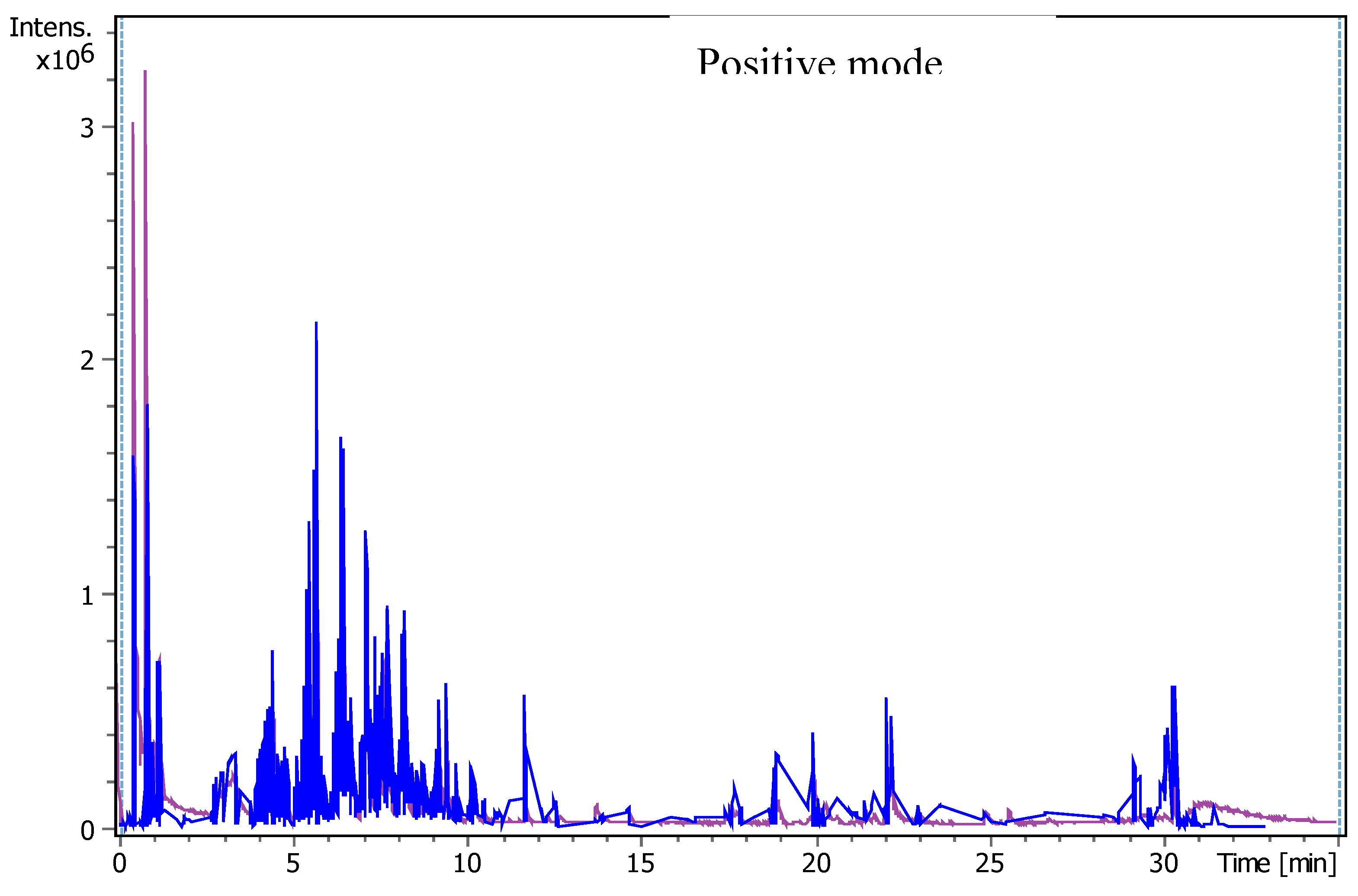
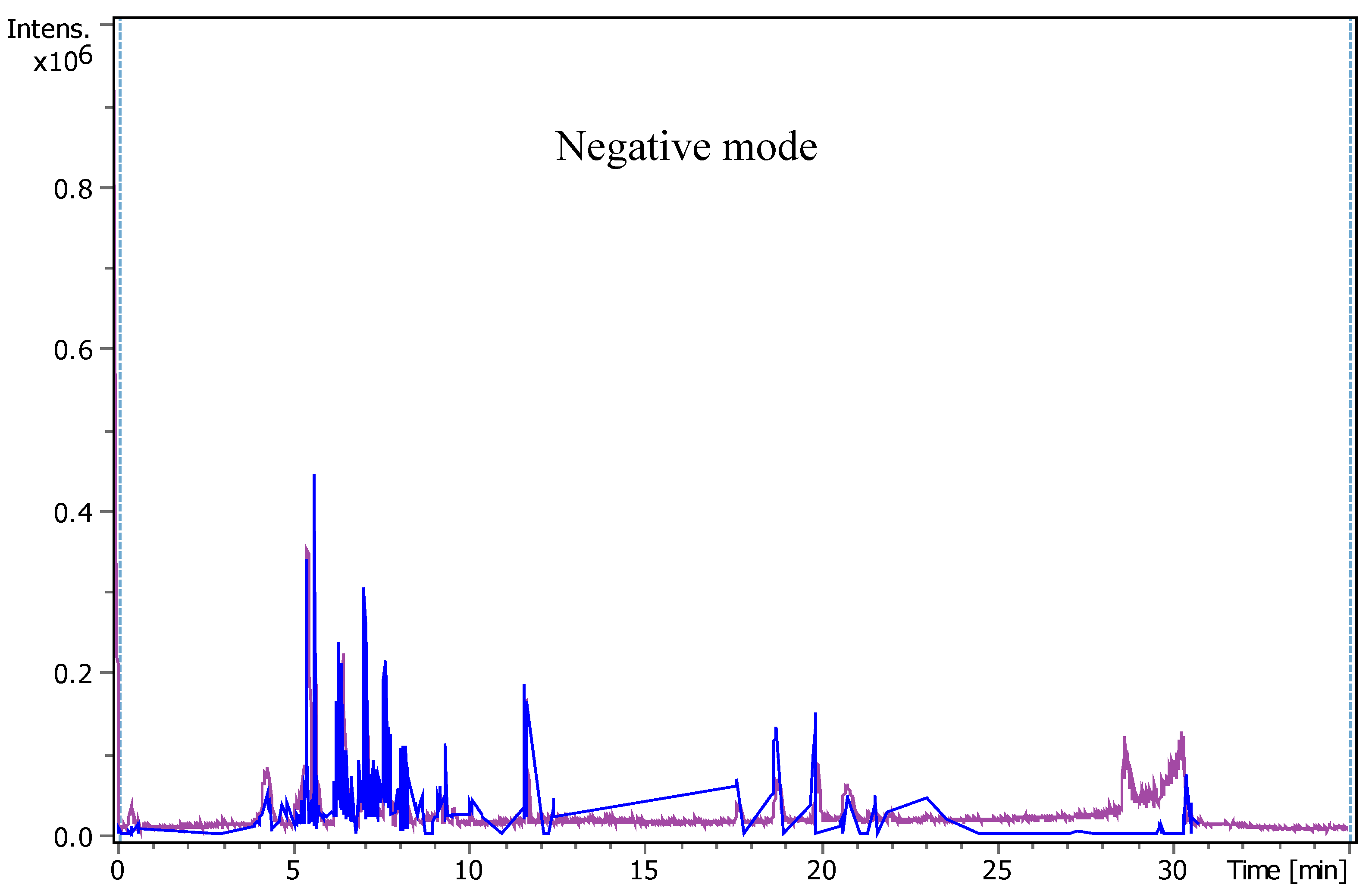
Figure 2.
The UHPLC-chromatograms show peaks and retention time of each compound detected in N. glauca MC extract.
Figure 2.
The UHPLC-chromatograms show peaks and retention time of each compound detected in N. glauca MC extract.
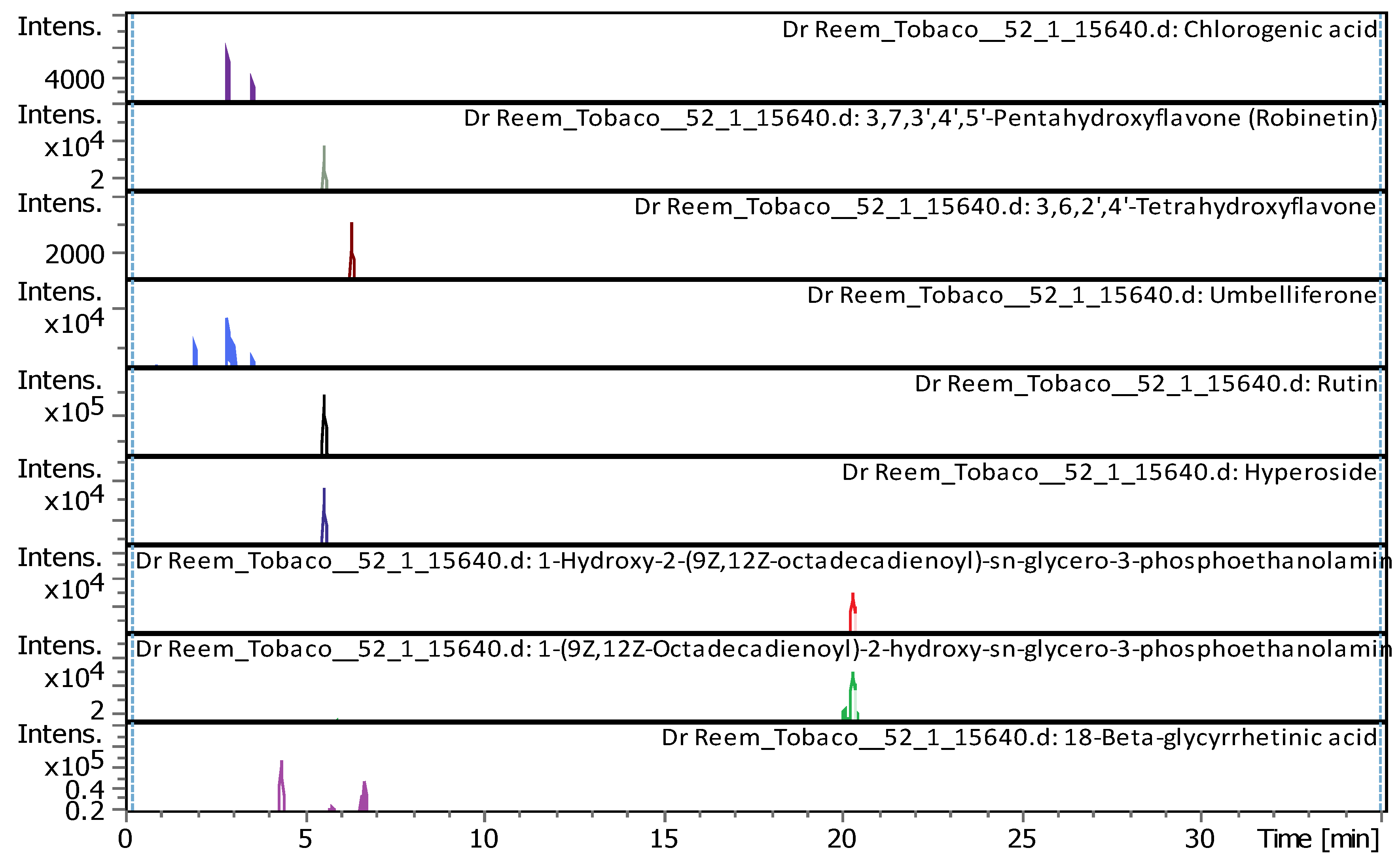
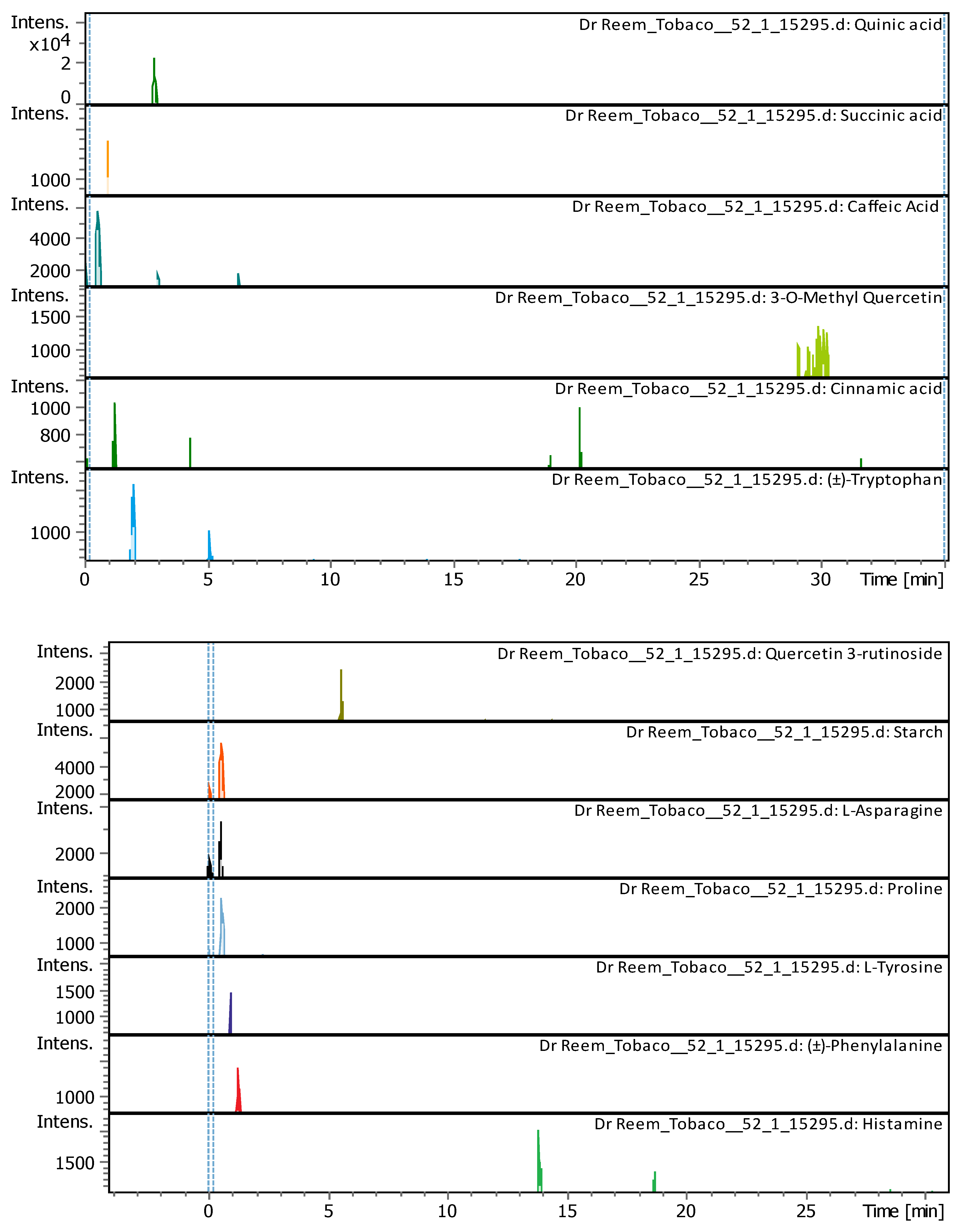
Figure 3.
Total ion chromatograms for all compounds detected in N. glauca DES extract.
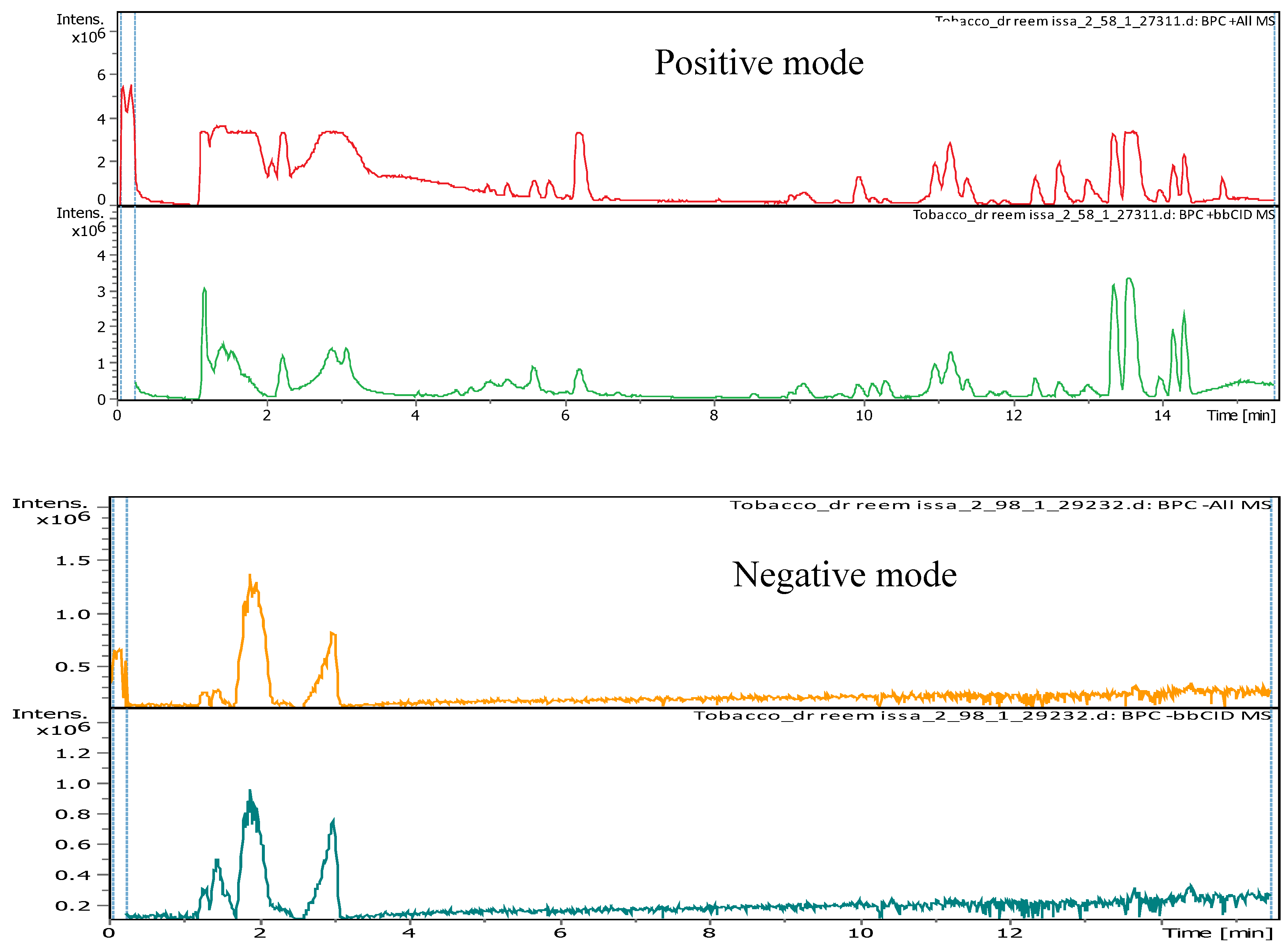
Figure 4.
The UHPLC-chromatograms show peaks and retention time of each compound detected in N. glauca DES extract.
Figure 4.
The UHPLC-chromatograms show peaks and retention time of each compound detected in N. glauca DES extract.
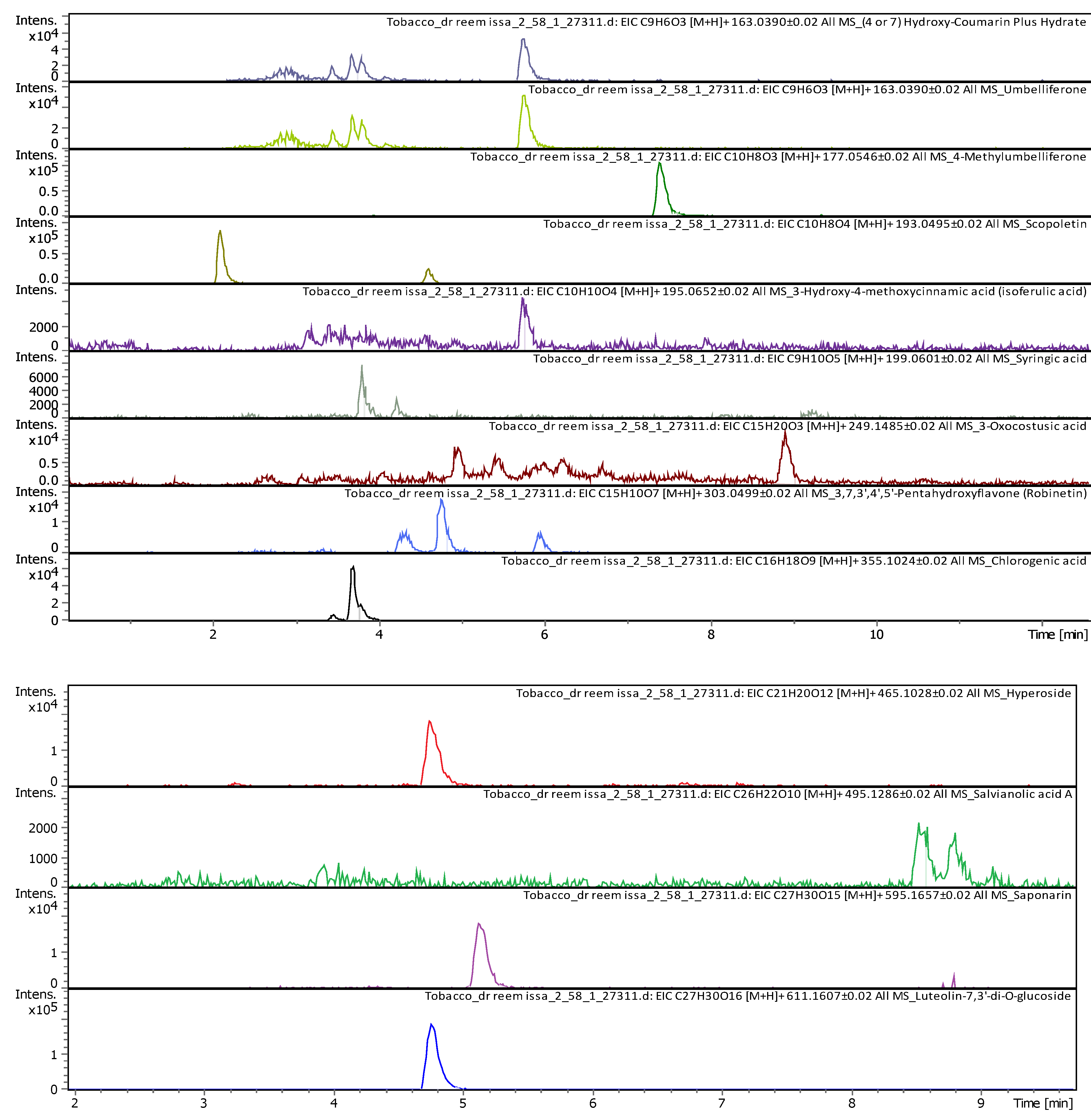
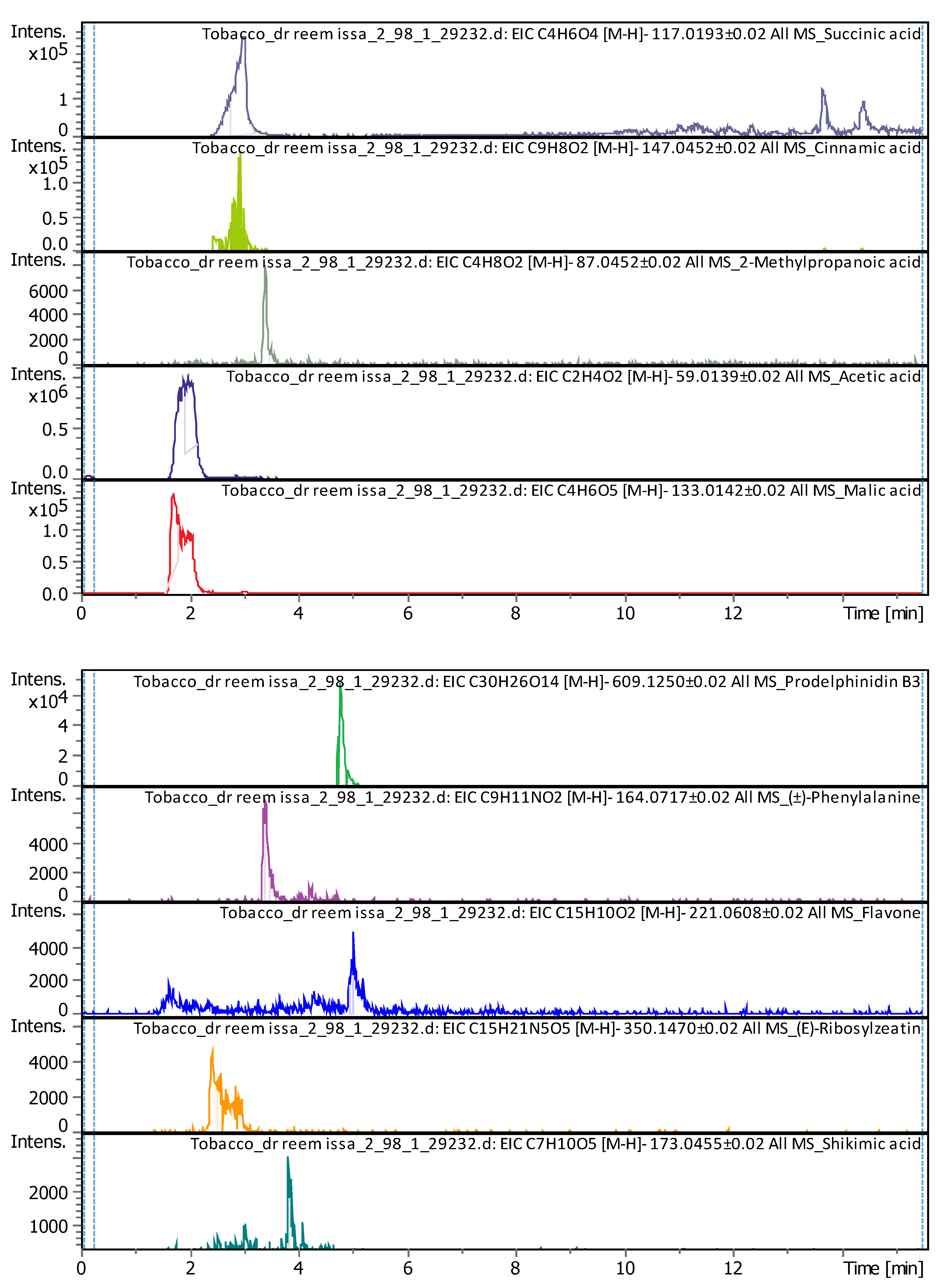
Figure 5.
The UHPLC-chromatogram and mass spectra show peaks and retention time of nicotine detected in N. glauca extract.
Figure 5.
The UHPLC-chromatogram and mass spectra show peaks and retention time of nicotine detected in N. glauca extract.

Table 1.
Total phenols content in MC and DES extracts at three extracts ratios.
| Extraction media | mg/mL ± SD (equivalent to gallic acid) |
|---|---|
| DES 30% | 0.326 ± 0.11 |
| DES 70% | 0.300± 0.03 |
| DES 90% | 0.311± 0.02 |
| MC | 0.119 ±0.01 |
Table 2.
Total flavonoid content in MC and DES extracts at three extract ratios.
| Extraction media | mg/mL ± SD (equivalent to rutin) |
|---|---|
| DES 30% | 0.128 ± 0.03 |
| DES 70% | 0.115 ± 0.14 |
| DES 90% | 0.928 ± 0.09 |
| MC | 0.011±0.01 |
Table 3.
UHPLC-MS/MS analysis (positive and negative modes) showing all components detected in N. glauca MC extract based on retention time (Rt), Mass-to-charge ratio (m/z) and molecular formula.
Table 3.
UHPLC-MS/MS analysis (positive and negative modes) showing all components detected in N. glauca MC extract based on retention time (Rt), Mass-to-charge ratio (m/z) and molecular formula.
| # | Rt [min] | m/z meas. | M meas. | Ions | Name | Molecular Formula |
|---|---|---|---|---|---|---|
| 1 | 0.61 | 131.04612 | 132.05339 | [M-H]- | L-Asparagine | C4H8N2O3 |
| 2 | 0.62 | 114.05616 | 115.06344 | [M-H]- | Proline | C5H9NO2 |
| 3 | 0.97 | 180.06594 | 181.07322 | [M-H]- | L-Tyrosine | C9H11NO3 |
| 4 | 1 | 117.01933 | 118.02661 | [M-H]- | Succinic acid | C4H6O4 |
| 5 | 1.28 | 147.04508 | 148.05236 | [M-H]- | Cinnamic acid | C9H8O2 |
| 6 | 1.28 | 164.0718 | 165.07908 | [M-H]- | (±)-Phenylalanine | C9H11NO2 |
| 7 | 2.03 | 203.08226 | 204.08954 | [M-H]- | (±)-Tryptophan | C11H12N2O2 |
| 8 | 2.9 | 191.05607 | 192.06335 | [M-H]- | Quinic acid | C7H12O6 |
| 9 | 2.96 | 355.10248 | 354.09521 | [M+H]+ | Chlorogenic acid | C16H18O9 |
| 10 | 3.05 | 179.03487 | 180.04214 | [M-H]- | Caffeic Acid | C9H8O4 |
| 11 | 5.07 | 163.03979 | 162.03251 | [M+H]+ | Umbelliferone | C9H6O3 |
| 12 | 5.12 | 203.08228 | 204.08955 | [M-H]- | (±)-Tryptophan | C11H12N2O2 |
| 13 | 5.57 | 609.1455 | 610.15278 | [M-H]- | Quercetin 3-rutinoside | C27H30O16 |
| 14 | 5.61 | 303.05014 | 302.04287 | [M+H]+ | Robinetin | C15H10O7 |
| 15 | 5.61 | 465.10293 | 464.09566 | [M+H]+ | Hyperoside | C21H20O12 |
| 16 | 5.62 | 611.16099 | 610.15353 | [M+H]+, [M+Na]+ | Rutin | C27H30O16 |
| 17 | 6.31 | 179.05592 | 180.0632 | [M-H]- | Starch | C6H12O6 |
| 18 | 6.37 | 287.0557 | 286.04842 | [M+H]+ | 3,6,2',4'-Tetrahydroxyflavone | C15H10O6 |
| 19 | 9.1 | 315.05061 | 316.05788 | [M-H]- | 3-O-Methyl Quercetin | C16H12O7 |
| 20 | 21.09 | 478.28898 | 477.2817 | [M+H]+ | 1-Hydroxy-2-(9Z,12Z-octadecadienoyl)-sn-glycero-3-phosphoethanolamine (NMR) | C23H44NO7P |
| 21 | 22.31 | 478.28916 | 477.28189 | [M+H]+ | 1-(9Z,12Z-Octadecadienoyl)-2-hydroxy-sn-glycero-3-phosphoethanolamine (NMR) | C23H44NO7P |
| 22 | 22.49 | 471.35137 | 470.34409 | [M+H]+ | 18-Beta-glycyrrhetinic acid | C30H46O4 |
| 23 | 28.62 | 221.15517 | 222.16244 | [M-H]- | Histamine | C10H18N6 |
Table 4.
UHPLC-MS/MS analysis (positive and negative modes) showing all components detected in N. glauca DES extract based on retention time (Rt), Mass (m/z) and molecular formula.
Table 4.
UHPLC-MS/MS analysis (positive and negative modes) showing all components detected in N. glauca DES extract based on retention time (Rt), Mass (m/z) and molecular formula.
| # | Rt [min] | m/z meas. | M meas. | Ions | Name | Molecular Formula |
|---|---|---|---|---|---|---|
|
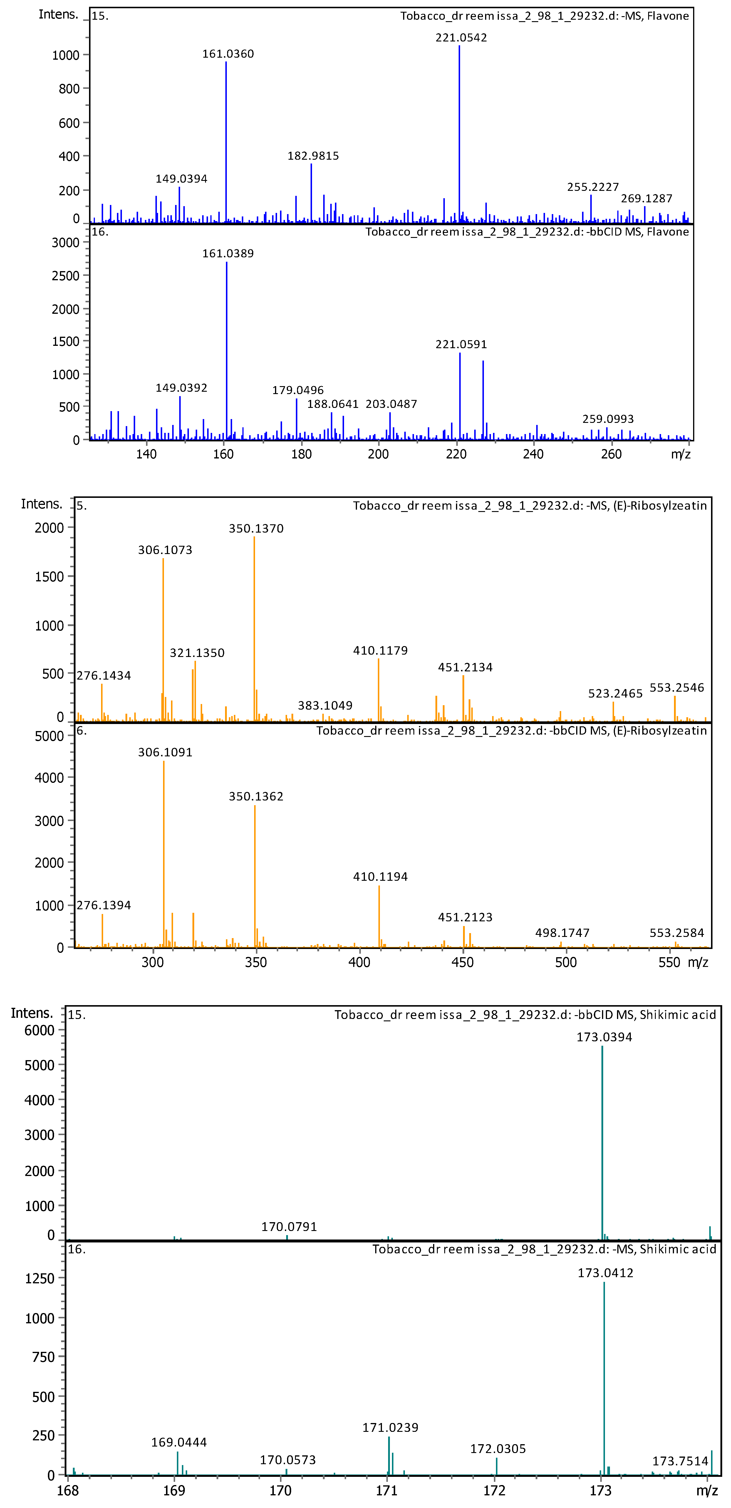
1 |
1.65 | 133.00998 | 134.0174 | [M-H]-, [M-H H2O]- |
Malic acid | C4H6O5 |
| 2 | 1.91 | 59.01128 | 60.01856 | [M-H]- | Acetic acid | C2H4O2 |
| 3 | 2.39 | 350.14397 | 351.15125 | [M-H]- | (E)-Ribosylzeatin | C15H21N5O5 |
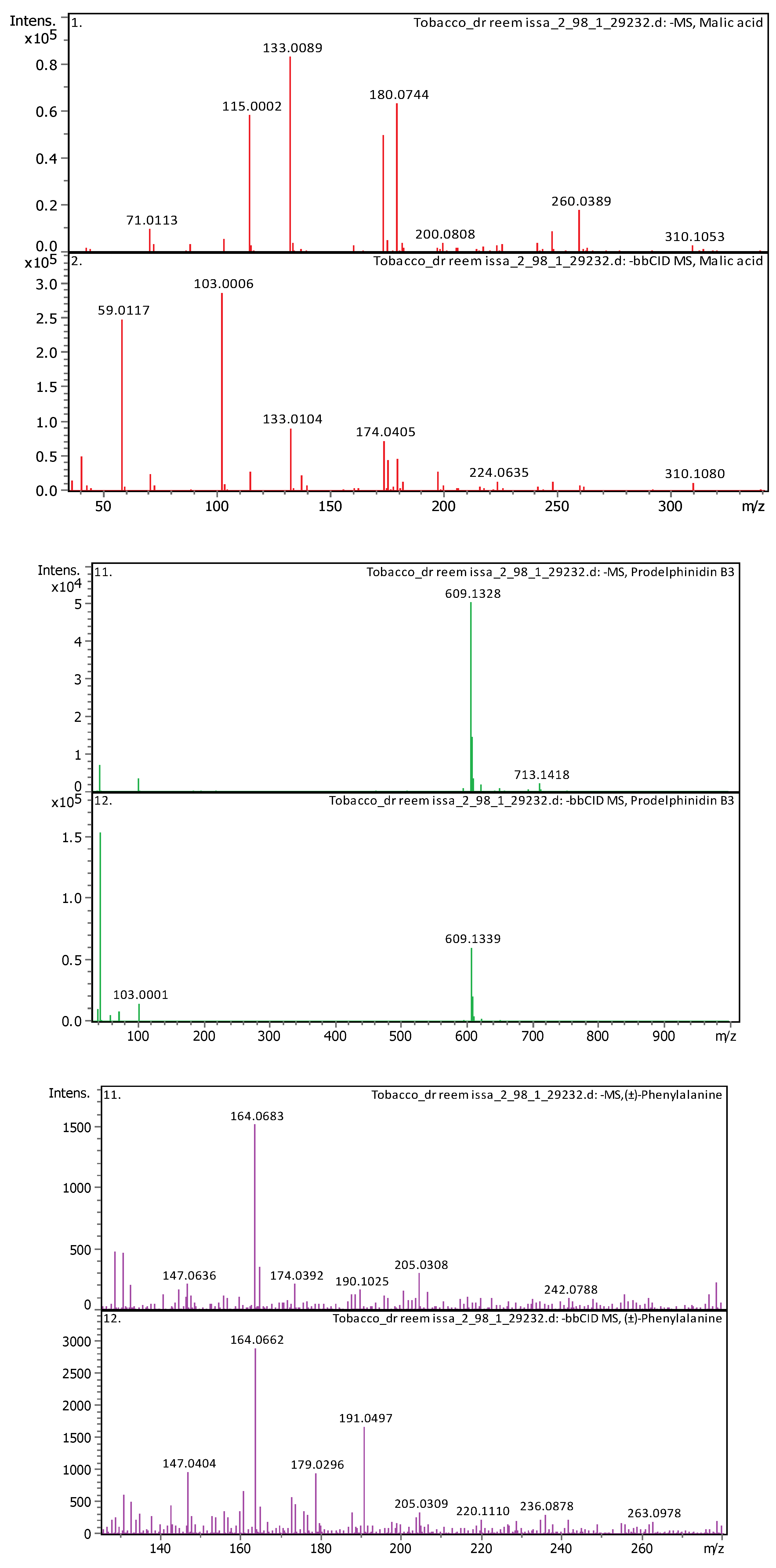
| 4 | 2.96 | 117.01473 | 118.022 | [M-H]- | Succinic acid | C4H6O4 |
| 5 | 3.32 | 147.04052 | 148.0478 | [M-H]- | Cinnamic acid | C9H8O2 |
|
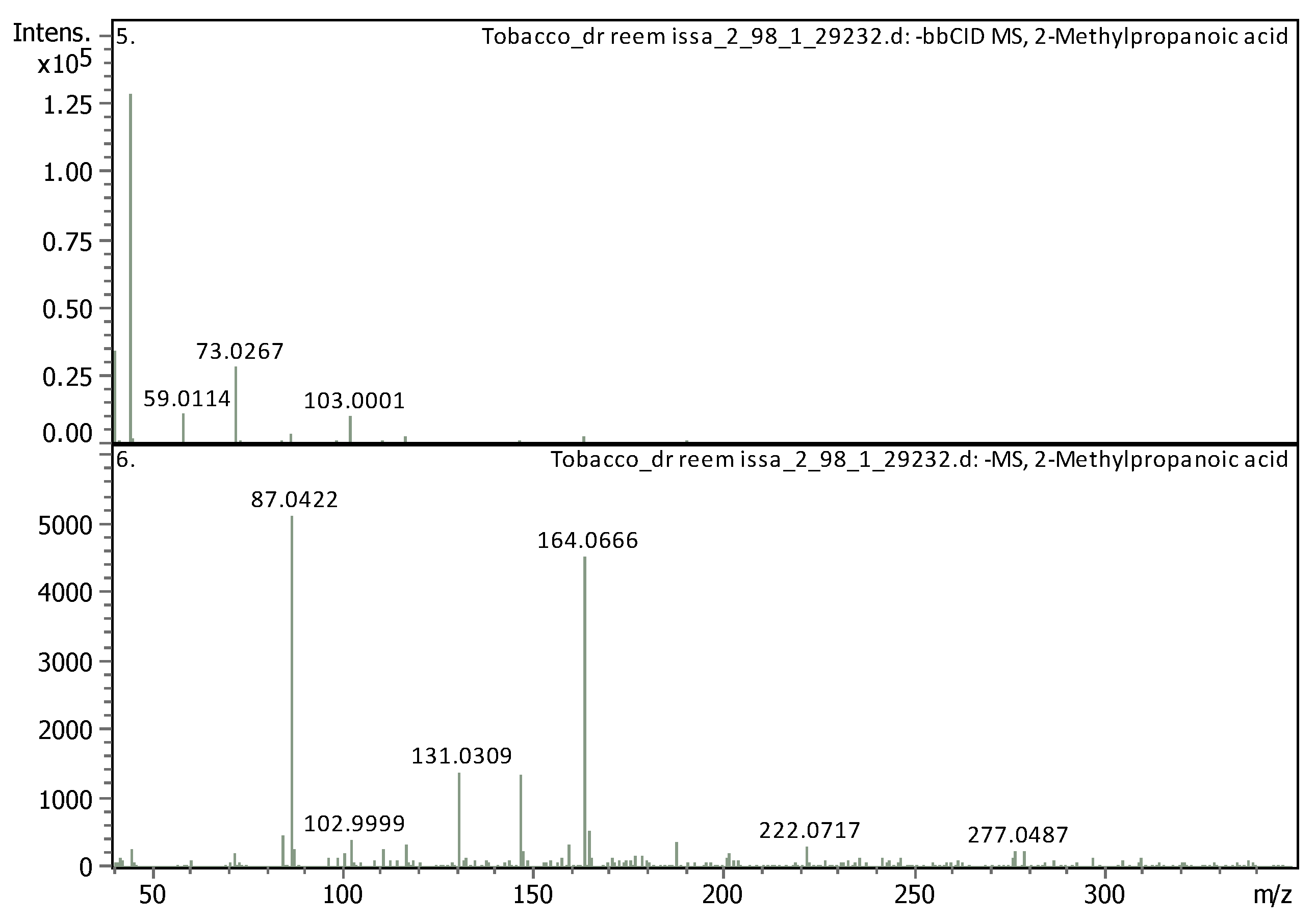
6 |
3.35 | 87.04212 | 88.04939 | [M-H]- | 2-Methylpropanoic acid | C4H8O2 |
| 7 | 3.36 | 164.06681 | 165.07409 | [M-H]- | (±)-Phenylalanine | C9H11NO2 |
| 8 | 3.69 | 163.038920 | 162.031640 | [M+H]+ | Umbelliferone | C9H6O3 |
|
9 |
3.7 | 355.102500 | 354.095190 | [M+H]+, [M+K]+, [M+Na]+ |
Chlorogenic acid | C16H18O9 |
| 10 | 3.79 | 173.04114 | 174.04841 | [M-H]- | Shikimic acid | C7H10O5 |
| 11 | 3.8 | 199.057710 | 198.050440 | [M+H]+ | Syringic acid | C9H10O5 |
| 12 | 4.6 | 193.049450 | 192.042170 | [M+H]+ | Scopoletin | C10H8O4 |
| 13 | 4.76 | 465.102820 | 464.095540 | [M+H]+ | Hyperoside | C21H20O12 |
| 14 | 4.77 | 303.050010 | 302.042740 | [M+H]+ | Robietin | C15H10O7 |
|
15 |
4.77 | 611.160240 | 610.152970 | [M+H]+ | Luteolin-7,3'-di-O-glucoside | C27H30O16 |
| 16 | 4.91 | 609.1281 | 610.13537 | [M-H]- | Prodelphinidin B3 | C30H26O14 |
| 17 | 5 | 221.05958 | 222.06686 | [M-H]- | Flavone | C15H10O2 |
|
18 |
5.15 | 595.165470 | 594.158150 | [M+H]+, [M+Na]+ | Saponarin | C27H30O15 |
| 19 | 5.74 | 195.065060 | 194.057780 | [M+H]+ | 3-Hydroxy-4-methoxycinnamic acid (isoferulic acid) |
C10H10O4 |
|
20 |
7.39 | 163.039120 | 162.031850 | [M+H]+ | (4 or 7) Hydroxy-Coumarin Plus Hydrate | C9H6O3 |
|
21 |
7.41 | 177.054460 | 176.047190 | [M+H]+ | 4-Methylumbelliferone | C10H8O3 |
| 22 | 8.54 | 495.125630 | 494.118360 | [M+H]+ | Salvianolic acid A | C26H22O10 |
| 23 | 11.6 | 249.148000 | 248.140730 | [M+H]+ | 3-Oxocostusic acid | C15H20O3 |
Table 5.
UHPLC-MS/MS analysis for nicotine detected in N. glauco extracts based on retention time.
| Rt [min] | m/z meas. | M meas. | Ions | Name | Molecular Formula |
|---|---|---|---|---|---|
| 2.89 | 163.12293 | 324.2313 | [M+H+H]2+ | Nicotine | C10H14N2 |
Table 6.
Area under the curve and concentration of nicotine (ppm) in the MC and DES extracts based on multiple external standards method.
Table 6.
Area under the curve and concentration of nicotine (ppm) in the MC and DES extracts based on multiple external standards method.
| Sample | MC extract | DES extract |
|---|---|---|
| Area of Nicotine in Sample | 2526713 | 4192477 |
| Concentration of Nicotine | 635.07 ppm | 1194.91 ppm |
Disclaimer/Publisher’s Note: The statements, opinions and data contained in all publications are solely those of the individual author(s) and contributor(s) and not of MDPI and/or the editor(s). MDPI and/or the editor(s) disclaim responsibility for any injury to people or property resulting from any ideas, methods, instructions or products referred to in the content. |
© 2024 by the authors. Licensee MDPI, Basel, Switzerland. This article is an open access article distributed under the terms and conditions of the Creative Commons Attribution (CC BY) license (http://creativecommons.org/licenses/by/4.0/).
Copyright: This open access article is published under a Creative Commons CC BY 4.0 license, which permit the free download, distribution, and reuse, provided that the author and preprint are cited in any reuse.


