Submitted:
27 December 2023
Posted:
27 December 2023
You are already at the latest version
Abstract
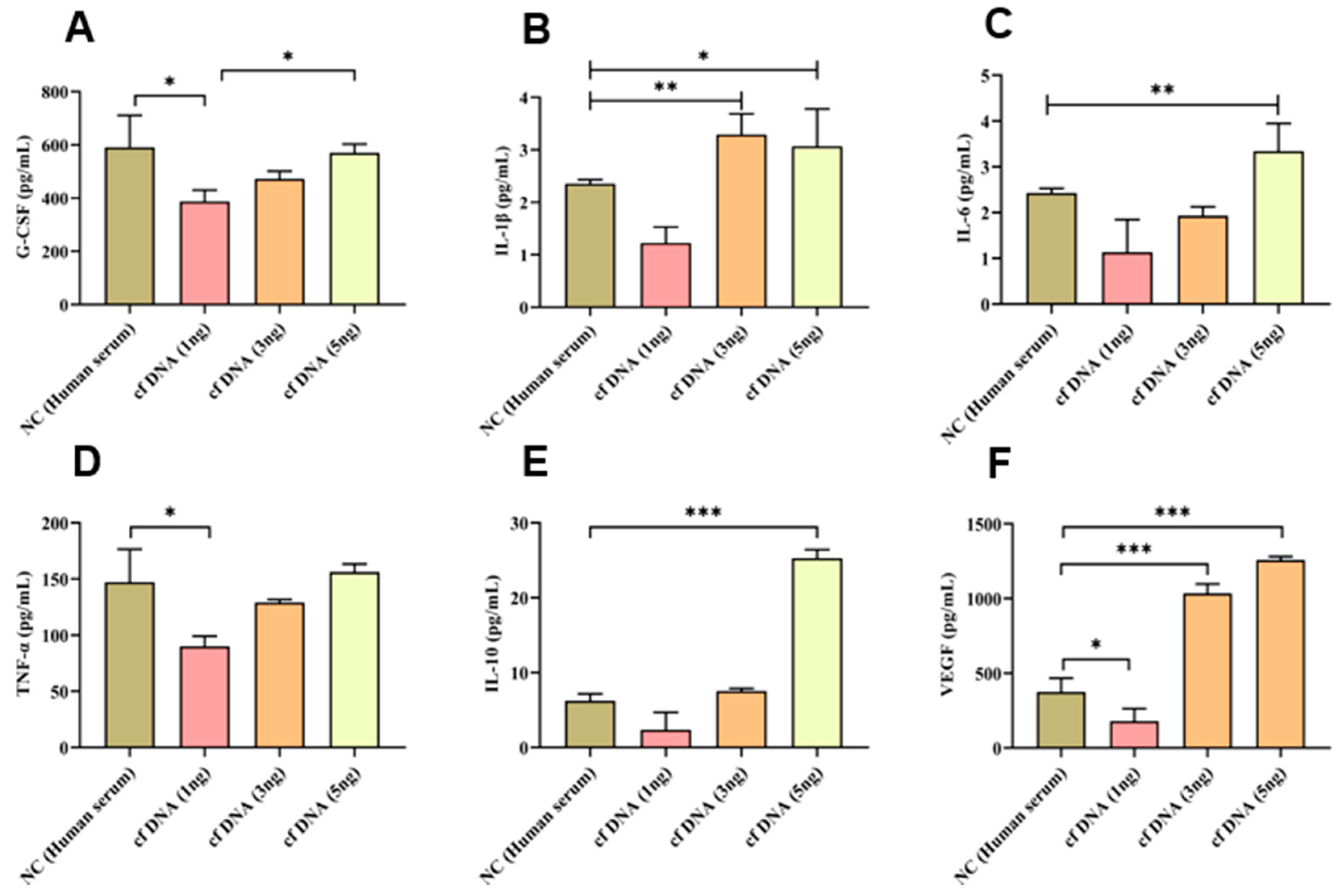
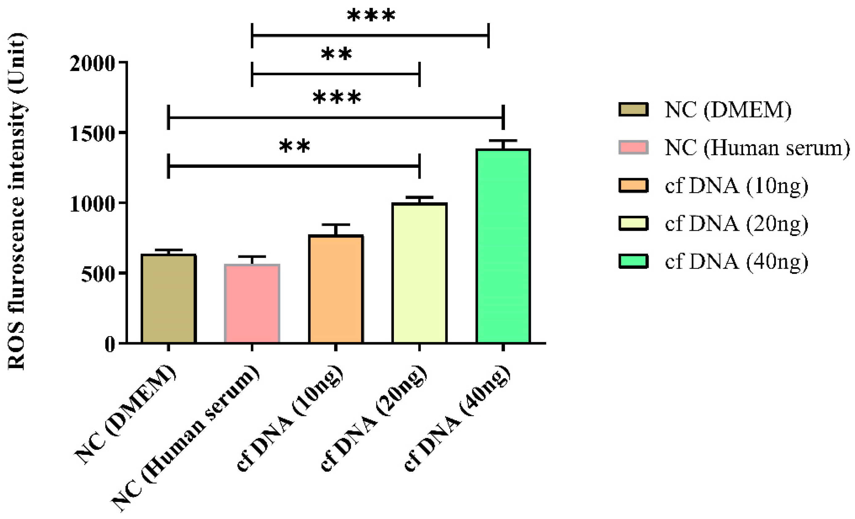
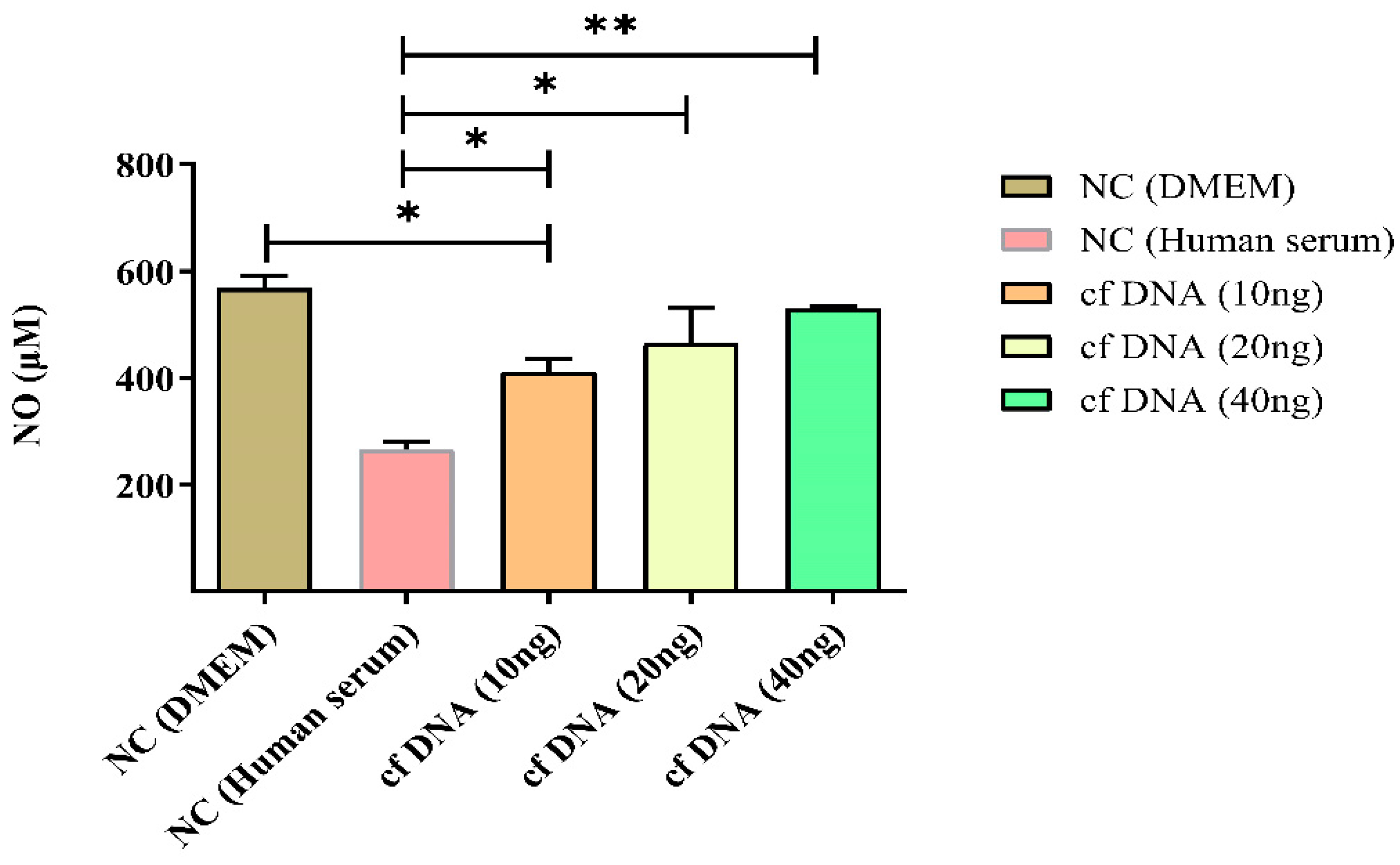
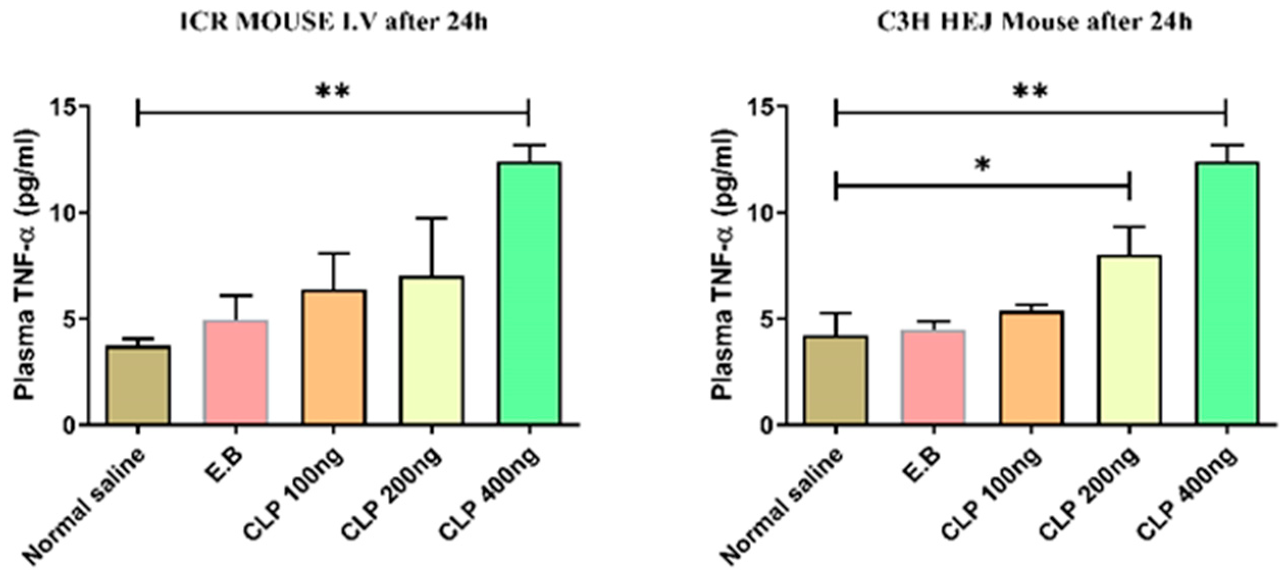
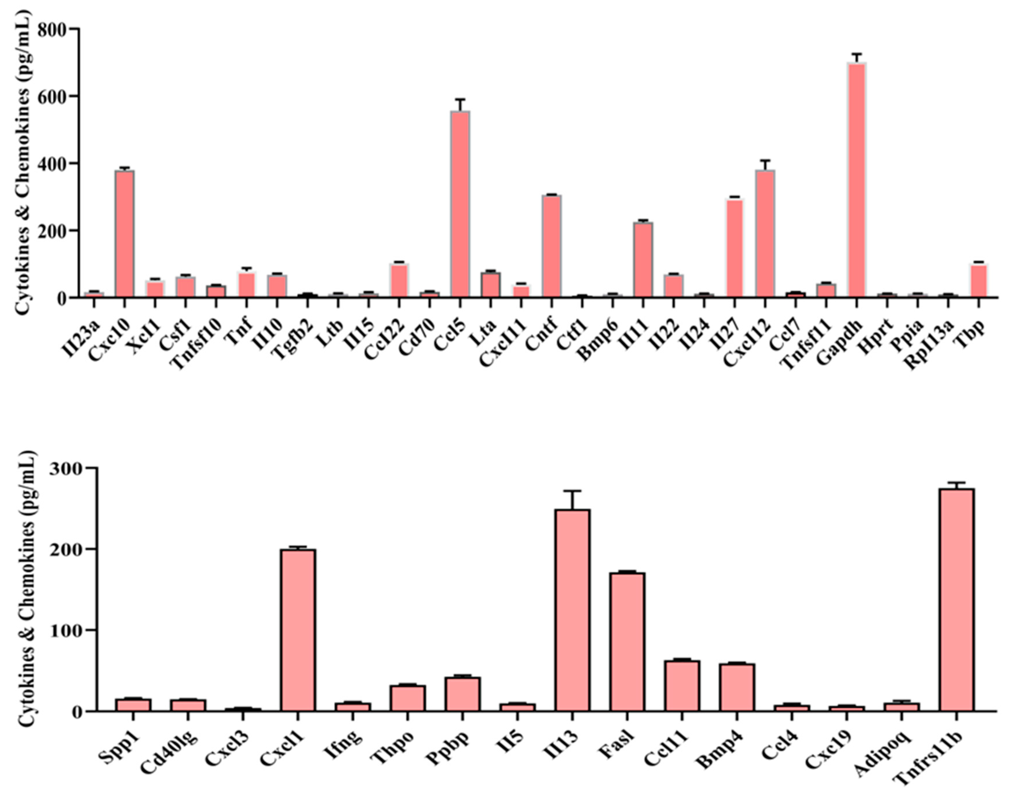
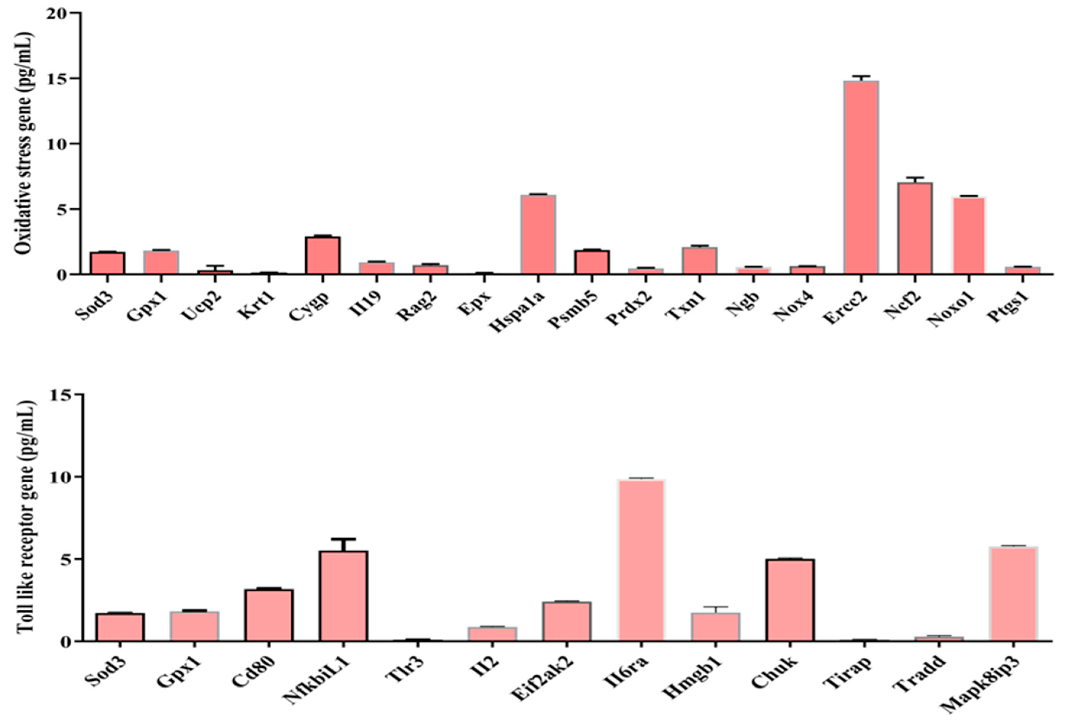
cfDNA is known to be a beneficial biomarker for the diagnosis, treatment response, and prognosis of sepsis. Previous cell line studies have shown that cfDNA in septic mice triggers inflammatory responses and tissue damage, and the mechanism occurs by binding to toll-like receptors (TLRs). Meanwhile, reactive oxidative species (ROS) and nitric oxide (NO) are increased in sepsis, which is known to be associated with organ failure and poor prognosis of sepsis through immunoredox imbalance. This study investigates the occurrence and mechanisms of inflammatory responses when cfDNA from septic mice is administered to macrophages (RAW 264.7 cells) and normal ICR mice aged 7-9 weeks. In addition, cfDNA-activated genes and their pathways are investigated and studied. Tumor necrosis factor-alpha (TNF-α) measured 24 hours after administration of 100, 200, and 400 ng/mL of sepsis-derived cfDNA to healthy mice was significantly higher in the 400 ng/mL group than in the control group. Furthermore, in macrophage cell line experiments, total ROS, NO, and catalase concentrations measured at 24 hours after cfDNA administration were significantly increased compared to the control group. In the meantime, cell viability studies revealed that the viability of the group treated with cfDNA from septic mice was much lower than that of the control group treated with normal human cfDNA. Cytokine levels such as granulocyte colony-stimulating factor (G-CSF), tumor necrosis factor-alpha (TNF-σ), interleukin-10 (IL-10), and vascular endothelial growth factor (VEGF) were significantly higher in the 5 ng/mL group when compared to the 1 ng/mL group, but levels of IL-1β and IL-6 were not significantly higher. Finally, we measured the expression levels of several oxidative stress genes and receptor (TLR) genes and found that 46 cytokines & chemokines genes, namely II23a, Cxc10, XcI1, Csf1, Tnfsf10, Tnf, II10, Tgfb2, Ltb, II15, CcI22, Cd70, Cc15, Lta, CxcI11, Cntf, Ctf1, Bmp6, II11, II22, II24, II27, CxcI12, Ccl7, Tnfsf11, Gapdh, Hprt, Ppia, RpI13a, Tbp, Spp1, Cd40lg, Cxcl3, Cxcl1, lfng, Thpo, Ppbp, II5, II13, Fasl, Ccl11, Bmp4, Ccl4, Cxc19, Adipoq and Tnfrs11b and 18 oxidative stress genes, namely Sod3, Gpx1, Ucp2, Krt1, Cygp, II19, Rag2, Epx, Hspa1a, Psmb5, Prdx2, Txn1, Ngb, Nox4, Ercc2, Ncf2, Noxo1, and Ptgs1 were expressed at various levels, and 13 receptor genes, namely Sod3, Gpx1, Cd80, NfkbiL1, Tlr3, II2, Eif2ak2, II6ra, Hmgb1, Chuk, Tirap, Tradd, and Mapk8ip3, were also expressed at various levels. cfDNA from septic mice and healthy humans exhibit different immunoredox responses through immunoredox-related gene pathways.
Keywords:
1. Introduction
2. Results
2.1. Proinflammatory(cytokines) effect of cfDNA) in vitro
2.2. Effects of cfDNA on immune redox system (total ROS production) in vitro
2.3. Effects of cfDNA on immune redox system (NO production) in vitro
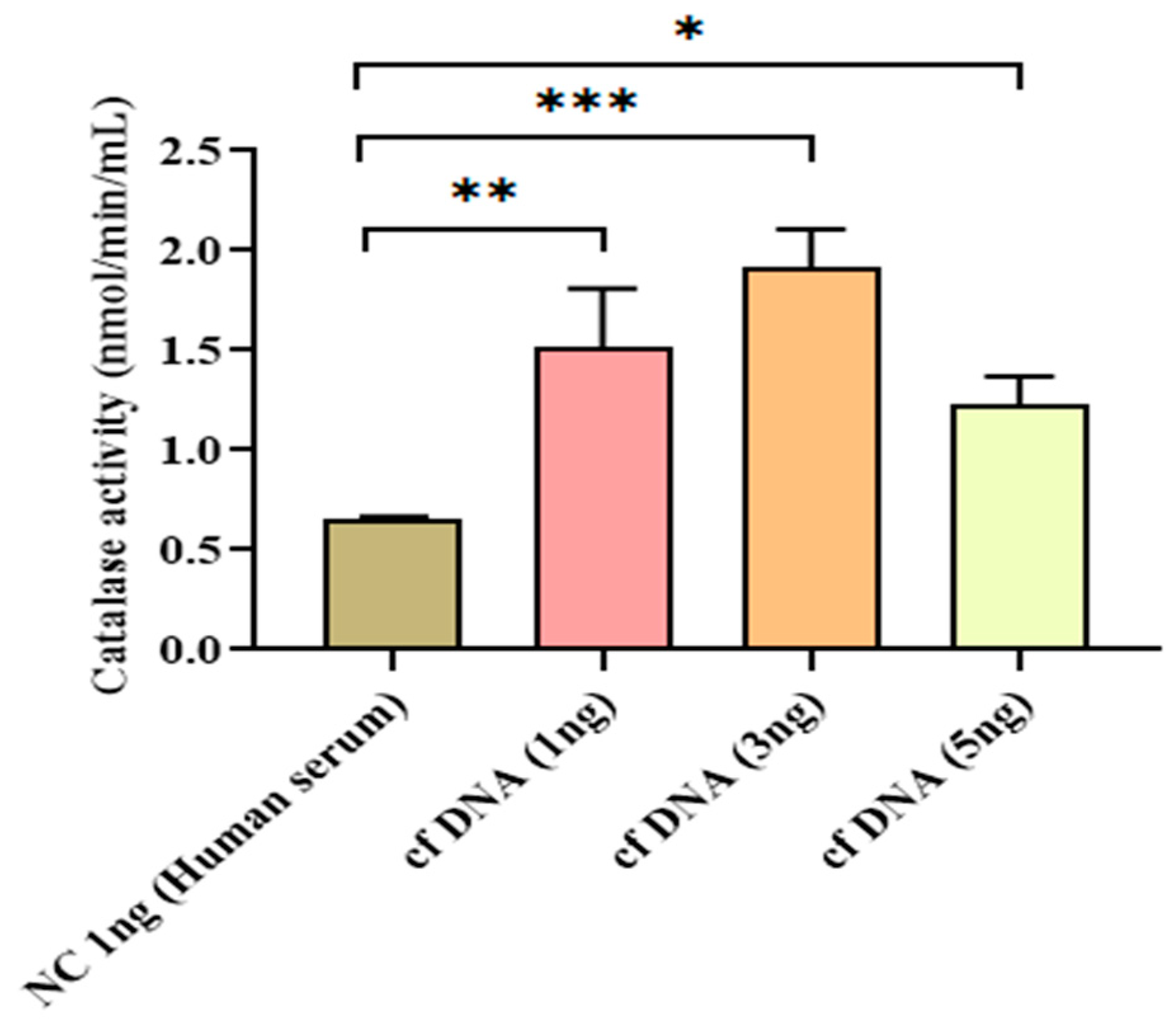
2.4. Effects of cell-free DNA on immune redox system [antioxidant (catalase) production] in vitro
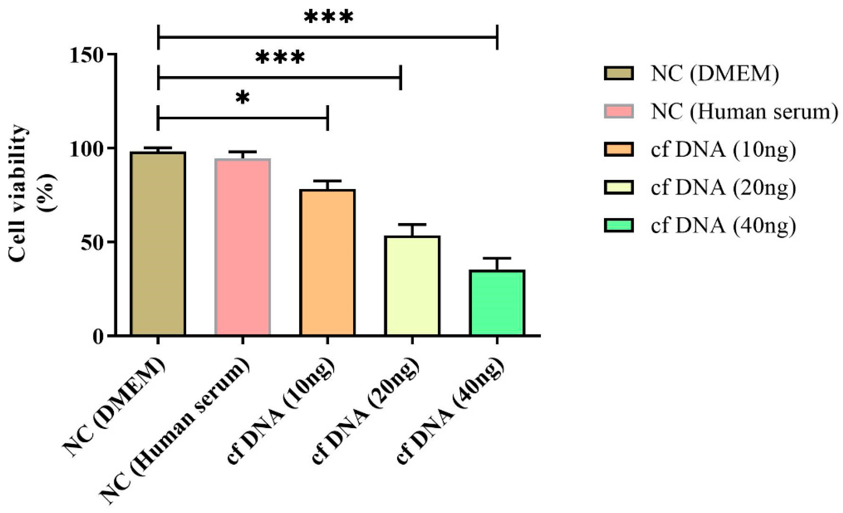
2.5. Cytotoxic effects of cell-free DNA (cell viability)
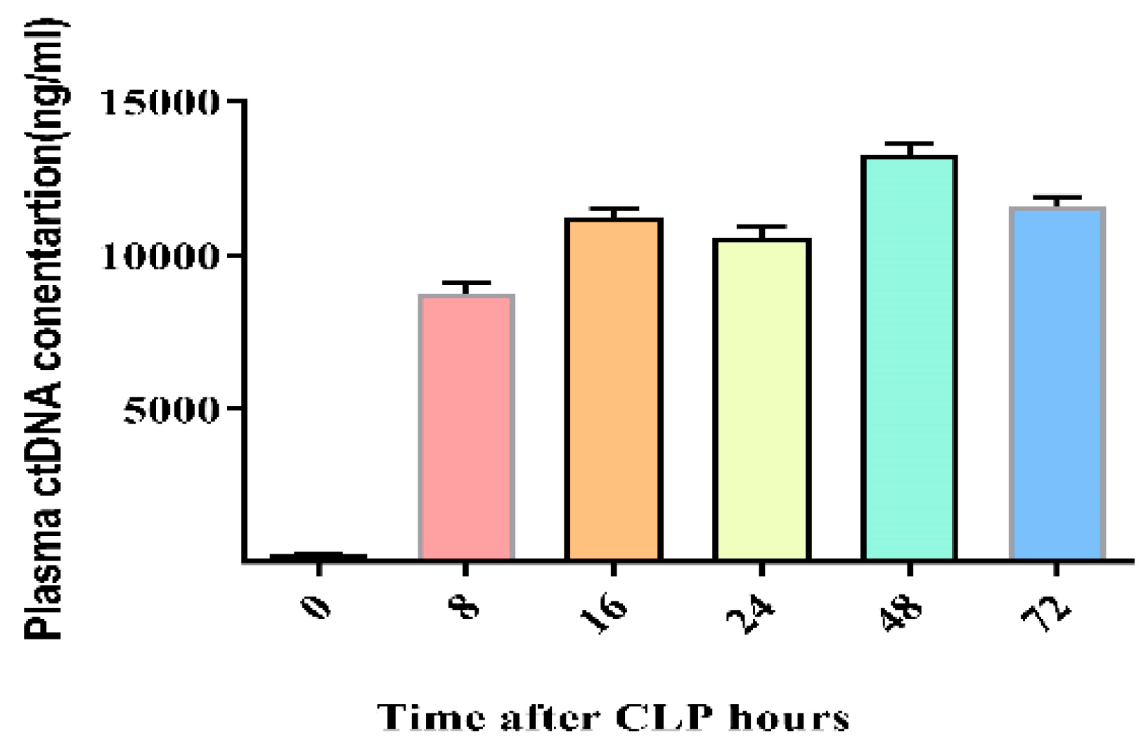
2.6. Time-dependent kinetics of cfDNA in CLP-induced mouse sepsis
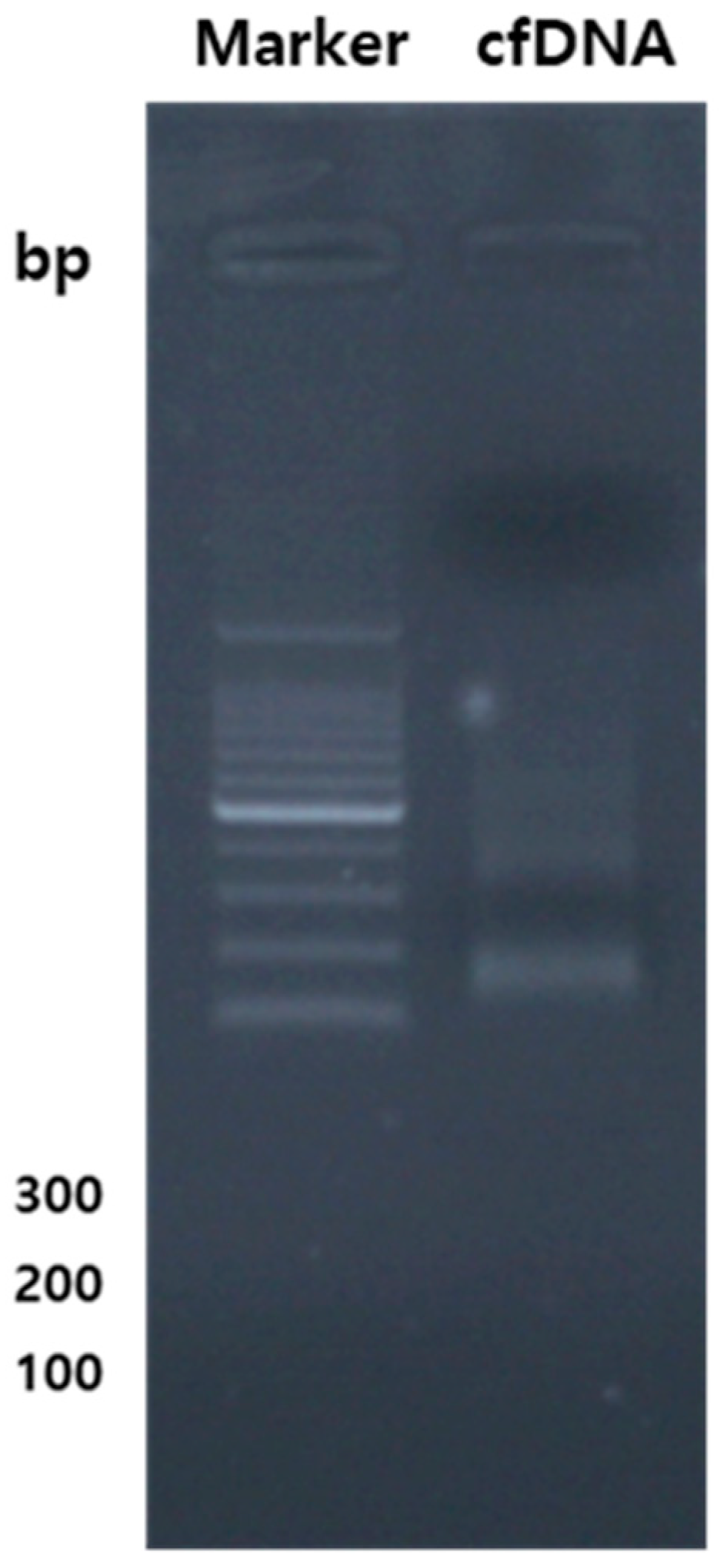
2.7. Chromatograph of cfDNA from CLP-induced mouse sepsis
3.8. Release of tumor necrosis factor-α at 24 hours after injection of cfDNA from septic mice into healthy ICR mice and C3H/HeJ mice
3. Discussion
4. Materials and Methods
4.1. Reagents
4.2. Macrophage RAW 264.7 cells culture and cfDNA hit into cells
4.3. Cell viability assay
4.4. Measurement of total ROS
4.5. Measurement of NO levels
4.6. Measurement of antioxidant activities (Catalase)
4.7. Extraction and purification of cfDNA, along with the quantification of genes related to cytokines & and chemokines, TLR, and oxidative stress using qPCR DNA.
4.8. Measurement of cytokines
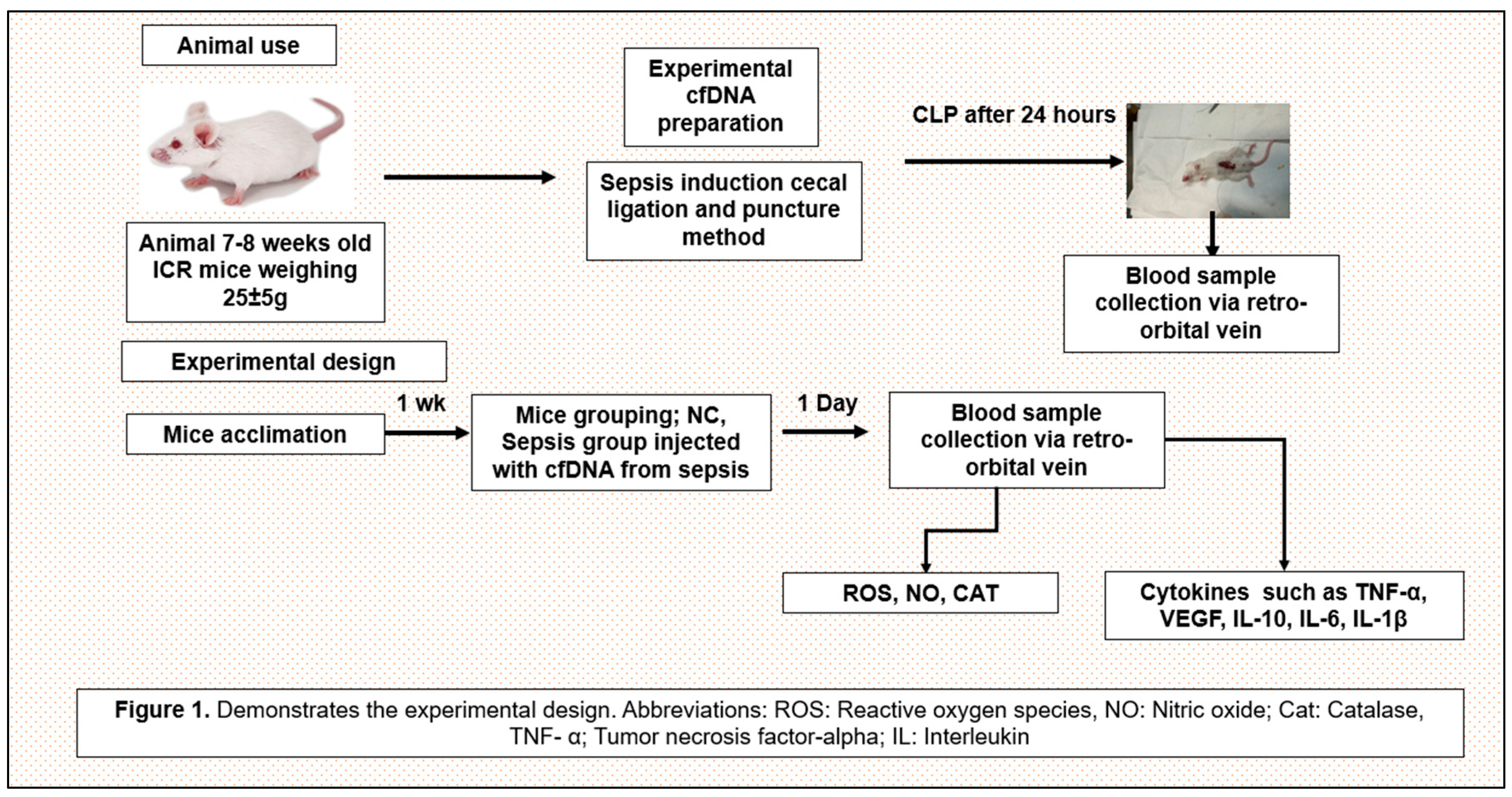
4.9. Experimental design
4.10. Data management and statistical analysis
5. Conclusion
Authors contributions
Funding
Institutional Review Board Statement
Informed Consent Statement
Data Availability Statement
Acknowledgments
Conflicts of Interest
References
- Korabecna, M.; Zinkova, A.; Brynychova, I.; Chylikova, B.; Prikryl, P.; Sedova, L.; Neuzil, P.; Seda, O. Cell-free DNA in plasma as an essential immune system regulator. Scientific reports 2020, 10, 17478. [Google Scholar] [CrossRef]
- Bauer, M.; Gerlach, H.; Vogelmann, T.; Preissing, F.; Stiefel, J.; Adam, D. Mortality in sepsis and septic shock in Europe, North America and Australia between 2009 and 2019—results from a systematic review and meta-analysis. Critical Care 2020, 24, 1–9. [Google Scholar] [CrossRef] [PubMed]
- Dawulieti, J.; Sun, M.; Zhao, Y.; Shao, D.; Yan, H.; Lao, Y.-H.; Hu, H.; Cui, L.; Lv, X.; Liu, F. Treatment of severe sepsis with nanoparticulate cell-free DNA scavengers. Science advances 2020, 6, eaay7148. [Google Scholar] [CrossRef]
- Wang, S.; Xie, T.; Sun, S.; Wang, K.; Liu, B.; Wu, X.; Ding, W. DNase-1 treatment exerts protective effects in a rat model of intestinal ischemia-reperfusion injury. Scientific reports 2018, 8, 17788. [Google Scholar] [CrossRef] [PubMed]
- Aucamp, J.; Bronkhorst, A.J.; Badenhorst, C.P.; Pretorius, P.J. The diverse origins of circulating cell-free DNA in the human body: a critical re-evaluation of the literature. Biological Reviews 2018, 93, 1649–1683. [Google Scholar] [CrossRef] [PubMed]
- Volpi, C.; Fallarino, F.; Pallotta, M.T.; Bianchi, R.; Vacca, C.; Belladonna, M.L.; Orabona, C.; De Luca, A.; Boon, L.; Romani, L. High doses of CpG oligodeoxynucleotides stimulate a tolerogenic TLR9–TRIF pathway. Nature communications 2013, 4, 1852. [Google Scholar] [CrossRef] [PubMed]
- Bellanti, F.; Pannone, G.; Tartaglia, N.; Serviddio, G. Redox control of the immune response in the hepatic progenitor cell niche. Frontiers in cell and developmental biology 2020, 8, 295. [Google Scholar] [CrossRef]
- Kaewduangduen, W.; Visitchanakun, P.; Saisorn, W.; Phawadee, A.; Manonitnantawat, C.; Chutimaskul, C.; Susantitaphong, P.; Ritprajak, P.; Somboonna, N.; Cheibchalard, T. Blood bacteria-free DNA in septic mice enhances LPS-induced inflammation in mice through macrophage response. International Journal of Molecular Sciences 2022, 23, 1907. [Google Scholar] [CrossRef]
- Joffre, J.; Hellman, J. Oxidative stress and endothelial dysfunction in sepsis and acute inflammation. Antioxidants & redox signaling 2021, 35, 1291–1307. [Google Scholar]
- Jing, Q.; Leung, C.H.C.; Wu, A.R. Cell-free DNA as biomarker for sepsis by integration of microbial and host information. Clinical Chemistry 2022, 68, 1184–1195. [Google Scholar] [CrossRef]
- Wiggins, J.M.; Ali, S.; Polsky, D. Cell-free DNA in dermatology research. Journal of Investigative Dermatology 2022, 142, 1523–1528.e1521. [Google Scholar] [CrossRef] [PubMed]
- Mortazavipour, M.M.; Shahbazi, S. The current applications of cell-free fetal DNA in prenatal diagnosis of single-gene diseases: A review. International Journal of Reproductive BioMedicine 2022, 20, 613. [Google Scholar] [CrossRef] [PubMed]
- Műzes, G.; Bohusné Barta, B.; Szabó, O.; Horgas, V.; Sipos, F. Cell-Free DNA in the Pathogenesis and Therapy of Non-Infectious Inflammations and Tumors. Biomedicines 2022, 10, 2853. [Google Scholar] [CrossRef] [PubMed]
- Roth, S.; Wernsdorf, S.R.; Liesz, A. The role of circulating cell-free DNA as an inflammatory mediator after stroke. In Proceedings of the Seminars in Immunopathology; 2023; pp. 1–15. [Google Scholar]
- Konkova, M.S.; Kaliyanov, A.A.; Sergeeva, V.A.; Abramova, M.S.; Kostyuk, S.V. Oxidized cell-free DNA is a factor of stress signaling in radiation-induced bystander effects in different types of human cells. International journal of genomics 2019, 2019. [Google Scholar] [CrossRef]
- Duvvuri, B.; Lood, C. Cell-free DNA as a biomarker in autoimmune rheumatic diseases. Frontiers in immunology 2019, 10, 502. [Google Scholar] [CrossRef] [PubMed]
- Pokrywka, A.; Zembron-Lacny, A.; Baldy-Chudzik, K.; Orysiak, J.; Sitkowski, D.; Banach, M. The influence of hypoxic physical activity on cfDNA as a new marker of vascular inflammation. Archives of Medical Science 2015, 11, 1156–1163. [Google Scholar] [CrossRef]
- Stawski, R.; Walczak, K.; Perdas, E.; Wlodarczyk, A.; Sarniak, A.; Kosielski, P.; Meissner, P.; Budlewski, T.; Padula, G.; Nowak, D. Decreased integrity of exercise-induced plasma cell free nuclear DNA–negative association with the increased oxidants production by circulating phagocytes. Scientific Reports 2019, 9, 15970. [Google Scholar] [CrossRef]
- He, L.; He, T.; Farrar, S.; Ji, L.; Liu, T.; Ma, X. Antioxidants maintain cellular redox homeostasis by elimination of reactive oxygen species. Cellular Physiology and Biochemistry 2017, 44, 532–553. [Google Scholar] [CrossRef]
- Zheng, D.; Liwinski, T.; Elinav, E. Inflammasome activation and regulation: toward a better understanding of complex mechanisms. Cell discovery 2020, 6, 36. [Google Scholar] [CrossRef]
- Burke, R.M.; Dale, B.L.; Dholakia, S. The NLRP3 Inflammasome: Relevance in Solid Organ Transplantation. International Journal of Molecular Sciences 2021, 22, 10721. [Google Scholar] [CrossRef]
- Dholakia, S.; De Vlaminck, I.; Khush, K.K. Adding insult on injury: immunogenic role for donor-derived cell-free DNA? Transplantation 2020, 104, 2266. [Google Scholar] [CrossRef]
- Nishimoto, S.; Fukuda, D.; Sata, M. Emerging roles of Toll-like receptor 9 in cardiometabolic disorders. Inflammation and Regeneration 2020, 40, 1–13. [Google Scholar] [CrossRef]
- Murata, H.; Kinoshita, M.; Yasumizu, Y.; Motooka, D.; Beppu, S.; Shiraishi, N.; Sugiyama, Y.; Kihara, K.; Tada, S.; Koda, T. Cell-Free DNA derived from neutrophils triggers type 1 interferon signature in neuromyelitis optica spectrum disorder. Neurology-Neuroimmunology Neuroinflammation 2022, 9. [Google Scholar] [CrossRef]
- Rahman, M.H.; Bajgai, J.; Cho, Y.; Fadriquela, A.; Sharma, S.; Thuy, T.T.; Cho, S.H.; Jeong, Y.J.; Goh, S.H.; Kim, Y. Immune Redox Modulation Effects of Non-Electrolyzed Hypochlorous Acid Water on Helicobacter pylori-Infected C57BL/6 Mouse Model. Processes 2023, 11, 1474. [Google Scholar] [CrossRef]
- Rahman, M.H.; Fadriquela, A.; Bajgai, J.; Hoon, G.S.; Hyun, C.S.; Kim, C.-S.; Lee, K.-J. Long-Term Skin Safety Effect of Chlorine-Rich Water Treatment on C57BL/6 Mice. Processes 2023, 11, 1914. [Google Scholar] [CrossRef]
- Rahman, M.H.; Bajgai, J.; Sharma, S.; Jeong, E.-S.; Goh, S.H.; Jang, Y.-G.; Kim, C.-S.; Lee, K.-J. Effects of Hydrogen Gas Inhalation on Community-Dwelling Adults of Various Ages: A Single-Arm, Open-Label, Prospective Clinical Trial. Antioxidants 2023, 12, 1241. [Google Scholar] [CrossRef] [PubMed]
- Dejager, L.; Pinheiro, I.; Dejonckheere, E.; Libert, C. Cecal ligation and puncture: the gold standard model for polymicrobial sepsis? Trends in microbiology 2011, 19, 198–208. [Google Scholar] [CrossRef] [PubMed]
Disclaimer/Publisher’s Note: The statements, opinions and data contained in all publications are solely those of the individual author(s) and contributor(s) and not of MDPI and/or the editor(s). MDPI and/or the editor(s) disclaim responsibility for any injury to people or property resulting from any ideas, methods, instructions or products referred to in the content. |
© 2023 by the authors. Licensee MDPI, Basel, Switzerland. This article is an open access article distributed under the terms and conditions of the Creative Commons Attribution (CC BY) license (http://creativecommons.org/licenses/by/4.0/).


