Submitted:
04 October 2023
Posted:
05 October 2023
You are already at the latest version
Abstract
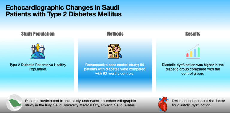
Background and Objectives: Cardiovascular disease is one of the leading causes of morbidity and mortality among the diabetic population. Given the high prevalence of diabetes mellitus (DM) in Saudi Arabia and the high prevalence of heart failure in the diabetic population, this study assesses the echocardiographic changes in Saudi patients with type 2 DM (T2DM) compared with healthy controls. Materials and Methods: In this retrospective case control study, 80 patients with diabetes (45 males, age: 58.78±10.2 years) were compared with 80 controls (45 males, age: 58.6±10 years) who underwent an echocardiographic study in the King Saud University Medical City, Riyadh, Saudi Arabia. Results: There were no significant differences between patients with diabetes and controls in terms of aortic root diameter, left atrium diameter, posterior wall, interventricular wall thickness, left ventricular diameters and ejection fraction. However, diastolic dysfunction was statistically significantly higher in the diabetic group compared with the control group (P <0.05). Conclusions: This is the first case control study in Saudi Arabia that assesses echocardiographic parameters in T2DM patients. DM is an independent risk factor for diastolic dysfunction regardless of its association with hypertension and dyslipidemia.
Keywords:
Diabetes mellitus
; diabetes cardiomyopathy
; echocardiography
1. Introduction
Diabetes mellitus (DM) is one of the most common chronic metabolic diseases, and, in recent decades, DM rates have significantly increased worldwide. The Saudi population has the second highest prevalence of diabetes in the Middle East and is ranked seventh in the world for diabetes rates reported by the World Health Organization, with a prevalence of 25.4% [1]. It has been estimated that 40.3% are unaware of their disease [1] and are thus prone to developing diabetes complications. DM complications significantly increase morbidity and mortality. Cardiovascular disease (CVD) is the leading cause of death among the diabetes population [2,3,4].
Echocardiography plays an important role in the diagnosis of cardiomyopathy. It is the gold standard for providing a comprehensive evaluation of myocardial structure and function [5,6] and is a valuable tool for facilitating early diagnosis and hence early medical management. It is widely used due to its proven safety and non-invasiveness.
It has been well-established that the heart is affected by DM. DM can contribute to CVD development through multiple factors, such as accelerated atherosclerosis, increased oxidative stress, endothelial dysfunction and cardiac autonomic neuropathy [7]. On the other hand, it was introduced to be a contributory risk factor to heart failure as well, a different entity of heart diseases. This is supported by the reported prevalence of HF in patients with diabetes being significantly higher than that reported in the general population. The prevalence in the diabetic population is 12%, whereas, in the general population, it is 1% to 4% [8]. Researchers of the Framingham study reported that diabetic women and men have a two- and five-fold increased risk, respectively, of congestive heart failure compared with the non-diabetic population [9]. However, it is unclear whether diabetes is a direct causative risk factor or related to confounders such as coexistent hypertension, ischemic or valvular heart disease.
Controversy has emerged concerning the effect of DM on echocardiographic parameters [10,11]. To the best of our knowledge, the relationship between DM and cardiac structural changes has not been studied in Saudi patients with type 2 DM (T2DM) without overt heart disease. Given the high prevalence of DM in Saudi Arabia, we examine the direct effect of DM on the heart and detect any myocardial changes via an echocardiographic study of the Saudi diabetic population and healthy population.
2. Materials and Methods
2.1. Study design and setting
This is a retrospective case control study of 80 T2DM patients in King Saud University Medical City’s diabetes clinics and 80 control subjects (age and gender matched) who were identified as non-diabetic in the same hospital; both groups underwent an echocardiographic study. Approval for the study was obtained from the Institutional Review Board, College of Medicine, King Saud University (project no.: E-21-5899), Riyadh, Saudi Arabia. The patients were selected using the systematic random sampling technique; their data were retrieved from electronic medical records using a structured data clinical sheet, which included demographic data, clinical history and vital signs (weight, height, body mass index, systolic blood pressure [SBP] and diastolic blood pressure) as well as metabolic laboratory parameters, including total cholesterol (TC), low-density lipoprotein-cholesterol (LDL-C), high-density lipoprotein-cholesterol (HDL-C), triglyceride, creatinine, haemoglobin A1c (HgA1c), fasting blood glucose (FBG) and estimated glomerular filtration rate (eGFR), which was calculated using CKD-EPI equation.
2.2. Inclusion criteria
The study included adult men and women aged between 18 and 75 with T2DM diagnosed based on ADA guidelines and had echocardiographic study from 2018–2020. Compared with healthy control group.
2.3. Exclusion criteria
The study excluded patients aged 75 years and older and 18 years and younger, patients with type 1 DM or gestational diabetes mellitus and patients previously diagnosed with comorbidities, such as heart failure, coronary artery disease, valvular heart disease or chronic kidney disease (eGFR of <30mL/min/1.73m2). This was documented from the patient history in the medical records.
2.4. Echocardiographic evaluation
Transthoracic echocardiographic imaging was performed using PHILIPS EPIQ 7 Ultrasound system, and the following parameters were attained for each subject:
1. Aortic root diameter (ARD; 20–29 mm)
2. Left atrium diameter (LAD; 15–37 mm)
3. Left ventricular internal diameter end diastole (LVIDd; 35–56 mm)
4. Left ventricular internal diameter end systole (LVIDs; 22–40 mm)
5. Posterior wall thickness (PWT; 7–12 mm)
6. Interventricular wall thickness (IVWT; 7–11 mm)
7. Ejection Fraction (EF; >50 %)
Left ventricular diastolic dysfunction (LVDD) diagnosis was done by specialists in echocardiography based on the diagnostic criteria of the American Society of Echocardiography and the European Association of Cardiovascular Imaging (annular e0 velocity: septal e0 < 7 cm/sec and lateral e0 < 10 cm/sec; average E/e0 ratio > 14, LA volume index > 34 ml/m2 and peak TR velocity > 2.8 m/sec).
2.5. Statistical analysis
Analysis was done using IBM-SPSS version 25. Descriptive statistics are presented in frequency, percentage, mean and standard divisions. For inferential statistics, CH-square was used to study the association between two categorical variables. A proportionality test and independent t-test were used to compare the diabetic and non-diabetic groups by percentage or mean. Categorical variables with a CH-square p-value less than 0.05 were further analysed using multivariate logistic regression to determine the risk factor or the odd ratio of each factor. A statistical test p-value less than 0.05 was considered significant.
3. Results
3.1. Characteristics of the subjects
A total of 80 patients with diabetes (cases) and healthy subjects (controls) with the echocardiographic study were enrolled in the study. Patients with T2DM had a mean age of 58.78±10.2 years, the controls had a mean age of 58.6±10 years and both had the same gender proportions: 45 males (56.3%) and 35 females (43.8%). The mean duration of DM among patients was 13±8.3 years. Compared to controls, T2DM had a higher prevalence of hypertension (HTN) and dyslipidemia (DLP) at 68% and 63%, respectively (Table 1). SBP, FBG and HgA1c were higher among the T2DM group (all p-value < 0.05) but TC, LDL-C, HDL-C and eGFR were lower (all p-value < 0.05; Table 1).
They also use more anti-hypertensive medications, including calcium channel blockers, angiotensin-converting enzyme inhibitors (ACEI), angiotensin II receptor blockers (ARBs) and diuretics along with statins and antiplatelets (all p-value < 0.05; Table 2).
3.2. Echocardiography parameters
As tabulated in Table 3, the diabetic group had higher but not statistically significant ARD, LAD, PWT and IVWT values compared with the control group (p-value > 0.05). In terms of left ventricular (LV) systolic function parameters, LV diameters and EF, the diabetic group had smaller values compared with the control group (Table 2), but no significant differences were found (p-value > 0.05). Patients with T2DM had significantly higher proportion of diastolic dysfunction at 48/80 patients (60%) compared to controls at 35/80 subjects (43.7%; p-value = 0.04; Table 3).
Multivariate logistic regression of risk factors associated with diabetes did not show any significant differences in the odd ratio between the two groups (table 4). However, the odd ratio for HTN was 2.060 with 95% CI (0.862, 4.926), and, for DLP, the odd ratio was 1.643 with 95% CI (0.797, 3.389). Moreover, lower eGFR levels were slightly associated with an increased risk of LVDD in patients with diabetes compared to the control group, but this was not statistically significant.
4. Discussion
Diabetic cardiomyopathy (DCM) was coined by Rubler et al. in 1972, who described the condition as myocardial dysfunction in the absence of coronary artery disease, hypertension, and alcohol [12]. It varies from subclinical ventricular dysfunction to overt clinical heart failure.
The present study evaluates the effect of DM on systolic function, LV internal dimensions, PWD and the thickness of the interventricular septum (IVS) based on an echocardiographic study. It compares asymptomatic patients with diabetes with a healthy matched control group.
In terms of echocardiographic parameters, ARD, LAD, IVS, PWT, LVIDs and LVIDd values were obtained in this study. Although IVS, PWT, LVIDd and LVIDs findings were located in the normal global range, ARD and LAD were slightly above the normal range in the diabetic group compared to non-diabetic group (29 cm vs 28 cm and 38.3 cm vs 37.6 cm, respectively). There were no significant differences between the two groups (p-value > 0.05).
In the literature, inconsistent results have been established. Our findings were in agreement with the Zhi et al. case control study, where 48 patients with diabetes were compared with 48 controls and non-statistically significant measurements were obtained for LVDD, IVSD and PWD: 4.6±0.5 cm vs 4.5±0.4 cm, 1.1±0.2 cm vs 1.0±0.2 cm and 1.0±0.1 cm vs 0.9±0.2 cm, respectively [10]. Another study agreed with our findings, where LVIDs and LVIDd values in 40 patients with diabetes were not statistically significant compared to 40 controls [11]. Although the Zheo et al. case control study reported no statistically significant differences in ARD between diabetic and healthy populations [13], statistically significant differences were observed in a study among Sudanese population [14].
EF was another parameter assessed in the current study. Lower values were observed among diabetics vs controls but there was no significant differences between the two groups (p-value > 0.05). This is in line with multiple studies in the literature. In the Halil study, EF in diabetic group did not differ significantly from the healthy group [11]. In addition, Mohammed et al. concluded no significant differences in EF between 113 patients with diabetes vs 130 controls [14]. Nevertheless, a large cohort study of 2,400 patients showed significantly lower EF in the diabetic population compared to controls (55±13% vs 53±13%, respectively), which can be attributed to the larger sample size in addition to the higher age group of their diabetic population (65±10 years) [15].
Our results show significantly higher diastolic dysfunction in the diabetic group compared to the control (60% vs 43.7%, respectively; p-value < 0.05). This finding is supported by the majority of studies in the literature. Virendra et al. studied similar patients’ mean HbA1c of 8.3 ± 1.91% in males and 8.1 ± 1.3% in females along with a disease duration of 11 ± 5 years in males and 10 ± 4 years in females. The results showed 54.33% of diastolic dysfunction among 127 diabetics with normal systolic function compared to 11% of 100 controls (p-value < 0.05); patients with a longer disease duration (defined as 11 to 15 years) and HbA1c of >7.5 % were significantly associated with higher diastolic dysfunction (p-value < 0.05) [18]. Similarly, significant higher LV diastolic dysfunction was observed among asymptomatic Indian patients with diabetes compared to the healthy population [17].
An additional observation was the prevalence of DLP and HTN among our diabetic population at 63% and 68%, respectively. Similar to previous studies, DLP and HTN were highly prevalent in T2DM patients compared to controls: Yaru et al. reported DLP prevalence of 59.3% in diabetic patients and 39.9% in patients with normal glucose levels [18], and a HTN prevalence of 86.2% was reported among a group of Saudi patients with T2DM by Khalid et al. [19] and 85.6% among Benghazi patients with T2DM by Faiza et al. [20]. The underlying pathophysiology of insulin resistance predisposes patients to metabolic abnormalities, which alter systemic lipid metabolism and result in the development of DLP along with HTN due to inappropriate activation of the renin-angiotensin-aldosterone system and sympathetic nervous system [21,22,23]. Coexistence of DM and HTN may accelerate or worsen several echocardiographic parameters reflecting heart structure or function. Ehud et al. demonstrated a greater LV mass index in hypertensive and diabetic patients compared to non-diabetic patients (158±45 g/m2 vs 113±20 g/m2) [26]. However, debate is ongoing concerning whether HTN is truly an affecting risk factor. We studied this relation among our population. Our result showed an independent effect of diabetes on diastolic dysfunction despite a significant higher proportion of DLP and HTN among diabetics vs non-diabetics. A similar outcome was reported by Micheal et al., who compared the systolic and diastolic function of 116 diabetic patients with 232 matched non-diabetics and coexistent hypertension (51% and 53%, respectively) using echocardiography and concluded that DM is an independent risk factor for cardiomyopathy [25].
Several factors could have contributed to the varying results in the literature, i.e., the study design, methods of echocardiographic parameters, age of the sample included, disease duration, coexistence of comorbidities and glycemic control. We relate our findings to the characteristics of our patients. The glycemic control of our diabetic population was moderately controlled (HgA1c: 8.3±1.8 %) in addition to the protective effect of statin and antihypertensive medications, including ACEI and ARBs. Poor glycemic control is an observation in multiple studies with positive findings. In the case control studies with significantly higher IVWT, LVDD, PWT and LAD in the diabetics group vs controls, patients were poorly controlled with a reported mean HgA1c of 11.77±3.53% [13]. A recent metanalysis has addressed multiple risk factors associated with DCM, highlighting a cut-off HbA1c of >9% [26]. It was suggested that regression of diastolic and systolic findings can be achieved with intensive glycemic control. In the study of Melissa et al., blood glucose, blood pressure and cholesterol were optimized to recommended targets for 12 months in subjects with T2DM and poor glycemic control. LV systolic and diastolic function, measured by LV global longitudinal strain and septal e’ velocities, have been shown to improve by 21% and 24% respectively with HgA1c improvements of 10.3%±2.4% to 8.3%±2.0%; patients who achieved a HbA1c of less than 7% have shown the greatest improvement [27].
There are several well-evidenced metabolic disturbances at the level of the myocardium causing DCM suggested, such as hyperglycemia, hyperinsulinemia and insulin resistance resulting in glucotoxicity and lipotoxicity, leading to inflammation, oxidative stress, myocellular hypertrophy, myocardial fibrosis and microvascular dysfunction within the diabetic heart [28,29,30]. This provides a comprehensive picture of subclinical cardiac dysfunction and how it can eventually lead to overt heart failure.
5. Study limitations
This study observed the cardiac structural and functional abnormalities at a single point in time, as data were collected retrospectively from medical records and no available follow up data were conducted. Further prospective studies are needed to study the progression and regression of these cardiac abnormalities over time.
6. Conclusions
This is the first retrospective case control study in Saudi Arabia to assess echocardiographic parameters in T2DM patients compared to a healthy population. Diastolic dysfunction among diabetic patients is significantly associated with DM independent of hypertension and dyslipidemia, a finding that supports the concept of DCM. In the asymptomatic period, early screening for DCM should be done to prevent cardiovascular complications, and preventive measures should be implemented to avoid overt heart failure.
7. Acknowledgement
Researchers Supporting Project number (RSPD2023R883), King Saud University, Riyadh, Saudi Arabia.
8. Conflict of Interest
Nothing to declare.
9. Ethical approval
King Saud University, College of Medicine Institutional Review Board (IRB) approval (E-21-5899) was obtained to conduct this study.
10. Statement of informed consent
All participants were informed about the study objectives, and their permission to enroll in the study was requested. A written consent form was obtained from all participants, indicating the study's purpose and the right to withdraw at any time. The anonymity was assured by assigning patients code numbers. No incentives or rewards were given.
11. Data Availability
The data used to support the findings of this study are restricted by the institutional review board at King Saud University Medical City to protect patient privacy.
12. Autor’s contribution
All authors have equally contributed, reviewed, and improved the manuscript. All authors have revised the final manuscript. All authors have read and agreed to the published version of the manuscript
13. Declarations
Not applicable.
References
- Al-Rubeaan, K.; Al-Manaa, H.; Khoja, T.; Ahmad, N.; Al-Sharqawi, A.; Siddiqui, K.; et al. Epidemiology of abnormal glucose metabolism in a country facing its epidemic: SAUDI-DM study. J. Diabetes 2014, 7, 622–632. [Google Scholar] [CrossRef] [PubMed]
- Cardiovascular Disease and Risk Management: Standards of Medical Care in Diabetes—2021. Diabetes Care 2020, 44 (Suppl. 1), S125–S150.
- Leon, B. Diabetes and cardiovascular disease: Epidemiology, biological mechanisms, treatment recommendations and future research. World J. Diabetes 2015, 6, 1246. [Google Scholar] [CrossRef]
- International Diabetes Federation. Diabetes and cardiovascular disease. Brussels, Belgium: International Diabetes Federation, 2016.
- Thomas, D.; Wheeler, R.; Yousef, Z.; Masani, N. The role of echocardiography in guiding management in dilated cardiomyopathy. European Journal of Echocardiography 2009, 10, iii15–iii21. [Google Scholar] [CrossRef] [PubMed]
- Ciampi, Q.; Villari, B. Role of echocardiography in diagnosis and risk stratification in heart failure with left ventricular systolic dysfunction. Cardiovasc. Ultrasound 2007, 5. [Google Scholar] [CrossRef] [PubMed]
- Dokken, B. The Pathophysiology of Cardiovascular Disease and Diabetes: Beyond Blood Pressure and Lipids. Diabetes Spectr. 2008, 21, 160–165. [Google Scholar] [CrossRef]
- Thrainsdottir, I.S.; Aspelund, T.; Thorgeirsson, G.; Gudnason, V. The association between glucose abnormalities and heart failure in the population-based Reykjavik study. Diabetes Care 2005, 28, 612–616. [Google Scholar] [CrossRef]
- Kannel, W.; Hjortland, M.; Castelli, W. Role of diabetes in congestive heart failure: The Framingham study. Am. J. Cardiol. 1974, 34, 29–34. [Google Scholar] [CrossRef]
- Fang, Z.; Yuda, S.; Anderson, V.; Short, L.; Case, C.; Marwick, T. Echocardiographic detection of early diabetic myocardial disease. J. Am. Coll. Cardiol. 2003, 41, 611–617. [Google Scholar] [CrossRef]
- Atas, H.; Kepez, A.; Atas, D.; Kanar, B.; Dervisova, R.; Kivrak, T.; Tigen, M. Effects of diabetes mellitus on left atrial volume and functions in normotensive patients without symptomatic cardiovascular disease. J. Diabetes its Complicat. 2014, 28, 858–862. [Google Scholar] [CrossRef]
- Rubler, S.; Dlugash, J.; Yuceoglu, Y.Z.; Kumral, T.; Branwood, A.W.; Grishman, A. New type of cardiomyopathy associated with diabetic glomerulosclerosis. Am. J. Cardiol. 1972, 30, 595–602. [Google Scholar] [CrossRef] [PubMed]
- Zhao, Z.; Hou, C.; Ye, X.; Cheng, J. Echocardiographic Changes in Newly Diagnosed Type 2 Diabetes Mellitus Patients with and without Hypertension. Medical Science Monitor 2020, 26. [Google Scholar] [CrossRef] [PubMed]
- Alhaj, M.; Gameraddin, M.; Ahmed, A.; Babiker, M. The assessment of echocardiographic findings of diabetes in Sudanese. Scholars Journal Of Applied Medical Sciences 2016, 4, 2790–2794. [Google Scholar]
- Ehl, N.; Kühne, M.; Brinkert, M.; Müller-Brand, J.; Zellweger, M. Diabetes reduces left ventricular ejection fraction-irrespective of presence and extent of coronary artery disease. Eur. J. Endocrinol. 2011, 165. [Google Scholar] [CrossRef]
- Patil, V.; Shah, K.; Vasani, J.; Shetty, P.; Patil, H. Diastolic dysfunction in asymptomatic type 2 diabetes mellitus with normal systolic function. J. Cardiovasc. Dis. Res. 2011, 2, 213–222. [Google Scholar] [CrossRef]
- Bendigeri, M.; Sadique, R.; Aziz, A.; Jaladhar, P. A study of cardiac changes in asymptomatic diabetic patients in comparison with normal population. Int. J. Adv. Med. 2019, 6, 291. [Google Scholar] [CrossRef]
- Li, Y.; Zhao, L.; Yu, D.; Ding, G. The prevalence and risk factors of dyslipidemia in different diabetic progression stages among middle-aged and elderly populations in China. PLOS ONE 2018, 13, e0205709. [Google Scholar] [CrossRef]
- Aljabri, K. Hypertension in Saudi Adults with Type 2 Diabetes. Interv. Obes. Diabetes 2018, 1. [Google Scholar] [CrossRef]
- Nouh, F.; Omar, M.; Younis, M. Prevalence of Hypertension among Diabetic Patients in Benghazi: A Study of Associated Factors. Asian J. Med. Health 2017, 6, 1–11. [Google Scholar] [CrossRef]
- Ormazabal, V.; Nair, S.; Elfeky, O.; Aguayo, C.; Salomon, C.; Zuñiga, F. Association between insulin resistance and the development of cardiovascular disease. Cardiovasc. Diabetol. 2018, 17. [Google Scholar] [CrossRef]
- Ohishi, M. Hypertension with diabetes mellitus: physiology and pathology. Hypertens. Res. 2018, 41, 389–393. [Google Scholar] [CrossRef] [PubMed]
- Jia, G.; Sowers, J. Hypertension in Diabetes: An Update of Basic Mechanisms and Clinical Disease. Hypertension 2021, 78, 1197–1205. [Google Scholar] [CrossRef] [PubMed]
- Grossman, E. Left ventricular mass in diabetes-hypertension. Archives Of Internal Medicine 1992, 152, 1001–1004. [Google Scholar] [CrossRef]
- Klajda, M.; Scott, C.; Rodeheffer, R.; Chen, H. Diabetes Mellitus Is an Independent Predictor for the Development of Heart Failure. Mayo Clin. Proc. 2020, 95, 124–133. [Google Scholar] [CrossRef]
- Negishi, K. Echocardiographic feature of diabetic cardiomyopathy: where are we now? Cardiovasc. Diagn. Ther. 2018, 8, 47–56. [Google Scholar] [CrossRef]
- Leung, M.; Wong, V.; Hudson, M.; Leung, D. Impact of Improved Glycemic Control on Cardiac Function in Type 2 Diabetes Mellitus. Circ. Cardiovasc. Imaging 2016, 9. [Google Scholar] [CrossRef]
- Gend, A.; Mizuno, S.; Nunoda, S.; Nakayama, A.; Igarashi, Y.; Sugihara, N.; et al. Clinical studies on diabetic myocardial disease using exercise testing with myocardial scintigraphy and endomyocardial biopsy. Clin. Cardiol. 1986, 9, 375–382. [Google Scholar] [CrossRef]
- Mordi. Non-Invasive Imaging in Diabetic Cardiomyopathy. Journal of Cardiovascular Development and Disease 2019, 6, 18. [CrossRef]
- Lorenzo-Almorós, A.; Tuñón, J.; Orejas, M.; Cortés, M.; Egido, J.; Lorenzo, Ó. Diagnostic approaches for diabetic cardiomyopathy. Cardiovasc. Diabetol. 2017, 16. [Google Scholar] [CrossRef]

Table 1.
Baseline Characteristics Comparison of Cases and Controls.
| Variables | Cases (n=80) | Controls (n=80) | P-value |
|---|---|---|---|
| Male, n (%) | 35 (43.8%) | 35 (43.8%) | 1 |
| Female, n (%) | 45 (56.3%) | 45 (56.3%) | |
| Duration of diabetes (years) | 13 ±8.3 years | - | - |
| HTN, n (%) | 68 (85%) | 47 (58.8%) | 0.000 |
| DLP, n (%) | 63 (78.8%) | 25 (31.3%) | 0.000 |
| Age (years) | 58.78±10.23 | 58.63±10.18 | 0.926 |
| Height (cm) | 159.92±9.56 | 160.13±11.32 | 0.898 |
| Weight (kg) | 81.82±15.89 | 78.03±16.41 | 0.14 |
| BMI (kg/m2) | 31.68±6.23 | 30.27±6.61 | 0.165 |
| SBP (mmHg) | 133.73±18.82 | 125.37±19.39 | 0.006 |
| DBP (mmHg) | 73.76±11.31 | 75.01±10.34 | 0.467 |
| FBG (mmol/L) | 9.21±3.52 | 5.99±0.90 | 0.000 |
| HbA1c (%) | 8.34±1.83 | 5.67±0.38 | 0.000 |
| TC (mmol/L) | 4.15±1.03 | 4.60±1.13 | 0.01 |
| HDL (mmol/L) | 1.23±0.32 | 1.39±0.34 | 0.003 |
| LDL (mmol/L) | 2.27±0.94 | 2.70±0.89 | 0.004 |
| TG (mmol/L) | 1.67±0.97 | 1.46±0.66 | 0.105 |
| Creatinine (μmol/L) | 79.71±24.93 | 73.15±17.12 | 0.054 |
| eGFR (mL/min/1.73m2) | 82.68±19.65 | 89.35±15.99 | 0.02 |
HTN: Hypertension, DLP: Dyslipidema, BMI: Body Mass Index, SBP: Systolic Blood Pressure, DBP: Diastolic Blood Pressure, FBG: Fasting Blood Glucose, TC: Total Cholesterol, HDL: High Density Lipoprotein Cholesterol, LDL-C: Low Density Lipoprotein Cholesterol, TG: Triglyceride, eGFR: Estimated Glomerular Filtration Rate.
Table 2.
Anti-Hypertensive Medications Comparison Between Cases and Controls.
| Medications | Cases | Controls | P-value |
|---|---|---|---|
| BB | 23 (28.7%) | 17 (21.2%) | 0.103 |
| CCB | 23 (28.7%) | 12 (15%) | 0.01 |
| ARB | 29 (36.2%) | 18 (22.5%) | 0.02 |
| ACEI | 15 (18.7%) | 7 (8.7%) | 0.03 |
| Diuretic | 28 (35%) | 14 (17.5%) | 0.006 |
| Statin | 68 (85%) | 28(35%) | 0 |
| Antiplatelet | 54 (67.5%) | 30(35.7%) | 0 |
BB: Beta Blocker, CCB: Calcium Channel Blocker, ARB: Angiotensin Receptor Blocker, ACEI: Angiotensin Converting Enzyme Inhibitor.
Table 3.
Echocardiographic Parameters Comparison Between Cases and Controls.
| Echocardiographic parameter | Cases (n=80) | Controls (n=80) | P-value |
|---|---|---|---|
| ARD (mm) | 29.03±3.64 | 28.05±3.39 | 0.982 |
| LAD (mm) | 38.30±5.40 | 37.63±6.87 | 0.499 |
| LVIDd (mm) | 46.46±5.61 | 47.11±6.15 | 0.486 |
| LVIDs (mm) | 27.73±6.99 | 28.46±5.53 | 0.469 |
| PWT (mm) | 9.32±9.9 | 8.33±1.57 | 0.381 |
| IVWT (mm) | 9.92±2.23 | 9.38±2.66 | 0.169 |
| EF (%) | 55.50± 8.84 | 56.25±8.28 | 0.581 |
| LVDD, n (%) | 48 (60%) | 35 (43.7%) | 0.04 |
ARD: Aortic Root Diameter, LAD: Left Atrium Diameter, LVIDd: Left Ventricular Internal Diameter End Diastole, LVIDs: Left Ventricular Internal Diameter End Systole, PWT: Posterior Wall Thickness, IVWT: Interventricular Wall Thickness, EF: Ejection Fraction.
Table 4.
Multivariate Logistic Regression of Risk Factors Associated With DM.
| Factor | B | Exp(B) OR | Sig. | 95% C.I. |
|---|---|---|---|---|
| HTN | 0.723 | 2.060 | 0.104 | 0.862 – 4.926 |
| DLP | 0.497 | 1.643 | 0.179 | 0.797 - 3.389 |
| SBP | 0.013 | 1.013 | 0.190 | 0.994 - 1.033 |
| TC | 0.457 | 1.580 | 0.184 | 0.805 - 3.102 |
| HDL-C | -0.015 | 0.985 | 0.977 | 0.352 - 2.756 |
| LDL-C | -0.589 | 0.555 | 0.143 | 0.252 - 1.220 |
| eGFR | -0.021 | 0.979 | 0.058 | 0.958 - 1.001 |
HTN: Hypertension, DLP: Dyslipidemia, SBP: Systolic Blood Pressure, TC: Total Cholesterol, HDL: High Density Lipoprotein Cholesterol, LDL-C: Low Density Lipoprotein Cholesterol, eGFR: Estimated Glomerular Filtration Rate.
Disclaimer/Publisher’s Note: The statements, opinions and data contained in all publications are solely those of the individual author(s) and contributor(s) and not of MDPI and/or the editor(s). MDPI and/or the editor(s) disclaim responsibility for any injury to people or property resulting from any ideas, methods, instructions or products referred to in the content. |
© 2023 by the authors. Licensee MDPI, Basel, Switzerland. This article is an open access article distributed under the terms and conditions of the Creative Commons Attribution (CC BY) license (http://creativecommons.org/licenses/by/4.0/).
Copyright: This open access article is published under a Creative Commons CC BY 4.0 license, which permit the free download, distribution, and reuse, provided that the author and preprint are cited in any reuse.


