Submitted:
16 June 2023
Posted:
16 June 2023
You are already at the latest version
Abstract
Femtosecond laser-induced fluorescence (FLIF) and femtosecond laser-induced optical breakdown spectroscopy (FIBS) are important tools for remote diagnostics of atmospheric aerosols using LiDAR techniques. They are based on light emission excitation in disperse medium via the multiphoton nonlinear processes in aerosol particles induced by high-power optical pulses. To date, the main challenge restraining the large-scale application of the FLIF and FIBS in atmospheric studies is the lack of valued theory of the stimulated light emission in liquid microparticles with sufficiently broad range of sizes. In this paper, we fill this gap and present the theoretical model of dye water droplets emission under high-intense laser exposure that adequately simulates the processes of multiphoton excited fluorescence and optical breakdown plasma emission in microparticles and gives quantitative estimates of the angular and power characteristics of the nonlinear emission. The model is based on the numerical solution to the inhomogeneous Helmholtz equations for the stimulating (primary) and nonlinear (secondary) waves provided by the random nature of molecule emission in particles. We show that droplet fluorescence stimulated by the multiphoton absorption generally becomes more intensive with increasing particle size. Moreover, far-field plasma emission from liquid particles demonstrates larger angular diversity when changing droplet radius in comparison with the multiphoton excited fluorescence, which is mainly due to the excitation of the internal optical field resonances in spherical particles.
Keywords:
ultrashort laser pulse
; water droplet
; fluorescence
; multiphoton absorption
; plasma emission
; angular diagram
; remote diagnostics
1. Introduction
Femtosecond nonlinear laser-stimulated emission
spectroscopy of disperse media, when the medium under study is a micron-sized
particle which is luminous due to either laser-induced fluorescence (FLIF) of
the active substance or because of the recombination of optical breakdown
plasma (FIBS) generated in particle volume, has received recently a new impetus
in its development due to the use of femtosecond laser sources. High intensity
achieved in such ultrashort laser pulses provides for the overcoming the energy
threshold for nonlinear-optical processes of multiphoton laser absorption and
optical breakdown of matter inside a particle and enables for reliable optical
signal receiving from emitting particles at considerable long ranges in the
atmosphere using the LiDAR method [1].
At sufficiently high irradiation intensities, far
above the optical breakdown threshold Ib , a laser-induced
plasma can arise in aqueous medium [2], which
produces spectrally-resolved light emission of elemental plasma components as a
result of radiative recombination of generated free electrons with ions. For
example, in an aqueous solution of table salt (sodium chloride) the spectral
doublet of sodium with wavelengths near 590 nm possesses the highest emission
intensity, which is reported in many experiments on laser-induced femtosecond
spectroscopy of model marine aerosol [3–10].
Importantly, the threshold for laser plasma formation in a spherical
microparticle in addition to the physical and chemical properties of particle
substance depends on the parameters of the laser radiation (wavelength, pulse
duration) also. According to the published data [3,11,12],
the threshold for optical breakdown in a water microdroplet exposed to a
femtosecond pulse of Ti:Sapphire laser (λ
≈ 800 nm) is about Ib
≈ 1011 W/cm2,
which is two orders above the breakdown intensity of similar water droplets
placed in air and illuminated by a nanosecond laser pulse at the wavelength of
second harmonic of Nd:YAG-laser (λ =
532 nm), i.e. Ib ≈ 2.5⋅109 W/cm2 [13], and simultaneously by two orders of magnitude
lower than the breakdown threshold of pure air (without particles) when Ib
≈ 5⋅1013
W/cm2 [14].
If the incident laser radiation has a sub-threshold
breakdown intensity, I < Ib , but a droplet contains
a fluorescent dye, it is quite likely that the laser stimulated fluorescence
can be excited. For example, for the greenish-yellow fluorescent dye uranine,
the center wavelength of a wide spectral region of fluorescence lies near 525
nm, while the center of the absorption band is located near 500 nm. Obviously,
since the photon energy in a stimulating IR laser radiation is usually less
than the energy gap between the absorption levels of a dye molecule, the
two-photon absorption fluorescence (TPA) through main radiative transition of
uranine will have the highest probability in such spectral range. As a result,
under the TPA a dye molecule absorbs two photons of incident radiation in a
single quantum event (at the same time instant) and then releases its lower
excited vibrational sublevel through a radiative transition to the ground
singlet state.
Secondary radiation arising in a droplet volume
(plasma emission, or TPA fluorescence) is localized mainly in the vicinity of
the internal focuses (“hot areas”) and by exiting the particle experiences
multiple refractions and reflections on the spherical liquid-air interface.
This causes a nonuniform character of the angular distribution of emission far
from the particle (the receiver area). Besides, the particular shape of droplet
emission phase function is also influenced by the character of emission
mechanism, namely, the multiphoton-order ( m ) of the excitation process.
As shown earlier [3,10,15], the higher the
values of m , the more forward- backward-directed becomes the angular
distribution of nonlinear emission from spherical particles. Moreover, light
emission of sufficiently large water droplets (particle radius a
>> λ) is always characterized by
an anomalously enhanced backward emission intensity [16].
In Refs. [3,10]
the angular characteristics of multiphoton excited fluorescence in ethanol and
methanol droplets with the radius from 25 to 40 μm are studied experimentally
and theoretically. The droplets contain different dyes, coumarin 510 or
tryptophan, and are irradiated by a Ti:Sapphire laser pulse with duration of
about 100 fs and energy per pulse of the order of several microjoules. For
one-, two-, and three-photon excitation of fluorescence in microdroplets,
central laser wavelengths of 400, 850, and 1200 nm are used, respectively. The
main finding of these works is that the maximum fluorescence intensity of
droplets is observed in the backward direction, i.e., toward the direction of
incidence of the excitation pulse. Meanwhile, the ratio of fluorescence
intensities in the backward and transverse directions increases when the order
of multiphoton fluorescence excitation increases. A qualitative explanation of
this effect is given based on the reciprocity principle of light rays emitted
by fluorescence sources within a spherical particle. Numerical simulations
established a relationship between the observed intensity of particle
luminescence in the backward direction and the spatial localization of
fluorescence sources, which increases with increasing the parameter m. Later, a
similar effect was reported for non-spherical micro-objects, particularly in
clusters formed by several micrometer polystyrene (PS) microparticles
containing dry tryptophan [17].
In our previous work [15],
the spatial location, effective volume, and intensity of the excitation optical
field in the “hot areas” of a micron spherical droplet are calculated within
the Lorentz-Mie formalism. Particularly, by the method of geometric optics we
show that the shape of the angular distribution of fluorescence excited in a
spherical microparticle by laser radiation significantly depends on the
morphology of the particle, in particular on the specific location within the
droplet and the effective power of the fluorescence sources. If the most
intense of these sources is located near the shadow surface of the particle,
the inelastic scattering phase function is elongated in the backward direction
to the direction of laser pulse incidence. As the fluorescence source moves
toward the particle center, the emission asymmetry disappears. In addition, the
fluorescence emitted from the rear and front hemispheres of the droplet is
characterized by different angular spreading. These results indicate that the
size of aerosol particle as well as the specific type of nonlinear process
causing the secondary emission are the key parameters affecting the angular
structure of emission.
In present work, the development and dynamics of
two-photon excited fluorescence and plasma emission inside an aqueous dyed
spherical droplet is considered theoretically in particles exposed to a
high-power laser radiation. Based on the numerical solution of the Helmholtz
equation by means of the finite element method, the angular structure of the
secondary emission is simulated and studied in droplets with different sizes
that allows us to determine the directions of the maximal emission from the
particles in the far-field.
2. Materials and Methods
Consider the following problem. A spherical droplet
is placed in the air and illuminated by laser radiation with a wavelength λ0 = 800 nm. Either saline water
(NaCl solution) or a diluted aqueous solution of fluorescein (uranine,
C₂₀H₁₂O₅) is considered as liquid. The droplet is characterized by the radius a
and the refractive index n = 1.33 in the visible and near-IR spectral
region (Figure 1a). In this spectral
region we neglect linear light absorption of a liquid particle. As a result of optical
wave diffraction incident on dielectric sphere, inside it the regions with
increased optical intensity are formed which usually are termed as the “hot areas”
(HAs). These HAs offer favorable conditions for optical nonlinearity
manifestation of particulate substance and serve as the main sources of nonlinear
light emission. Light emitted from HAs experiences constructive/destructive interference
both inside and outside the particle provided by wave refraction and reflection
at the boundaries. The resulting stationary emission field at blue-shifted
wavelength λ1 is analyzed in
terms of angular intensity distribution in the far-field region.
From now on, we make several simplifications of the
problem under consideration. First, a stationary scattering problem is
considered as we reduce the dimensionality of the original problem to only two
spatial dimensions by assuming that all the optical fields (primary and
secondary) possess azimuthal symmetry. This reduction neglects only the
azimuthal field variation in the direction transverse to the direction of pump
radiation incidence but the angular emission structure along the polar angle θ is preserved.
The second problem simplification considers only a monochromatic
optical radiation for both the excitation optical field and the nonlinear
emission from the droplet. This assumption is based on the fact that, even in
the case of a high-power femtosecond pulse upon its filamentation in air and
supercontinual spectral broadening, the bulk of the optical energy is
transported in a fairly narrow spectral range near the central wavelength [18]. Consequently, in the problem of droplet
emission instead of a realistic wide-spectrum optical excitation one can
consider some artificial monochromatic radiation at certain effective
wavelength.
A similar approximation is adopted when modeling
the nonlinear optical emission from the particle, i.e., the spatial dynamics of
the optical field at only single wavelength from the emission spectrum is
considered. In the case of the breakdown plasma emission this approximation
corresponds to the receiving the optical signal from only one selected chemical
constituent of particle substance, e.g., sodium in a table salt solution. For the
situation with the multiphoton-excited fluorescence which is characterized by
broad spectrum of dye luminescence, the spectral averaging of the working
transition of the fluorophore molecule over energy sublevels as shown in [19] does not lead to critical errors in the
measured emission signal if the fluorescing particles are of the mesowavelength
scale ( a < 10 λ). Practically,
this means the absence of simultaneous excitation of several high-quality Mie resonances
(the "whispering gallery" modes [20])
within the broadband spontaneous fluorescence spectrum inside the
microparticle, which can significantly modify the angular distribution of the
emission. The requirement of the absence of strong resonances in the secondary
emission allows one neglecting the modification of quantum yield of spontaneous
dipole emission excited by the resonant field, which is known as the Purcell
effect [21].
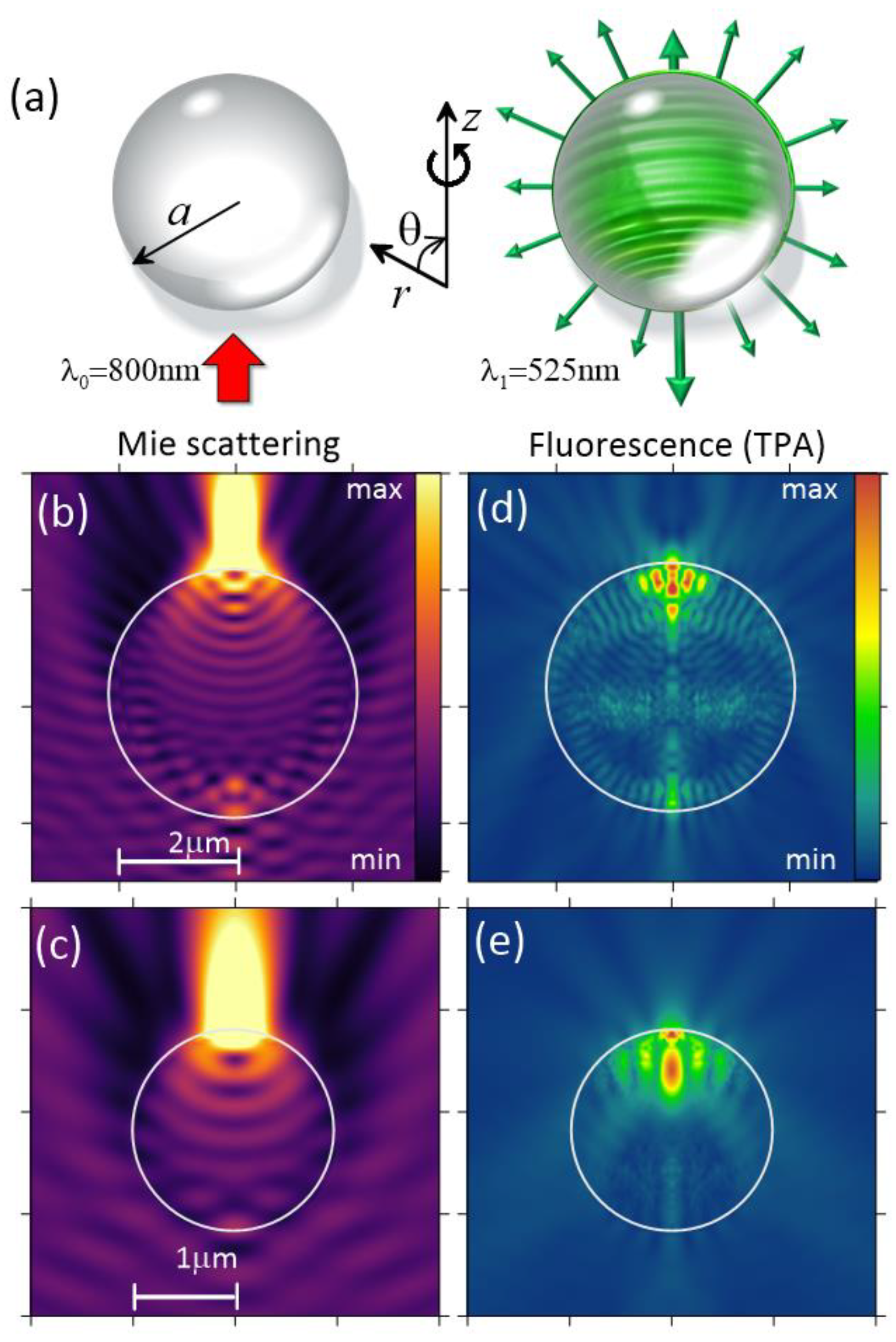
Figure 1.
(a) Schematics of the problem statement on laser-stimulated nonlinear emission from a droplet. (b-e) Relative intensity distribution of (b, c) pumping radiation at λ0 and (d, e) TPA fluorescence at λ1 near water droplets with a = 2 μm (b, d) and 1 μm (c, e).
Figure 1.
(a) Schematics of the problem statement on laser-stimulated nonlinear emission from a droplet. (b-e) Relative intensity distribution of (b, c) pumping radiation at λ0 and (d, e) TPA fluorescence at λ1 near water droplets with a = 2 μm (b, d) and 1 μm (c, e).
Thus, within the framework of the approximations made it
is considered that each molecule of particulate substance located at the point r
inside a particle represents a dipole that first absorbs a certain portion of optical
energy from the incident electromagnetic field E i (r )
at the wavelength λ0 and
then spontaneously (stochastically) emits an optical quantum E 1(r )
at a shifted wavelength λ1.
Without considering the nonradiative relaxation mechanisms, the rate γ d of spontaneous dipole emission
(probability of dipole transition) is proportional to the scalar product of the
dipole moment p d of the corresponding energy transition
and local field amplitude  , where m is
the number of simultaneously absorbed photons [22,23].
, where m is
the number of simultaneously absorbed photons [22,23].
, where m is
the number of simultaneously absorbed photons [22,23].
The governing equation describing the spontaneous
emission from a spherical particle is the following inhomogeneous Helmholtz (wave)
equation which is solved within the stationary problem formulation:
Here, k 0
is the wave number in free space, ε1
= n 2, and the polarization source P 1 depends
randomly on the radiating dipole spatial position :
where we use the notation
for the optical intensity, , and denotes a randomly-oriented unit vector of the
dipole (). It follows from Equation (2), that the energy
exchange rate between the optical field and radiating dipole in a unite volume,
, in the case of, e.g., two-photon absorption (m = 2) depends on the squared intensity of the pump field as: .
Worthwhile, when simulating plasma emission from a droplet, the volume density of secondary emission sources in the right-hand side of Equation (1) should be proportional to free electron concentration ρe (laser plasma is considered in equilibrium) generated through the photoionization of particle medium induced by the incident (“primary”) radiation. The plasma polarization source reads as:
where the random character of dipole emission is assumed also.
In turn, the density of free electrons in the medium can be determined from the kinetic plasma equation accounting for the field (multiphoton) and impact (avalanche) medium ionization, as well as the decreasing in the concentration of electrons due to the recombination with ions, as follows [2,24]:
whereWI is the photoionization rate (probability), ρnt is the density of neutrals (molecules, atoms), νc – avalanche ionization rate, νr is the electron recombination rate. In Equation (4), the diffusion of free electrons from the region occupied by plasma is not accounted during the characteristic time of radiative recombination of femtosecond plasma [12]. Additionally, further consideration is carried out in approximation of a short optical pulse, when its duration is much less than characteristic decay time of free electron plasma which allows neglecting the term responsible for plasma relaxation in Equation (4).
Then, under the apparent condition, ρe << ρnt (ρnt ≈ 1022 cm-3 is the critical density of free electrons when plasma begins resonantly absorbing optical energy), one obtains the following solution to Equation (4) for a rectangular pulse with the duration tp of stimulating laser radiation [24]:
Here, , while m-photon water molecule photoionization with a certain rate is assumed: .
By substituting (5) into (3), one obtains an expression for plasma polarization source in a droplet, which is similar to Equation (2). In contrast to TPA fluorescence, the degree of multiphoton ionization of water molecule at wavelength 800 nm is considerably higher and equals to m = 5 because bound electrons in a molecule need to overcome a high energy potential barrier of 6.5 eV to enter the conduction band and become free. In other words, not two but five photons of incident optical radiation are involved in a single excitation event of one plasma electron.
The exponential multiplier in (5) takes into account the “warming up” of free electrons by the optical field in a series of elastic collisions with heavy multicharged particles by the inverse Bremsstrahlung mechanism. This energy excess is used for increasing the kinetic energy of free electron and is converted into electron chaotic drift, which contributes to the development of the electron avalanche. The avalanche rate is proportional to the optical energy density (Ii⋅tp) at a selected point in the particle. For typical breakdown intensities of a femtosecond pulse, Ii ~ 5⋅1013 W/cm2, with the duration tp = 100 fs, by accounting the data on νc from [12] one obtains the following estimate for the exponent index: (νc⋅Ii⋅tp) ≈ 10 >> 1. This indicates that in the area of optical breakdown, the dynamics of free electron concentration is not a power dependence, but rather an avalanche-like (exponential) growth with a linear increase in the pulse intensity.
Theoretical simulation of the nonlinear droplet emission is based on the numerical solution of the equation (1) by means of the finite element method implemented in the COMSOL Multiphysics software package. Since the emission of photons by a molecule/ion occurs spontaneously, in the numerical model the emitting dipoles at each point of particle generally are not correlated either in phase or in the direction. Hereafter, in the steady-state approximation we consider only the random direction of the dipole moments by specifying a random polar angle of the dipole within the range in the (r-z) plane. Then, at each point r inside the particle the unit dipole moment vector can be represented as a function of a single random parameter: , where are unit vectors along the corresponding coordinate axes. In the simulation, the statistics on the droplet emission is gathered by setting different (usually a hundred variants) spatial distributions of dipole moments inside a particle having random dipole moment directions . For each of these variants the solution of the problem (1)-(5) is carried out separately. All the calculation series are eventually averaged over the spatial coordinates and the resulting angular distribution of the nonlinear droplet emission is obtained in the far-field region (r >> a) by using Stratton-Chu integrals [25].
As known, this formalism is based on the assumption that the Green's function for the vector Helmholtz equation (1) in the far-field region is known and the medium in this region is homogeneous. Then, the vectorial electric field of the emission Efar in the far-field region at any point r = (r,θ) located on the surface of certain speculative sphere S with radius r and normal n which encompasses the particle is expressed through the integral of the near-field as follows:
Here, is the medium impedance, and j stays for . Using Equation (6) one can calculate the angular distribution of the emission intensity of a water droplet by calculating the electromagnetic fields E1 and H1 directly near the particle surface.
3. Results
3.1. Polarization sources of droplet emission
This section is devoted to the study of the spatial configuration of nonlinear polarization sources formed in a liquid microparticle by incident optical radiation and causing droplet emission in the anti-Stokes spectral wing. As an example, Figure 1b–e show 2D distributions of the relative electric field intensity at the incident Ii and shifted (TPA) wavelengths calculated for two radii of a spherical particle. Here and hereafter, due to the model geometry used, the initial radiation is assumed to be circularly polarized relative to the incidence direction.
Analysis of the fundamental wave intensity Ii profiles shows that inside an optically small particle with a radius a ~ λ only a single HA is formed with absolute maximum intensity located near the surface of the shadow hemisphere (Figure 1c). This HA serves as a source for TPA fluorescence providing the fluorescence distribution is also concentrated predominantly in the shadow part of the droplet (Figure 1e), although exhibiting more complex spatial structure due to the interference of fields at multiple reflections from the inner particle rim.
In a larger droplet (Figure 1b), intensity distribution at the incident wavelength is characterized already by two HAs located symmetrically in both hemispheres. At the same time, the intensity maximum in the shadow part of the drop still has larger amplitude, which leads to the predominant concentration of the fluorescence also in the shadow droplet part. Additionally, besides two main HAs located along the main sphere diameter, Figure 1d also shows a ring structure of the fluorescence extending along the entire particle surface, which is evidence of quasi-resonant excitation of an eigenmode (or several modes) in spherical microresonator at shifted emission wavelength [26]. As shown below, exactly such situations cause sharp enhancement of transverse directed light emission of particles with certain resonant sizes.
The difference in the physical mechanisms causing the nonlinear polarizability of the droplet substance causes the different nature of the luminescence regions distribution. As seen in Figure 2a,b, inside a large water droplet (5 μm) the areas of maximal fluorescence excited by two-photon absorption (m = 2) are distributed along the sphere diameter with the front (illuminated) hemisphere fluorescing also quite strongly. Meanwhile, in the shadow hemisphere one can see the luminous area of the “Descartes ring", which formation is typical during plane optical wave focusing by a transparent sphere [27].
Increasing the order m of multiphoton absorption, in case of particle matter ionization and plasma emission shown in Figure 2(b) leads to a drastic change in the polarization source configuration. Here, there only one highly localized HA arises located in the rear (shadow) hemisphere at a distance r ~ 0.8a from the particle center. In accordance with the reciprocity principle between the electromagnetic fields of the source and receiver [10], such a position of the nonlinear polarization maximum should lead to a sharply pronounced backward directed plasma emission of the spherical particle.
Figure 3a–d summarize the results on the HA parameters inside water droplets with different radii formed by optical radiation at the fundamental wavelength (800 nm). HA parameters are obtained separately for the illuminated and shadow parts of a spherical particle. The figures show the maximum intensity Im, the rms-radius , the z-coordinate of the maximum intensity zm for corresponding hot area, and the effective absorbed in HA power via the two-photon excitation of the molecules.
From Figure 3a,c is clear, that the relative size of the regions with dominating light absorption generally decreases monotonically with increasing particle size. Meanwhile, HA position along the droplet diameter behaves oscillatory, but always staying in the range of 0.75 ≤ ≤ 1. The peak intensity Im of the optical field inside the HA (Figure 3b) demonstrates on average an increase with droplet radius increasing, that can be explained by the increasing of collecting ability of a spherical optical lens (spherical droplet) with increasing its linear aperture (radius). At the same time, the quasi-monotone dependence Im(a) for certain particle radii are superposed with sharp intensity bursts. Analysis of the internal optical field structure shows that these bursts correspond to resonant excitation of particle eigenmodes which field distribution follows that of a WGM. Some of these resonances are indicated in Figure 3(b) in “(TE/TM)l,p” notation, showing the state of wave polarization, as well as azimuthal (l) and radial (p) mode orders.
Generally, the dependence of the effective power for two-photon absorption PTPA (Figure 3d) on the droplet radius follows the proportionality PTPA ∝ a3 with superimposed peaks coming from the internal field resonances. Worth noting, in practice when measuring the integral emission signal received from a polydisperse (in size) water aerosol, the relative contribution of the selected aerosol fraction to the received optical signal will be proportional not to the number of particles with the given sizes but to their volume fraction (water content).
3.2. Angular structure of droplet emission (phase function)
Now consider the simulation results of far-field laser-induced emission from water droplets. As mentioned above, the angular distribution of the particle emission intensity is calculated by integral expressions (6) obtained within the assumption that in the Fraunhofer diffraction zone, i.e. far enough from a particle, only the scattered field exists, while the field of the incident wave is completely vanished. With this assumption one can transit from cylindrical to polar coordinate system and consider the change of field vectors only in the polar angle θ. The results of our calculations of the phase function I(θ) for the elastic scattering and nonlinear emission of different size uranine-dyed water droplets are shown in Figure 4a–d. Recall that all of the calculated data on the angular droplet emission presented below are statistically averaged over approximately one hundred variants of the random configuration of the emitting dipoles at each point of the particle. The standard deviation of the emission intensity for each angular direction depends on the nature of the simulated process and is about 30% for the TPA fluorescence and 12% for the plasma emission.
The fundamental differences between the angular distribution of the linear scattering and the nonlinear two-photon excited fluorescence are clearly illustrated in Figure 4a,b. One can see, that as the particle radius increases the Mie scattering angular diagram evidently straightens up in the forward direction (θ→ 0°), while TPA angular distribution on the contrary sharpens in the opposite direction, i.e., it has a maximum in the direction of the incident radiation (θ→ 180°). At the same time, in the transverse direction (θ→ 90°) both types of optical scattering show a decrease in intensity.
From a comparison of the angular structure of the fluorescence and plasma emission from micron-sized droplets shown in Figure 4c,d, it follows that the plasma radiation demonstrates a sharper backward directionality. Obviously, this is caused by the formation of only single HA inside a spherical particle, which is located in particle shadow part and acts as a source of medium nonlinear polarization for the emission field. Worthwhile noting, the TPA fluorescence of a droplet almost always is characterized by the presence of two polarization sources in both particle hemispheres.
Now we discuss an important quantitative characteristic of the angular diagram of the nonlinear particle emission, namely, the backward angular directivity D of emission into the rear (illuminated) hemisphere bounded by the polar angles π/2 ≤ θ ≤ π, which is determined in the standard way:
where θm is the angle at which the emission signal is maximal in amplitude, and the integration in the denominator (7) does not include the angle θm. The parameters θmand D as a function of particle size are presented in Figure 5(a) and (b), respectively. Here for comparison, the simulation results for three different physical processes of nonlinear droplet emission are shown, differing in the number of simultaneously absorbed photons.
As seen in Figure 5(a), except for small droplet sizes a ≤ 1 μm where the emission has a dipole character (θm ≈ 90°), the emission maximum for all types of nonlinear scattering in general is observed at angles close to the backward direction, θm > 150°. The change in the multiphoton order of dye fluorescence is affected the angle θm only for small- and medium-size particles and this dependence is not monotonic when one transits from one- to two-photons and then to a sharper plasma absorption. Meanwhile, the angular emission directivity D from the droplets (Figure 5b) normally increases with the increase of their size and is practically independent on m. One can conclude that on average, the emission of medium-size and large water droplets has a pronounced angular orientation in the reversed direction, i.e., towards the source of primary excitation radiation.
Interestingly, the angular distribution of plasma emission from droplets exhibits a sharply nonmonotonic dependence versus particle size, that is not observed for TPA fluorescence. As can be seen in Figure 5(a), the angle of maximum emission intensity demonstrates three local minima when changing the drop radius from 1 μm to 6 μm. At these angles of minimal emission, a noticeable decrease of emission angular directivity is realized also. As our detailed analysis shows, this is caused by the specificity of the spatial position of the sources of nonlinear polarization in the particle.
Indeed, recalling Figure 3(c) one can see that as the drop radius increases there is a general tendency for the HA to move to the particle center. We carried out additional simulation of droplet emission when artificially setting the polarization (emitting) source in the form of a pointwise dipole, which physically corresponds to a single plasma region forming in the shadow part of a micron-sized drop as that in Figure 2(b). The angular dependence of droplet emission obtained in this case is shown in Figure 6. Evidently, as the dipole coordinate zm decreases the angular distribution of the particle emission becomes more homogeneous while the angle of emission maximum θm apparently decreases. This means that when a droplet size increases the plasma emission originated by the optical breakdown inside particle volume becomes more homogeneous.
At the same time as noted above, for certain sizes of spherical particles there are possible situations when the internal optical field is drastically enhanced by the resonance WGM excitation. In this case, instead of a single point polarization source one should consider two separated HAs located diametrically opposed near the surface of the spherical droplet. In Figure 6(a) this situation corresponds to the case with zm/a = 0.9. Under WGM resonance the angular distribution of droplet emission becomes appreciably elongated and quasi-symmetric in the forward/backward directions, and the value of maximal angle θm approaches 180°.
As seen in Figure 3b,c, the cases of excitation of the internal field resonances in particle are observed near radii a = 2.5, 4.5, and 6.5 μm. As a result, the oscillatory character of the dependence θm(a) for the plasma emission is caused by two opposing physical trends: (a) shifting the nonlinear polarization source closer to the particle center which leads to θm decrease, and (b) increasing the probability of WGM excitation inside a spherical particle which causes an increase in the emission maximum angle. For more “smooth” processes of single- and two-photon excited fluorescence such oscillatory behavior of the maximal emission angle θm with droplet radius is not observed because of the absence of such sharp disbalance of polarization sources in shadow and illuminated hemispheres during nonresonant excitation of the internal optical field.
As the key result of our studies, the normalized emission power Pe of aqueous dye droplets of different radius is plotted in Figure 7. The values of Pe are presented in arbitrary units for each type of nonlinear optical emission considered in the direction of maximal intensity θm. For each type of the nonlinearity, the normalization of Pe values is done in such a way that the different curves in the graph are clearly distinguishable. Importantly, a quantitative comparison of the results is possible only within each individual dependence.
4. Discussion
Generally, the analysis of Figure 7 shows that the droplet emission via multiphoton absorption processes becomes more intense with increasing size of particles. Moreover, for the case of TPA fluorescence, this tendency can be approximated as: Pe ∝ a4.14, which is steeper than the above established increment for TPA polarization source power (PTPA ∝ a3 in Figure 3d) and indicates better emission confinement within the angle θm. A similar tendency for classical single-photon fluorescence yields a lower power degree, Pe ∝ a2.47. And in the case of plasma droplet fluorescence, this dependence becomes considerably sharper with high influence of WGM resonance excitations and cannot be fitted by a power law.
5. Conclusions
In conclusion, the angular distribution of dye water droplets emission is theoretically examined when the droplets are exposed to high-power infrared laser radiation stimulating nonlinear optical processes of multiphoton absorption. Specifically, we consider dye fluorescence at two-photon absorption and emission of recombining optical breakdown plasma created inside the droplet by incident laser radiation. Using numerical simulation providing the random distribution of nonlinear polarization sources in the droplet volume, we calculate the angular diagram of emission in the far-field and obtain the dependence of emission angular directivity on the particle size and type of optical nonlinearity. It turns out that the droplet emission at the multiphoton absorption generally becomes more intensive with increasing droplet size. Meanwhile, backward directed droplet emission (toward the laser incidence) exhibits better angular directivity with increasing particle radius providing the maximum emission angle approaches the value 180° (backward direction). Compared to the two-photon excited fluorescence (TPA), plasma emission of droplets demonstrates stronger angular variability with particle radius, which is due to the specific spatial position of the emitting plasma regions inside spherical particle and the significant influence of eigenmode resonances.
We expect the results of this work can be useful particularly in elaborating a theoretical model for the nonlinear emission of liquid aerosol particles stimulated by high-intensive ultrashort laser radiation which can improve the femtosecond LiDAR technique for remote diagnostics of atmospheric aerosols.
Author Contributions
All authors have read and agreed to the published version of the manuscript.
Funding
This research was funded by Russian Science Foundation (RSF), grant number 21-12-00109.
Data Availability Statement
Data available on request due to restrictions e.g. privacy or ethical.
Conflicts of Interest
The author declares no conflict of interest.
References
- Kasparian, J.; Rodriguez, M.; Mejean, G.; Yu, J.; Salmon, E.; Wille, H.; Bourayou, R.; Frey, S.; Andre, Y.-B.; Mysyrowicz, A.; Sauerbrey, R.; Wolf, J.-P.; Woeste, L. White-light filaments for atmospheric analysis. Science 2003, 301, 61–64. [Google Scholar] [CrossRef] [PubMed]
- Vogel, A.; Noack, J.; Nahen, K.; Theisen, D.; Busch, S.; Parlitz, U.; Hammer, D.X.; Noojin, G.D.; Rockwell, B.A.; Birngruber, R. Energy balance of optical breakdown in water at nanosecond to femtosecond time scales. Appl. Phys. B 1999, 68, 271–280. [Google Scholar] [CrossRef]
- Boutou, V.; Favre, C.; Hill, S.C.; Pan, Y.-L.; Chang, R.K.; Wolf, J.-P. Backward enhanced emission from multiphoton processes in aerosols. Appl. Phys. B 2002, 75, 145–152. [Google Scholar] [CrossRef]
- H. L. Xu, W. Liu, and S. L. Chin, “Remote time-resolved filament-induced breakdown spectroscopy of biological materials,” Opt. Lett. 31(10), 1540–1542 (2006). [CrossRef]
- T. Fujii, N. Goto, M. Miki, T. Nayuki, and K. Nemoto, “Lidar measurement of constituents of microparticles in air by laser-induced breakdown spectroscopy using femtosecond terawatt laser pulses,” Opt. Lett. 31(23), 3456–3458 (2006). [CrossRef]
- J. F. Daigle, G. Méjean, W. Liu, F. Théberge, H. L. Xu, Y. Kamali, J. Bernhardt, A. Azarm, Q. Sun, P. Mathieu, G. Roy, J. R. Simard, and S. L. Chin, “Long range trace detection in aqueous aerosol using remote filament-induced breakdown spectroscopy,” Appl. Phys. B 87(4), 749–754 (2007). [CrossRef]
- S.S. Golik, A.Y. Mayor, V.V. Lisitsa, Y.S. Tolstonogova, A.A. Ilyin, A.V. Borovskiy, and O.A. Bukin, “Limits of Detection of Chemical Elements in an Aqueous Aerosol in Filament-Induced Breakdown Spectroscopy,” J. Appl. Spectrosc. 88(2), 337–342 (2021). [CrossRef]
- P.A. Babushkin, G.G. Matvienko, and V.K. Oshlakov, “Determination of the elemental composition of aerosol by femtosecond laser-induced breakdown spectroscopy,” Atmospheric and Oceanic Optics 35(1), 19–26 (2022). [CrossRef]
- Z. Zhang, N. Zhang, Y. Wang, B. Xie, Y. Xiang, J. Guo, B. Shang, L. Guo, X. Zhao, M. Xie, L. Lin, W. Liu, “Detection of 1.4 μg/m3 Na+ in aerosol at a 30 m distance using 1 kHz femtosecond laser filamentation in air,” Opt. Express 31, 6464 (2023). [CrossRef]
- S.C. Hill, V. Boutou, J. Yu, S. Ramstein, J.-P. Wolf, Y.-L. Pan, S. Holler, R.K. Chang “Enhanced-backward directed multi-photon-excited fluorescence from dielectric microcavities,” Phys. Rev. Lett. 85, 54-57 (2000). [CrossRef]
- C.H. Fan, J. Sun, J.P. Longtin, “Breakdown threshold and localized electron density in water induced by ultrashort laser pulses,” J. Appl. Phys. 91, 2530-2536 (2002). [CrossRef]
- A.A Zemlyanov, Yu.E. Geints, “Optical breakdown thresholds for transparent microparticles irradiated with laser pulse of the nano-, pico-, and femtosecond duration,” Atmospheric and oceanic optics 17, 268-273 (2004): https://ao.iao.ru/en/content/vol.17-2004/iss.04/4.
- P. Chýlek, M.A. Jarzembski, V. Srivastava, R.G. Pinnick, J.D. Pendleton, J.P. Cruncleton, “Effect of spherical particles on laser-induced breakdown of gases,” Appl. Opt. 26, 760-762 (1987). [CrossRef]
- A, Talebpour, J. Yang, S.L. Chin, “Semi-empirical model for the rate of tunnel ionization of N2 and O2 molecule in an intense Ti:sapphire laser pulse,” Opt. Commun. 163, 29-32 (1999). [CrossRef]
- Yu. E. Geints, A. A. Zemlyanov, and E. K. Panina, “Modeling of Multiphoton Excited Fluorescence from a Spherical Droplet Irradiated by an Ultrashort Laser Radiation Using the Method of Computation Electrodynamics,” Atmospheric and oceanic optics 24, 294 (2011). [CrossRef]
- J. Xue, X. Zeng, L. Guo, R. Guo, Z. Zhang, C. Chu, Z. Cheng, N. Zhang, L. Lin, W. Liu, “High directional aerosol fluorescence distribution affected by Mie scattering during femtosecond laser filamentation in air,” Optics & Laser Technology 161, 109175 (2023). [CrossRef]
- Y.-L. Pan, S.C. Hill, J.-P. Wolf, S. Holler, R.K. Chang, and J.R. Bottiger, “Backward-enhanced fluorescence from clusters of microspheres and particles of tryptophan,” Appl. Opt. 41, 2994-2999 (2002). [CrossRef]
- Y.E. Geints, A.A. Ionin, D.V. Mokrousova, G.E. Rizaev, L.V. Selesnev, E.S. Sunchugasheva, A.A. Zemlyanov, “Energy, spectral and angular properties of post-filamentation channels during propagation in air and condensed media,” JOSA B 36, G19-G24 (2019). [CrossRef]
- S.C. Hill, H.I. Saleheen, M.D. Barnes, W.B. Whitten, and J. M. Ramsey, “Modeling fluorescence collection from single molecules in microspheres: effects of position, orientation, and frequency,” Appl. Opt. 35, 6278 (1996). [CrossRef]
- A.B. Matsko and V.S. Ilchenko, “Optical Resonators With Whispering-Gallery Modes—Part I: Basics,” IEEE J. Sel. Topics Quantum Electron. 12 (1), 3-14 (2006). [CrossRef]
- H. Chew, “Transition rates of atoms near spherical surfaces,” J. Chern. Phys. 87 1355–1360 (1987). [CrossRef]
- D.M. Friedrich, “Two-photon molecular spectroscopy,” J. Chem. Educ. 59, 472 (1982). [CrossRef]
- S.D. Druger, P.J. McNulty, “Radiation pattern of fluorescence from molecules embedded in small particles: general case,” Appl. Opt. 22, 75 (1983). [CrossRef]
- Yu. E. Geints and A. A. Zemlyanov, “Phase explosion of a water drop by a femtosecond laser pulse: I. Dynamics of optical breakdown,” Atmospheric and Oceanic Optics 22(6), 581–589 (2009). [CrossRef]
- Zhdanov, M.S. Stratton-Chu Type Integrals. In Integral Transforms in Geophysics. Springer: Berlin, Heidelberg, Germany, 1988. [CrossRef]
- A. Biswas, H. Latifi, R. L. Armstrong, and R. G. Pinnick, “Double-resonance stimulated Raman scattering from optically levitated glycerol droplets,” Phys. Rev. A 40, 7413(R). [CrossRef]
- A.A. Zemlyanov, Yu.E. Geints, R.L. Armstrong, “Stimulated light scattering in transparent liquid particles: Effect of the Descartes ring,” Appl. Opt. 39(36), 6888 (2001). [CrossRef]
Figure 2.
Relative intensity distribution of (a) TPA fluorescence (525 nm) and (b) plasma emission (590 nm) inside a water droplet with a = 5 μm.
Figure 2.
Relative intensity distribution of (a) TPA fluorescence (525 nm) and (b) plasma emission (590 nm) inside a water droplet with a = 5 μm.
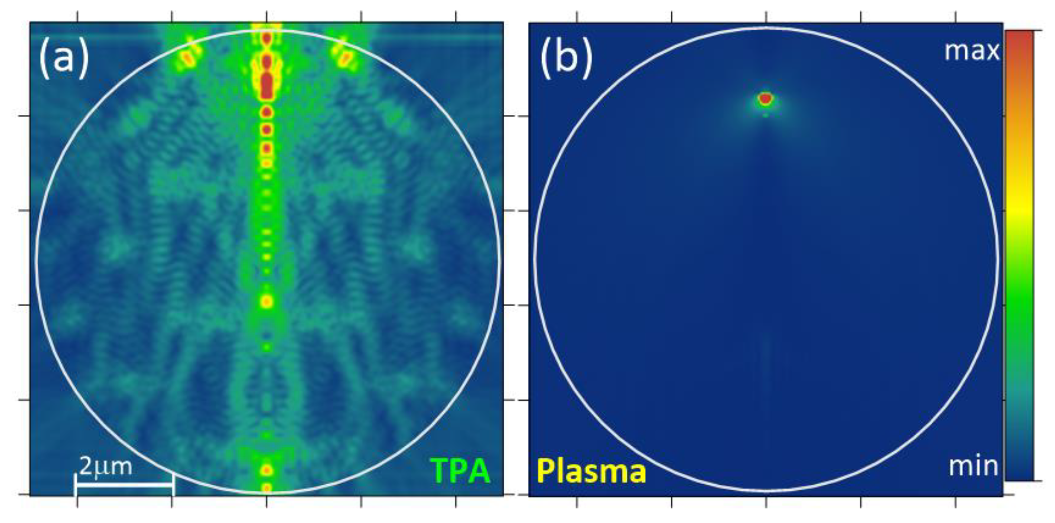
Figure 3.
HA parameters (λ= 800 nm) in water droplet of different radii. (a) rms-radius R, (b) maximal intensity Im, (c) HA position zm, (d) effective power of TPA source PTPA. The parameter values in upper and lower hemispheres are shown by circles and squares, respectively.
Figure 3.
HA parameters (λ= 800 nm) in water droplet of different radii. (a) rms-radius R, (b) maximal intensity Im, (c) HA position zm, (d) effective power of TPA source PTPA. The parameter values in upper and lower hemispheres are shown by circles and squares, respectively.
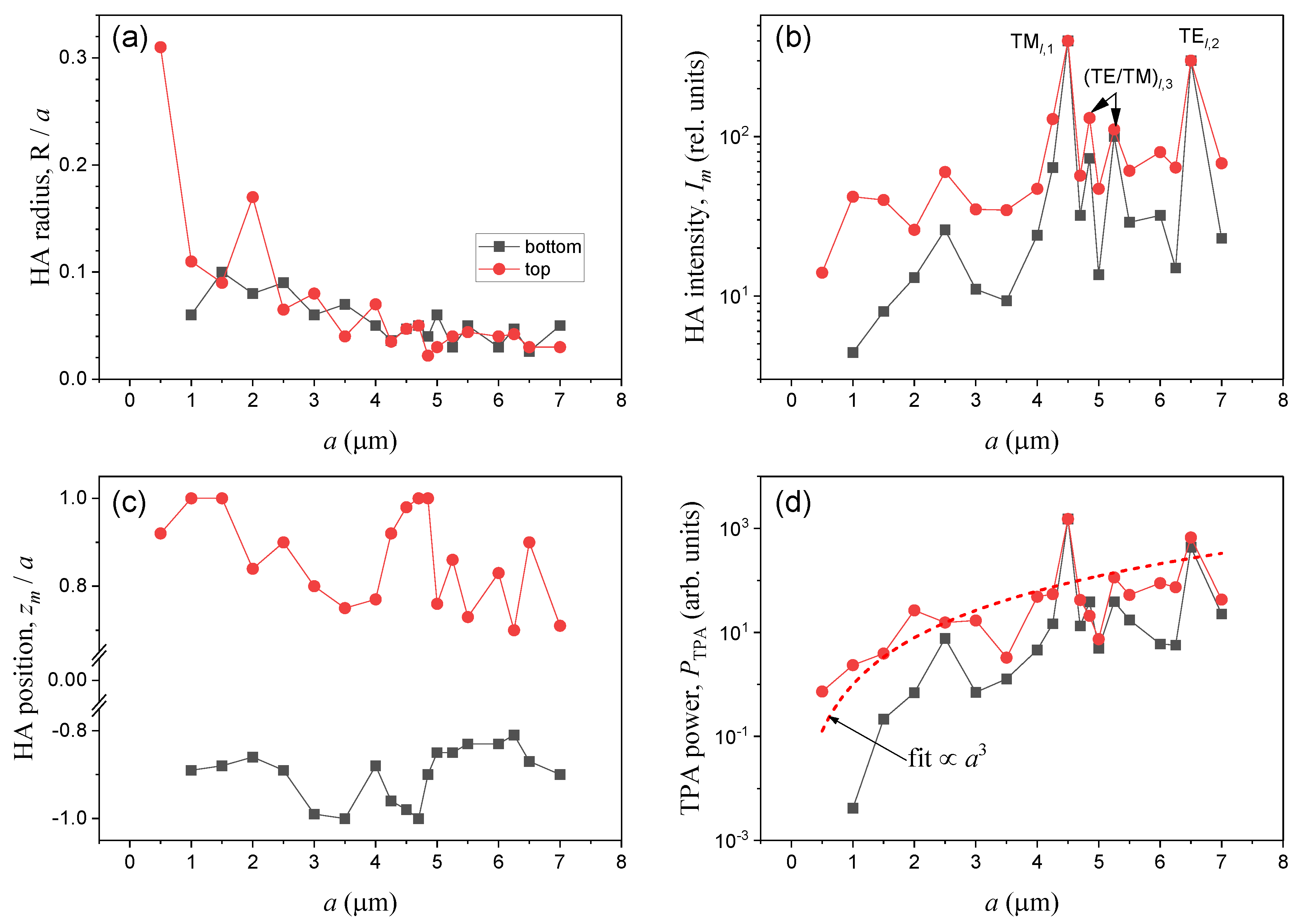
Figure 4.
(a-d) Angular distribution of the far-field scattering intensity from dye water droplets of different radii. 3D diagram of (a) elastic Mie scattering and (b) two-photon fluorescence. (c, d) Intensity distribution of (c) TPA fluorescence and (d) plasma emission on the scattering angle θ (logarithmic scale). Arrows show the direction of incident excitation radiation.
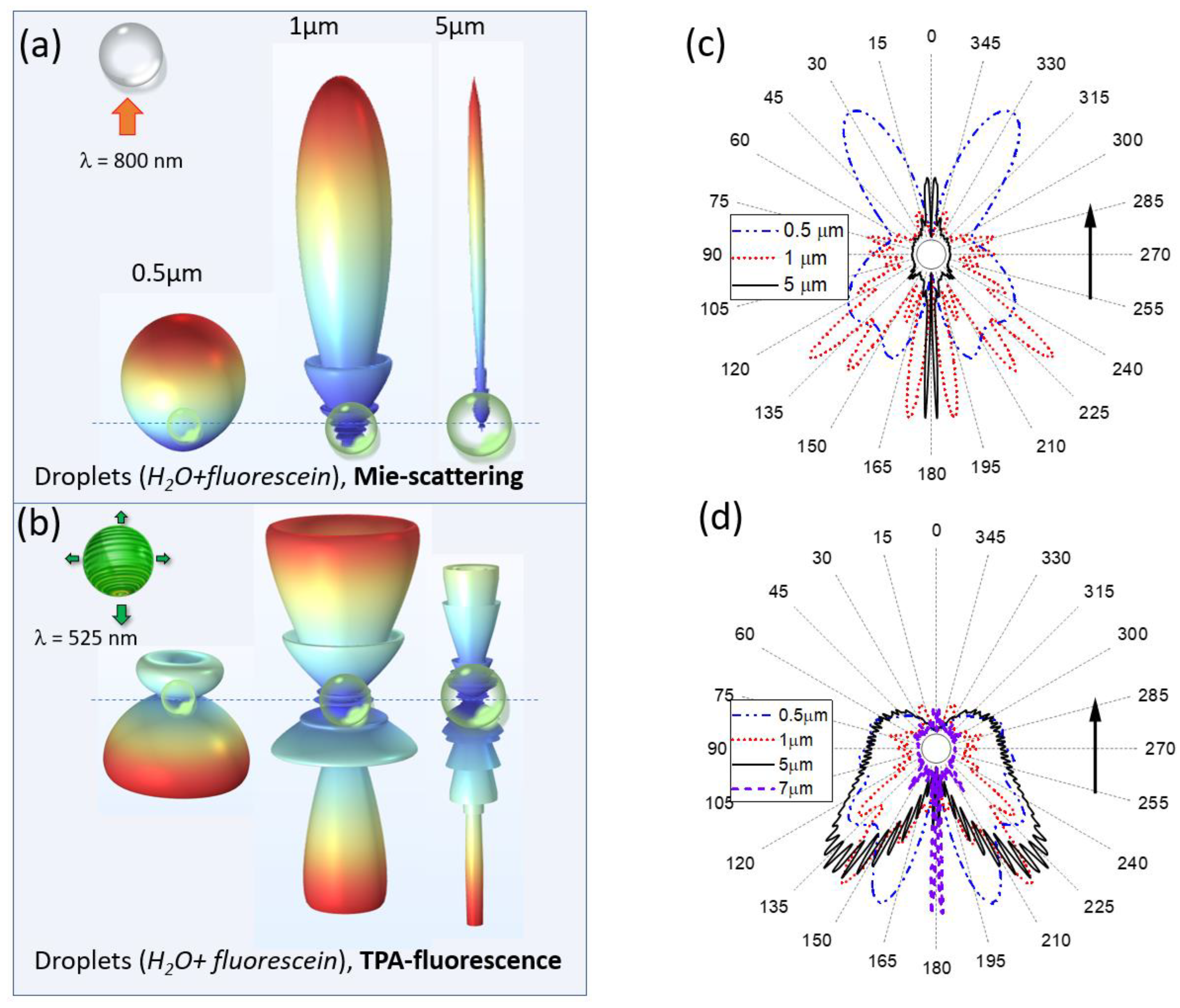
Figure 5.
(a) Maximum emission angle θm and (b) backward angular directivity D of water droplet emission with different radius resulting from the fluorescence of single- (1-PA), two-photon (TPA) absorption, as well as from the optical breakdown emission (plasma).
Figure 5.
(a) Maximum emission angle θm and (b) backward angular directivity D of water droplet emission with different radius resulting from the fluorescence of single- (1-PA), two-photon (TPA) absorption, as well as from the optical breakdown emission (plasma).
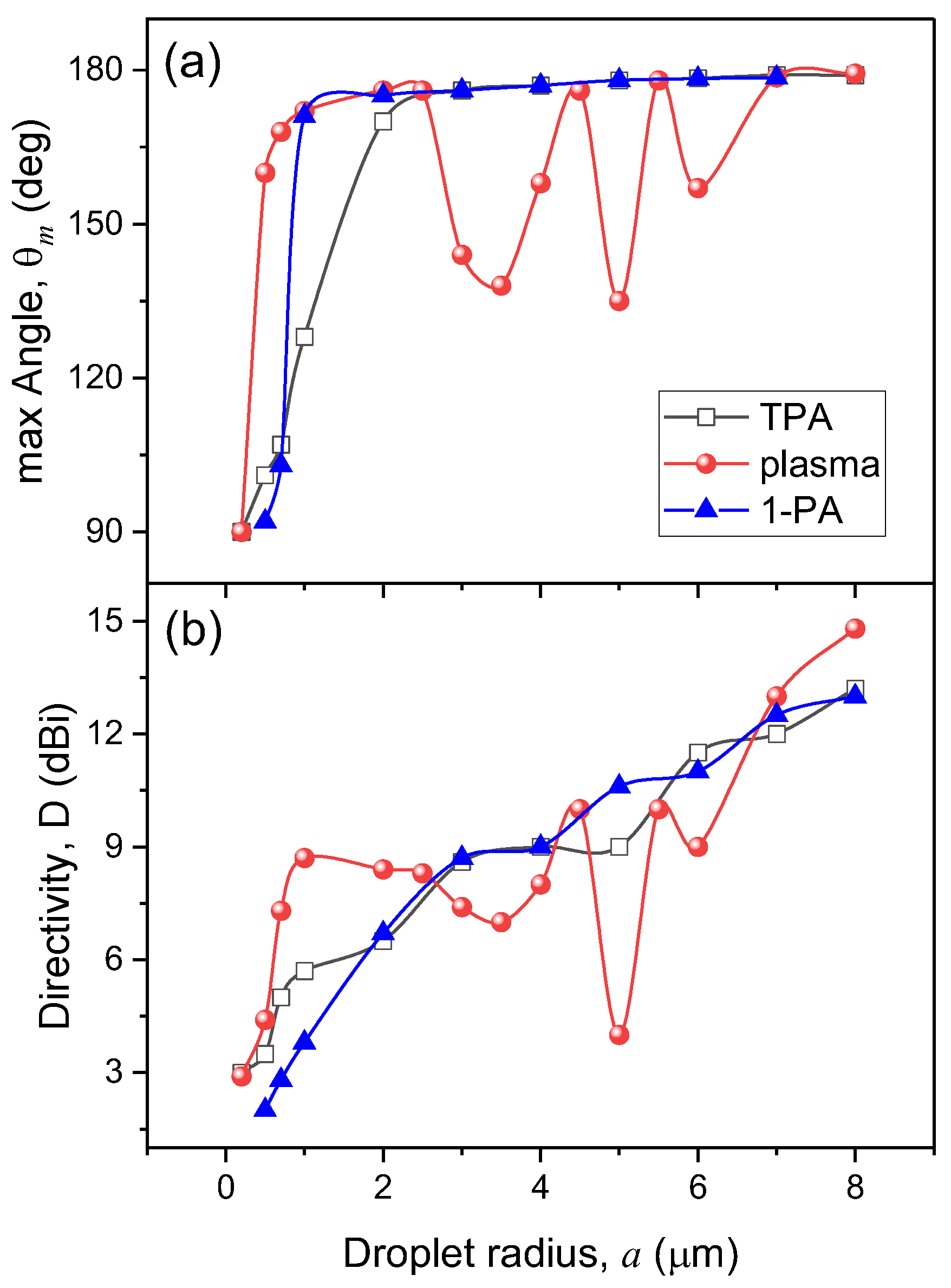
Figure 6.
Model calculations of the angular emission distribution of a 5 µm droplet at different positions of nonlinear polarization source represented by an electric dipole (left column).
Figure 6.
Model calculations of the angular emission distribution of a 5 µm droplet at different positions of nonlinear polarization source represented by an electric dipole (left column).
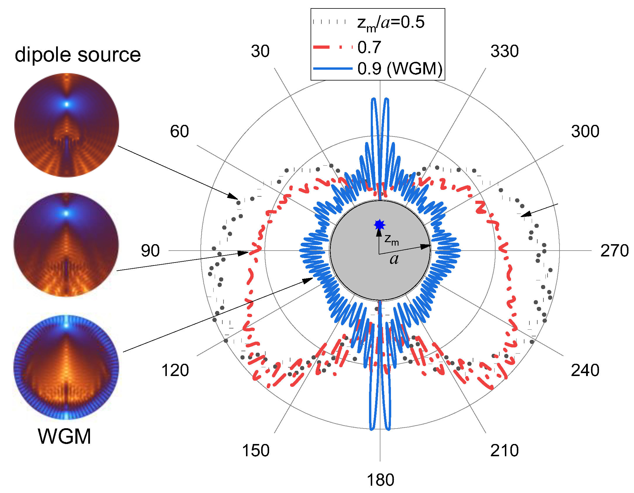
Figure 7.
Relative emission luminance (power within θm) of dye water droplets for different physical processes. Fitted dependence for TPA fluorescence is shown by dashed curve; the situations of WGM excitation in droplets are indicated for plasma emission.
Figure 7.
Relative emission luminance (power within θm) of dye water droplets for different physical processes. Fitted dependence for TPA fluorescence is shown by dashed curve; the situations of WGM excitation in droplets are indicated for plasma emission.

Disclaimer/Publisher’s Note: The statements, opinions and data contained in all publications are solely those of the individual author(s) and contributor(s) and not of MDPI and/or the editor(s). MDPI and/or the editor(s) disclaim responsibility for any injury to people or property resulting from any ideas, methods, instructions or products referred to in the content. |
© 2023 by the authors. Licensee MDPI, Basel, Switzerland. This article is an open access article distributed under the terms and conditions of the Creative Commons Attribution (CC BY) license (http://creativecommons.org/licenses/by/4.0/).
Copyright: This open access article is published under a Creative Commons CC BY 4.0 license, which permit the free download, distribution, and reuse, provided that the author and preprint are cited in any reuse.


