Introduction
Ingrown toenails (IGTN) are a commonly occurring foot problem. The condition occurs when the nail plate pierces the sulcus, giving rise to pain, inflammation and infection1. The skin’s attempt at healing leads to the formation of hypergranulation tissue (HGT) - vascularized oedematous stroma containing a mixed infiltrate of inflammatory cells2, which continues to be produced until the nail is removed3. Multiple staging systems for IGTNs exist, but most describe a variation of the original classification put forward by Heifetz4:
Stage I: erythema and swelling of the lateral nail fold,
Stage II: infection with accompanying oedema and drainage,
Stage III: chronic infection characterised by hypertrophy of granulation tissue.
Nail surgery techniques for permanent removal of part or all of a nail fall into two categories, the latter with the use of phenol being the most popular5:
Excision of the pathological nail/soft tissue by the use of sharp instrumentation (cold steel/incisional procedures),
Destruction of the pathological tissue by physical means such as topical chemotherapy (phenol), freezing, electro-galvanism, burring or lasering.
Reilly and Burt6 note that only a brief mention is made in the literature of performing sharp resection of the HGT at the time of nail surgery1,3,7–11. Within the UK Podiatry profession, concurrent sharp resection of the HGT is not common practice12. This is a case report of phenolic nail surgery with sharp resection of HGT in only one of the two affected sulci, with the aim of noting any differences between the two sulci in their post-procedure healing.
Case report
MT is a 22-year-old male who was referred for nail surgery by a local podiatrist. The original referral for nail surgery was made to the local School of Podiatry, which undertakes the majority of the county’s nail surgery as adult nail surgery has been decommissioned from the NHS podiatry contract. The referral was diverted to the author, who is able to see patients under the NHS consultant contract.
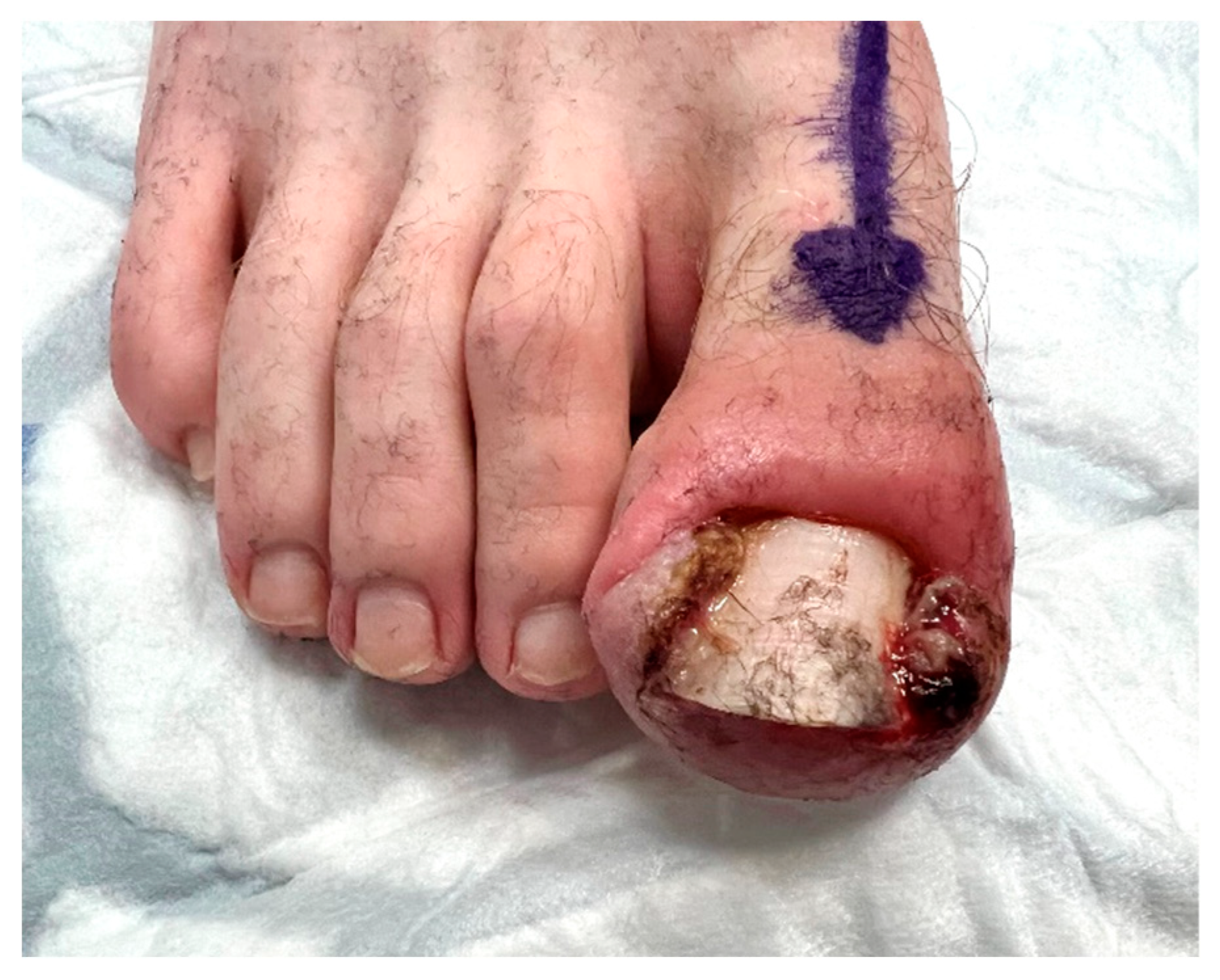
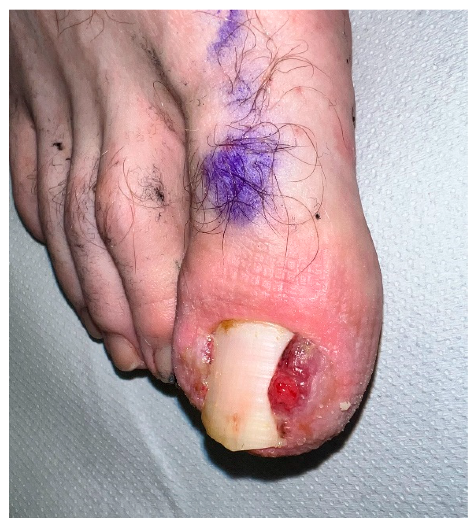
MT attended a primary assessment on 01.12.22 with a stage 3 bilateral ingrown toenail of the right hallux and severe HGT (see Figure 1). He had no relevant medical or pharmacological history other than penicillin allergy. The IGTN had been present for over 12 months and was resistant to self-care and non-invasive conservative care from the local NHS Podiatry team. Following his initial assessment, he was recommended for nail surgery and issued with the author’s standard nail surgery patient information leaflet (PIL) that outlined the process of nail surgery, healing rates and the complication profile.
01.12.22
I see that MT had been seen by JC, one of our Extended Scope Practitioners, who had referred MT on to the School of Podiatry for an ingrown toenail. He has come through to see me at Three Shires Hospital today. He has a fairly long history of an ingrown toenail in the right 1st hallux with a stage 3 (out of 3) presentation. He is beyond the point where I can do anything for him from a conservative point of view and he is indicated for nail surgery.
I have explained that nail surgery involves removing part of the nail, and is done as an out-patient procedure under local anaesthetic. This is performed using a tourniquet (to stop any bleeding). The nail matrix/root is then destroyed with phenol (acid) to prevent regrowth. The toe is bandaged after the operation and a sandal or roomy shoe should be worn to leave the hospital (as I apply a large bandage in case of bleeding). Over-the-counter analgesia is usually adequate for pain control.
He will see us after 2 days and again after 2/3 weeks for an operative site check. Full operative site healing normally takes 4-6 weeks. The possible complications associated with nail surgery are:
Regrowth of the nail in about 5% of cases
Infection (a 2% risk), possibly requiring antibiotics
A loose nail plate after the surgery
We will book MT in to have this done on 2nd February, and I will keep you updated.
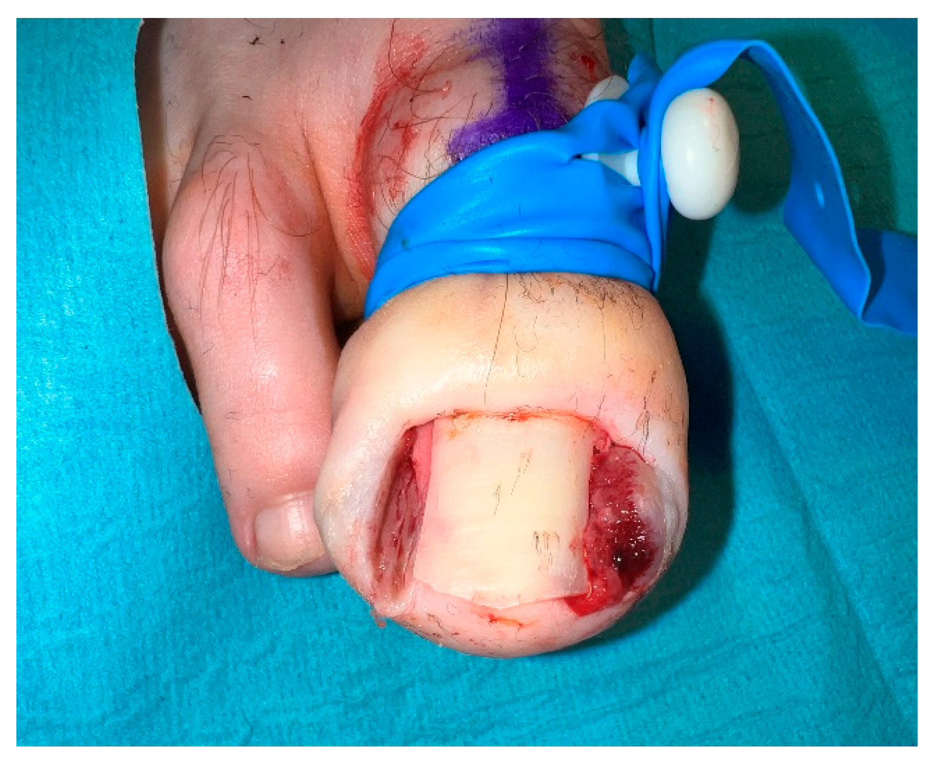
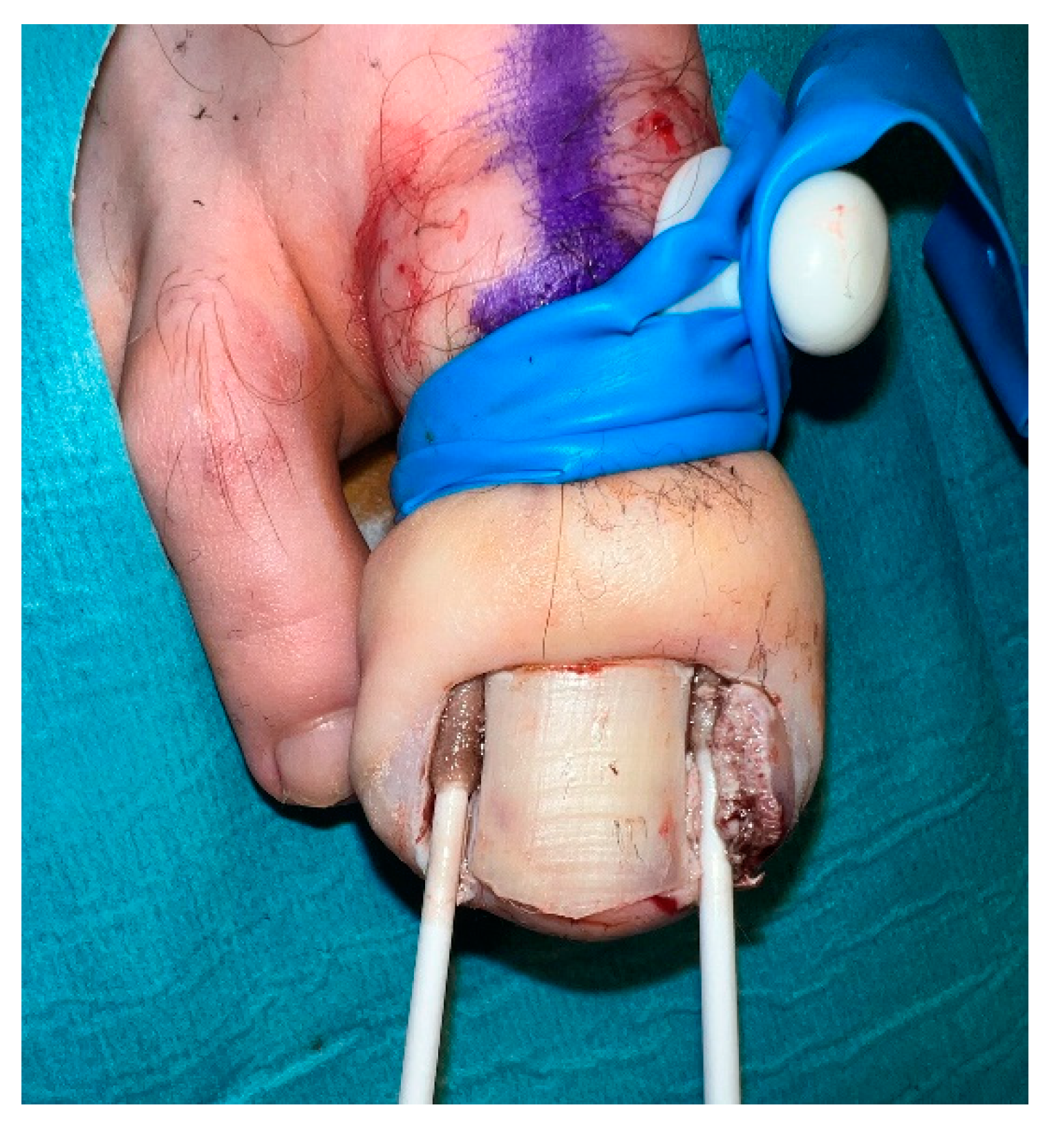
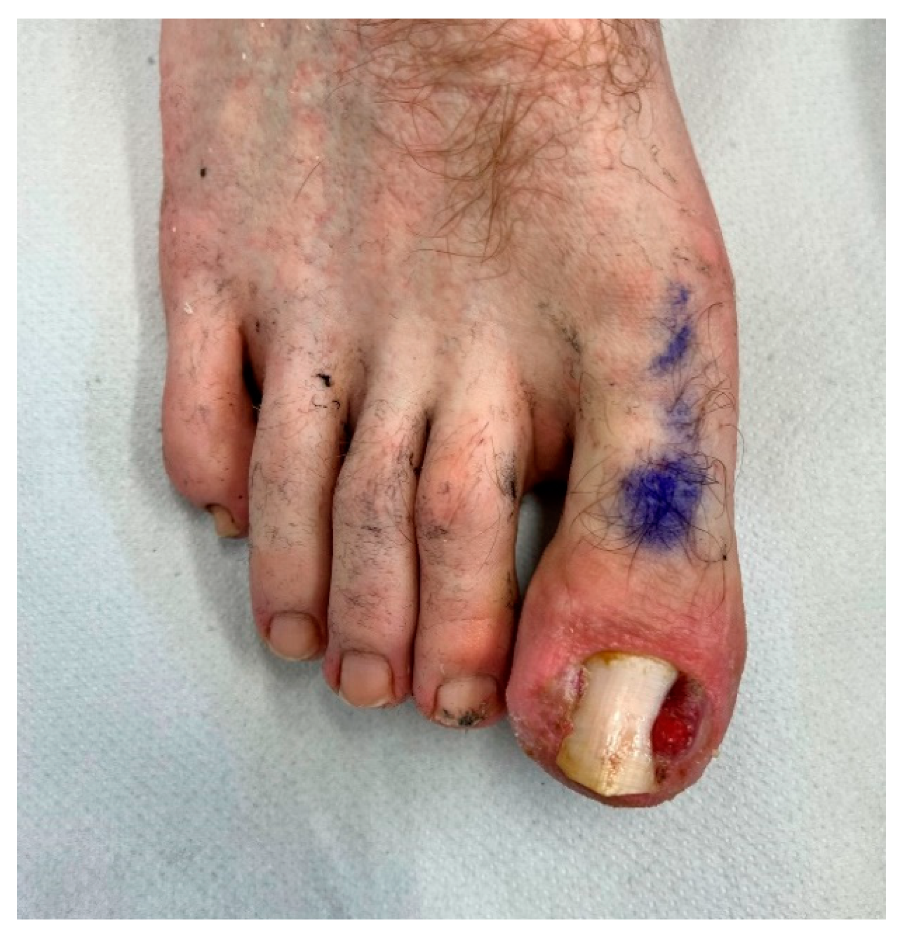
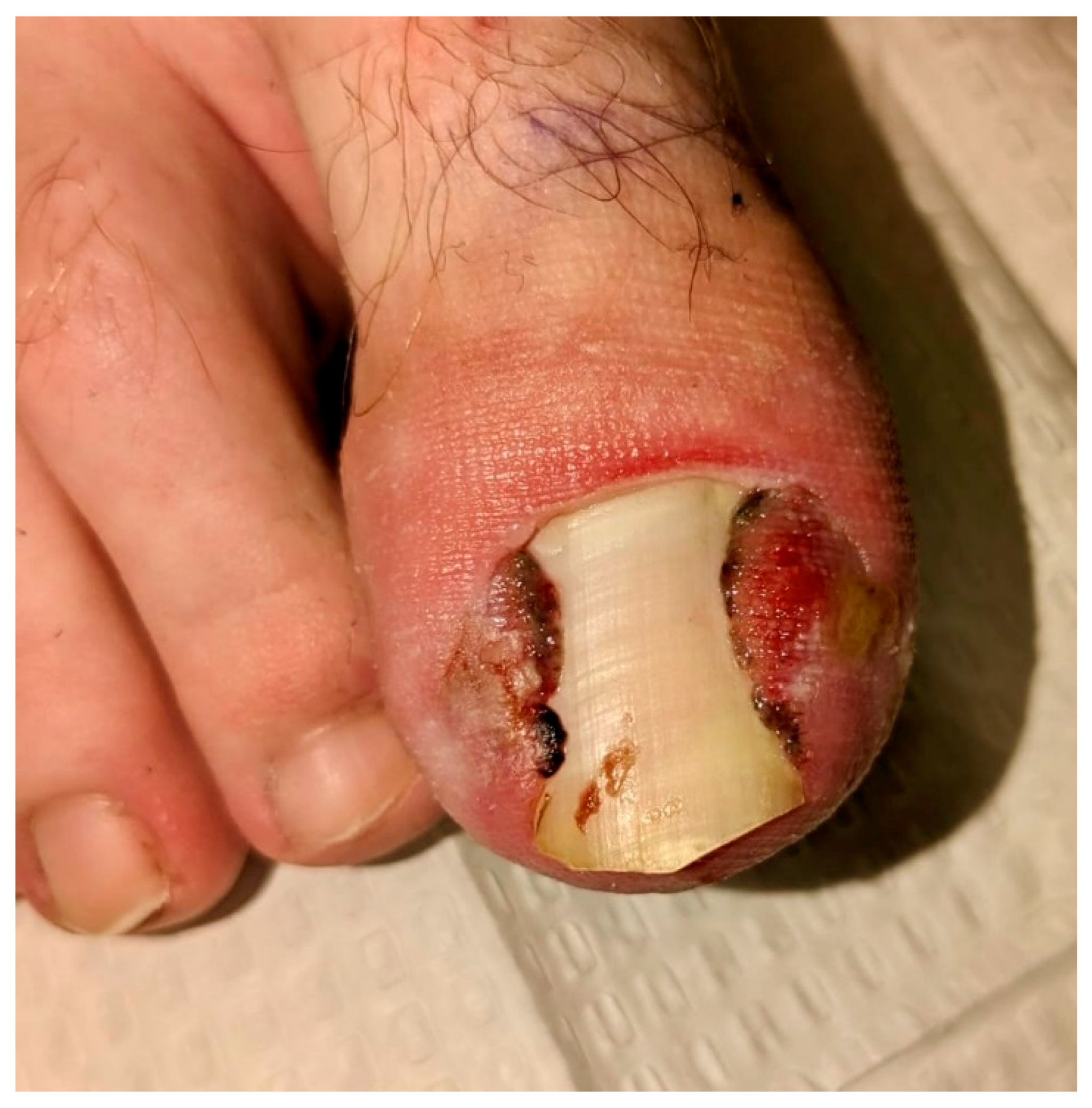
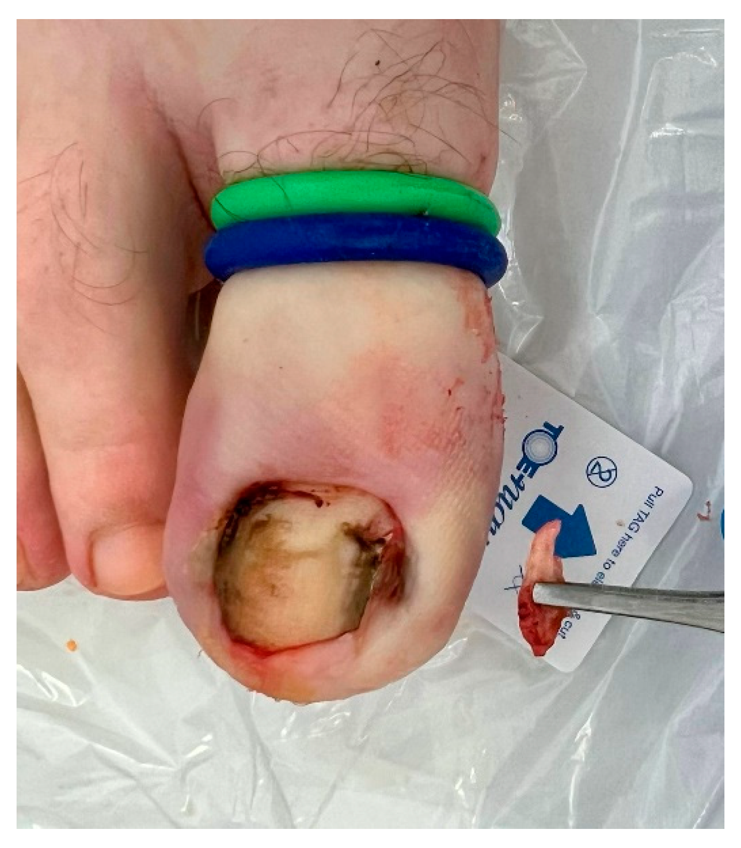
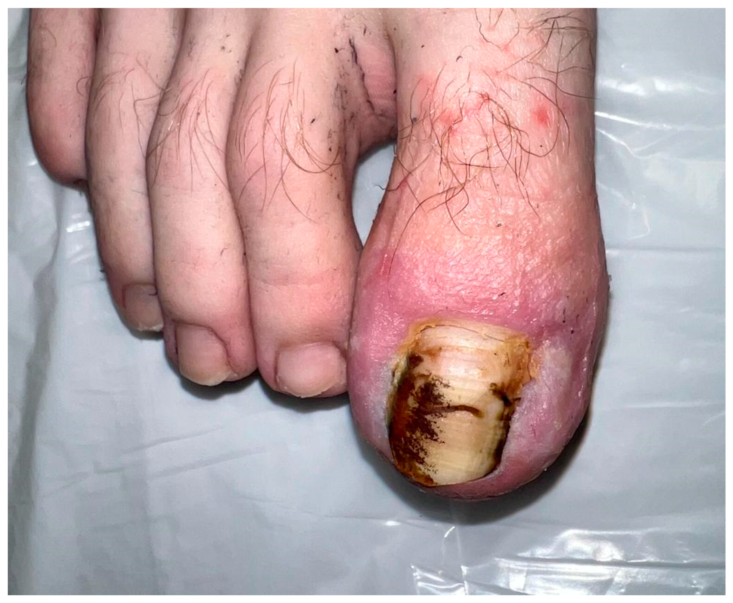
Nail surgery took place on 02.02.23; see
Figure 1,
Figure 2,
Figure 3 and
Figure 4. A standard bilateral partial nail avulsion under a digital tourniquet (TQ) was performed. With informed consent, sharp resection of the lateral hypergranulation tissue only was performed using a scalpel blade (see
Figure 2). Phenolisation of both sulci for three minutes was then completed using two 89% phenol EZ
TM swabs per sulcus (four in total: 90-second applications, twice) (see
Figure 3). An absorbent, compressive dressing was applied (see
Figure 4) with two post-operative appointments made for one day and two weeks post-procedure.
02.02.23
Nail Surgery Technique:
Consent form signed
Discussed milestones – healing, regrowth rate (5/10%), infection
ANTT of the toe
Digital block – 4ml 0.75% Naropin plain
Digital TQ
Nail sulcus released – no pain
PNA
HGT resected lateral sulcus
Phenol EZ swab – 3 mins
Irrigated
TQ off – revasc. noted – 6 mins total
Dry dressing
Advice re analgesia/bleeding
Dressing regime arranged
SOS any problems
MT attended for his nail surgery today as planned. Under digital anaesthesia and tourniquet he had a right 1st bilateral partial nail avulsion. The procedure went very well. He will return to see the nurses tomorrow and myself in two weeks time.
MT was seen on 16.02.23 for a re-dressing appointment, see
Figure 5 and
Figure 6. He had had minimal pain or bleeding.
16.02.23
MT returned for his nail surgery re-dressing appointment with myself. He has seen the nurses on one occasion since the surgery. The lateral sulcus is settling down well but the medial sulcus still has quite an amount of hypergranulation tissue. Typically this will settle down once the offending part of the nail unit has been removed but I would like to check him in a month’s time to ensure that this is so.
It is noteworthy that despite the aggressive resection of the HGT from the lateral sulcus, new HGT had formed (see
Figure 7). The medial HGT remained unchanged. Both sulci were treated with a topical application of 95% silver nitrate on 02.03.23.
MT returned three weeks later: the new (vascular) HGT in the lateral circus had resolved, but the more (fibrotic) medial tissue remained unchanged, and it was suggested that sharp resection to remove the residual medial tissue was required, which was performed one week later (see
Figure 8 and
Figure 9).
23.03.23
MT has returned to see me today. I am very pleased that the silver nitrate that I applied last time has settled down the new friable lateral hypergranulation tissue but it has not completely settled down the more chronic medial hypergranulation. It is therefore time to do a small excision to settle this down and I will do this for him next week under a brief local anaesthetic.
30.03.23
MT attended for his right 1st medial hypergranulation resection today. Under a brief local anaesthesia and tourniquet, I have resected the final portion of hypergranulation tissue for MT today. I have put an Inadine dressing place for two days and he can remove this himself at the weekend. I will see him in a month for a review but I think we have got on top of the remaining issues now.
A final review took place on 27.04.23, where both sites of the previous IGTN and HGT had completely resolved (see
Figure 10), but the residual erythema has yet to settle fully. MT was satisfied with the outcome and continues to remain IGTN- and HGT-free.
He will be reviewed later in the year for a further follow-up.
Discussion
Granulation tissue forms in the proliferation phase of wound healing and comprises newly growing capillaries from the base of the wound, leading to angiogenesis13. Fibroblasts from the surrounding tissue are activated by growth factors released in the inflammatory phase, which rapidly replicate and produce a collagen-rich matrix that builds strength and elasticity into the wound. Hypergranulation is defined as an excess of granulation tissue and usually presents in wounds healing by secondary intention. It is precipitated by an aberrant inflammatory phase caused by infection or foreign bodies13.
Richert et al.14 believe that the HGT associated with IGTN is not true granulation tissue, but rather it is fibrous tissue, similar to what would be tissue observed in an early keloid scar formation. This might may account for the improvement following the application of silver nitrate in the new lateral vascular HGT two months post-index procedure but without effect on the more established fibrotic medial HGT. The histopathology of chronic HGT has received little attention in the literature and will be the subject of further study by the author. To further muddy the waters, some authors conflate peri-ungual HGT with a pyogenic granuloma (PG), but from a dermatopathological perspective, the PG is synonymous with lobular capillary haemangioma15. That said, true PGs often occur in the ungual region2.
Some authors suggest that the application of phenol or silver nitrate to the HGT or removal of the offending nail spike is sufficient to allow for the resolution of the peri-ungual swelling3,16,17. In contrast, Markinson18 asserts that sharp resection speeds up the overall healing process, improves the cosmetic result, and states that this is a common practice in American podiatry clinics, but as noted by Reilly and Burt6, this technique is not well examined in the literature. Kang et al. 7 conducted a randomised controlled trial (RCT) on ingrown toenails treated by partial nail avulsion followed by matricectomy with or without removal of the HGT. They found no significant difference in recurrence rates between the two groups but a significant difference in the wound healing time between the granulation removal group (13.8 days) and the control group (17.6 days). HGT resection performed concurrently with nail surgery received just one ‘citation’ (unreferenced) for ‘excision of the granuloma where necessary’ in the Cochrane Review1, and this case study adds to the argument.
Building on the work of Kang et al.7, ethical approval has been granted for a prospective RCT to be carried out at the University of Northampton. The two-year study will randomise patients into two groups: those who will have sharp resection of HGT (associated with IGTN) and those who will not. Their overall healing metrics will be recorded and pooled for analysis. This clinical research underscores and supports advancing the skill set of the ordinarily skilled podiatric practitioner in the UK, which is a current objective of the UK Royal College of Podiatrists19.
This is a short-term follow-up of a single case study but highlights the author's experience of sharp HGT resection and the potential role of HGT management in the overall care of a severe IGTN. It could be that the medial HGT would have settled down with the tincture of time, but as Reilly and Burt6 point out in their study, that is not always the case. A technique tip given to the author by a retired colleague in cases of bilateral IGTN is to perform a standard bilateral partial nail avulsion with phenolisation but to then avulse the remaining total nail plate, which will regrow. In the short term, this removes any potential irritation from the remaining nail plate on the healing sulci. On reflection, this might have been a useful adjunct to perform for MT regarding the new GHT in the lateral sulcus in particular.
Conclusion
This case report highlights the potential role of sharp resection in the management of HGT in association with IGTN surgery and the possible delay in healing or the need for further surgery if this is not carried out. Further work is underway proceeding with the University of Northampton via a prospective randomised controlled trial of sharp resection versus no resection of HGT in stage 3 IGTNs, and to identify the histopathological features of chronic HGT.
Funding
this research received no grant from funding agencies in the public, commercial, or not-for-profit sectors.
Informed Consent Statement
written consent was obtained from the patient (MT) to publish digital imagery and details of his treatment.
Conflicts of Interest
the author has no competing interests to declare.
Availability of data and material
consent for publication is available on request, a copy of which is retained in MT’s medical records. .
References
- Eekhof JAH, Van Wijk B, Neven AK, van der Wouden JC. Interventions for ingrowing toenails. Cochrane Database of Systematic Reviews. 2012 Apr;4(4). [CrossRef]
- Piraccini BM, Bellavista S, Misciali C, Tosti A, De Berker D, Richert B. Periungual and subungual pyogenic granuloma. British Journal of Dermatology. 2010 Nov;163(5):941–53. [CrossRef]
- Haneke, E. Controversies in the treatment of ingrown nails. Dermatol Res Pract. 2012;1–12. [CrossRef]
- Heifetz, CJ. Ingrown toenail. A clinical study. The American Journal of Surgery. 1937;38(2):298–315. [CrossRef]
- Laco, JE. Nail Surgery. In: Hetherington V, editor. Hallux Valgus and Forefoot Surgery. Churchill Livingstone; 1994. p. 481–96.
- Reilly I, Burt N. Periungual soft tissue resection. The Podiatrist. 2021 Mar;24(2):44–8.
- Kang MH, Seo YJ, Park EJ, Kim CW, Jo HJ, Kim KH, et al. The effect of removal of granulation tissue on ingrown toenails associated with granulation tissue. Korean Journal of Dermatology. 2008;46(4):453–8.
- Perez CI, Maul XA, Heusser MC, Zavala A. Operative technique with rapid recovery for ingrown nails with granulation tissue formation in childhood. Dermatologic Surgery. 2013 Mar;39(3 PART 1):393–7. [CrossRef]
- de Almeida Figueiras D, Ramos TB, de Oliveira Ferreira Marinho AK, Bezerra MSM, Cauas RC. Paronychia and granulation tissue formation during treatment with isotretinoin. An Bras Dermatol. 2016 Mar 1;91(2):223–5. [CrossRef]
- Reyzelman AM, Trombello KA, Vayser DJ, Armstrong DG, Harkless LB. Are antibiotics necessary in the treatment of locally infected ingrown toenails? Arch Fam Med. 2000;9(9):930.
- Zhang N, Huang Z, Cao SH, Wang Y, Hu Y. Cosmetic, minimally invasive, partial matricectomy of ingrown toenails with granulation tissue. Vol. 71, Journal of Plastic, Reconstructive and Aesthetic Surgery. Churchill Livingstone; 2018. p. 773–4. [CrossRef]
- Huang, CK. Case study. Ingrowing cancer. Podiatry Now. 2020;23(9):33–5.
- Mitchell A, Llumigusin D. The assessment and management of hypergranulation. British Journal of Nursing. 2020;30(5 TV Supp):S6–10. [CrossRef]
- Richert et al. Chapter 2: Definition – Pathogenesis Risk Factors – Classification – Scoring. In: Richert B., Di Chiacchio N., Caucanas M., Di Chiacchio N.G., editors. Management of Ingrowing Nails. Springer; 2016. p. 35–56.
- Bakotik, BW. Tumors of the soft tissue of the lower extremity. In: Levy LA, Hetherington V, editors. Principles and Practice of Podiatric Medicine. 2nd ed. Data Trace Publishing Company; 2006. p. 17.1-17.55.
- Erdogan, FG. A simple, pain-free treatment for ingrown toenails complicated with granulation tissue. Dermatologic Surgery. 2006 Nov;32(11):1388–90. [CrossRef]
- Harada K, Yamaguchi M, Matsuzawa M, Shimada S. Ingrown nail with a giant granulation tissue successfully treated with the gutter method. Int J Dermatol. 2015;54:e191-192. [CrossRef]
- Markinson, B. Personal Communication. 2020.
- Reilly I, Longhurst B, Chadwick P. A cut above. A deeper dive into the development of a College-accredited module on skin surgery. The Podiatrist. 2022;25(1):37–41.
|
Disclaimer/Publisher’s Note: The statements, opinions and data contained in all publications are solely those of the individual author(s) and contributor(s) and not of MDPI and/or the editor(s). MDPI and/or the editor(s) disclaim responsibility for any injury to people or property resulting from any ideas, methods, instructions or products referred to in the content. |
© 2023 by the authors. Licensee MDPI, Basel, Switzerland. This article is an open access article distributed under the terms and conditions of the Creative Commons Attribution (CC BY) license (http://creativecommons.org/licenses/by/4.0/).
