
Submitted:
27 February 2023
Posted:
06 March 2023
You are already at the latest version
Abstract
The importance of understanding the corrosion mechanisms of excavated metal artefacts help in determining the physico-chemical parameters of the burial environment and the formation of different corrosion products. These products can be observed and analysed with the help of various techniques that provide information on their morphology, chemical composition and structure. The analysis of ancient coins is extremely challenging in the presence of heavily corroded surfaces; as quantitative information may not exactly concur with its bulk composition. In the case of silver coins, the use of surface information can be used as a guide for bulk composition only. The current study carries out investigation and characterization of selected coins from a large coin hoard excavated from Amheida, Dakhla oasis, Egypt. The study and analysis of the alloy composition and corrosion products was performed using a multi-technique approach which included Light optical microscopes, Scanning Electron microscope (SEM) coupled with energy dispersive spectroscopy (EDS) and X-ray diffraction analysis. Investigation of the coins revealed the presence of a thick active inhomogeneous corrosion crust, while analysis showed that the coins were made from a binary silver copper alloy (billon) while the corrosion crust was rich in chlorides and carbonates, later identified by XRD analysis as Paratacamite, Malachite and Chrysocolla.
Keywords:
Amheida
; Coin hoard
; Silver-copper alloy
; Billon
; Corrosion products
1. Introduction
Corrosion or mineralization of metals is defined as a process resulting from the electrochemical reactions in which metal or alloys react with the surrounding environment to form chemical compounds that are similar to the original mineral ores from which the metal was extracted [1]. This process can take many forms depending on the composition of the metal, environmental conditions [2] and the existence of internal stresses [3,4]. Ancient metal coins recovered from archaeological sites are subjected to several corrosion processes, resulting in a nearly composite material consisting of metal remnants and mineral alteration products [5]. Normally corrosion produces a buildup of insoluble products, both within and overlying the original metal volume. Billon is an alloy of silver and copper [6] used in the manufacture of coins, medals, and tokens. The use of billon coins date from ancient Greece and continued through the Middle Ages, but are perhaps best known from the Roman Empire [7]. The addition of copper to silver was to increase its hardness and durability as silver is a very soft metal [8,9]. Surface corrosion products can occur, when breaks in the oxide film or the buildup of surface scale change the nature of the surface [10], in addition to the effects of the free exchange of ions [11]. However, the rate of corrosion can be increased through galvanic process that occurs when objects made of different metals are in contact with each other in high relative humidity (RH) [12,13]. Silver alloys are sensitive to exposure to atmospheric aggressive agents such as H2S, carbonyl sulphide (COS) and SO2, combined with the presence of high relative humidity (RH), can rapidly form an adherent layer of tarnish [14]. Ancient silver copper alloys are also susceptible to intergranular corrosion-induced embrittlement due to the segregation of copper to grain boundaries (Smith, 1965). Over time, the corrosion of archaeological objects becomes extensive, and its removal may affect their appearance and cause compositional changes on the object’s surface [15]. Therefore, the removal of corrosion products from excavated metal objects should be always performed with extreme caution [16]. A preliminary examination and condition assessment of the object is of great importance [17]. It is necessary to form some estimate of the thickness and regularity of the encrusted layer, and the degree of penetration of corrosion into the core [18].
1.1. The study area
The study area “Amheida”, (Figure 1-a, b) is located on the western edge of Dakhla oasis in the western desert of Egypt about 750 km south-west of Cairo at Lat. 25°25′56″ N to 25°55′11″ N and Long. 28°28’37’’ E to 29°22’14’’ E) [19]. According to Brookes [20] and Kuciewicz, et al. [21] this area hosts potentially unique features that were noted by some previous researchers. This area is a vast archaeological site that has a long history that goes back to at least the old Kingdom [22]. It can be reached via the loop road running from Mut to el-Qasr. Geologically, this oasis corresponds to the interior of the African continent where Mesozoic and Cenozoic rocks prevail [23]. According to Said [24] the study area is a part of the stable shelf that generally includes the horizontal strata forming the Western desert plateau. These strata are mainly covered by sedimentary rocks ranging in age from Jurassic to Quaternary [25,26].
1.2. The study objects
According to Bagnall [27] the discovered coin hoard (Figure 2-a) contained around 850 coins, which was studied to investigate the different effects of the surrounding environment [28]. The coin hoard is the part of a project aiming to perform the conservation and authentication of the archaeological coins’ hoards discovered in Amheida. The hoard was divided into three textile bags (traces of fabrics are visible) lying on top of a mudbrick debris layer filling Area (Figure 2-b). The hoard (Figure 2-c) was covered with a thick layer of corrosion products and soil encrustations, resulting from their long-term underground burial. Their surface features and inscriptions were obscured and distorted to such an extent that no detail of the original surface could be retrieved. Most ancient coins are subjected to various corrosion processes [29], resulting in the formation of different corrosion products [30]. Corrosion can gradually alter their aspect, shape, and nature, up to a stage where it is impossible to use them as historical evidence of human civilizations [31].
2. Materials and Methods
Three Roman billon (Ag-Cu alloy) coins were selected for this research; coins no.(66, 68 & 73) with the dimensions (Ø2.5cm, 0,4cm) for coin number (66), (Ø2,4cm, 0,5cm) for coin number (68) and(Ø2,3cm, 0,4cm) for coin number (73) were studied, (Figure 3-a, b, c). They are from the excavations at Amheida, Dakhla oasis, Egypt. The coins are covered with a thick layer of corrosion and soil encrustations (Figure 3).
The coins were studied using different techniques for defining surface and morphological features, different elemental and mineralogical components, in addition to structure and nature of the patina according to Constantinides, et al. [32]. Light optical microscope (LOM) was used because it allows a rapid and representative characterization of the morphological features of the corrosion products, the nature of the patina, and the associated burial remains. Optical observations were primarily carried out without any preparation in order to keep the surface intact.
Scanning electron microscopy coupled with energy dispersive spectrometry (SEM- EDS), was used according to Borges, et al. [33] and Di Turoa. et al. [34] to investigate the surface and the metallic core and to detect the distribution of the chemical elements in the corrosion layer and in the core. SEM micrographs and EDS spectra of the selected coins were obtained by using a JEOL/EO, JSM-6380 device, equipped with an EDS link operating up to an accelerating voltage of 20 kV and a working distance of 9 mm.
X-Ray diffraction analysis (XRD) was used to identify the corrosion products and to understand the corrosion mechanism [35,36]. The studied coins surfaces were carefully scraped with a small spatula, to collect the fine corrosion particles for the X-ray diffraction analysis. The analysis of the corrosion products was performed with an Ultima IV, multipurpose X-ray diffraction system equipped with a copper anticathode. The measuring conditions were set as follows: Cu target, 40 kV accelerating voltage, 40 mA current, the scanning range of 2θ was from 5 to 70° and the scanning speed was 2°/min.
3. Results
3.1. Optical Microscope
OM observations (Figure 4) revealed that the coins showed a rough corrosive surface with cracks and pits. It was covered with different corrosion products with colors of dark green, light green, greenish blue and metallic gray blackish surface covered with soil residues. In some parts, there was a thick active inhomogeneous green corrosion crust, full of pores and cavities.
3.2. SEM-EDS
Coin no.66 is of a binary silver copper alloy otherwise known as Billon, with the bulk composition of around 63% Ag, 15.4% Cu, (Figure 5-a, b). The surface of Coin no. 68 shows distinct layered corrosion structure rich in chlorides, (Figure 5-c, d). Within the same context, the investigation of the coin no. 73 proved that it is covered with thick corrosion crust rich in copper chlorides, oxides and soil deposits, (Figure 5-e, f).
3.3. XRD
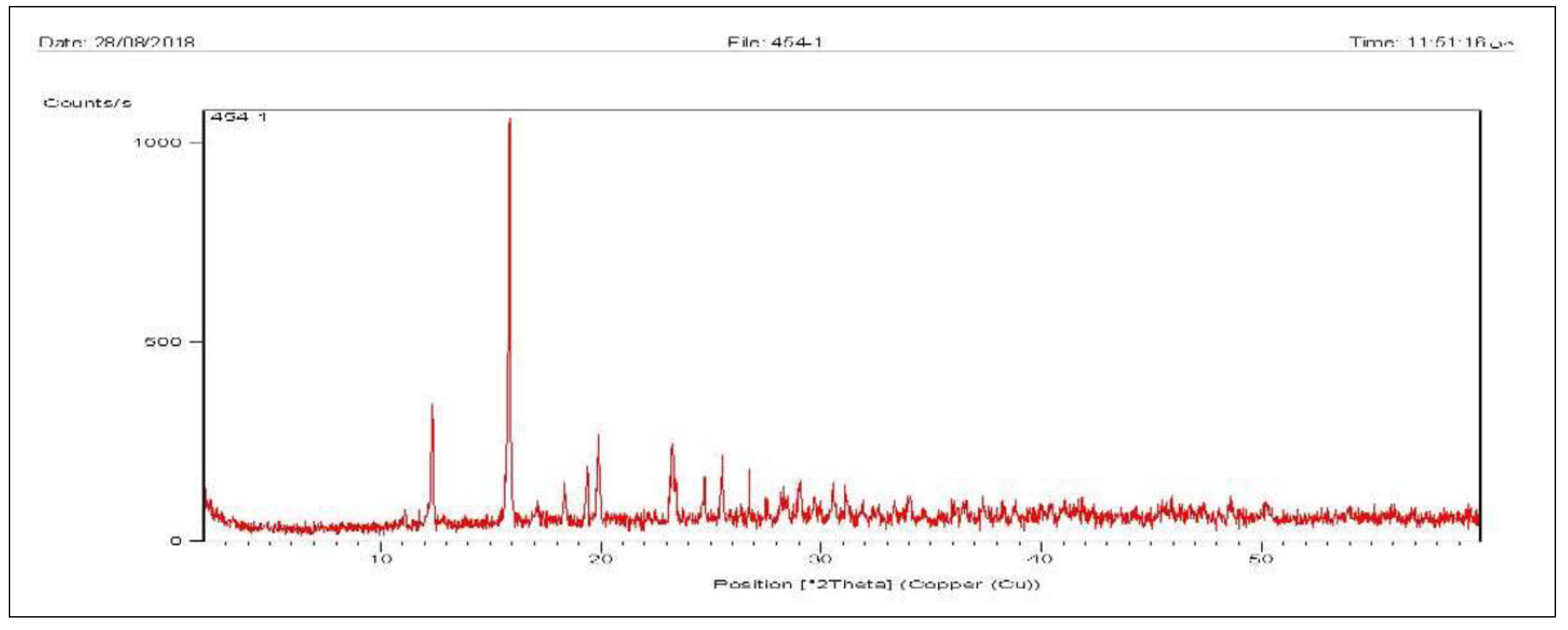
The results showed that the major compound of the light green patina of the sample is paratacamite (Cu2(OH)3Cl), which exists with little amounts of malachite (Cu2CO3(OH)2), chrysocolla (CuSiO2.H2O) and metallic copper (Cu), ( Figure 6).
4. Discussion
Base silver alloyed with copper usually displays a variety of copper corrosion products on the surface. All metals have specific relative electrical potential. When metals of different electrical potential are in contact in the presence of moisture, a low energy electric current flows from the metal having the higher position in the galvanic series. This is called "galvanic action or corrosion", which is considered a form of electrochemical corrosion [37] that occurs when two dissimilar metals come together in the presence of an electrolyte to form an electrical couple, known as a galvanic couple [12]. The more noble or cationic the metal, the less likely it will corrode relative to the other metal it is in contact with [38] Billon is a Roman silver copper alloy[39] composed of a noble and base metal, it is used chiefly for making coins and medals [40]. Billon alloy is susceptible to galvanic corrosion and this explains the fact that most of the corrosion products identified by X-ray diffraction are of copper. Corrosion of archaeological alloys is also essentially due to ambient environments, particularly soil and groundwater [41,42]. The corrosion of billon coins in the soil is a complicated process caused by interaction between metals and surrounding soil [43], especially with presence of moisture and high salt contents [44].
This process is generated as moisture is commonly present within the inter-granular spaces and organic matter of soil, which alters the ambient pH [45]. This leads to two main mechanisms; physical and chemical, which, result from the aggressive deterioration factors, such as soil external stress and internal strains. According to Merk [46]; Agrawal [47]; Quaranta [48], research work has been carried out on their interaction with metal alloys. In our case, this mechanism is primarily attributed to the combined environmental effects such as O, Cl within the soil as argued previously by Schweizer [49]. As well as, the effects of urban pollution and ion migration in the study area [50,51]. Physical corrosion mechanism is one of the most important mechanisms affecting the metal corrosion process [52], which is attributed to the pressure on the overlying corrosion products that cause physical damage. This damage is visually evident in some serious forms, such as cracking, which develops into lamination and exfoliation [53,54]. This is mostly linked to the rate of oxygen consumption and dominated RH. It controls the metal corrosion products as attested by Matthiesen [55].
Post-excavation, archaeological silver alloy coins usually display a central core of uncorded metal surrounded by a layer of predominantly silver corrosion products. Some archaeological silver artifacts may also become brittle as a long-term consequence of corrosion and microstructural changes [56,57]. Corrosion-induced embrittlement results from selective corrosion that penetrates the metal and eventually causes it to crack and fracture. The surface of corroded silver alloys is slowly converted to silver chloride [58]. Silver chloride forms a brittle, finely granular layer but it does not affect the remaining metal, while copper diffuses out of the alloy and forms green copper corrosion products on the surface [56]. Other common types of corrosion in silver copper alloys are intergranular, interdendritic, along segregation bands that are the remains of coring and interdendritic segregation and along slip lines and deformation twin boundaries in objects not annealed after their final mechanical working [43]. Intergranular corrosion is very common in silver copper alloys. It is the selective dissolution of the grain boundary zone, while the bulk grain is not attacked. Intergranular corrosion is caused by the action of micro-galvanic cells at the grain boundaries. Grain boundaries are preferred sites for segregation and precipitation, which makes them physically and chemically different from the matrix. Furthermore, a zone adjacent to the grain boundary is depleted of the solute elements. Consequently, a ‘galvanic cell’ is formed [59].
4.1. Optical microscope
Based on the surfaces features’, (Figure 4), it could be asserted that the noted rough corrosive surface and weakness areas mostly resulted from aggressive factors dominating the study area, especially the abundance of chlorides in the soil. Cracks and pits on the coin surfaces owed essentially to the developing of macroscopic activity between the corrosion layers or fatigue cracks [60]. In our case, it is attributed to covering the surfaces of the original objects by complex corrosion compounds (dark-green areas), due to defects in the protecting oxide film [61]. Furthermore, the crack propagation in the coins could result from acid production and saline water/soils in the study area as mentioned by Tylecote [62] in his case study, in addition to the effect of stress concentration and the physical properties of the metal itself [63]. The presences of corrosion products; on one hand, dark green carbonates, malachite and light green paratacamite covered with soil residues are attributed to the burial crusts dominated in the area because of the long interaction of soil environment [64]. The latter compounds are inherently unstable and convert into more stable compounds due to active chlorine [65]. Finally, the occurrence of black spots with microscopic appearance is mostly attributed to local migration of copper ions from the alloy to form chalcocite crystals on the surface as mentioned previously by Eggert & Sobottka-Braun in their case study [66].
4.2. SEM-EDS
Through evaluating the SEM, (Figure 5-a, b & c) it could be noted that they show some variations in layered corrosion structure characterized by distinct layers rich in chlorides covered with thick corrosion crust. This crust contains copper chlorides, carbonates, sulphides and soil deposits due to direct influence of the burial effects for a long time as mentioned previously by Cura, et al. [67]. Although, it wasn’t possible to prepare a cross-section, the flaking of the outer corrosion products made it possible to examine and analyze these layers. These corrosion layers are heterogeneous and composed of different elements with different ratios. Within the same context, it could be asserted that there are main differences between the investigated samples according to their deterioration states. In coin 66, there is a distinct enrichment of silver at the surface with the composition of Ag 63.15%, stating clearly that it’s a billon alloy, however, the bulk analysis couldn’t be determined. In coin 68, (Figure 5-e) only copper corrosion products were detected, the surface being rich in malachite and paratacamite as confirmed by XRD. The surface of Coin 73, shows signs of active corrosion rich in chlorides and soil deposits.
4.3. XRD
The studied coins were found in a desert environment, enough moisture is present to interact with the soil salts, with the result that the movement of free ions produces different corrosion products as evident from the microscopic examination. XRD analysis identified the presence of several mineral species representing the corrosion products and soil deposits. Chalcocite (Cu2S) is one of corrosion products formed on copper and copper alloys exposed outdoors at sites with high hydrogen sulphide (H2S) or buried in soils where sulfate-reducing bacteria may generate (H2S) during respiration. Studies have shown that copper sulfides are readily produced in moist, anoxic soil environments or deoxygenated seawater [68,69,70]. Copper alloys (except possibly arsenical bronzes) are also subject to sulfide-induced corrosion by SRB within a biofilm. Under these circumstances chalcocite forms easily [71,72,73]. The corrosion layer may contain other sulfides buried under other corrosion products [74,75]. Nonetheless, the poor adherence and mechanical properties of the sulfides make these layers non-protective.
Paratacamite Cu2(OH)3Cl, occurs as a powdery light green secondary corrosion layer found on the patina surface [76]. The conditions for its formation include the presence of a deposit of insoluble cuprous chloride (CuCl) under a layer of cuprite (Cu2O) which acts as a bipolar electrode. This means that the corrosion (anodic) reactions occur on the metal side of the cuprous oxide membrane while oxygen reduction (cathodic) reactions occur on the environmental side. This coincides with the fact that XRD data gave a clear indication that this mineral is in fact present at the interface of the outer-layer and the inner-layer (metallic substrate) of corrosion surface. Furthermore, it could be claimed that it was formed principally when copper alloys come in contact with soil in the presence of moisture formed by surface condensation and charged with carbon dioxide [48]. The existence of the cuprous chloride layer in the pit will depend on the relative rates of copper corrosion and the hydrolysis of cuprous. In the presence of chlorine ions CuCl can form a series of soluble complexes such as CuCl2- and CuCl3- in the pit. These copper (I) species diffuse through the cracks in the Cu2O membrane and are then oxidized by molecular oxygen to cupric ions while oxygen is reduced to hydroxide ions. Some of the cupric ions will be precipitated in the form of basic cupric chlorides (Cu2(OH)3Cl), while other cupric ions can be reduced to form cuprous ions, which are subsequently oxidized by molecular oxygen away from the surface of the pit. The anodic reaction inside the pit is the oxidation of cuprous to cupric ions at the Cu2O surface, which in turn attack the copper metal to form more cuprous ions causing the pit to deepen. The driving force for the pitting reaction is the concentration gradient of copper (I) species between the bottom of the pit and the corrosion mound formed above the Cu2O film [75]. In addition, the presence of this mineral indicates that the formation of the internal chloride layer could be linked to the contamination in the soil due to artificial fertilizers as attested by Gerwin & Baumhauer [4]. Cuprous chloride may lie dormant until reaction with moisture or oxygen causes this stable compound to expand in volume on conversion to one of the copper trihydroxychlorides. This creates physical stress within the object affected, resulting in cracking or fragmentation. Cl- (chloride ions) is very active when subjected to moisture or high relative humidity. Deterioration occurring to alloys rich in copper is often attributed to the presence of chloride, and there is no doubt that most, if not all, corrosion products on archaeological copper alloy artifacts contain chloride ions.
Malachite, CuCO3.Cu(OH)2 is a significant component of patinas that develops during burial in the soil [77], practically all copper alloys buried in soil form a cuprite crust that is adjacent to the metal and overlaid with malachite [78]. It is formed principally when copper alloys come in contact with soil waters or with water formed by surface condensation and charged with carbon dioxide. Malachite can be formed in two ways: by reaction of cupric ions with carbonate ions from a super-saturated aqueous solution 10,17,18, deposited on a substrate 19 , or by the reaction of cupric oxide (tenorite) or cuprous oxide (cuprite) with carbon dioxide and water 17,20,21. Malachite usually can form above the initial cuprite layer due to the direct chemical reaction of the carbonate/ bicarbonate anions and the copper and/or cuprous oxide patina in the presence of the high humid environment [79]. The carbonate/ bicarbonate anions can result from the dissolution of the salts in the burial environment.
Chrysocolla, CuSi02.nH20, a copper silicate is not a common corrosion product on copper alloy artefacts; however, its formation could be due to the reaction of leached copper ions with the surrounding soil. The sandy soil (the place which the coins found) allows good penetration of air, because the large spaces between the grains of sand make it easy for air and moisture to pass through. As a result, the corrosion process becomes very active, and some coins display spongy corrosion.
5. Conclusions
Three silver copper alloy (billon) coins from the Amheida excavation site in Dakhla oasis, Egypt were chosen for this study. Various investigative and non-destructive analytical techniques were used to assess the condition of the coins including a light optical microscope and a Smart-Eye USB Digital microscope to characterize the morphological features of the corrosion products and the associated burial soil remains. These showed evidence of the presence of active localized pitting corrosion known as “bronze disease”. Scanning electron microscopy (SEM) coupled with energy dispersive spectrometry (EDS), showed that the coins were made from a binary silver copper alloy while the corrosion crust was rich in chlorides and carbonates, later identified by XRD analysis as Paratacamite, Malachite, Chalcocite and Chrysocolla. These corrosion products are typical for silver copper alloys, whereby copper diffuses out of the alloy and forms different copper corrosion products. The coins were found in a sandy soil which allows good penetration of air, and moisture. As a result, corrosion becomes very active, and the coins display thick inhomogeneous corrosion crust. Moreover, the presence of copper chlorides expressed as paratacamite in the outer corrosion crust suggests the objects were recovered from partly aerobic conditions, while the presence of chalcocite suggests an anaerobic reducing environment which could suggest fluctuating conditions.
Author Contributions
Authors’ El-Gohary & Rifai designed, directed, and carried out this study, in addition to the analyzing of the results and wrote the paper. Hussein prepared the samples and helped the authors during defining their characteristics. All authors have read and approved the manuscript to be published
Institutional Review Board Statement
Not applicable.
Informed Consent Statement
Not applicable.
Data Availability Statement
Not applicable.
Conflicts of Interest
The authors declare no conflict of interest
References
- Abebe, A.; Misganaw, W. Experimental Analysis of Environmental Effects on Corrosion and Degradation of Metals. High Technology Letters 2021, 27, 389–399. [Google Scholar]
- Nord, A.; Mattsson, E.; Tronner, K. Factors Influencing the Long-term Corrosion of Bronze Artefacts in Soil. Protection of Metals 2005, 41, 309–316. [Google Scholar] [CrossRef]
- Akimov, G. Factors Influencing Corrosion. Corrosion 1959, 15, 23–36. [Google Scholar] [CrossRef]
- Gerwin, W.; Baumhauer, R. Effect of Soil Parameters on the Corrosion of Archaeological Metal Finds. Geoderma 2000, 96, 63–80. [Google Scholar] [CrossRef]
- Scharff, W.; Huesmann, L. Accelerated Decay of Metal Soil Finds Due to Soil Pollution: First Report. In Proceedings of the Int. Conf. on Metal Conservation (Metal 95), Semur-en-Auxois, France (25-28/9/1995). [Google Scholar]
- Skogvold, S.; Mikkelsen, Ø.; Billon, G.; Garnier, C.; Lesven, L.; Barthe, J-F. Electrochem-ical Properties of Silver-copper Alloy Microelectrodes for Use in Voltammetric Field Apparatus. Anal Bioanal Chem 2006, 384, 1567–1577. [Google Scholar] [CrossRef] [PubMed]
- Mavridis, D.; Vatalis, K. Entrepreneurial Aspects in East Roman Empire-The Imperial Kommerkiarioi as Quasi-entrepreneurs. Procedia Economics and Finance 2013, 5, 552–561. [Google Scholar] [CrossRef]
- Prashanth, M.; Satish, N.; Ajay kumar, B. Effect of Brass and Silver on Mechanical Properties of Copper. Materials Today: Proceedings 2018, 5, 25404–25411. [Google Scholar] [CrossRef]
- Felicia, D., Rochiem, R. & Laia, S. (2018). The Effect of Silver (Ag) Addition to Mechanical and Electrical Properties of Copper Alloy (Cu) Casting Product. In Proceedings of the 3rd Int. Conf. on Materials and Metallurgical Engineering and Technology (ICOMMET 2017): Advancing Innovation in Materials Science, Technology and Applications for Sustainable Future, Surabaya, Indonesia (30-31/10/2017).
- Mattson, E. Localized Corrosion'. Br. Corros. J 1978, 13, 5–12. [Google Scholar] [CrossRef]
- Macleod, I. Formation of Marine Concretions on Copper and Its Alloys'. Int. J. Naut. Arch. 1982, 11, 267–275. [Google Scholar] [CrossRef]
- Selwyn, L. Metals and Corrosion: A Handbook for the Conservation Professional, Canadian Conservation Institute, Ottawa, 2004; p.1.
- Watkinson, D.; Rimmer, M.; Emmerson, N. The Influence of Relative Humidity and Intrinsic Chloride on Post-excavation Corrosion Rates of Archaeological Wrought Iron. Studies in Conservation 2019, 64, 456–471. [Google Scholar] [CrossRef]
- Marchand, G.; Guilminot, E.; Lemoine, S.; Rossetti, L.; Vieau, M.; Stephant, N. Degradation of archaeological horn silver artefacts in burials. Heritage Science 2014, 2. [Google Scholar] [CrossRef]
- Denker, A.; Bohne, W.; Opitz-Coutureau, J.; Rauschenberg, J.; Röhrich, J.; Strub, E. Influence of Corrosion Layers on Quantitative Analysis. Nucl. Instrum. Methods. Phys. Res. B. am Interactions with Materials and Atoms 2005, 239, 65–70. [Google Scholar] [CrossRef]
- Grimmer, A. Dangers of Abrasive Cleaning to Historic Buildings: Preservation Briefs, U.S. Department of the Interior, National Park Service, Cultural Resources, Heritage Preservation Services, Washington, D.C., 1979.
- El-Gohary, M.; Al-Naddaf, M. Characterization of Brick Used in the External Casing of Roman Bath Walls "Gedara-Jordan". MAA 2009, 9, 29–46. [Google Scholar]
- Domingo, R. Corrosion Control for Aircraft: AC No: 43-4B, US. Department of Transportation Federal Aviation Administration Advisory Circular Subject, USA, 2018.
- Hereher, M.; Ismael, H. The Application of Remote Sensing Data to Diagnose Soil Deg-radation in the Dakhla Depression – Western Desert, Egypt. Geocarto Int 2016, 31, 527–543. [Google Scholar] [CrossRef]
- Brookes, I. Geomorphology and Quaternary Geology of the Dakhla Oasis Region, Egypt. Quat Sci Rev 1993, 12, 529–552. [Google Scholar] [CrossRef]
- Kuciewicz, E.; Polkowski, P.; Kobusiewicz, M. Dakhleh Oasis Project, Petroglyph Unit: Seasons 2012 and 2013. Polish Archaeology in the Mediterranean 2015, 24, 275–296. [Google Scholar] [CrossRef]
- Cribiore, R.; Davoli, P. New Literary Texts from Amheida, Ancient Trimithis (Dakhla Oasis, Egypt). Zeitschrift für Papyrologie und Epigraphik 2013, 187, 1–14. [Google Scholar]
- Abdel Maksoud, K.h.; Elfeky, H.; Ruban, D.; Ermolaev, V. A Unique Coincidence of Geom-orphological, Geological, and Geoarchaeological Features in the Valley of Camels (Dakhla Oasis, Western Desert, Egypt). Geoheritage 2020, 12, 81. [Google Scholar] [CrossRef]
- Said, R. The Geology of Egypt; Elsevier: Amsterdam, 1962; pp. 140–146. [Google Scholar]
- El-Desoky, H.; Khalil, A.; Farouk, S.; Fahmy, W. Dakhla–Kharga Iron-rich Paleosols, Western Desert, Egypt: Geology, Geochemistry, and Mineralization. Arab J. Geosci 2017, 10, 74. [Google Scholar] [CrossRef]
- Masoud, A.; El-Horiny, M.; Atwia, M.; Gemail, K.; Koike, K. Assessment of Groundwater and Soil Quality Degradation Using Multivariate and Geostatistical Analyses, Dakhla Oasis, Egypt. Afr. Earth Sci 2018, 142, 64–81. [Google Scholar] [CrossRef]
- Bagnall, R. Documentary Material: Excavations at Amheida, Preliminary Report, New York University. 2012.
- Ismael, H.; Abdel-Motamed, M.; Abbas, W. Environmental and climatic hazards and their impacts on the cultural heritage of El-Kharga oasis, Western desert, Egypt. EJARS 2020, 10, 135–152. [Google Scholar]
- Doménech-Carbó, A. Electrochemical Analysis of Metallic Heritage Artefacts: Voltammetry of Microparticles (VMP). In Corrosion and Conservation of Cultural Heritage Metallic Artefacts, Dillmann, P., Watkinson, D., Angelini, E., Adriaens, A. Eds.; Woodhead Pub. Limted: Oxford, 2013; pp. 165–189. [Google Scholar]
- Tidblad, J. Tidblad, J. & Swerea, A. Atmospheric corrosion of heritage metallic artefacts: Processes and prevention. In Corrosion and Conservation of Cultural Heritage Metallic Artefacts, Dillmann, P., Watkinson, D., Angelini, E., Adriaens, A. Eds.; Woodhead Pub. Limted: Oxford, 2013, pp. 37–52.
- Ioanida, E.; Ioanida, A.; Rusub, D.; Dorofteia, F. Surface Investigation of Some Medieval Silver Coins Cleaned in High-Frequency Cold Plasma, J. of Cultural Heritage 2011, 12, 220–226. [Google Scholar] [CrossRef]
- Constantinides, I.; Gritsch, M.; Adriaens, A.; Hutter, H.; Adams, F. Microstructural Characterisation of Five Simulated Archaeological Copper Alloys Using Light Microscopy, Scanning Electron Microscopy, Energy Dispersive X-Ray Microanalysis and Secondary Ion Mass Spectrometry. Analytica Chimica Acta 2001, 440, 189–198. [Google Scholar] [CrossRef]
- Borges, R.; Alves, L.; Silva, R.; Araujo, M.; Candeias, A.; Corregidor, V.; Valerio, P.; Barrulas, P. Investigation of Surface Silver Enrichment in Ancient High Silver Alloys by PIXE, EDXRF, LA-ICP-MS and SEM-EDS, Microchem. J 2017, 131, 103–111. [Google Scholar]
- Di Turoa, F.; Colettic, F.; De Vitob, C. Investigations on Alloy-Burial Environment Interaction of Archaeological Bronze Coins. Microchemical J 2020, 157. [Google Scholar] [CrossRef]
- Dinnebier, R.; Fischer, A.; Eggert, G.; Runčevski, T.; Wahlberg, N. X-Ray Powder Diffraction in Conservation Science: Towards Routine Crystal Structure Determination of Corrosion Products on Heritage Art Objects. J. of Visualized Experiments 2016, 112, e54109. [Google Scholar] [CrossRef]
- Dowsett, M.; Sabbe, P.-J.; Anjos, J.; Schofield, E.; Walker, D.; Thomas, P.; York, S.; Brown, S.; Wermeille, D.; Adriaens, M. Synchrotron X-ray Diffraction Investigation of the Surface Condition of Artefacts from King Henry VIII’s Warship the Mary Rose. J. Synchrotron Radiation 2020, 27, 653–663. [Google Scholar] [CrossRef] [PubMed]
- Makhlouf, A.; Herrera, V.; Muñoz, E. Corrosion and Protection of the Metallic Structures in the Petroleum Industry Due to Corrosion and the Techniques for Protection. In Handbook of Materials Failure Analysis With Case Studies from the Construction Industries, Makhlouf, A., Aliofkhazraei, M., Eds. Elsevier: Amsterdam, 2018; pp. 107–122. [Google Scholar]
- Schlamp, G. Noble Metals and Noble Metal Alloys". In Springer Handbook of Materials Data. Warlimont, H., Martienssen, W. Eds.; Springer Cham, Switzerland, 2018; pp. 339–412.
- Bude, R.; Bigelow, E. Nano - CT Evaluation of Totally Corroded Coins a Demonstrations Study to Determine if Might Still Detail be Discernible Despite the Lack of Internal Non –Corroded, Metal. Archaeometry 2020, 62, 1195–1201. [Google Scholar] [CrossRef]
- Wilson, A. The Metal Supply of the Roman Empire. J. of Roman Archaeology 2007, 69, 110–125. [Google Scholar]
- Levard, C.; Hotze, E.; Lowry, G.; Brown, G. Environmental transformations of silver nanoparticles: impact on stability and toxicity. Environ. Sci. Technol 2012, 46, 6900–6914. [Google Scholar] [CrossRef]
- El-Gohary, M. A Holistic Approach to the Assessment of the Groundwater Destructive Effects on Stone Decay in Edfu Temple Using AAS, SEM-EDX and XRD. Environ Earth Sci 2016, 75, 13. [Google Scholar] [CrossRef]
- Vassiliou, P.; Novakovic, J.-M. Corrosion of Ancient Silver Alloys. In Proceedings of 17th Int. Corrosion Congress: Corrosion Control in the Service of Society, Curran Associates, Inc., NY (6-10/10/2008).
- El-Gohary, M.; Redwan, M. Alteration Parameters Affecting the Luxor Avenue of the Sphinxes-Egypt. Science of the Total Environment 2018, 626, 710–719. [Google Scholar] [CrossRef] [PubMed]
- Dracott, J. Piezoelectric Printing and Pre-corrosion, Electrical Resistance Corrosion Monitors for the Conservation of Heritage iron. PhD, University of Manchester, UK, 2014.
- Merk, L. A Study of Reagents Used in the Stripping of Bronzes. Studies in Conservation 1978, 23, 15–22. [Google Scholar] [CrossRef]
- Agrawal, O. Conservation of Metals—Problems and Prospects. In Conservation of Metals in Humid Climate, Agrawal, O., Ed.; 1CCROM & NRLC: Rome & Lucknow, 1987; pp. 1–17. [Google Scholar]
- Quaranta, M. On the Degradation Mechanisms under the Influence of Pedological Factors Through the Study of Archaeological Bronze Patina. PhD, Alma Mater Studiorum – Università di Bologna, Italy, 2009.
- Schweizer, F. Bronze Objects from Lake Sites: From Patina to Biography. In Ancient & Historic Metals: Conservation and Scientific Research, Scott, D., Podany, J., Considine, B. Eds.; Paul Getty Museum & Getty Conservation Institute: USA, 1991, pp. 33–50.
- Robbiola, L. Characterisation de L’alteration de Bronzes Archeologiques Enfouis a Partir d’un Corpus D’objets de L’age du Bronze: Mechanismes de Corrosion. PhD, Matériaux. Université Pierre et Marie Curie, Paris, 1990.
- Chase, W. Chinese Bronzes: Casting, Finishing, Patination, and Corrosion. In Ancient & Historic Metals: Conservation and Scientific Research, Scott, D.; Podany, J., Considine, B., Eds.; Paul Getty Museum & Getty Conservation Institute: USA, 1991; pp. 95–117. [Google Scholar]
- Argyropoulos, V.; Boyatzis, S.; Giannoulaki, M.; Polikreti, K. The Role of Standards in Conservation Methods for Metals in Cultural Heritage. In Corrosion and Conservation of Cultural Heritage Metallic Artefacts, Dillmann, P., Watkinson, D., Angelini, E., Adriaens, A. Eds.; Woodhead Pub. Limted: Oxford, 2013, pp. 478–517.
- Wang, Q. An Investigation of Deterioration of Archaeological Iron. Studies in Conservation 2007, 52, 125–134. [Google Scholar] [CrossRef]
- Watkinson, D. (2013). Conservation, corrosion science and evidence-based preservation strategies for metallic heritage artefacts. In Corrosion and Conservation of Cultural Heritage Metallic Artefacts, Dillmann, P., Watkinson, D., Angelini, E., Adriaens, A. Eds.; Woodhead Pub. Limted: Oxford, 2013, pp. 9–36.
- Matthiesen, H. (2013). Oxygen monitoring in the corrosion and preservation of metallic heritage artefacts. In Corrosion and Conservation of Cultural Heritage Metallic Artefacts, Dillmann, P., Watkinson, D., Angelini, E., Adriaens, A. Eds.; Woodhead Pub. Limted: Oxford, 2013, pp. 368–391.
- Organ, R. The Current Status of the Treatment of Corroded Metal Artifacts, Corrosion and Metal Artifacts, Special Pub. 479; National Bureau of Standards: Washington, D.C, 1977; pp. 107–142. [Google Scholar]
- Wanhill, R.; Steijaart, J.; Leenheer, R.; Koens, J. Damage Assessment and Preservation of an Egyptian Silver Vase (300-200 BC). Archaeometry 1998, 40, 123–137. [Google Scholar] [CrossRef]
- Schweizer, F.; Meyers, P. A New Approach to the Authenticity of Ancient Silver Objects: The Discontinuous Precipitation of Copper from a Silver-Copper Alloy. In Proceedings of the 18th Int. Symp. on Archaeometry and Archaeological Prospection, Rheinland– Verlag GmbH, Cologne, (14-17/3/1978).
- Bloeck, M. Aluminium Sheet for Automotive Applications. In Advanced Materials in Automotive Engineering, 1st ed.Rowe, J. Ed. Woodhead Pub.: UK, 2012; pp. 85–108. [Google Scholar]
- Bardal, E. Different Forms of Corrosion Classified on the Basis of Appearance. In Corrosion and Protection. Engineering Materials and Processes, Bardal, E. Ed.; Springer: London, 2004; pp. 89–191. [Google Scholar]
- He, L.; Liang, J.; Zhao, X.; Jiang, B. Corrosion Behavior and Morphological Features of Archeological Bronze Coins from Ancient China. Microchemical J 2011, 99, 203–212. [Google Scholar] [CrossRef]
- Tylecote, R. The effect of soil conditions on the long-term corrosion of buried tin-bronzes and copper. J. of Archaeological Science 1979, 6(4), 345–368. [Google Scholar] [CrossRef]
- Baker, H. Properties of Metals. In Metals Handbook, 2nd ed.; Davis, J. Ed., ASM Int.: Ohio, 1998. [Google Scholar] [CrossRef]
- Gouda, V.; Youssef, G.; Abdel Ghany, N. Characterization of Egyptian Bronze Archaeological Artifacts, Surf. Interface Anal 2012, 44, 1338–1345. [Google Scholar] [CrossRef]
- Mereu, M.; Basilissi, V.; Guida, G.; Vidale, M.; Pia Casaletto, M.; Maria Ingo, G.; Drago, L.; Greco, E. Conservation of Copper Alloys Artefacts from Archaeological Excavation. In YOCOCU: Contribute and Role of Youth in Conservation of Cultural Heritage, Macchia, A.; Greco, E., Chiarandà, B., Barbabietola, N., Eds.; Italian Association of Conservation Scientist: Rome, 2011; pp. 163–175. [Google Scholar]
- Eggert, G.; Sobottka-Braun, U. Black Spots on Bronzes and Elemental Sulphur. In: 12th ICOM-CC Triennial Meeting, Vol. 2, ICOM Eds., James & James: London, 1999; pp. 823–827.
- Cura, J.; Junior, D.; Freitas, V.; Lins, C. Surface Characterization of a Corroded Bronze-leaded Alloy in Salt Sprays Cabinet. Applied Surface Science 2007, 253, 1104–7107. [Google Scholar]
- Young, M.; Casadio, F.; Marvin, J.; Chase, W.; Dunand, D. An Ancient Chinese Bronze Fragment Re-examined after 50 Years: Contributions from Modern and Traditional Techniques. Archaeometry 2010, 52, 1015–1043. [Google Scholar] [CrossRef]
- Smith, J.; Qin, Z.; King, F.; Werme, L.; Shoesmith, D. Sulfide Film Formation on Copper under Electrochemical and Natural Corrosion Conditions. Corrosion Science Section 2007, 63, 135–144. [Google Scholar] [CrossRef]
- Trentelman, K.; Stodulski, L.; Lints, R.; Kim, C. Comparative Study of the Composition and Corrosion of Branches from Eastern Han Dynasty Money Trees. Studies in Conservation 1999, 44, 170–183. [Google Scholar] [CrossRef]
- Syrett, B. Accelerated Corrosion of Copper in Flowing Pure Water Contaminated with Oxygen and Sulfide. Corrosion 1977, 33, 257–262. [Google Scholar] [CrossRef]
- Syrett, B. The Mechanism of Accelerated Corrosion of Copper-Nickel Alloys in Sulphide-Polluted Seawater. Corrosion Science 1981, 21, 187–209. [Google Scholar] [CrossRef]
- Kato, C.; Pickering, H.; Castle, J. Effect of Sulfide on the Corrosion of Cu-9.4 Ni-1.7 Fe Alloy in Aqueous NaCl Solution. J. of the Electrochemical Society 1984, 131, 1225–1229. [Google Scholar] [CrossRef]
- Mor, E.; Beccaria, A. Behaviour of Copper in Artificial Seawater Containing Sulphides. British Corrosion J 1975, 10, 33–38. [Google Scholar] [CrossRef]
- North, N.; MacLeod. I. Corrosion of Metals. In Conservation of Archaeological Objects, Pearson, C. Ed., Butterworths: London, 1987; pp. 669–698.
- Gharib, A. A Scientific Study of the Patina, Corrosion Morphology, and Conservation of Egyptian Brass Object. EJARS 2014, 4, 25–33. [Google Scholar]
- Oudbashi, O.; Emami, S.; Ahmadi, H.; Davami, P. Micro-stratigraphical investigation on corrosion layers in ancient Bronze artefacts by scanning electron microscopy energy dispersive spectrometry and optical microscopy. Heritage Science 2013. [Google Scholar] [CrossRef]
- Scott, D. Copper and Bronze in Art: Corrosion, Colorants, Conservation, Getty Conservation Institute, USA, 2012; p.35.
- Vink, B. Stability Relations of Malachite and Azurite. Mineralogical Magazine 1986, 50, 41–47. [Google Scholar] [CrossRef]
Figure 1.
(a) map of Dakhla oasis; (b) the Amheida study area.
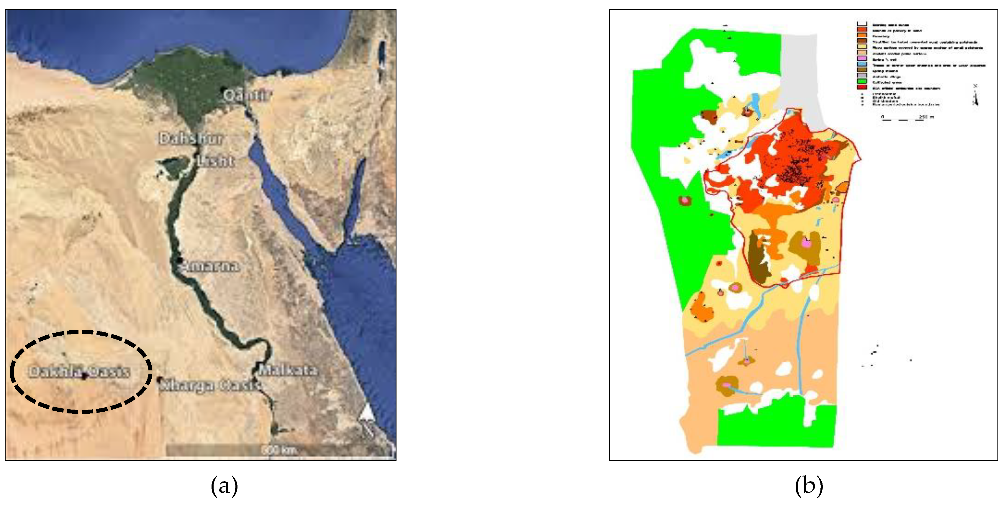
Figure 2.
(a) The area where the excavated coins were found (b & c) the coin hoard.
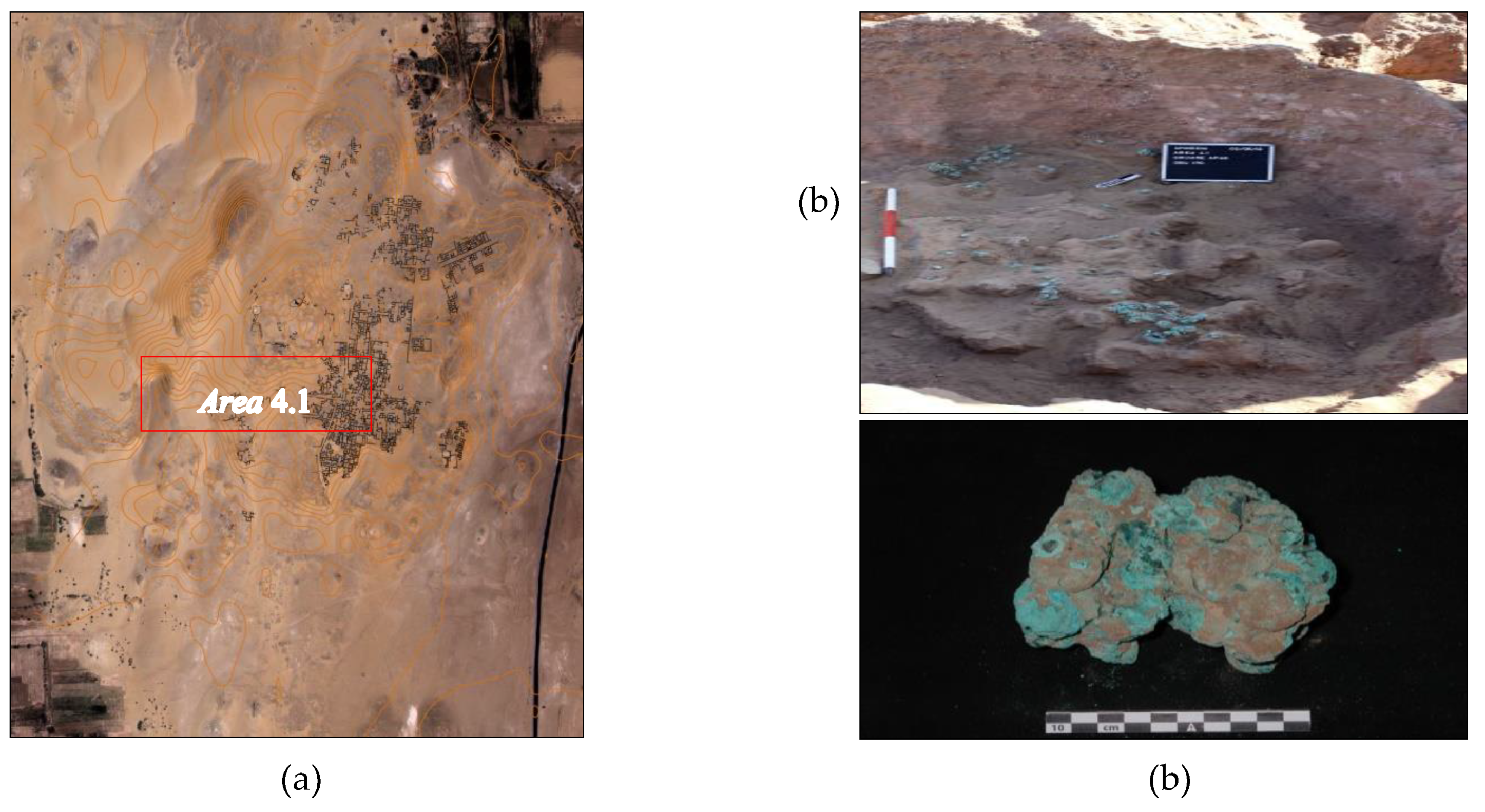
Figure 3.
(a) Coin number 66 (b) coin number 68 (c) coin number (73).
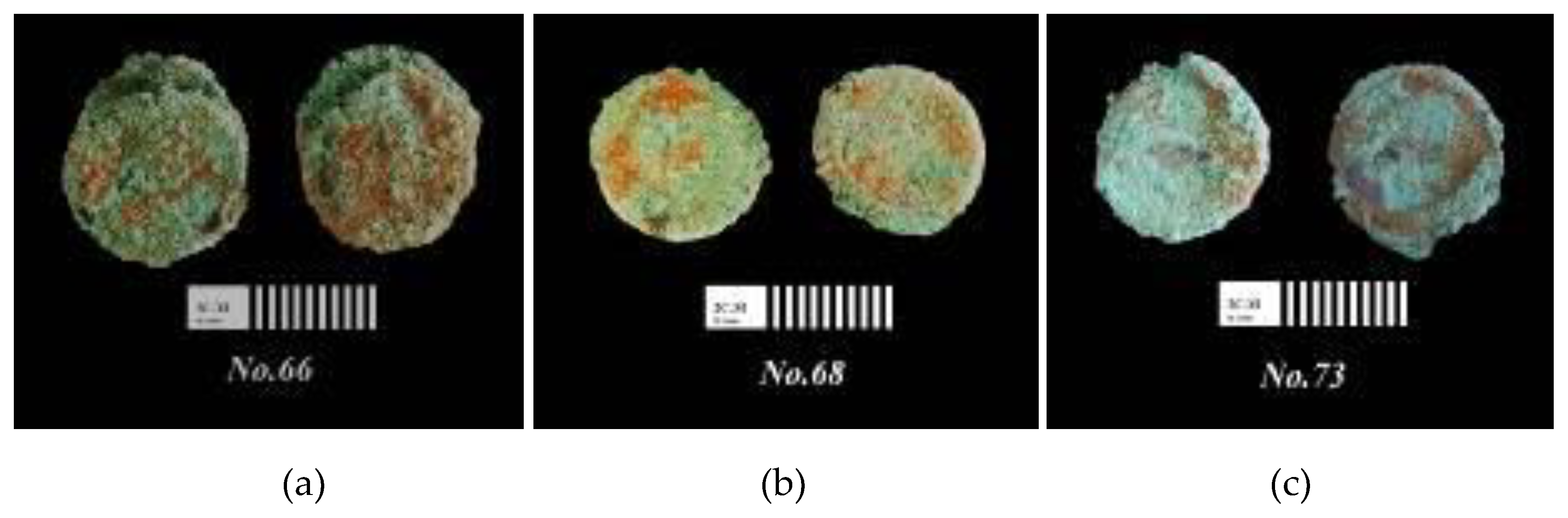
Figure 4.
Optical observation of (a) corrosion layers (170-x) (b) schematic representation of the corrosion layers (c) corrosion pitting (bronze disease) with pale green color (150-x).
Figure 4.
Optical observation of (a) corrosion layers (170-x) (b) schematic representation of the corrosion layers (c) corrosion pitting (bronze disease) with pale green color (150-x).
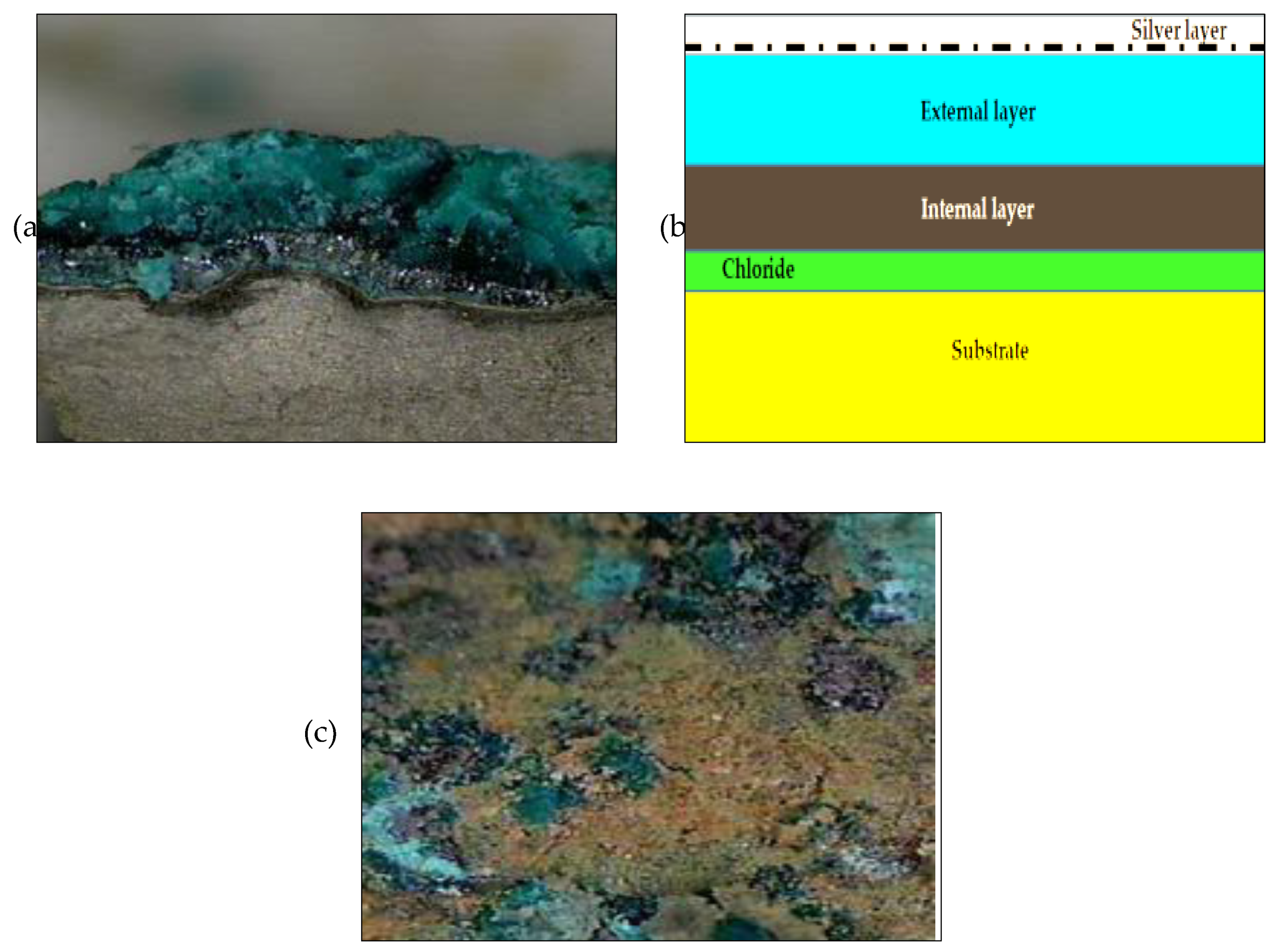
Figure 5.
SEM photomicrographs of (a) the bright areas on the surface of coin (66), (b) the thin superficial layer on the surface of coin (68), (c) the corrosion products on the surface of coin (73), (d, e. & f) EDS spectrum of the same samples.
Figure 5.
SEM photomicrographs of (a) the bright areas on the surface of coin (66), (b) the thin superficial layer on the surface of coin (68), (c) the corrosion products on the surface of coin (73), (d, e. & f) EDS spectrum of the same samples.
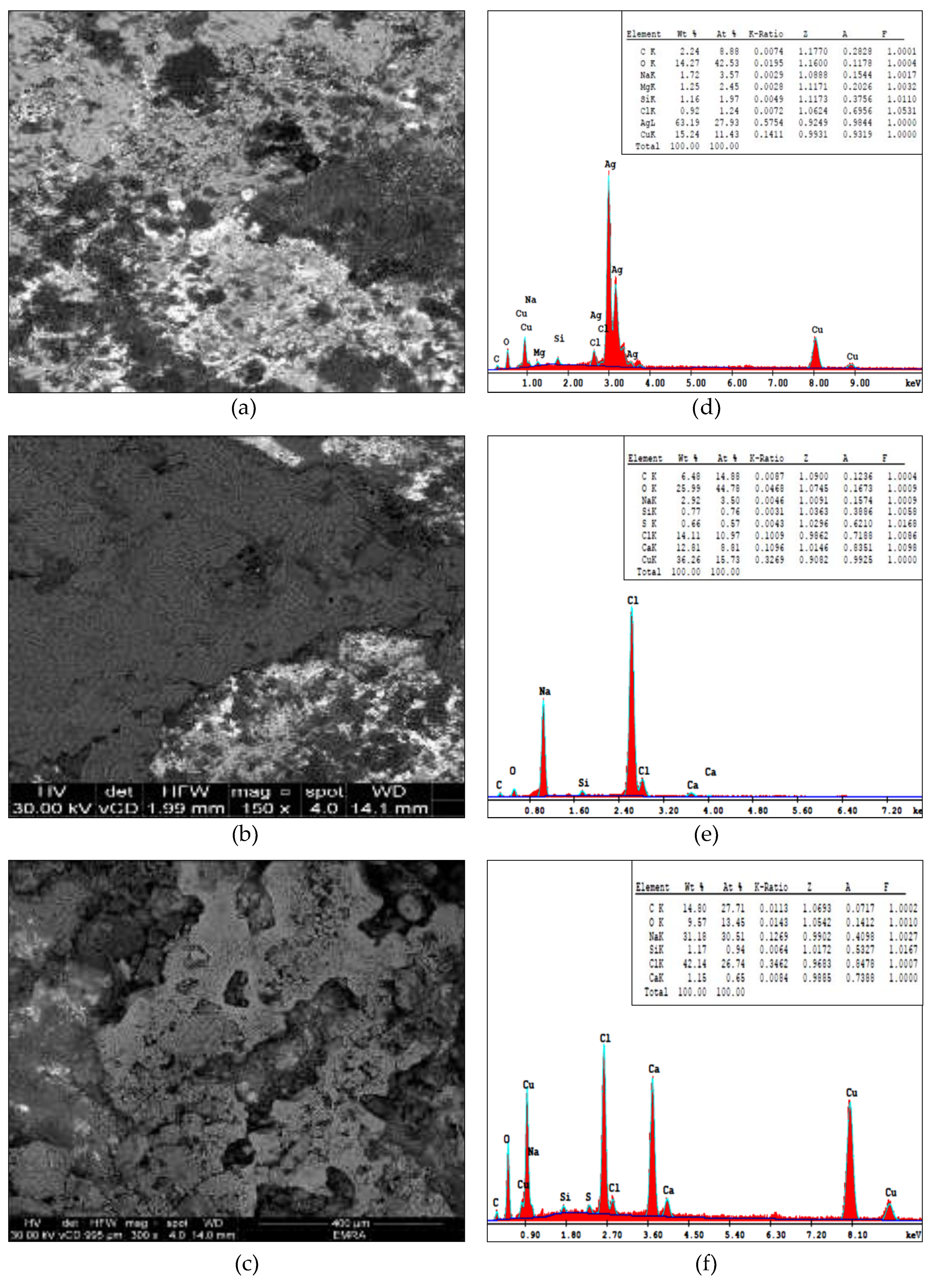
Figure 6.
XRD patterns of the studied samples of corrosion powder.
Disclaimer/Publisher’s Note: The statements, opinions and data contained in all publications are solely those of the individual author(s) and contributor(s) and not of MDPI and/or the editor(s). MDPI and/or the editor(s) disclaim responsibility for any injury to people or property resulting from any ideas, methods, instructions or products referred to in the content. |
© 2023 by the authors. Licensee MDPI, Basel, Switzerland. This article is an open access article distributed under the terms and conditions of the Creative Commons Attribution (CC BY) license (http://creativecommons.org/licenses/by/4.0/).
Copyright: This open access article is published under a Creative Commons CC BY 4.0 license, which permit the free download, distribution, and reuse, provided that the author and preprint are cited in any reuse.


