
Submitted:
04 April 2022
Posted:
07 April 2022
You are already at the latest version
Abstract
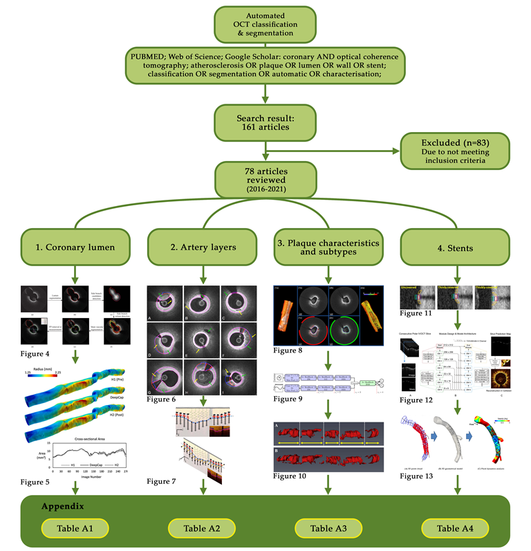
Coronary optical coherence tomography (OCT) is an intravascular, near-infrared light-based imaging modality capable of reaching axial resolutions of 10-20 µm. This resolution allows for accurate determination of high-risk plaque features, such as thin cap fibroatheroma; however, visualisation of morphological features alone still provides unreliable positive predictive capability for plaque progression or future major adverse cardiovascular events (MACE). Biomechanical simulation could assist in this prediction, but this requires extracting morphological features from intravascular imaging to construct accurate three-dimensional simulations of patients’ arteries. Extracting these features is a laborious process, often carried out manually by trained experts. To address this challenge, numerous techniques have emerged to automate these processes while simultaneously overcoming difficulties associated with OCT imaging, such as its limited penetration depth. This systematic review summarises advances in automated segmentation techniques from the past five years (2016-2021) with a focus on their application to the three-dimensional reconstruction of vessels and their subsequent simulation. We discuss four categories based on the feature being processed, namely: coronary lumen; plaque characteristics and subtypes; artery layers; and stents. Areas for future innovation are also discussed as well as their potential for future translation.
Keywords:
atherosclerosis
; biomechanics
; border detection
; coronary artery disease
; optical coherence to-mography
; stents
; vulnerable plaque
Copyright: This open access article is published under a Creative Commons CC BY 4.0 license, which permit the free download, distribution, and reuse, provided that the author and preprint are cited in any reuse.


