
Submitted:
23 January 2026
Posted:
02 February 2026
You are already at the latest version
Abstract
Dengue infection places over 40% of the world’s population at direct risk. This was a cross-sectional study involving collection of socio-demographic and risk factors data via self-designed structured questionnaires. Also, blood samples were collected from 600 consenting febrile patients of different ages, in three selected Government public health facilities of Adamawa State. Furthermore, serum was screened for dengue antibodies by ELISA. Reverse Transcriptase Polymerase Chain Reaction (RTPCR) was used to detect dengue virus serotypes. Data obtained was analyzed using SPSS version 20, Odds Ratios (OR) for associations between seroprevalence and all variables studied was determined. Of the 600 participants, 65% (390/600) were IgM positive for DENV infection with the highest prevalence obtained in Mubi (81.5%:163/200) and the lowest prevalence in Numan (52%:104/200). Higher insignificant prevalence was obtained in the males (66.8%:181/271) than females (63.6%:201/316). There was significant association of DENV antibodies, in terms of Tertiary education (χ2 = 4.594, p=0.032; OR = 1.502; 95%CI= 1.050-2.151), among those who uses well water (p=0.023; OR=2.021, 95% CI 1.131-3.611), those who use traditional medicine for treatment of fever (p=0.014, OR=3.126; 95% CI=1.289-7.582), mosquito protection methods used against bite and those who never travel outside the state. Other risk factors studied were not significantly associated with DENV infection. DENV 1 and 4 serotypes were detected. The study reveals that low economic disadvantage and negligence could serve as predisposing factors to the DENV infection. Therefore, it identified potential virus reservoir which may likely spread in humans and cause epidemics.
Keywords:
DENV 1-4
; serotypes
; pool screen
; dengue virus
; co-infection
; seroprevalence
1. Introduction
Dengue is an important mosquito-borne viral infection in humans, caused by Dengue Virus (DENV) globally. It is found in tropical and sub-tropical climates worldwide, mostly in urban and semi-urban areas (WHO, 2021). There are four serotypes of DENV; DENV1-DENV4 which is transmitted by infected female mosquitoes mainly of the species Aedes aegypti and to lesser extend Ae. albopictus. These are also vectors of chikungunya, yellow fever and Zika viruses (WHO, 2020). Infection with one serotype provides lifelong im munity against that serotype but confers only partial or transient protection against subsequent infection with any of the other three serotypes and secondary infection usually become severe (Oladipo et al., 2014).
Dengue virus is a positive sense, single-stranded RNA virus of the family Flaviviridae which comprised of more than 70 viruses. The viruses have four antigenically distinct serotypes, DENV1-4 distributed in tropical and subtropical areas (Nedjadi et al., 2015).
Infection may be asymptomatic and symptomatic. Patients that are symptomatic may present with fever, severe headache, backache, joint pains, nausea and vomiting, eye pain, rash, liver enlargement and haemorrhagic manifestations (Oyero and Ayukekbong, 2014). Infection with these viruses typically leads to antibody production in the serum. Immunoglobulin M (IgM) develops acutely and is short-lived, while immunoglobulin G (IgG) develops shortly thereafter and is long-lasting (Luke et al., 2011).
Dengue is an emerging and re-emerging arboviral disease (Marcos et al., 2019) and is a major public health concern throughout tropical and sub-tropical regions of the world. It has been estimated that 3.9 billion people in 129 countries are at risk of DENV infection with 390 million dengue infections annually, while 96 million manifest clinical symptoms. There is unprecedented global burden of dengue in 2024 with over 14.1 million reported cases worldwide, dengue has exceeded the historic milestone of 7 million cases reported in 2023. This highlights the alarming growth trajectory of this mosquito-borne disease (Haider et al., 2025). About half million people with dengue heamorrhagic fever and dengue shock syndrome requires hospitalization each year and 5% death (Khan and Bhatti, 2020). In addition, the COVID-19 pandemic has place immense pressure on health care and management systems worldwide. The combined impact of COVID-19 and dengue epidemics can potentially result in devastating consequences for the populations at risk (WHO, 2021).
There are many predisposing factors attributed to vector breeding and the rise in dengue outbreaks; these include demographic changes (global population increase and urbanization), changes in sociological behaviour (sudden increased in migration), agricultural development (new irrigation techniques and deforestation), possible global climate change, urban slums, changes in government public health strategies and evolutionary’ changes in the pathogen genomes (Malhotra et al., 2014).
There is substantial under-reporting of dengue within the health systems in Africa (Adeleke et al., 2015). In Nigeria, most cases of dengue are undiagnosed or misdiagnosed as malaria or referred to as fever of unknown cause (Oyero and Ayukekbong, 2014). The neglect of these viruses in Africa could be due to the high burden of malaria and to the more priority and attention given to other neglected tropical diseases. Therefore, this study aimed at assessing the serological and Molecular Surbey of dengue virus among febrile patients in the study area.
Serological and Molecular Survey of Dengue Virus Among Febrile Patients Attending Public Health Facilities in Some Part of North East, Nigeria
2. Materials and Methods
2.1. Study Area
The study was carried out in three purposively selected Local Government Area (LGAs) of Adamawa State. The state is located between latitude 9°45’06”N - 10°19’ 60”N and longitude 12° 03’ 18” E- 13° 29’ 59”E. Adamawa State is bordered by the Cameroon Republic on the East (Figure 1). The state is transverse by large rivers: Benue, Gongola and Yadzarem that are surrounded with water puddles and discarded containers due to human activities, which provided suitable breeding sites for Aedes mosquitoes (Pukuma et al., 2011). The study area is also characterized by high population movements due to insurgency in the North-east. Also, people store water around their homes, they dump solid waste in an open gutter and close to water bodies which provide suitable environment for breeding of Aedes species and the spread of DENV (Anthony, 2014). The study populations included both male and female febrile patients of all ages, attending the three selected public health facilities. The sample size used for the study was determined using the equation {n = Z2(1-p)/2d2} of Cochrane as modified by Kogi (2017) and DENV prevalence rate of 74.4% from previous studies was adopted (Oyinloye, et al., 2016).
2.2. Ethical Clearance
Ethical approval for the study was obtained from the Ethical Committee of the Adamawa State Ministry of Health and the Ahmadu Bello University Ethical Committee on the Use of Human and Animal Subject for Research, ABU, Zaria (Ref. N0. S/MoH/1131/1 and ABUCUHSR/2017/004).
2.3. Inclusion and Exclusion Criteria
The inclusion criteria are all consented patients of all ages, presenting with symptoms of fever (˃38 °C), severe headache, neck pain, back and abdominal pains in three selected hospitals of the study locations. Parents’ consented for their children to participate.
The exclusion criteria are all patients who did not consent to the study and those who did not present with febrile symptoms of fever.
2.4. Data Collection Using Questionnaires
A self-designed structured questionnaire was administered by trained interviewers to obtain possible risk factors and clinical information. The questionnaire was piloted around the actual communities of study.
2.5. Blood Samples Collection and Processing
Five millilitres (5ml) of blood was collected with the help of Phlebotomists from the consenting patients attending public health facilities that were selected for the study by venepuncture, placed in clean labelled sample bottles and allowed to clot at room temperature (15-250C). To obtain serum, each blood sample was separated by centrifugation at 1000rpm for 10 minutes (Cheesbrough, 2006). All the serum samples were transported in cold box and stored at -200C in the Parasitology and Entomology Laboratory of the Department of Zoology, Ahmadu Bello University, Zaria until test.
2.6. Detection of IgM Antibodies by ELISA
Dengue IgM ELISA test is an Enzyme-Linked Immunosorbent Assay (ELISA) for the qualitative identification of antibodies to dengue in human serum. Dengue virus was detected in patient serum using ELISA kit (Diagnostics Automation/Cortez Diagnostics, Inc. Woodland Hills, USA) following manufacturers’ protocols. All the Samples and reagents were brought to room temperature (15-25℃) before use. A 25ml Wash Buffer (20x) was diluted with 250ml of reagent grade water. A 96 Well Microplate was used. The wells were labelled as follows: a reagent blank well, 2 controls well and samples wells. Hundred micro litters (100µl) of controls, samples and 40µl Rheumatoid Factor (RF) Absorbent was dispensed into the microplate wells and thoroughly mixed using pipette. The plate was incubated for 10minutes at room temperature. The contents were shaken and washed 3 times with the diluted Wash Buffer and 2 drops of enzyme conjugate were added to the wells. Hundred micro litters (100µl) of Enzyme Conjugate was added to each well and incubated at room temperature for 10 minutes. The plate was vigorously washed 3 times with the Wash Buffer and blotted to dry on an absorbent paper. Two (2) drops of substrate Tetramethyl-benzidine (Chromogen) was then added to the wells and incubated for 10 minutes at room temperature after which Two (2) drops of Stop Solution was added to the wells and mixed gently by tapping the strip holder. The Optical Density (O.D) of the wells was read by setting the Microplate Reader wavelength at 450nm.
2.7. Interpretation of Results
An Optical Density ratio of 0.3-1.0 was interpreted as Negative, but when greater than 1.0 was interpreted as a positive result. The negative result indicates that there is no detectable antibody in the specimen while the positive result revealed antibody against DENV.
2.8. Extraction of Viral RNA from Serum
The RNA extraction of patient serum was performed following the methods by Chomczynski and Sacchi (1987). The 200µl of the serum and 0.5ml of Solution D (5M guanidium thiocynate, 31.25mM Sodium citrate, 0.625% sarcosyl and 0.45mL mercaptoethanol) were poured in 2ml tube. Followed by the addition of 100µl of sodium acetate, 500µl phenols (H2O saturated) and 100µl chloroform isoamylethanol. The mixture was vigorously shaken and placed on ice for 45 minutes and the reaction mixtures was centrifuged for 20min at 13000rpm. Then, 750µl upper aqueous phase was carefully pipetted and dispensed into a clean 1.5 tube to which 37.5µl glycogen and 750µl cold isopropanol was added and kept in -80℃ for 60 minutes. The reaction mixture was centrifuged for 20 minutes at 13000rpm, after which the supernatant was discarded and the pellet was washed with 75% cold ethanol, vortex and centrifuged for 20 minutes at 13000rpm. The tubes were air dried using speed Vacuum for 5minutes. The pellet was dissolved in 32.5µl of DEPC H2O. The extracted RNA was stored at -80℃ in Laboratory for Viral Research, University of Bremen, Germany for onward downstream application.
2.9. Synthesis of Complementary DNA (cDNA) from the Extracted RNA
The complementary DNA (cDNA) was synthesized using 80ng of RNA. The extracted RNA was mixed with 1µl (0.2µg/µl) random hexamer primer and 1µl (10 mM) dNTP mix in a 0.2 ml PCR tube. The mixture was made up to 15µl with nuclease-free water on ice. The following component was then added to the PCR tube: 4µl of 5X Reaction Buffer, 0.5 µl of Ribolock RNase inhibitor (40U/µl) and 0.5µl of Maxima H Minus Reverse Transcriptase (10000U, 100 U/µl) making up to a total volume mixture of 20µl.The reaction was vortexed, centrifuged and incubated in a thermocycler with the following conditions: 25℃ for 5 mins, 50℃ for 30 mins. The reaction mixture was then terminated by heating at 85℃ for 5 mins (Thermo scientific). The cDNA was stored in -80℃ for downstream application.
2.10. Reverse Transcriptase Polymerase Chain Reaction (RT-PCR) Amplification
To perform RT-PCR, the cDNA was amplified using DENV serotypes-specific consensus primers D1 and D2 that target the envelope and non-structural protein 1 (E/NS1) junction of the DENV genome (Table 1). The multiplex semi nested PCR was performed according to a previous protocol (Hakami et al., 2018) with some adjustment. The master mix reaction was prepared for detection of DENV. The reagents were vortex and kept on ice for the duration of the master mix preparation.
In 25 μl of final volume reaction mixture containing 2.5µl of Aedes species cDNA template, 2.5 μl (10 x dreamTaq buffer containing 20 MgCl2), 0.5 μl of dNTP Mix (10 mM, final concentration of 0.2 mM), 1μl of 25 mM MgSO4 (final concentration 1.0 mM), 0.5 μl of dream Taq polymerase (final concentration 5U/µl), 0.875µl of forward D1 and reverse D2 primers (100 μM; final concentration 0.7 μM), which was designed against the consensus cDNA sequence of DENV1-4 serotypes that amplify a fragments of 511 base pair. The reaction volume was made up to 25μl with double-distilled water (ddH20). The thermo cycling conditions were as follows: Initial denaturation at (94 °C, 3 mins), 35 cycles of denaturation (94℃, 30 secs), primers annealing (55℃, 1 min), primer extension (72 °C, 2 mins) and final extension at 72 °C for 5 mins.
2.11. Serotyping of DENV Serotypes
The PCR product obtained from the RT-PCR amplification using D1 and D2 as primers was further used as the template for DENV1-4 serotypes. Based on the above temperature conditions and reagent volumes, 0.5 µl and 1µl of the Nested 1 RT-PCR product (1:10 and 1:1000 in ddH20 for DENV serotypes 2 and DENV serotypes 1,3 and 4, respectively) was used as template in the subsequent nested PCR reaction (Table 1). In 25 µl reaction volume containing the forward primer D1 and the type-specific (TS) reverse primers; TS1, TS3 and TS4 (reverse primers for DENV serotypes 1, 3 and 4), and the forward primer, DV1 and DSP2, the reverse primers for serotype 2 was further amplified by nested PCR step II. The entire PCR product was resolved on a 3% agarose gel containing 0.5 μg/ml ethidium bromide (SERVA, Heidelberg, Germany). The expected sizes of the amplicons were 482 for DENV1, 362 for DENV2, 290 for DENV3 and 392 for DENV4 (Hakami et al., 2018).
2.12. Data Analyses
The data obtained from the questionnaire and the result of the laboratory tests was analyzed using Statistical Package for Social Sciences (SPSS) software (version 24 for Windows; SPSS Inc., Chicago, IL, USA). The odds ratios (OR) for the association of various variables in relation to dengue virus detection was analyzed using Epi-InfoTM. Descriptive statistics was used to determine the prevalence of dengue in human’s population. All data was analyzed and tested at p ≤ 0.05 and p ≤ 0.05 was considered significant in all the analysis.
3. Results
Dengue Virus Antibodies (IgM) Among Patients in Adamawa State Using ELISA
Serological results of the 600 individuals assay for IgM antibodies of DENV, 200 from each location, comprising of Mubi, Yola and Numan. Of the 600 febrile patients, 65% (390/600) of the sera were IgM positive for DENV (Table 2). Highest prevalence was observed in Mubi (81.5%: 163/200) and lower prevalence in Numan (52%:104/200). Dengue virus infection was significantly detected in Mubi (χ2 = 34.821, p=0.000; 95%CI= 0.304-0.614). Patients residing in Mubi were 3.4 times likely to be infected with DENV (OR = 3.35).
Results of other risk factors that might be associated with DENV infection is presented in (Table 3). Based on the methods people use for protecting themselves against the Aedes mosquito’s bite namely, mosquito coil/repellant, net and insecticide. The highest prevalence (87.3%:55/63) was observed among those who use all these protective methods while lower prevalence (62.3%:149/239) was obtained among those who use net. Also, there was a significant association between DENV IgM detection with all the protective methods used (χ2 = 13.634, p=0.000; 95%CI=1.883-8.664). There were higher odds of association (OR=4.039) of protective methods use and DENV infection.
Dengue virus IgM was highest (73.0%:46/63) in febrile patients that use well as their source of water and lower (50.0%:2/4) among those who use rain water as their sources of water. There was observed significant association (χ2 = 5.197, p=0.023; 95% CI =1.131-3.611) between well water and prevalence to DENV infection. Febrile patients who use well water showed high odds of association (OR=2.021) with DENV infection.
There was no significant association (χ2 = 0.227, p=0.634; 95% CI =0.227-0.634) between storage of water in the compound and DENV infection. Majority of the positive cases were observed among those that do not store water in the compound (69.8%:30/43). Febrile patients who store water in their compound showed higher likelihood (OR=1.247) of infection with DENV.
Highest prevalence (66.2%:96/145) was obtained among those who do not empty water from discarded containers while lowest prevalence (65.2%:281/431) was observed among those who empty water from discarded containers.
However, there was no significant association of DENV prevalence with those who do not empty water from discarded containers (χ2 = 0.015, p=0.904;95% CI =0.703-1.556).
Highest prevalence (68.2%:116/170) was observed among those that have damaged drainage system/Septic tanks and lowest prevalence (64.5%:262/406) among those that have good drainage system/Septic Tanks. There was no significant association in the prevalence of DENV infection (χ2 = 0.574, p=0.449) with the presence of damaged drainage system/septic tanks. However, those with damaged drainage system/septic tanks were 1.18 times likely to be infected with DENV(OR=1.18).
Febrile patients that traveled outside the country had the highest prevalence (70.7%:70/99) while individuals that never travel outside the state had the lowest prevalence (60.3%:70/116). There was a significant association in the prevalence obtained among individuals that never travels outside the state and the presence of the virus (χ2 = 16.237, p=0.000; 95% CI=1.532-3.402). However, febrile patients who had travelled outside were 1.3 times likely to be infected with DENV (OR=1.340).
Higher percentage (85.0%:34/40) of the patients indicated using traditional medicine for treatment of fever, while lowest prevalence (61.8%:226/366) were obtained among those who usually see doctor for treatment of fever. There was significant association between those who used traditional medicine for treatment and DENV infection (χ2 = 6.102, p=0.014, 95% CI=1.289-7.582). Patients who use traditional medicine for treatment showed higher likelihood of infection with DENV (OR=3.126).
Symptoms presented by the febrile patients were considered (Table 4). The highest prevalence (72.9%:97/13; 72.9%: 27/37) of DENV IgM was observed among individuals who had fever and pains behind the eyes while lowest prevalence (53.3%:8/15) was obtained among those who had nausea. There was no significant association (χ2= 0.098, 0.438, p=0.508) between fever, pains around the eyes and DENV infection. Patients who reported symptoms of fever (OR= 1.472) and pains around the eyes (OR= 1.378) had more likelihood of been infected with DENV.
In order to confirm dengue infection and to determine the dengue serotypes circulating among human patients in Adamawa State. Nine (9) serum samples positive for IgM ELISA were selected tested by semi-nested RT-PCR. Dengue virus 1,2, 3 and 4 serotypes specific primer sets was used with specific amplicons of 482 bp, 362bp, 290 bp and 392 bp respectively.
The Electrophoregram of the amplified cDNA by RT-PCR is presented in Figure 2. Lanes M: Standard size marker of 50bp, C- = negative control, C+ = positive control of DENV-4 (392bp) from cell culture propagation in Vero cells, Lanes 107,113,149 (Mubi), Lanes 9,15,89 (Yola) and Lanes 23,36, 44 (Numan) were test samples. Dengue virus 1, was detected in all the representative samples (100%:9/9), DENV 4 was detected in 6 samples (66.7%). Mixed infection of DENV1 and DENV4 occurred in 6 samples (66.7%). However, DENV 3 and DENV 2 were not detected in any of the samples tested (Figure 3.
4. Discussion
4.1. Screening of IgM Antibodies Against DENV Infection Among Patients in Adamawa State
This study was conducted to screen IgM antibodies against dengue virus infection among febrile patients in Adamawa State, Nigeria. Results obtained showed 65% of the febrile patients studied have been exposed to DENV infection and was significantly distributed by location during the study. The high prevalence obtained in the study could be attributed to the following reasons. Firstly, the samples were collected from febrile patients with DENV indicative symptoms during period of peak of Aedes mosquito breeding, thus transmission due to DENV was in corroboration with Baba et al. (2011) who reported dengue antibodies to be significantly higher during the raining season, than during the cold harmattan and dry periods in the North-east, Nigeria. Secondly, the insurgency in North-east, Nigeria might have forced infected people from neighbouring states and some of the affected areas to settle in these cities for safety which might have led to over population and possible surge in the risk of transmission. Thirdly, trans-border transmission due to socio-economic activities as the state shares borders with Cameroon, that have been reported to have prevalence of 61.4% in Douala, 24.2% in Garoua and 9.8% in Yaounde (Demanou et al., 2014).
The 65% prevalence reported in this study is higher than the 0.5% reported in Borno (Baba and Talle, 2011), 51.9% reported from three selected hospitals in Kaduna State (Bello, et al., 2016), 35% reported in Ibadan by Oyero and Ayukekbong (2014), 30.8% in Kwara State by Adedayo et al. (2013) and 17.2% reported among healthy individuals, in Ogbomoso, Oyo State (Oladipo et al., 2014). In contrast, lower than the prevalence of 78.3% reported in the febrile patients attending secondary health facilities in Kano (Abdulaziz et al., 2020), the 77.1% prevalence reported among children with febrile illness in Nnewi, Anambra State, the 67.71% prevalence reported in sera samples collected from febrile, clinically suspected malaria/typhoid patients in Borno State, (Baba et al., 2013) and 74.4% prevalence among Internally Displaced Persons (IDPs) in North-east, Nigeria (Oyinloye, et al., 2016). Thus, the variations may be related to the fact that infections tend to vary from one locality to another and from one country to another depending on the level of the associated risk factors, seasons of the samples collection, methods of analysis and climatic conditions.
Highest seroprevalence was obtained in Mubi (81. 5%).This correlates with DENV infection detected in Aedes mosquitoes, which was also highest in Mubi. Therefore, there is high endemicity of DENV IgM, in the study area which could lead to epidemic if left unchecked.
4.2. The Risk Factors Associated with DENV Transmission in Adamawa State
Despite the use of mosquito protection methods against bite such as mosquito coil, net, insecticide and repellant by individual’s participants, infection was still found to be significantly associated with all the methods used. Probably, the people have been exposed to Aedes mosquitos’ bites during outdoors activities such as leisure hours, field activities, relaxation centers and public places, since Aedes species are known to bite during the day time and more Aedes mosquitoes were collected in the study areas between 6:45am-6:23pm.
The study consent with that of Ndenga et al. (2017) who reported that most human-vector contact occurs during the time of the day. The study also agrees with that of Liu et al., (2019) who found out that people who participated in outdoor sports activities were at a significantly higher risk of contracting DENV than those who did not. Also, the Aedes species might have developed resistance over time against the commercially available aerosols and repellant in the study area.
The infection was significantly associated and highest among individuals who uses well water as their sources of water. Many of the study population depend mainly on personal wells dug in or around their houses. However, some of the wells in the study areas were shallow as well as open. Similarly, some of the wells were surrounded with water puddles, discarded containers and water troughs for domesticated animals such as poultry, dogs, cats and caged birds. These might have increased a potential breeding spots for Aedes species, since Aedes mosquitoes are known to breeds in any small water collection available to them. World Health Organization has established that Aedes mosquitos’ breeds in small water collections, in and around houses made up of drinking water containers, discarded car tyres, flower vases and ant traps (WHO, 2020). The Aedes species after breeding in the water paddles and discarded containers might have acquired DENV from infected humans and eventually aid the spread of the DENV in the area.
The consequence of rapid urbanization due to the upheavals in the North-east might have contributed to the overstretching of infrastructure, thereby reducing the possible supply of clean treated drinking water in the area, thus encouraging digging of shallow wells and storage of water in the houses. Beside, most of the study participants are poor who largely depend on public water supply systems (borehole) or well water. The low prevalence obtained among those that uses rain water may not be unconnected to the fewer number of individuals who indicated using rain water as sources of drinking water. Thus, this finding established that the use of well water in the area is important in the spread of DENV infection.
Dengue infection was highest and insignificantly associated with individuals who do not store water inside containers in the study locations. It is possible that many of the people despite storage of water in containers might have unknowingly been practicing dengue eradication programme by covering of water storage tanks and vessels with lids or mesh to prevent mosquitoes from breeding. It is also clear that the high dengue infection obtained in the study may not be due to water storage in containers. However, febrile patients who store water in their compound were likely to be infected with DENV.
The study also established highest prevalence of DENV infection among individuals who do not empty discarded containers free from water as part of DENV controls measures. Although not statistically significant, but is an indicator of possible ground for Aedes mosquitoes breeding in the study area. It also signifies the importance of emptying discarded containers free from water for prevention of outbreaks.
Dengue virus was insignificantly detected with highest prevalence among those whose drainage/Septic Tank were opened or broken. Those with damaged drainage system/septic tanks indicated likelihood of infection with DENV. The study area was observed to have poorly constructed septic tanks/drainage systems which were broken and blocked with solid waste due to human activities over time and overflown with stagnant water. Some of the Aedes species were collected in damaged septic tanks around the residential areas. This might have provided favourable environmental conditions for the breeding of DENV vector and thereby increasing the transmission of DENV infection in the area. The study agrees with that of Dhimal et al. (2014) who established association of dengue virus vectors and poor septic tanks/drainage systems in Nepal.
In terms of travel history, the study showed significant difference with those that never travel outside the state. It means that individuals were highly susceptible to the infection despite not travelling outside the country. This indicates that the disease is domicile within the population and those participants might have gotten it from asymptomatic individuals through the bite of infected Aedes mosquito.
However, there was strong association between travel history and the prevalence of DENV infections in the study area. Highest prevalence was observed among those who had the privilege of travelling outside the country but not statistically significant. This study further confirms possible cross boarder transmission. This is not surprising since Adamawa State shares borders with Cameroon and people usually visit for educational, vocational or business activities. Thus, increasing the risk of importing and distributing DENV infection. Similarly, Ratnam et al. (2013) confirm dengue as the common cause of fever and accounting to about 16% of all febrile illnesses in returned travelers. Also, Raut et al. (2015), further stressed a connection between dengue prevalence and history of travels; in which, DENV infection was detected in a young man attending college in India after returning from Nigeria.
This study also showed highest prevalence and significant association among those who use traditional medicine for treatment of fever. The high prevalence obtained could have been due to the ineffectiveness of the remedies used or the poor method of administering a correct dosage. The people of Adamawa State, uses medicinal plants for treatment of infectious diseases, some of the people preferred it to the orthodox drugs (Thagriki et al., 2015). However, despite the wide acceptance of traditional medicine in Nigeria for treatment, it has its attendant consequences. The safety and effectiveness of a sizeable number of such plant-based formulations have not been scientifically validated and proven by researchers for treatment of DENV fever (Singh and Rawat, 2017). In addition, there is problem in ensuring safety, effectiveness, efficacy and quality of traditional medicine in Nigeria (Augustine et al., 2017).
In respect to symptoms of DENV infection, each study participants indicated at least one symptoms of DENV infection. However, highest prevalence was observed among those who came with complain of fever and pains around their eyes but not significant. This agrees with several reports that stated fever and pains around the eye as one of the commonest symptoms of DF (CDC, 2020). This is a further confirmation to the fact that the target populations were truly febrile patient who came to seek medical assistance. However, this is a community that perceives most fever to be due to malaria and many usually resolve to self-medication.
4.3. Dengue Virus Prevalence and Serotypes Circulating in Humans in Adamawa State
The study also considered DENV serotypes circulating among febrile patient in Adamawa State. The RT-PCR results of the positive samples showed DENV1 to be the most dominant serotype circulating currently among humans in Adamawa State. This could be due to the vector competency in transmission of DENV 1 serotype to humans in the study area compared to the other serotypes. This finding agrees with that of Mwanyika et al. (2019) who reported DENV1 as the most dominant serotypes among febrile patient in Tanzania. The finding however, contrast Ayolabi et al. (2019) who detected DENV 1 and 3 to be actively circulating in Lagos, Nigeria and Shah et al. (2020) who found DENV 1-4 serotypes among children with undifferentiated fever in Kenya. The variation observed could be due to differences in study locations, populations and time of the study.
Even though, we found DENV1-4 serotypes circulating in Aedes species in the same study locations, we did not isolate DENV-2 and 3 serotypes in humans. This suggests an inefficient transmission of DENV-2 and DENV-3 by the vectors in Adamawa State even though DENV 1, 2 and 3 serotypes have been reported among febrile patients in Borno State (Baba et al., 2011). However, we did not screen all the positive samples obtained by IgM ELISA using multiplex semi nested RT-PCR for DENV serotypes, rather we only used representative samples.
The study reported DENV serotypes co-circulating among humans in Adamawa State for the first time. Simultaneous infection of DENV serotypes is possible especially during outbreaks and this suggest hyper endemic of DENV infection in the study areas. The mixed-infection of DENV serotypes could increase or worsen the severity of the disease leading to dengue haemorrhagic fever and dengue shock syndrome. The mixed infection could also create difficulty in the management of patient (Mwanyika et al., 2019).
5. Conclusions
The seroprevalence of DENV among febrile patients attending public health facilities in the study locations was found to be 65% and highest in Mubi (81.5%). This is an indication of the potential endemicity of DENV infection in the North-east, Nigeria.
The highest and significant association of DENV infection was observed among those using well water (73.0%; OR=2.021;95% CI:1.131-3.611), the mosquito protection methods used against Aedes mosquitoes bite (87.3%; OR= 4.039;95%CI:1.883-8.664), those that never travel outside the state (60.3%; χ2 = 16.237, OR=2.28; p=0.000; 95% CI=1.532-3.402) and those who use traditional medicine for treatment of fever (85.00%; OR= 3.13 ;95%CI:1.289-7.582). Mixed infection of DENV 1 and 4 were detected in humans with DENV-4 serotype not previously described in Nigeria. Therefore, these individuals could serve as a potential reservoir for the transmission of DENV infection.
6. Recommendation
Given the high prevalence obtained in our study, there is the need to strengthen the surveillance system (humans, animal and vectors) to prevent outbreaks and/or to ensure the early detection of a potential epidemic.
Acknowledgments
We wish to appreciate the support of all the phlebotomists who assisted in the blood collection in the various locations. A special appreciation goes to Africa Centre of Excellence and Forensic Biotechnology (ACENTDFB) and Erasmus plus EU Program for funding support. Also, Sorge Kelm, Centre for Biomolecular Interaction Bremen (CBIB) University of Bremen for laboratory space, Jonas Schmidt-Chanasit, Bernhard-Nocht-Institute, Hamburg for providing positive controls for DENV-2 and 4 and Ibrahim Mahamat, CBIB for guidance and technical support.
Conflicts of Interest
The authors declare no conflicting interest regarding the publication of this paper.
References
- Abdulaziz, M.M.; Ibrahim, A.; Ado, M.; Ameh, C.; Umeokonkwo, C.; Sufyan, M.B.; Balogun, M.S.; Ahmed, S.A. Prevalence and factors associated with dengue fever among febrile patients attending secondary health facilities in Kano metropolis, Nigeria. African Journal of Clinical and Experimental Microbiology 2020, 21(4), 340–348. [Google Scholar] [CrossRef]
- Adedayo, F.; Nioma, I.; Olanrewaju, M.B.; Adeyinka, A.; Ebele, A. Serological evidence of recent dengue virus infection among febrile children in a semi-arid zone. American Journals of Infectious Diseases 2013, 9, 7–10. [Google Scholar] [CrossRef]
- Adeleke, M.A.; Sam-Wobo, S.O.; Garza-Hernandez, J.A.; Oluwole, A.S.; Mafiana, C.F.; Reyes-Villanueva, F.; Rodriguez-Perez, M.A. Twenty-three years after the first record of Aedes albopictus in Nigeria: its current distribution and potential epidemiological implications. African Entomology 2015, 23(2), 348–355. [Google Scholar] [CrossRef]
- Anthony, G.T. Assessing Web Compliance of Base Map using the Open Street Map: The Case of Adamawa State, Nigeria. Journal of Environment and Earth Science 2014, 4(11), 73–82. Available online: https://www.iiste.org/Journals/index.php/JEES/article/view/13994.
- Augustine, E.C.; Ugoha, R.; Azubuike, M.I. The Contributions of African Traditional Medicine to Nigeria’s Health Care Delivery System. IOSR Journal of Humanity and Social Sciences 2017, 22(5), 32–43. [Google Scholar]
- Ayolabi, C. I.; Olusola, B. A.; Ibemgbo, S. A.; Okonkwo, G. O. Detection of Dengue viruses among febrile patients in Lagos, Nigeria and phylogenetic of circulating Dengue serotypes in Africa. Infection, genetics and evolution: journal of molecular epidemiology and evolutionary genetics in infectious diseases 2019, 75, 103947. [Google Scholar] [CrossRef] [PubMed]
- Baba, M.; Logue, C. H.; Oderinde, B.; Abdulmaleek, H.; Williams, J.; Lewis, J.; Laws, T. R.; Hewson, R.; Marcello, A.; D’ Agaro, P. Evidence of arbovirus co-infection in suspected febrile malaria and typhoid patients in Nigeria. Journal of infection in developing countries 2013, 7(1), 51–59. [Google Scholar] [CrossRef]
- Baba, M.M.; Muhammad, T. The Effect of Climate on Dengue Virus Infections in Nigeria. New York Science Journal 2011, 4(1), 28–33. Available online: http://www.sciencepub.net/newyork.
- 9. Centre for Disease Control and Prevention Symptoms and Treatment. 23 November 2020. Available online: https://www.cdc.gov/dengue/symptoms/index.html.
- Cheesbrough, M. District Laboratory Practice in Tropical Countries, 2nd ed.; Cambridge University, Press: USA, 2006; pp. Pp 295–297. [Google Scholar]
- Demanou, M.; Pouillot, R.; Grandadam, M.; Boisier, P.; Kamgang, B.; Hervé, J. P.; Rogier, C.; Rousset, D.; Paupy, C. Evidence of dengue virus transmission and factors associated with the presence of anti-dengue virus antibodies in humans in three major towns in Cameroon. PLoS Neglected Tropical Diseases 2014, 8(7), e2950. [Google Scholar] [CrossRef]
- Dhimal, M.; Aryal, K.K.; Dhimal, M.L.; Gautam, I.; Singh, S.P.; Bhusal, C.L. Knowledge, Attitude and Practice Regarding Dengue Fever among the Healthy Population of Highland and Lowland Communities in Central Nepal. PLoS ONE 2014, 9(7), e102028. [Google Scholar] [CrossRef]
- Haider, N.; Hasan, M.N.; Onyango, J.; Billah, M.; Khan, S.; Papakonstantinou, D.; Paudyal, P.; Asaduzzaman, M.D. Global dengue epidemic worsens with record 14 million cases and 9000 deaths reported in 2024. International Journal of Infectious Diseases 2025, 158, 107940. [Google Scholar] [CrossRef]
- Hakami, A.M.; Mohammed, I.; Qadri, M.I.; Al-Ghamdi, K.; Alkenani, N.A.; Eisa, Z.M.; Matabi, A.M.; Bakri, M. Molecular Identification and Characterization of Different Dengue Virus Serotypes Reported in Jazan Area, Kingdom of Saudi Arabia. Journal of Experimental Biology and Agricultural Sciences 2018, 6(5), 828–835. [Google Scholar] [CrossRef]
- Huang, Y.; Higgs, S.; Horne, K.; Vanlandingham, D. Flavivirus-Mosquito Interactions. Viruses 2014, 6(11), 4703–4730. [Google Scholar] [CrossRef] [PubMed]
- Khan, N.; Bhatti, J. M. A Case Report on Dengue Encephalitis with Optic Neuropathy. Cureus 2020, 12(8), e9592. [Google Scholar] [CrossRef] [PubMed]
- Kogi, E. A Proposed Modification to the Formula for Determination of Sample Size in Prevalence Studies. A Paper Presented at the 39th Annual Conference of the Parasitology and Public Health Society of Nigeria Held at the Federal University, Lafia, Nasarawa State, Nigeria, 2nd to 5th September, 2017; 2017. [Google Scholar]
- Lanciotti, R.S.; Charles, H.C.; Duane, J.G.; Gwong-Jen, C.; Vorndam, A.V. Rapid Detection and typing of Dengue Viruses from Clinical Samples by Using Reverse Transciptase-Polymerase Chain Reaction. Journal of Clinical Microbiology 1992, 30(3), 545–551. [Google Scholar] [CrossRef]
- Liu, J.; Tian, X.; Deng, Y.; Du, Z.; Liang, T.; Hao, Y.; Zhang, D. Risk Factors Associated with Dengue Virus Infection in Guangdong Province: A Community-Based Case-Control Study. International Journals of Environmental Research and Public Health 2019, 16, 617. [Google Scholar] [CrossRef] [PubMed]
- Luke, EM; Rodney, LC; Musila, AL; Trish, P; Fredrick, O; Victor, OO; Randal, JS; Cindy, AR; Nicholas, A. Seroprevalence and distribution of arboviral infections among rural Kenyan adults: A cross-sectional study. Virol J 2011, 8, 371. [Google Scholar]
- Marcos A. Espinal, MD, Dr PH, Jon K. Andrus, MD, Barbara Jauregui, MD, Stephen Hull Waterman, MD, MPH, David Michael Morens, MD, Jose Ignacio Santos, MD, Olaf Horstick, Lorraine Ayana Francis and Olson, D. Emerging and Reemerging Aedes-Transmitted Arbovirus Infections in the Region of the Americas: Implications for Health Policy. America Journal of Public Health Pan American Health Organization 2019, 109(3), 387–392. [CrossRef]
- Malhotra, G.; Yadav, A.; Dudeja, P. Knowledge, awareness and practices regarding dengue among rural and slum communities in north Indian city, India. International Journals of Medical Science and Public Health 2014, 3(3), 295–9. [Google Scholar] [CrossRef]
- Mwanyika, G. O.; Mboera, L.; Rugarabamu, S.; Makange, M.; Sindato, C.; Lutwama, J.J.; Paweska, J.T.; Misinzo, G. Circulation of dengue serotype 1 viruses during the 2019 outbreak in Dar es Salaam, Tanzania. In Pathogens and global health; Advance online publication, 2021; pp. 1–9. [Google Scholar] [CrossRef]
- Ndenga, B.A.; Mutuku, F.M.; Ngugi, H.N.; Mbakaya, J.O.; Aswani, P.; Musunzaji, P.S. Characteristics of Aedes aegypti adult mosquitoes in rural and urban areas of western and coastal Kenya. PLoS ONE 2017, 12(12), e0189971. [Google Scholar] [CrossRef]
- Nedjadi, T.; El-Kafrawy, S.; Sohrab, S.S.; Desprès, P.; Damanhouri, G.; Azhar, E. Tackling dengue fever: Current status and challenges. Virology Journal 2015, 12(1), 212. [Google Scholar] [CrossRef]
- Oladipo, E.K.; Amanetu, C.; Gbadero, T.A.; Oloke, J.K. Detectable anti-dengue virus IgM antibodies among Healthy Individuals in Ogbomoso, Oyo State, Nigeria. American Journal of Infectious Diseases 2014, 10(2), 64–67. [Google Scholar] [CrossRef]
- Oyero, O.G; Ayukekbong, J.A. High dengue NS1 antigenemia in febrile patient’s in Ibadan, Nigeria. Virus Resources 2014, 191, 59–61. [Google Scholar] [CrossRef] [PubMed]
- Oyinloye, S.O.; Wajiroko, M.; Lawan, A.M.; Umar-Faruq, A.; Samuel-Bumba, M.; Yusuf, S.; Anjikwi, N.; Abu-Mohammad, K. Dengue virus infection in northeast Nigeria: Case study of a squatters’ Camp. International Journals of Perception in Public Health 2016, 1(1), 59–65. [Google Scholar] [CrossRef]
- Pukuma, S. M.; James-Rugu, N. N.; Sale, M. A study on tick borne infections of cattle in Yola locality of Adamawa State. African Journal of Agricultural Research 2011, 6(29), 6208–6211. [Google Scholar] [CrossRef]
- Ratnam, I.F.; Karin, L.F.; Jim, B.F.; Joseph, T.F. Dengue Fever and International Travel. Journal of Travel Medicine 2013, 20(6), 384–393. [Google Scholar] [CrossRef] [PubMed]
- Raut, C. G.; Rao, N. M.; Sinha, D.P.; Hanumaiah, H.; Manjunatha, M.J. Chikungunya, dengue, and malaria co-infection after travel to Nigeria, India. Emerging infectious diseases 2015, 21(5), 908–909. [Google Scholar] [CrossRef]
- Shah, M. M.; Ndenga, B. A.; Mutuku, F. M.; Vu, D. M.; Grossi-Soyster, E. N.; Okuta, V.; LaBeaud, A. High Dengue Burden and Circulation of 4 Virus Serotypes among Children with Undifferentiated Fever, Kenya, 2014–2017. Emerging Infectious Diseases 2020, 26(11), 2638–2650. [Google Scholar] [CrossRef]
- Singh, K.P.; Rawat, P. Evolving herbal formulations in management of dengue fever. Journal of Ayurveda and Integrate 2017, 8, 207–210. [Google Scholar] [CrossRef] [PubMed]
- Thagriki, D.; Dahiru, D.; Yaduma, W.G. Survey on Some Indigenous Selected Medicinal Plants Used for the Treatment of Malaria Found in Sangere, Girei Local Government Area of Adamawa State, Nigeria. International Journals of Research in Pharmaceutical and Bioscience 2015, 2(1), 21–26. [Google Scholar]
- World Health Organization. Dengue and severe dengue. May 2021. Available online: https://www.who.int/news-room/fact-sheets/detail/dengue-and-severe-dengue.
- World Health Organization. Dengue and severe dengue. June 2020. Available online: https://www.who.int/news-room/fact-sheets/dengue-and-severe-dengue.
Figure 1.
Sampling locations in Adamawa State. Source: Modified from the Administrative Map of Nigeria.
Figure 1.
Sampling locations in Adamawa State. Source: Modified from the Administrative Map of Nigeria.
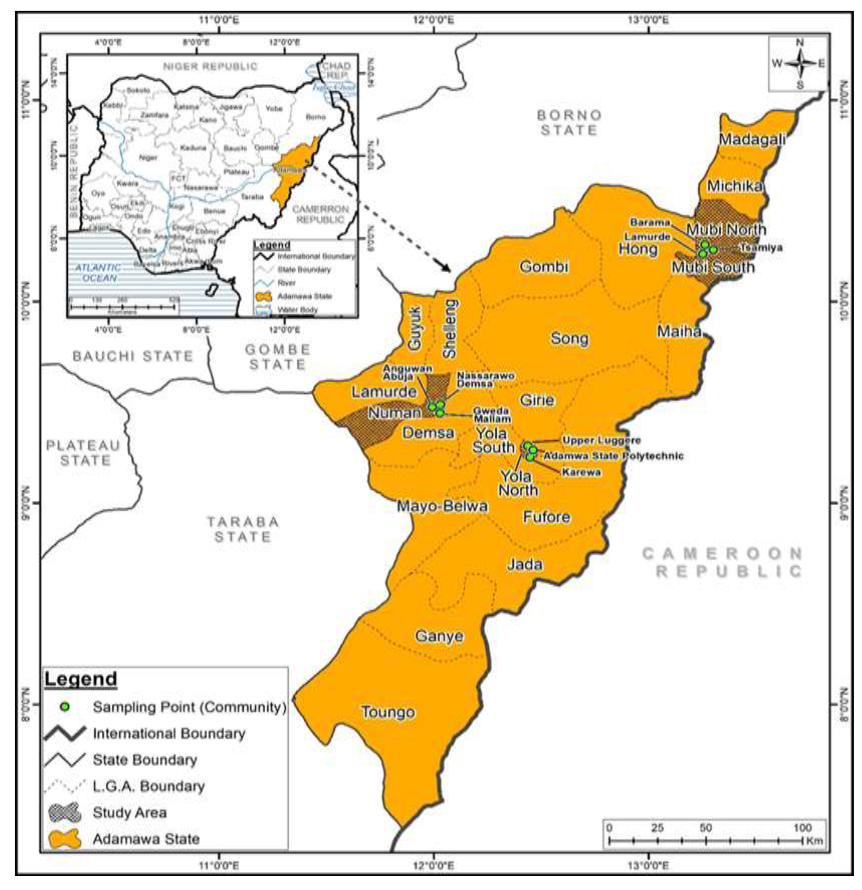
Figure 2.
Electrophoregram of amplified cDNA by RT-PCR isolated from human serum.
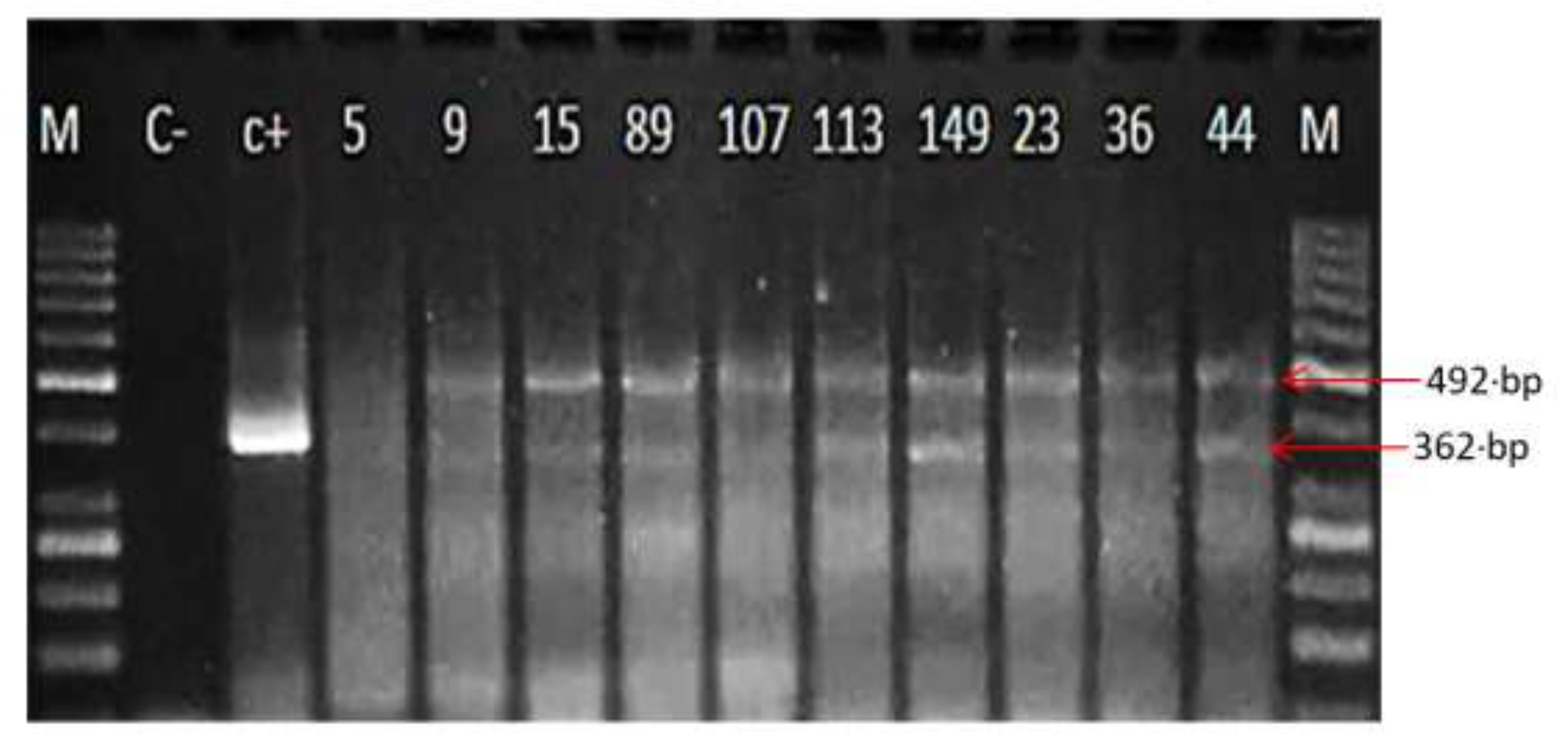
Figure 3.
DENV Serotypes circulating among patients in Adamawa State.
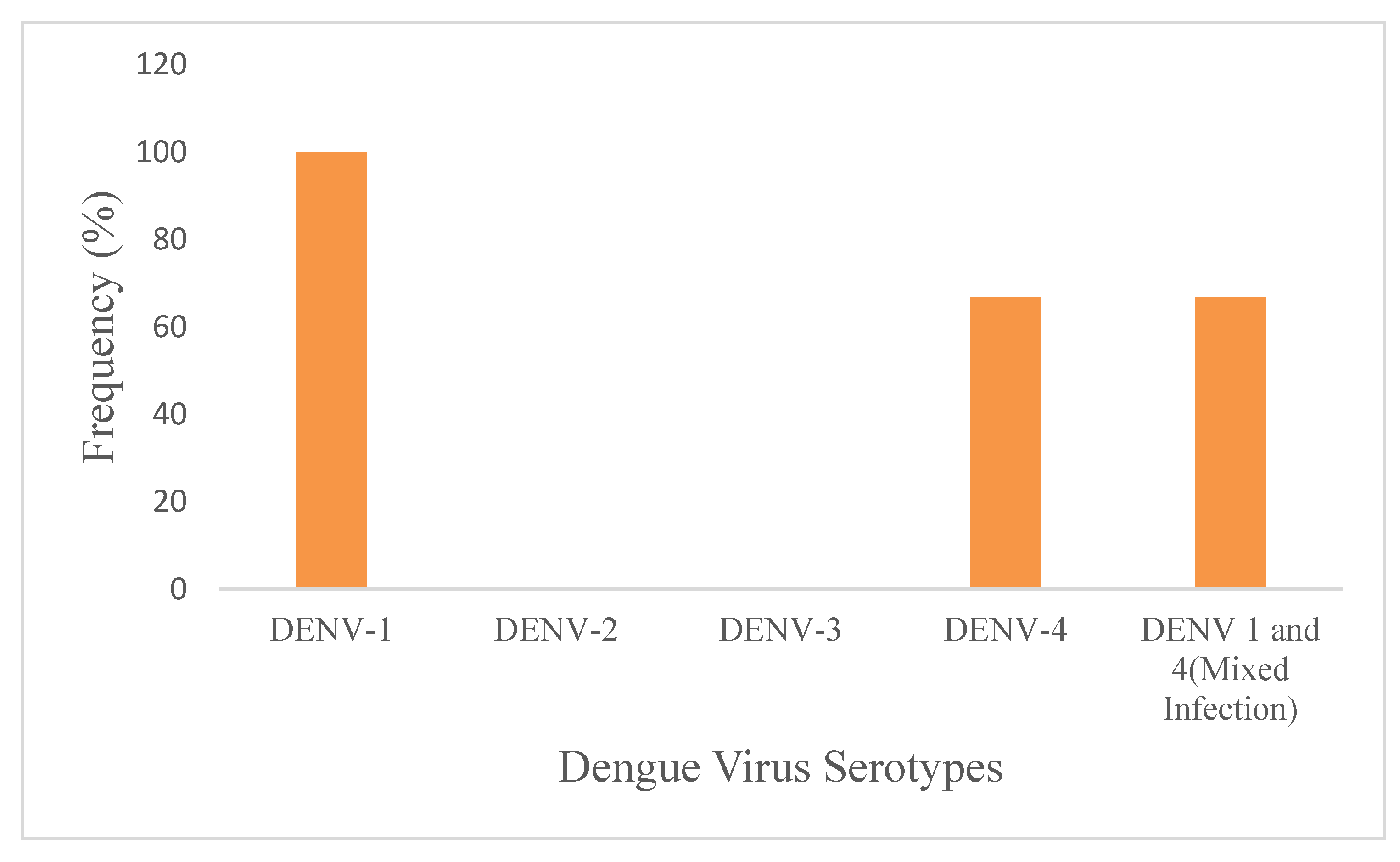

Table 1.
Primer sequences used for dengue consensus and serotype specific RT-PCR reactions.
| Primer | Sequence | Base pair size of amplified product | References |
|---|---|---|---|
| D1 D2 |
5′-TCAATATGCTGAAACGCGCGAGAAACCG-3′ 5′-TTGCACCAACAGTCAATGTCTTCAGGTTC-3′ |
511 Dengue concensus | (Lanciotti et.al., 1992) |
| TS1 | 5′-TCAATATGCTGAAACGCGCGAGAAACCG-3′ 5′-CGTCTCAGTGATCCGGGGG-3′ |
482 (Dl and TS1) DENV1 |
(Lanciotti et.al., 1992) |
| TS3 | 5′-TCAATATGCTGAAACGCGCGAGAAACCG-3′ 5′-TAACATCATCATGAGACAGAGC-3′ |
290 (Dl and TS3) DENV3 |
(Lanciotti et.al., 1992) |
| TS4 | 5′-TCAATATGCTGAAACGCGCGAGAAACCG-3′ 5′-CTCTGTTGTCTTAAACAAGAGA-3′ |
392 (Dl and TS4) DENV4 | (Lanciotti et.al., 1992) |
| DV1 | 5’-GGRACKTCAGGWTCTCC-3’ 5’-CCG GTGTGCTCRGCYCTGAT-3’ |
362 | (Hakami et.al., 2018) |
Table 2.
Prevalence of Dengue infection in the study Locations.
| Location | No. examined | No. positive(%) | χ2 | 95%CI | OR | p-value |
|---|---|---|---|---|---|---|
| Mubi | 200 | 163 (81.50) | 34.821 | 2.233-5.049 | 3.357 | 0.000 |
| Yola | 200 | 123 (61.50) | 1.393 | 0.559-1.132 | 0.796 | 0.238 |
| Numan | 200 | 104 (52.00) | 21.437 | 0.304-0.614 | 0.432 | 0.000 |
| Total | 600 | 390 (65.00) |
Table 3.
Prevalence of dengue virus infection among febrile patients, in relation to some risk factors.
Table 3.
Prevalence of dengue virus infection among febrile patients, in relation to some risk factors.
| Variables | No. examine | No. positive (%) | χ2 | CI | OR | p-value |
|---|---|---|---|---|---|---|
| Mosquito’s protection methods used | ||||||
| Mosquito coil/repellant | 129 | 82(63.6) | 4.952 | 1.097-2.997 | 1.813 | 0.026 |
| Net | 239 | 149(62.3) | 1.777 | 0.548-1.101 | 0.777 | 0.182 |
| Insecticide | 137 | 87(63.5) | 0.261 | 0.591-1.317 | 0.882 | 0.609 |
| All of the above | 63 | 55(87.3) | 13.634 | 1.883-8.664 | 4.039 | 0.000 |
| Others (Fan, AC) | 3 | 2(66.7) | 0.000 | 0.094-11.604 | 1.046 | 1.000 |
| Sources of water supply | ||||||
| Well | 63 | 46(73.0) | 5.197 | 1.131-3.611 | 2.021 | 0.023 |
| Borehole | 305 | 202(66.2) | 0.182 | 0.776-1.545 | 1.095 | 0.669 |
| River/stream | 25 | 17(68.0) | 0.006 | 0.482-2.683 | 1.137 | 0.938 |
| Tap | 68 | 45(66.2) | 0.001 | 0.613-1.787 | 1.047 | 0.974 |
| Rain | 4 | 2(50.0) | 0.014 | 0.074-3.788 | 0.529 | 0.907 |
| Sachet | 108 | 62(57.4) | 3.215 | 0.431-1.014 | 0.661 | 0.073 |
| Water storage in the compound | ||||||
| Yes | 533 | 346(64.9) | 0.227 | 0.408-1.574 | 0.802 | 0.634 |
| No | 43 | 30(69.8) | 0.227 | 0.227-0.634 | 1.247 | 0.634 |
| Emptying of discarded containers | ||||||
| Yes | 431 | 281(65.19) | 0.015 | 0.643-1.422 | 0.956 | 0.825 |
| No | 145 | 96(66.21) | 0.015 | 0.703-1.556 |
1.046 | 0.904 |
| Good drainage system/Septic Tanks | ||||||
| Yes | 406 | 262(64.53) | 0.574 | 0.578-1.241 | 0.847 | 0.449 |
| No | 170 | 116(68.24) | 0.574 | 0.806-1.729 | 1.181 | 0.449 |
| History of Travel | ||||||
| Travel out | 99 | 70(70.71) | 1.218 | 0.836-2.147 | 1.340 | 0.269 |
| Travel within the country | 363 | 238(65.56) | 0.000 | 0.716-1.454 | 1.02 | 0.985 |
| Never travel outside the state | 116 | 70(60.34) | 16.237 | 1.532-3.402 | 2.283 | 0.000 |
| Action during Fever | ||||||
| Self-prescription from chemist | 160 | 113(70.62) | 1.932 | 0.909-2.006 | 1.350 | 0.165 |
| Traditional medicine | 40 | 34(85.00) | 6.102 | 1.289-7.582 | 3.126 | 0.014 |
| See doctor | 366 | 226(61.75) | 7.433 | 0.399-0.849 | 0.582 | 0.006 |
Table 4.
Distribution of dengue virus IgM according to the symptoms presented by patients.
| Variables | No. examined | No. positive (%) | χ2 | CI | OR | p-value |
|---|---|---|---|---|---|---|
| Symptoms | ||||||
| Fever | 133 | 97(72.93) | 2.739 | 0.956-2.268 | 1.472 | 0.098 |
| Headache | 142 | 90(63.38) | 0.389 | 0.551-1.225 | 0.822 | 0.742 |
| Skin rash | 18 | 12(66.67) | 0.000 | 0.369-2.709 | 0.369 | 1.000 |
| Bleeding from nose | 10 | 6(60.00) | 0.013 | 0.208-2.677 | 0.746 | 0.910 |
| Joint pain | 90 | 57(63.33) | 0.374 | 0.524-1.343 | 0.839 | 0.541 |
| Pain around the eye | 37 | 27(72.97) | 0.438 | 0.652-2.912 | 1.378 | 0.508 |
| Nausea | 15 | 8(53.33) | 0.694 | 0.201-1.574 | 0.562 | 0.405 |
| Vomiting | 19 | 12(63.16) | 0.007 | 0.329-2.203 | 0.852 | 0.934 |
| Others | 85 | 57(67.06) | 0.00 | 0.625-1.669 | 1.021 | 1.000 |
Disclaimer/Publisher’s Note: The statements, opinions and data contained in all publications are solely those of the individual author(s) and contributor(s) and not of MDPI and/or the editor(s). MDPI and/or the editor(s) disclaim responsibility for any injury to people or property resulting from any ideas, methods, instructions or products referred to in the content. |
© 2026 by the authors. Licensee MDPI, Basel, Switzerland. This article is an open access article distributed under the terms and conditions of the Creative Commons Attribution (CC BY) license.
Copyright: This open access article is published under a Creative Commons CC BY 4.0 license, which permit the free download, distribution, and reuse, provided that the author and preprint are cited in any reuse.