
Submitted:
15 December 2025
Posted:
17 December 2025
You are already at the latest version
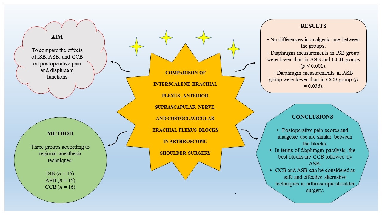
Abstract
Background: Interscalene brachial plexus block (ISB) remains gold standard anesthesia method in shoulder surgery. However, risk of diaphragm paralysis is themajor concern among anesthesiologists. Recent studies on anterior suprascapular nerve block (ASB) and costoclavicular brachial plexus block (CCB) have given promising results for preventing diaphragm paralysis and providing sufficient analgesia. Materials and Methods: Forty-six patients who underwent arthroscopic shoulder surgery under one of three regional anesthesia techniques, including ISB (n = 15), ASB (n = 15), and CCB (n = 16), were included in the study. Diaphragmatic excursion was measured by ultrasonography 30th minutes after block. Postoperative pain was assessed with a numerical rating scale. The groups were compared between each other in terms of diaphragm paralysis and postoperative pain status. Results: The groups were similar in basic patient and surgical characteristics, motor and sensory block scores. There was no difference in analgesic use between the groups. Diaphragm measurements in the ISB group were found significantly lower compared to the ASB and CCB groups (p < 0.001). In addition, diaphragm measurements in the ASB group were found lower than in the CCB group (p = 0.036). When compared diaphragm measurements between the initial and 30th minute of block, significant decreases were observed in the ISB and ASB groups (p < 0.001) whereas no difference was found in the CCB group. Conclusions: Postoperative pain scores and analgesic use were similar between there blocks. In terms of diaphragm paralysis, the best blocks were CCB followed by ASB. CCB and ASB can be considered as safe and effective alternative blocks in arthroscopic shoulder surgery.
Keywords:
anterior suprascapular nerve block
; costoclavicular brachial plexus block
; diaphragm paralysis
; interscalene brachial plexus block
; postoperative pain
1. Introduction
Although arthroscopic technique is associated with good cosmetic results, shorter hospitalization, and increased patient satisfaction, postoperative pain remains a significant problem in a portion of patients undergoing shoulder surgery [1]. Pain is not only impairs the patient’s comfort but also affects postoperative functional results by hindering early rehabilitation. Intravenous (IV) opioids are among the most commonly used analgesia tools for postoperative pain control. However, several undesirable side effects such as respiratory depression, prolonged sedation, constipation, allergic reactions, nausea, and vomiting limit its wide use [2]. In this context, different regional anesthesia techniques are being introduced into the routine practice for minimizing those problems and providing better analgesia. Recent advances in ultrasonography (US) techniques, such as the use of high-frequency probes in identifying peripheral nerves and providing clearer imaging of superficial tissues, have allowed safer, faster, and more comfortable blocks [3].
Interscalene brachial plexus block (ISB) is considered as gold standard anesthesia method in shoulder surgery; however, risk of diaphragm paralysis due to phrenic nerve injury is its leading disadvantage [4,5]. Changes in diaphragm functions can be measured and monitored by sonographic examination of the respiratory system, allowing the decision of correct block type on a patient-by-patient basis [6]. The recent studies on anterior suprascapular nerve block (ASB) and costoclavicular brachial plexus block (CCB) have given promising results for providing sufficient analgesia and minimizing diaphragm paralysis [7,8]. There is no study in the literature comparing these three regional blocks using US and nerve stimulators.
Based on these considerations, the present study aims to compare the effects of ISB, ASB and CCB techniques on postoperative pain and diaphragm functions in patients undergoing arthroscopic shoulder surgery.
2. Materials and Methods
2.1. Study Design
This prospective observational study was conducted at the Department of Anesthesiology and Reanimation, XXX University after receiving approval from the Instutional Clinical Research Ethics Committee (date: 09.02.2023, no: 47). The patients were informed in detail about the stages of the study, and both verbal and written informed consents were obtained from all.
Forty-six patients who underwent arthroscopic shoulder surgery under one of three regional anesthesia techniques, including ISB (n = 15), ASB (n = 15), and CCB (n = 16), were included in the study. The primary outcome was the differences in postoperative pain scores and analgesic use between the three regional blocks. The secondary outcomes were the differences in diaphragm functions, motor and sensory block scores, postoperative nause and vomiting, patient and surgeon satisfaction between the blocks.
Inclusion criteria were being above 18 years old, having American Society of Anesthesiologists (ASA) status of 1-3 and body mass index (BMI) between 20 and 40. The patients with neuropathic disorder, coagulopathy (preoperative platelet <100 μl, Inr >1.5, or prothrombin time >50 sec), obstructive or restrictive lung disease, renal/hepatic failure, allergy to local anesthetics, history of previous neck surgery, opioid use due to chronic pain syndrome, and pregnancy were excluded from the study.
2.2. Assessment of Diaphragm Excursion Before Regional Block
The patients were placed on the operating table in a supine position. Standard monitoring included heart rate (HR), peripheral oxygen saturation (SpO2), and blood pressure (BP). Nasal oxygen (2 L/min) was administered and IV access was established with a 20-gauge branula. Patients were premedicated with IV midazolam (2 mg). Diaphragm excursion on the shoulder side to be operated was assessed with a 2-5 MHz convex US probe (Philips Affiniti 50, Philips Medical Systems, Seattle, WA, United States) through subcostal acoustic windows between the anterior axillary midclavicular line adjacent to the liver or spleen. Inspiratory and expiratory craniocaudal displacements were measured as a bright line (hyperechoic waves) during normal breathing, by switching from B mode to M mode.
2.3. Basic Data Regarding Regional Block
After the sonographic assessment of diaphragm excursion, the patients were randomly divided into three groups; ISB, ASB or CCB. A high-frequency 5-13 MHz linear US probe, a 50 mm long 22-G short-beveled regional block needle, and a nerve stimulator (Vygon Ltd., Swindon, UK) were used in all blocks. The nerve stimulator was set to provide 1.5–2.5 mA current at a frequency of 1 Hz and a pulse duration of 0.1 ms. The needle was directed downward and inward through the skin. Local anesthetic was administered when stimulation was received at 1 mA and when stimulation was lost at 0.3 mA.
2.4. Interscalene Brachial Plexus Block (ISB) (Figure 1A)
In the supine position, with the arm adducted, the probe was placed transversely in the supraclavicular fossa just proximal to the midpoint of the clavicle. The brachial plexus was identified as a bright echogenic structure posterolateral to the subclavian artery (bunch of grapes sign). Then, the nerves were held in the center of the screen and the probe was moved in a cephalic direction up to the interscalene groove until a “string of pearls” image was obtained of the nerve roots located between the anterior and middle scalene muscles. After optimizing the image of the nerve roots as hypoechoic round or oval structures, the needle was placed posterolateral to the probe and moved in the plane. The needle was then located between the middle scalene muscle and the brachial plexus roots, and 15 ml of 0.25% bupivacaine was slowly injected into the area between the C5-6 roots.
Figure 1.
Sonographic views of three blocks. (A) ISB: 1. C5 nerve root, 2. C6 nerve root, 3. C7 nerve root, 4. Long thoracic nerve, 5. Supraclavicular nerves, ASM: Anterior scalene muscle, MSM: Middle scalene muscle. (B) ASB: 1. Subclavian artery, 2. Upper trunk, 3. Suprascapular nerve, OHM: omohyoid muscle. (C) CCB: 1. Posterior cord, 2. Lateral cord, 3. Medial cord, 4.Axillary artery, 5.Axillary vein.
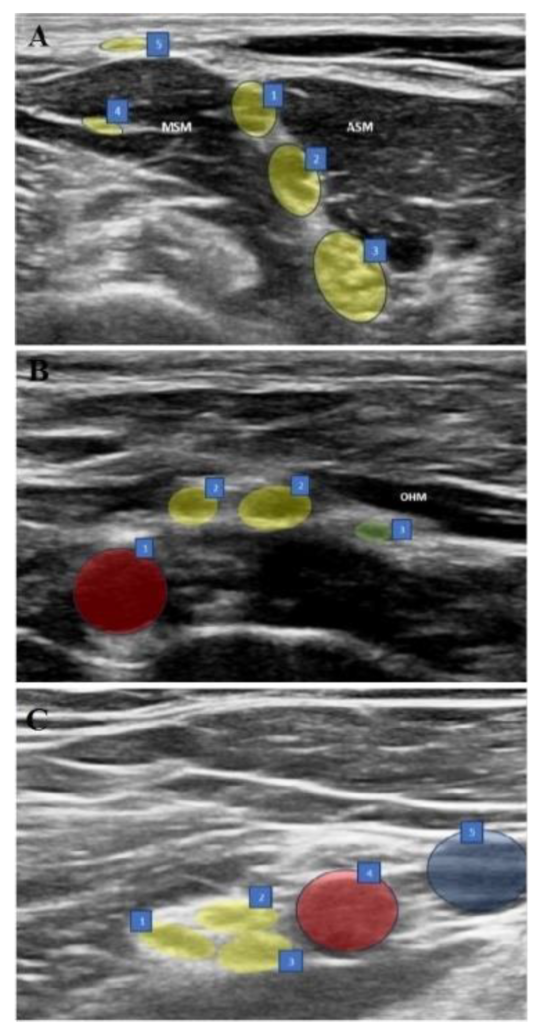
2.5. Anterior Suprascapular Nerve Block (ASB) (Figure 1B)
In the supine position, with the arm adducted, the probe was placed in the supraclavicular fossa to identify the brachial plexus. The plexus was then followed until the suprascapular nerve branching from the upper trunk was seen. The needle was advanced from posterolateral to anteromedial, and the most lateral transverse image of the nerve was obtained with the in-plane technique. The nerve was entered between the superficial cervical fascia and prevertebral fascia under the omohyoid muscle from the lateral side and 5 ml of 0.5% bupivacaine was injected following hydrodissection with DW5 and negative aspiration.
2.6. Costoclavicular Brachial Plexus Block (CCB) (Figure 1C)
The ipsilateral arm was abducted at 90 degrees with the palm facing the ceiling, and the ultrasound transducer was positioned parallel to the mid-clavicle. The long axis of the transducer was tilted slightly cephalad and the ultrasound beam was directed into the costoclavicular space. The ultrasound image was optimized so that all three cords of the brachial plexus were seen together lateral to the axillary artery, anteriorly between the clavicular head of the pectoralis major and the subclavius muscle and posteriorly between the serratus anterior muscle overlying the second rib. The needle was then advanced laterally and medially in the plane. With the needle tip in its intended position, a small volume (0.5-1 mL) of DW5 was injected before each injection to ensure that the needle tip was within the hyperechoic connective tissue matrix underlying the brachial plexus sheath and between the cords of the brachial plexus and not intraneurally. After verification, the local anesthetic was injected into multiple sites in 4-5 mL aliquots by redirecting the needle, for a total of 20 mL of 0.25% bupivacaine to homogeneously distribute the local anesthetic.
2.7. Motor and Sensory Assessments of the Regional Blocks
Thirty minutes after the block, ipsilateral hemidiaphragmatic excursion was measured and recorded at normal depth of breath. Motor and sensory functions of the shoulder were evaluated by a blind observer at 5-minute intervals for 30 minutes after the block. Motor function was evaluated with shoulder abduction (axillary and suprascapular nerves) and lateral rotation of the humerus against manual resistance (suprascapular nerve) while the arm was adducted and the elbow was flexed to 90°, with a score between 0 and 3 (0 = no motor block, 1 = no shoulder abduction, 2 = no shoulder abduction or elbow flexion, 3 = complete motor block). At the same time, sensory function was evaluated with the “pinprick test” (2 = feeling touch, 1 = reduced sense of touch, 0 = no sensation) on the skin over the clavicle (supraclavicular nerves) and the lateral surface of the deltoid (axillary nerve), and when the response to the pinprick test disappeared, it was considered ready for general anesthesia.
2.8. Induction and Maintenance of Anesthesia
In each group, induction was performed with IV propofol (1.5-2.5 mg/kg), IV fentanyl (1.5-3 μg/kg), and IV rocuronium (0.6 mg/kg). Anesthesia was maintained with sevoflurane (1-1.3 MAC) in 50%/50% O2/air mixture and remifentanil infusion (0.01-0.1 μg/kg/min). Mechanical ventilator settings were adjusted to tidal volume of 6-8 mL/kg and end-tidal CO2 of 30-35 mmHg. Arthroscopic shoulder surgery was performed on the patients in the beach bed position by the same surgical team. The patients’ hemodynamic data were recorded throughout the operation.
2.9. Postoperative Assessments
All patients were followed up at the 1st, 2nd, 6th, 12th and 24th hours postoperatively. The patients’ alertness was recorded at the postoperative 1st, 2nd, 6th, 12th and 24th hours with the Ramsay sedation scale. Scores of 1-2-3 on this scale indicate that the patient is awake, and scores of 4-5-6 indicate that the patient is under sedation.
Postoperative pain level was assessed with an 11-point numerical rating scale (NRS) ranging from 0 to 10. As part of multimodal pain management, all patients were given routine 1 g IV paracetamol (3 times daily). If NRS was >4, IV dexketoprofen (50 mg/2 ml) was administrated. If there was insufficient response to dexketoprofen, IV tramadol (1 mg/kg) was added.
Patients’ motor and sensory complaints in the ipsilateral upper extremity, side effects and complications were recorded at 1st, 2nd, 6th, 12th and 24th hours. Postoperative nausea and vomiting were assessed with Likert scale (Table 1). Patient and surgeon satisfaction were assessed with a 5-point scale at 24th hour postoperatively (5- very satisfied, 4 – satisfied, 3 – undecided, 2 – dissatisfied, 1 - very dissatisfied).
2.10. Statistical Analysis
The sample size and power analysis of the study were performed using G power (version 3.1.9.4), based on the study by Auyong et al. comparing the effects of interscalene, supraclavicular, and anterior suprascapular blocks on postoperative pain and diaphragm functions in shoulder surgery patients [9]. It was calculated that 80% power and 5% type I error could be achieved if at least 30 patients were included in the study (at least 10 patients for each group). The groups were determined as 15 people, considering possible follow-up losses. Data were analyzed using the SPSS 26 (SPSS Inc, Chicago, IL, USA) package program. Continuous and categorical variables were expressed as mean ± standard deviation (minimum-maximum) and numbers (percentages), respectively. Variables that conformed to normal distribution were examined with parametric tests, and variables that did not conform to normal distribution were examined with non-parametric tests. When parametric test assumptions were met, one-way analysis of variance (ANOVA), which examines the differences between three groups, was used in comparing independent group differences; when parametric test assumptions were not met, the Kruskal-Wallis test, which is a non-parametric test that examines the differences between three groups, was used in comparing independent group differences. A p value less than 0.05 was considered statistically significant.
3. Results
Data of 46 patients with a mean age of 51.4 years old were analyzed. There were three groups according to the regional anesthesia technique: ISB (n = 15), ASB (n = 15), and CCB (n = 16). All groups were similar in basic characteristics (Table 2).
In terms of motor and sensorial block examinations performed at the 30th minute, no significant differences were found between the groups (p = 0.345 and p = 0.230, respectively). Complete motor and sensorial blocks were observed in all patients.
There was no significant difference between the groups in the use of 0-6th, 6-12th and 6-12th hours non-steroidal anti-inflammatory drug (NSAID), and rescue analgesia (tramadol) (p > 0.05) (Table 3).
The mean initial normal breath diaphragm measurements were 1.97±0.22 cm in the ISB group, 1.81±0.15 cm in the ASB group and 1.75±0.17 cm in the CCB group. Initial values were significantly higher in the ISB group compared to the CCB group (p = 0.006). No significant difference was observed in pairwise comparisons of other initial values (p > 0.05).
At 30 minutes after the block, diaphragm measurements in the ISB group were found to be significantly lower compared to the ASB and CCB groups (p < 0.001). In addition, diaphragm measurements in the ASB group were found to be significantly lower compared to the CCB group (p = 0.036). In each group, changes in diaphragm measurements between the beginning and the 30th minute were compared; significant decreases were observed in the ISB and ASB groups (p < 0.001), while no difference was found in the CCB group (p > 0.05). (Table 4).
No significant difference was observed in Ramsay sedation scores (RS) among the groups (p > 0.05). All groups were similar in terms of pre-block NRS scores (p > 0.05). At 1 and 2 hours, the NRS scores of the ISB group were significantly lower than those of the ASB and CCB groups (p < 0.001), while there was no significant difference between the ASB and CCB groups (p > 0.05). No significant difference was found in the pairwise comparisons of the 3 groups in terms of NRS scores at 6 hours (p > 0.05). NRS scores at 12 and 24 hours were lower in the ASB group than in the ISB group (p < 0.05). At 12 and 24 hours, no significant differences were found between CCB and ISB and between CCB and ASB (p > 0.05).
No differences were found between the groups in terms of postoperative nausea (p = 0.764) and vomiting (p = 0.852) scores. There were also no differences between the groups in terms of patient satisfaction (p = 0.611) and surgeon satisfaction (p = 0.450).
One patient in the CCB and ISB groups had paresthesia, and one patient in the ISB group developed Horner syndrome. No significant difference was found between the groups in terms of complications (p = 0.607).
4. Discussion
In the present study, three different brachial plexus blockade techniques, ISB, ASB and CCB, were compared in terms of diaphragm functions and analgesia in patients undergoing shoulder arthroscopy. The results showed that three techniques provided effective shoulder analgesia but had different effects on diaphragm functions.
Shoulder innervation is provided by the distal branches of the C5-6 roots including suprascapular, axillary, subscapular, and lateral pectoral nerves [10]. Recently, the suprascapular nerve has been demonstrated not to be the dominant nerve of shoulder innervation as previously described [11]. In clinical studies, posterior suprascapular blockade, which blocks only the innervation of the posterior upper quadrant of the shoulder, has been shown to have limited advantage over placebo and has no clinically significant effect on pain scores and opioid consumption [12]. Currently, the subscapular and axillary nerves are known to contribute to the innervations of the anterior upper quadrant of the shoulder joint and the lower half of the joint, respectively [11]. These findings have led researchers to combine posterior suprascapular blockade with additional blocks such as supraclavicular [13] and infraclavicular [14] techniques or to find new block locations. ASB, which exposes the suprascapular nerve more proximally and anteriorly, has been presented as a reliable alternative to the shortcomings of the posterior approach to the suprascapular nerve [15].
In the present study, there was a significant difference in favor of ISB in the first two hours; however, this difference disappeared at 6 hours, and it was found that the ASB group showed significantly better analgesia at 12-24 hours. These results supported the previous finding that the upper trunk is consistently involved in ASB.
Contrary to previous belief, the fact that posterior rather than the anterior portion of the upper trunk is located close to the suprascapular nerve may explain the analgesic efficacy of the ASB [16]. Posterior portion of the upper trunk and thus the axillary and subscapular nerves innervating the shoulder can be blocked by directing a low volume of local anesthetic to the proximal origin of the suprascapular nerve [9].
The fact that the distance between the phrenic nerve and the C5 root (and also the suprascapular nerve) enlarges towards the root of the neck is another avantage of ASB, resulting to minimize the effect of the local anesthetic on the phrenic nerve. In parallel, the diaphragm measurements at 30 min were lower in the ISB group compared to the ASB group. The incidence of hemidiaphragmatic paresis after ISB increases to 100% with local anesthetic volumes of 20 ml or more [17]. This can be reduced by up to 45% by reducing the local anesthetic volume to 5 -10 ml, but is associated with a clinically significant decrease in the duration and efficacy of perioperative analgesia and carries a risk of block failure [17,18,19]. Therefore, in our study, a standard dose of 15 ml of 0.25% bupivacaine was used for ISB. The local anesthetic concentration was determined as 0.25% in order to reduce the differences in total drug doses administered between the groups. In our study, 5 ml of 0.25% bupivacaine was used in the ASB group as in previous studies [8]. The diaphragm function at 30 minutes was better in ASB group than in the ISB group. However, the significant decrease in the 30th minute value compared to the initial level was a remarkable result. In three studies where the upper trunk block was evaluated in terms of preserving diaphragmatic and respiratory functions, the rates of hemidiaphragmatic paralysis were reported to be significantly different (4.8%, 5.3%, and 54.3%) using the same volume (15 ml) and the same diagnostic tool (ultrasound evaluation of diaphragm excursion) [20,21,22]. In a recent cadaver study, it was reported that ASB performed with a mixture containing 5 ml of methylene blue stained the superior trunk 100% and caused 20% retrograde staining in the phrenic nerve [23]. However, it should be kept in mind that the results of cadaver studies may not always be consistent with clinical findings. Different volumes and injection techniques for ASB should be evaluated in future studies for optimization of diaphragm function.
Injury to the dorsal scapular nerve and long thoracic nerves in ISB is another concern, because these nerves pass through the middle scalene muscle. In ASB, the needle does not pass through this muscle, but courses between the superficial cervical and prevertebral fascias under the omohyoid muscle. Despite all these advantages, the anatomical difficulties of ASB, such as the proximity to vascular structures such as the suprascapular artery and transverse cervical artery and the inability to see the suprascapular nerve branching off from the upper trunk, should not be forgotten.
CCB is a relatively new infraclavicular block technique that has become widespread due to its rapid and reliable blockade [24]. The advantages that make CCB popular are that it can reliably block the lateral and posterior cords and the supraclavicular brachial plexus and suprascapular nerve via the retrograde channel, and that the needle entry site is located far enough from the phrenic nerve and brachial plexus to prevent the risk of hemidiaphragmatic paralysis [25]. As in other blocks, the dose and concentration of local anesthetic is an important factor for the effectiveness of CCB. In our study, the volume was selected as 20 ml for blockade of the lateral and posterior cords (not the medial cord) as suggested by Karmakar et al. [26], and the concentration was set at 0.25% to reduce the difference between the total amounts of local anesthetic. It should be noted here that further studies are necessary to determine the optimal (adequate analgesia and diaphragm-protecting) local anesthetic volume and concentration in CCB.
In terms of analgesia, a significant difference was found in favor of ISB at the 2nd hour. However, no difference was found between the analgesic efficacies of ISB and CCB at the 6th, 12th and 24th hours. Also, no significant difference was found between ASB and CCB in terms of pain scores at any of the 24-hour follow-ups. Our results clearly showed the stable and sufficient analgesic efficacy of CCB. During CCB, the lateral pectoral, subscapular and axillary nerves are completely blocked by lateral and posterior cord blockade [27]. However, the suprascapular nerve can only be blocked by supraclavicular spread of local anesthesic. Several anatomical structures that may limit this spread have been defined, such as the presence of more than one intraplexus septum in the supraclavicular fossa [5]. In addition, the presences of a paraneural sheath and an intraplexus septum between the three cords in the costoclavicular area have been reported both in cadavers and living organisms [27,28]. Those are believed to reduce the spread of the injection to the surrounding cords and proximal to the brachial plexus.
In our study, the diaphragm measurements at 30 minutes after block were found to be significantly lower in the ISB group compared to the CCB group. Interestingly, the diaphragm measurements at 30 minutes after block in the ASB group were also significantly lower than in the CCB group. It is known that moving from the supraclavicular fossa to the infraclavilar fossa during shoulder analgesia further reduces the risk of hemidiaphragmatic paralysis [25,29]. The paraneural sheath and fascial compartments surrounding the costoclavicular space cords play a critical bilateral role in the spread of local anesthetic. The connective tissue here and the three-cord organization create a potential retrograde channel, forcing the local anesthetic to spread around the cords and in a cephalic direction. On the other hand, the increase in the distance between the costoclavicular space and the interscalene groove with local anesthetic injection acts as a physical barrier that prevents the spread of local anesthetic to the phrenic nerve, stellate ganglion, or recurrent laryngeal nerve [30]. In our study, unlike the other groups, no significant change was detected between the beginning and the 30th minute of diaphragm measurements in the CCB group. This proves the protective role of CCB against hemidiaphragmatic paralysis in the local anesthetic volumes used in our study, similar to previous studies [8,30].
There were several limitations to our study. First, all blocks were performed by a single anesthesiologist in a single center, which facilitated standardization but limited the generalizability of the results. Blocks were performed for analgesia in patients undergoing general anesthesia. Further studies are needed to determine the anesthetic adequacy of the ASB and CCB groups. In the study, blocks were performed only in patients undergoing shoulder arthroscopy, and the results may not be generalizable to other surgeries such as shoulder arthroplasty. All blocks were performed with a single injection technique, and the side effects that may develop due to catheter-based blocks and local anesthetic accumulation are the subject of other new studies. Since patient-controlled analgesia was not used in the postoperative period, the postoperative opioid consumption of the patients could not be measured. Finally, only diaphragm excursion values were measured, and detailed respiratory function tests were not performed. Although no serious side effects were observed, it is important to be cautious in generalizing the results of the study, especially to patients with serious respiratory comorbidities.
5. Conclusions
Although the NRS score at the postoperative 2nd hour was significantly lower in the ISB group than in the ASB and CCB groups, subsequent measurements showed that adequate analgesia was provided in all groups. In terms of diaphragm paralysis, the best block was CCB. Similarly, ASB was found to be associated with a reduced risk of diaphragm paralysis. All patients in ISB group developed diaphragm paralysis. These results showed that CCB and ASB can be considered as safe and effective alternative blocks in arthroscopic shoulder surgery.
Author Contributions
Y.K. and B.T.S. wrote the main manuscript text. All authors contributed to the study conception and design. Material preparation, data collection and analysis were performed by B.T.S., I.E.S., and Y.K. All authors have read and agreed to the published version of the manuscript.
Funding
The authors declare that no funds, grants, or other support were received during the preparation of this manuscript.
Institutional Review Board Statement
The study was approved by the XXX XXX University Clinical Research Ethics Committee (date: 09.02.2023, no: 47), in accordance with the principles of the Declaration of Helsinki.
Informed Consent Statement
Informed consent was obtained from all the individual participants included in the study.
Data Availability Statement
Datasets analyzed during the current study are not publicly available due to patient privacy limitations, but are available from the corresponding author on reasonable request.
Acknowledgments
The authors would like to thank for their contributions on surgical interventions and for his contribution on statistical analysis.
Conflicts of Interest
The authors declare no conflicts of interest.
Abbreviations.
The following abbreviations are used in this manuscript:
| IV | Intravenous. |
| ISB | Interscalene brachial plexus block. |
| ASB | Anterior suprascapular nerve block. |
| CCB | Costoclavicular brachial plexus block. |
| US | Ultrasonography. |
| ASA | American Society of Anesthesiologists. |
| BMI | Body mass index. |
| HR | Heart rate. |
| SpO2 | Peripheral oxygen saturation. |
| BP | Blood pressure. |
| NRS | Numerical rating scale. |
References
- Goertz, A.W.; Lindner, K.H.; Schütz, W.; Schirmer, U.; Beyer, M.; Georgieff, M. Influence of phenylephrine bolus administration on left ventricular filling dynamics in patients with coronary artery disease and patients with valvular aortic stenosis. Anesthesiology 1994, 81, 49–58. [Google Scholar] [CrossRef]
- D’Alessio, J.G.; Rosenblum, M.; Shea, K.P.; Freitas, D.G. A retrospective comparison of interscalene block and general anesthesia for ambulatory surgery shoulder arthroscopy. Reg. Anesth. 1995, 20, 62–68. [Google Scholar] [CrossRef]
- Klaastad, O.; Sauter, A.R.; Dodgson, M.S. Brachial plexus block with or without ultrasound guidance. Curr. Opin. Anaesthesiol. 2009, 22, 655–660. [Google Scholar] [CrossRef] [PubMed]
- Liu, S.S.; Gordon, M.A.; Shaw, P.M.; Wilfred, S.; Shetty, T.; Yadeau, J.T. A prospective clinical registry of ultrasound-guided regional anesthesia for ambulatory shoulder surgery. Anesth. Analg. 2010, 111, 617–623. [Google Scholar] [CrossRef] [PubMed]
- Tran, D.Q.; Elgueta, M.F.; Aliste, J.; Finlayson, R.J. Diaphragm-Sparing nerve blocks for shoulder surgery. Reg. Anesth. Pain Med. 2017, 42, 32–38. [Google Scholar] [CrossRef]
- Aliste, J.; Bravo, D.; Layera, S.; Fernández, D.; Jara, Á.; Maccioni, C.; Infante, C.; Finlayson, R.J.; Tran, D.Q. Randomized comparison between interscalene and costoclavicular blocks for arthroscopic shoulder surgery. Reg. Anesth. Pain Med. 2019, rapm–2018–100055. [Google Scholar] [CrossRef]
- Scheibe, N.; Sosnowski, N.; Pinkhasik, A.; Vonderbank, S.; Bastian, A. Sonographic evaluation of diaphragmatic dysfunction in COPD patients. Int. J Chron Obstruct. Pulmon. Dis. 2015, 10, 1925–1930. [Google Scholar]
- Abdallah, F.W.; Wijeysundera, D.N.; Laupacis, A.; Brull, R.; Mocon, A.; Hussain, N.; Thorpe, K.E.; Chan, V.W.S. Subomohyoid anterior suprascapular block versus interscalene block for arthroscopic shoulder surgery: a multicenter randomized trial. Anesthesiology 2020, 132, 839–853. [Google Scholar] [CrossRef]
- Auyong, D.B.; Hanson, N.A.; Joseph, R.S.; Schmidt, B.E.; Slee, A.E.; Yuan, S.C. Comparison of anterior suprascapular, supraclavicular, and interscalene nerve block approaches for major outpatient arthroscopic shoulder surgery: a randomized, double-blind, noninferiority trial. Anesthesiology 2018, 129, 47–57. [Google Scholar] [CrossRef] [PubMed]
- Eckmann, M.S.; Bickelhaupt, B.; Fehl, J.; Benfield, J.A.; Curley, J.; Rahimi, O.; Nagpal, A.S. Cadaveric study of the articular branches of the shoulder joint. Reg. Anesth. Pain Med. 2017, 42, 564–570. [Google Scholar] [CrossRef]
- Tran, J.; Peng, P.W.H.; Agur, A.M.R. Anatomical study of the innervation of glenohumeral and acromioclavicular joint capsules: implications for image-guided intervention. Reg. Anesth. Pain Med. 2019, rapm–2018–100152. [Google Scholar] [CrossRef]
- Cho, N.; Kang, R.S.; McCartney, C.J.L.; Pawa, A.; Costache, I.; Rose, P.; Abdallah, F.W. Analgesic benefits and clinical role of the posterior suprascapular nerve block in shoulder surgery: a systematic review, meta-analysis and trial sequential analysis. Anaesthesia 2020, 75, 386–394. [Google Scholar] [CrossRef] [PubMed]
- Trabelsi, W.; Ben Gabsia, A.; Lebbi, A.; Sammoud, W.; Labbène, I.; Ferjani, M. Suprascapular block associated with supraclavicular block: An alternative to isolated interscalene block for analgesia in shoulder instability surgery? Orthop. Traumatol. Surg. Res. 2017, 103, 77–83. [Google Scholar] [CrossRef]
- Aliste, J.; Bravo, D.; Finlayson, R.J.; Tran, D.Q. A randomized comparison between interscalene and combined infraclavicular-suprascapular blocks for arthroscopic shoulder surgery. Can. J Anaesth. 2018, 65, 280–287. [Google Scholar] [CrossRef]
- Siegenthaler, A.; Moriggl, B.; Mlekusch, S.; Schliessbach, J.; Haug, M.; Curatolo, M.; Eichenberger, U. Ultrasound-guided suprascapular nerve block, description of a novel supraclavicular approach. Reg. Anesth. Pain Med. 2012, 37, 325–328. [Google Scholar] [CrossRef]
- Hanna, A. The SPA arrangement of the branches of the upper trunk of the brachial plexus: a correction of a longstanding misconception and a new diagram of the brachial plexus. J Neurosurg. 2016, 125, 350–354. [Google Scholar] [CrossRef] [PubMed]
- Riazi, S.; Carmichael, N.; Awad, I.; Holtby, R.M.; McCartney, C.J. Effect of local anaesthetic volume (20 vs 5 ml) on the efficacy and respiratory consequences of ultrasound-guided interscalene brachial plexus block. Br. J Anaesth. 2008, 101, 549–556. [Google Scholar] [CrossRef]
- Lee, J.H.; Cho, S.H.; Kim, S.H.; Chae, W.S.; Jin, H.C.; Lee, J.S.; Kim, Y.I. Ropivacaine for ultrasound-guided interscalene block: 5 mL provides similar analgesia but less phrenic nerve paralysis than 10 mL. Can. J Anaesth. 2011, 58, 1001–1006. [Google Scholar] [CrossRef] [PubMed]
- Fredrickson, M.J.; Abeysekera, A.; White, R. Randomized study of the effect of local anesthetic volume and concentration on the duration of peripheral nerve blockade. Reg. Anesth. Pain Med. 2012, 37, 495–501. [Google Scholar] [CrossRef]
- Kim, D.H.; Lin, Y.; Beathe, J.C.; Liu, J.; Oxendine, J.A.; Haskins, S.C.; Ho, M.C.; Wetmore, D.S.; Allen, A.A.; Wilson, L. Superior trunk block: a phrenic-sparing alternative to the interscalene block: A randomized controlled trial. Anesthesiology 2019, 131, 521–533. [Google Scholar] [CrossRef]
- Kang, R.; Jeong, J.S.; Chin, K.J.; Yoo, J.C.; Lee, J.H.; Choi, S.J.; Gwak, M.S.; Hahm, T.S.; Ko, J.S. Superior trunk block provides noninferior analgesia compared with interscalene brachial plexus block in arthroscopic shoulder surgery. Anesthesiology 2019, 131, 1316–1326. [Google Scholar] [CrossRef]
- Kim, H.; Han, J.U.; Lee, W.; Jeon, Y.S.; Jeong, J.; Yang, C.; Uhm, J.W.; Kim, Y. Effects of local anesthetic volume (standard versus low) on incidence of hemidiaphragmatic paralysis and analgesic quality for ultrasound-guided superior trunk block after arthroscopic shoulder surgery. Anesth. Analg. 2021, 133, 1303–1310. [Google Scholar] [CrossRef] [PubMed]
- Sehmbi, H.; Johnson, M.; Dhir, S. Ultrasound-guided subomohyoid suprascapular nerve block and phrenic nerve involvement: a cadaveric dye study. Reg. Anesth. Pain Med. 2019, 44, 561–564. [Google Scholar] [CrossRef] [PubMed]
- Songthamwat, B.; Karmakar, M.K.; Li, J.W.; Samy, W.; Mok, L.Y.H. Ultrasound-guided infraclavicular brachial plexus block: prospective randomized comparison of the lateral sagittal and costoclavicular approach. Reg. Anesth. Pain Med. 2018, 43, 825–831. [Google Scholar] [CrossRef] [PubMed]
- Petrar, S.D.; Seltenrich, M.E.; Head, S.J.; Schwarz, S.K. Hemidiaphragmatic paralysis following ultrasound-guided supraclavicular versus infraclavicular brachial plexus blockade: a randomized clinical trial. Reg. Anesth. Pain Med. 2015, 40, 133–138. [Google Scholar] [CrossRef]
- Karmakar, M.K.; Sala-Blanch, X.; Songthamwat, B.; Tsui, B.C. Benefits of the costoclavicular space for ultrasound-guided infraclavicular brachial plexus block: description of a costoclavicular approach. Reg. Anesth. Pain Med. 2015, 40, 287–288. [Google Scholar] [CrossRef]
- Areeruk, P.; Karmakar, M.K.; Reina, M.A.; Mok, L.Y.H.; Sivakumar, R.K.; Sala-Blanch, X. High-definition ultrasound imaging defines the paraneural sheath and fascial compartments surrounding the cords of the brachial plexus at the costoclavicular space and lateral infraclavicular fossa. Reg. Anesth. Pain Med. 2021, 46, 500–506. [Google Scholar] [CrossRef]
- Monzó, E.; Boezaart, A.P.; Tubbs, R.S.; Sanromán-Junquera, M.; Nin, O.C.; Reina, M.A. A reliable septum exists between the lateral cord and medial and posterior cords in the costoclavicular region: clinical and microanatomical considerations in brachial plexus anesthetic blockade. Clin. Anat. 2021, 34, 411–419. [Google Scholar] [CrossRef]
- Georgiadis, L.; Vlassakov, K.V.; Patton, M.E.; Lirk, P.B.; Janfaza, D.R.; Zeballos, J.L.; Quaye, A.N.; Patel, V.; Schreiber, K.L. Ultrasound-guided supraclavicular vs. retroclavicular block of the brachial plexus: comparison of ipsilateral diaphragmatic function: a randomised clinical trial. Eur. J Anaesthesiol. 2021, 38, 64–72. [Google Scholar] [CrossRef]
- Luo, Q.; Zheng, J.; Yang, C.; Wei, W.; Wang, K.; Xiang, X.; Yao, W. Effects of the costoclavicular block versus interscalene block in patients undergoing arthroscopic shoulder surgery under monitored anesthesia care: a randomized, prospective, non-inferiority study. Korean J Anesthesiol. 2023, 76, 413–423. [Google Scholar] [CrossRef]

Table 1.
Postoperative nause and vomiting (Likert scale).
| nause | vomiting |
|---|---|
| 0 - None 1 - Does not prevent eating 2 - Significantly reduces oral intake 3 - Requires IV fluid administration |
0 - None 1 - Once in 24 hours 2 - 2-5 times in 24 hours 3 - ≥6 times in 24 hours or IV fluid requirement 4 - Requires hospitalization |
Table 2.
Comparison of basic characteristics between the groups.
| ISB (n = 15) | ASB (n = 15) | CCB (n = 16) | p | |
|---|---|---|---|---|
| Age (mean, y) | 46.7±10.6 | 53.6±10.1 | 52.7±14.8 | 0.167 |
| Gender (F/M) | 5/10 | 6/9 | 7/9 | 0.835 |
| BMI (kg/m2) | 27.2±3 | 29.5±5.7 | 26.6±4.3 | 0.188 |
| Lateralization (R/L) | 9/6 | 9/6 | 9/7 | 0.970 |
| ASA status | 0.279 | |||
| ASA 1 | 7 | 4 | 2 | |
| ASA 2 | 8 | 10 | 13 | |
| ASA 3 | 0 | 1 | 1 |
Age, and BMI are presented as mean ± SD; other variables are presented as number.y: Year, F: Female, M: Male, BMI: Body Mass Index, R: Right, L: Left, ASA: American Society of Anesthesiologists.
Table 3.
Comparison of additional analgesic use between the groups.
| ISB (n = 15) | ASB (n = 15) | CCB (n = 15) | p | |
|---|---|---|---|---|
| 0-6th hour NSAID | 5 | 6 | 3 | 0.441 |
| 6-12th hour NSAID | 6 | 2 | 5 | 0.304 |
| 12-24th hour NSAID | 3 | 2 | 1 | 0.497 |
| Rescue analgesia (tramadol) | 2 | 1 | 2 | 0.814 |
All variables are presented as number. NSAIDs: Non-steroidal anti-inflammatory drugs.
Table 4.
Comparison of the initial and 30th minute diaphragm measurements in each group (during normal breathing).
Table 4.
Comparison of the initial and 30th minute diaphragm measurements in each group (during normal breathing).
| Initial | 30th minute of block | p | |
|---|---|---|---|
| ISB | 1.97±0.22 cm | 0.2±0.1 cm | < 0.001 |
| ASB | 1.81±0.15 cm | 1.36±0.5 cm | < 0.001 |
| CCB | 1.75±0.17 cm | 1.67±0.2 cm | 0.312 |
All variables are presented as mean ± SD. cm: centimeter.
Disclaimer/Publisher’s Note: The statements, opinions and data contained in all publications are solely those of the individual author(s) and contributor(s) and not of MDPI and/or the editor(s). MDPI and/or the editor(s) disclaim responsibility for any injury to people or property resulting from any ideas, methods, instructions or products referred to in the content. |
© 2025 by the authors. Licensee MDPI, Basel, Switzerland. This article is an open access article distributed under the terms and conditions of the Creative Commons Attribution (CC BY) license (http://creativecommons.org/licenses/by/4.0/).
Copyright: This open access article is published under a Creative Commons CC BY 4.0 license, which permit the free download, distribution, and reuse, provided that the author and preprint are cited in any reuse.


