
Submitted:
26 October 2025
Posted:
28 October 2025
You are already at the latest version
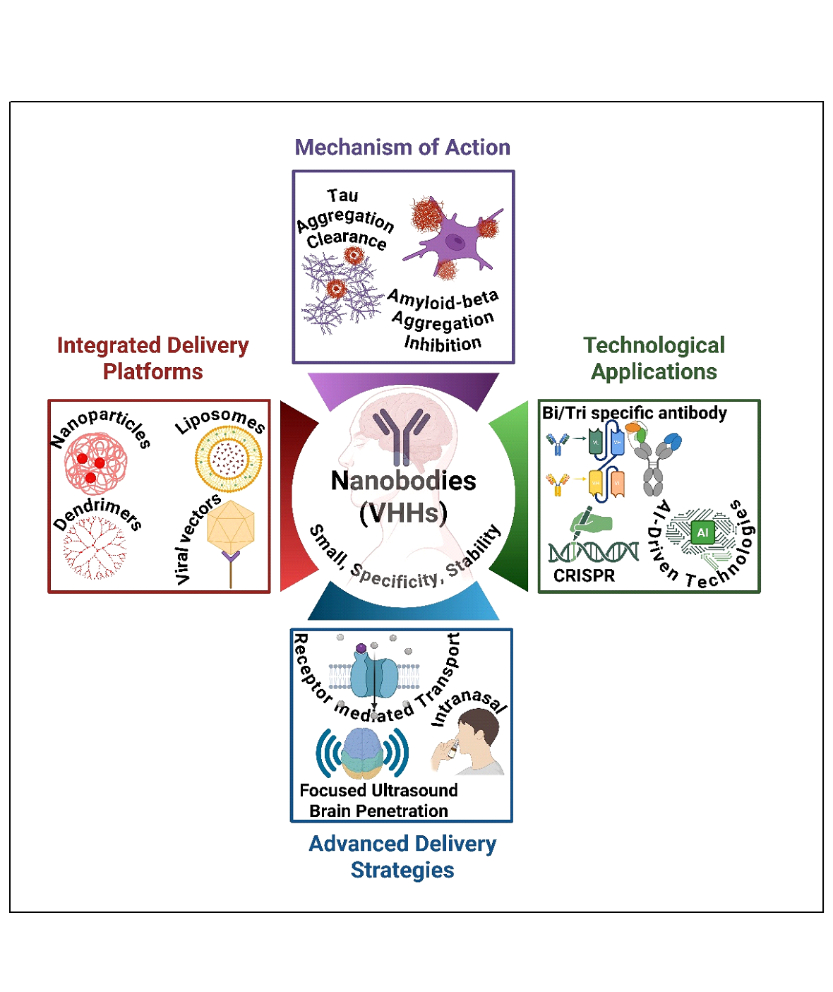
Abstract
Nanobodies (single-domain antibodies, VHHs) have emerged as versatile tools for evaluating and treating Alzheimer’s disease (AD). They offer unique advantages over traditional antibodies and small molecules, including small size, stability, and specificity. In AD, nanobodies were used to neutralize toxic amyloid-β oligomers, inhibit tau generation and aggregation, and modulate neuroinflammation, thereby demonstrating significant therapeutic potential. The delivery of nanobodies requires advanced strategies, including intranasal and intrathecal routes, receptor-mediated transport, plasma protein binding with albumin, and focused ultrasound to facilitate brain penetration. Additionally, to improve nanobody delivery precision, half-life, and efficacy, strategies such as integrating nanobodies with nanoparticles, dendrimers, liposomes, and viral vectors are being employed. In fact, nanobodies are applied beyond monotherapy across multiple technological platforms to optimize brain delivery and target multiple targets. Nanobodies have been used on bispecific and trispecific antibody platforms, as well as in CRISPR/Cas9 editing and AI-driven technologies, to expand their applications. Recently, preclinical evidence has been mounting on the efficacy of nanobodies in clearing Aβ and tau, preserving synapses, and normalizing biomarkers. Notably, clinical trials of bispecific antibodies, including trontinemab, are signaling translational progress and regulatory approvals, and further support would validate this class of therapeutic molecules.
This review critically delineates the current molecular mechanisms, emerging strategies, and delivery platforms, and emphasizes the potential of nanobodies as promising therapeutic and diagnostic molecules in AD therapeutics.
Keywords:
Amyloid-β and tau targeting
; Nanobodies
; Alzheimer’s disease
; Brain delivery
; Therapeutics
1. Introduction
Alzheimer’s disease (AD) is a progressive neurodegenerative disorder characterized by cognitive decline, memory loss, and impaired daily functioning. With an aging global population, AD prevalence is increasing, affecting approximately 33–39 million people worldwide in 2025 and projected to reach 152 million by 2050, highlighting the need for innovative therapeutic strategies. The hallmarks of AD pathology include amyloid-beta (Aβ) plaques, neurofibrillary tangles (NFTs) formed by hyperphosphorylated tau, and chronic neuroinflammation (Figure 1) [1,2,3].
Traditional therapeutic approaches, such as cholinesterase inhibitors, provide symptomatic relief but fail to address the underlying pathological mechanisms of the disease [4]. Immunotherapy, particularly antibody-based therapies, has gained attention for targeting Aβ and tau. However, conventional antibodies face challenges, including poor BBB penetration and off-target effects [5,6].
Nanobodies (Nbs) or single-domain antibodies derived from camelid heavy-chain antibodies offer a promising alternative due to their unique structural and functional properties [7,8]. This review examines the role of Nbs in AD immunotherapy, focusing on their mechanisms of action and translational potential.
2. Nanobodies: Structure and Advantages
Nbs are small antigen-binding fragments that typically range from approximately 12 to 15 kDa [9]. They are derived from the variable domain of heavy-chain-only antibodies found in camelids, with similar single-domain antibodies present in cartilaginous fish [10]. Key characteristics of Nbs include their compact, single-domain structure, which maintains high affinity and specificity, and their long complementarity-determining region 3 (CDR3) loops, which provide access to hidden or conformational epitopes. They also exhibit exceptional thermal and chemical stability and can be easily expressed in microbial systems [11].
Compared with full-length immunoglobulin G (IgG), Nbs offer advantages such as faster tissue penetration, reduced immunogenicity due to diminished Fc-mediated effector functions, and greater flexibility for multivalent or bispecific constructs|. These attributes make Nbs especially appealing for targeting the central nervous system (CNS), where large antibodies struggle due to steric access and limited brain penetration [12,13].
The compact size of Nbs enables enhanced tissue penetration, including crossing the BBB, a critical barrier for brain-targeted therapeutics [13]. Nbs exhibit high solubility, thermal stability, and resistance to aggregation, making them suitable for long-term storage and administration [14]. Additionally, they can be engineered for enhanced specificity, conjugated to imaging agents, or fused with other proteins to improve functionality [15]. These properties make nanobodies ideal candidates for targeting AD-related pathologies.
Figure 2.
Schematic representation of single domain antibody (VHH), showing the three complementarity-determining regions (CDR1- CDR3) highlighted as protruding loops (Created using Biorender).
Figure 2.
Schematic representation of single domain antibody (VHH), showing the three complementarity-determining regions (CDR1- CDR3) highlighted as protruding loops (Created using Biorender).

3. Mechanisms of Nanobody-Based Immunotherapy in Alzheimer’s Disease
Nb-based immunotherapy in AD focuses on three primary pathological targets: Aβ aggregates, tau pathology, and neuroinflammation [16].
3.1. Amyloid-Beta Aggregates
Nbs targeting Aβ species have demonstrated efficacy by binding to toxic oligomers, blocking oligomerization and fibril formation, and neutralizing neurotoxicity [17] [18]. Specific Nbs (e.g., A4, E1, V31-1) inhibit the aggregation of Aβ oligomers, thereby preventing progression to mature fibrils implicated in plaque formation. Some Nbs can enzymatically degrade Aβ at secretase cleavage sites, thereby offering a direct reduction in amyloid burden [17]. Delivery of Nbs constructs fused with blood-brain barrier (BBB) shuttle domains, such as FC5, which targets a sialo glycoprotein receptor on brain endothelium to induce receptor-mediated transcytosis, enables efficient brain uptake [19]. This modality has demonstrated reductions in brain Aβ levels, improvements in hippocampal volume, and normalization of cerebrospinal fluid (CSF) Aβ ratios in AD model mice. Multifunctional Nb conjugates with imaging agents (e.g., gadolinium) provide diagnostic capabilities for noninvasive tracking of amyloid pathology [20].
3.2. Tau Pathology
Tau proteins aggregate into paired helical filaments, which form neurofibrillary tangles, thereby driving neurodegeneration. Nbs that recognize critical amyloidogenic sequences of tau (e.g., PHF6 core) inhibit tau aggregation and propagation, as demonstrated by work using VHH Z70, which reduces tau spreading in mouse models [21,22]. These Nbs can be delivered via viral vectors such as lentivirus or adeno-associated virus (AAV) for sustained intracerebral expression, offering a gene therapy approach to reduce tau pathology. Imaging probes targeting phosphorylated tau using nanobodies facilitate early detection of tauopathy [23].
3.3. Neuroinflammation
Crossing the BBB remains a significant hurdle for brain therapeutics. Nbs exploit receptor-mediated transcytosis (e.g., via the transferrin receptor and insulin-like growth factor receptor) and adsorptive-mediated transcytosis to enter the brain [24]. FC5 is a pioneering Nb that binds the α (2,3)-sialo-glycoprotein receptor on brain endothelial cells, triggering clathrin-mediated endocytosis and enhancing BBB crossing [25]. Techniques to increase brain delivery include the use of multivalent nanobody constructs to enhance avidity, fusion to Fc fragments or albumin-binding domains to extend half-life, and conjugation to nanoparticles or cell-penetrating peptides [26]. Physical methods, such as ultrasound combined with microbubbles, transiently open the BBB to enhance nanobody delivery [27]. Alternative administration routes, such as intranasal, intrathecal, and intracerebroventricular delivery, directly target the CNS, resulting in higher local nanobody concentrations [28]. Gene therapy via AAV vectors encoding nanobodies provides a strategy for sustained brain exposure [29].
3.5. Translational Approaches and Challenges
Nb-based imaging agents have been developed to detect pathological Aβ and tau species with improved target specificity and rapid clearance, offering higher contrast and reduced toxicity compared to conventional antibodies [30]. For example, the Nb pa2H labeled with technetium-99m shows brain uptake that correlates with amyloid burden in AD models [31]. Nbs detecting retinal Aβ oligomers propose a minimally invasive early diagnostic eye test for AD. Therapeutically, Nanobodies inhibit toxic aggregation, promote clearance, and can be coupled to effector molecules or enzymes to degrade pathological proteins [32,33]. Studies report the reversal of hippocampal atrophy and normalization of biomarkers following Nb therapy in mice [34]. Clinical translation will depend on optimizing delivery, half-life, and multifunctionality while minimizing immunogenicity [35].
4. Nanobody-Based Drug Delivery Systems
Nb-based drug delivery systems can be broadly categorized into two principal approaches (Figure 3) [36]. The first approach involves the direct conjugation of therapeutic agents or toxic molecules to Nbs, exploiting their high specificity and affinity for targeted delivery. This strategy enables precise neutralization or payload delivery via direct binding interactions with Nb. The second approach centers on the functionalization of nanocarriers, such as liposomes, polymeric nanoparticles, and other nanostructures, with Nbs to enhance their targeting capabilities [37]. In this method, therapeutic agents are first encapsulated or incorporated into nanocarriers, which are subsequently conjugated to Nbs to facilitate selective delivery to disease-relevant sites [38]. This approach improves drug stability, bioavailability, and controlled release kinetics. Together, these strategies highlight the versatility of Nb-based platforms in optimizing therapeutic delivery, particularly in overcoming biological barriers such as the blood–brain barrier in central nervous system disorders [39].
4.1. Direct Injection of Nanobodies
Direct injections may optimize therapeutic outcomes and efficacy while leveraging the beneficial small size of Nbs [40,41]. A common matrix protein in malignancies, collagen is a suitable target for immunotherapies applied locally. By using the collagen-binding ectodomain of murine leukocyte-associated immunoglobulin-like receptor-1 (LAIR) to generate specific Nb variants, Nbs are tailored for treatments targeting collagen within cells [40]. To reduce systemic toxicity and localize the therapeutic effect, these modified Nbs are subsequently fused with Interleukin-2 (IL-2), a cytokine known to activate immune cells [40,41]. Similarly, a delay in tumor growth may result from the combination of small-format IL-2 immunocytokines with high-affinity Nbs that target the tumor-specific Extra domain-B of fibronectin (EIIIB), which is present in the tumor extracellular matrix. These immunocytokines have effects on tumor growth that are comparable to those of untargeted IL-2 when administered intravenously. On the other hand, better survival outcomes are achieved with intratumoral administration, which relies on the affinity for EIIIB [42]. Additionally, Escherichia coli bacteria with a type III protein secretion system may be able to engineer Single-domain antibodies (sdAbs) for injection into human cells, thereby bypassing the requirement that the Abs pass through the plasma membrane, a significant obstacle when targeting intracellular proteins for therapeutic purposes [41]. Two particular sdAbs: Variable single-domain antibody targeting amylase (Vamy) and Variable single-domain antibody targeting green fluorescent protein (GFP), used for intracellular tracking or delivery (Vgfp), which target amylase and GFP, respectively, are used in the engineering of these sdAbs [42].
4.2. Nanobodies Conjugated to the Drug and Toxin
The majority of Nbs must be coupled with a toxic load or another effector function since they lack innate therapeutic activity. Whether the conjugation is a single effector domain or a drug-containing nanocargo, the conjugated Nbs are used for drug delivery in various applications [43,44]. Because of its larger size, the conjugation of Nbs with an enzyme or toxin molecule lengthens the period that Nb circulates in the blood. As a result, the construct's ability to transport its load to tumors or sick tissues improves. There are two methods for conjugation: chemical conjugation and cloning in an expression vector by fusing the Nb gene with a hazardous protein [45,46]. Because the Nbs contain multiple lysines, the effector moiety's conjugation to the Nb, mostly to lysine residues, may be heterogeneous in chemical conjugation. If a Lys in the CDR reacted, the reaction could block the CDR's access to the antigen, reducing or eliminating the Nbs' ability to recognize it. In a different tactic, the addition of a single cysteine to the Nb's C-terminal end enables site-directed conjugation of a hazardous load far from the paratope, thereby minimizing disruption of antigen binding [47]. To connect chelators and nuclides, the C-terminal end of Nbs has recently been modified by Sortase A [48]. In any case, following conjugation, the antigen-binding qualities need to be verified. One of the most effective non-viral gene transfer methods, polyethyleneimine (PEI), is limited in its in vivo use due to several issues, including non-specific cell binding, interaction with blood components, and relatively high cytotoxicity [49]. In a work by Saqafi et al., a branched polyethyleneimine polymer (25 kDa) was attached to MAL-PEG3500-NHS (bi-functional polyethylene glycol molecules) to create a nano-carrier system based on PEI derivatives. Three distinct molar ratios of 10, 20, and 30 (polyethylene glycol: polyethylene imine) were used to create this chemical. The copolymer composed of PEI and polyethylene glycol was attached to the anti-HER2 nanobody as a targeting agent. The impact of these modifications on the cytotoxicity, physicochemical characteristics, cell absorption, and gene transfer efficiency of PEI polymers was evaluated as follows. Compared to unconjugated PEI, PEGylated PEI copolymers were less cytotoxic in vitro [39].
4.3. Nanobodies Attached to Nanocarriers
Nano-sized drug carriers, or NPs, with diameters less than 200 nm, are also used in drug delivery systems. One of the most innovative approaches in pharmaceutical technology and medicine has been the development of nanoscale drug-delivery vehicles. Inorganic, magnetic, and polymeric NPs are among the various drug delivery strategies developed using NPs [50]. By lowering the effective dose, these systems can boost a drug's bioavailability, shield it from oxido-reduction and enzymatic processes, and lessen its potential immunogenicity [51]. Toxic substances that are packaged and delivered can prevent harm and adverse effects on healthy tissues. They can also dissolve hydrophobic medications in lipidic bilayers, such as liposomes, or hydrophobic cores, such as micelles. Because of the NPs' delayed but sustained drug release, more drug can be administered in a single dose, and the frequency of administration will decrease [52].
4.4. Nanobody-Based Immunotoxin
Nb-based immunotoxins combine molecular targeting precision with cytotoxic function, enabling selective intervention against key pathological proteins in AD. These engineered chimeric proteins link single-domain antibody fragments, which penetrate the blood–brain barrier more efficiently, to toxin moieties designed to disrupt pathogenic protein aggregation or aberrant neuronal signaling [33]. Recent work has shown that Nb targeting tau and amyloid precursor protein (APP) domains fused with Pseudomonas exotoxin fragments can effectively reduce toxic aggregates in cellular and animal AD models. This targeted cytotoxicity addresses challenges inherent in conventional therapies, such as off-target effects and limited brain penetration, providing a strategic advantage for precise neurotherapeutics [16]. EGFR dysregulation is increasingly recognized as a key feature of AD pathophysiology, and EGFR-specific Nb immunotoxins offer a novel approach to modulating this pathway and mitigating neuroinflammation. Biparatopic humanized Nb constructs improve binding avidity and therapeutic potential, demonstrating robust efficacy in experimental systems [53]. Advances in recombinant protein engineering, including α-sarcin ribotoxin-linked Nb platforms and trimeric/bispecific designs, enhance delivery efficiency and therapeutic potency. These developments position Nb-based immunotoxins as promising candidates for future theranostic applications that integrate disease-specific targeting with diagnostic imaging in AD [54].
4.5. Nanoparticles or Liposomes-Conjugated Nanobodies
Liposomes have been regarded as a functional drug-carrier system since Bangham discovered them in the 1960s. Their appearance and properties are strikingly comparable to those of cellular membranes [54,55]. Given the significance of size in tumor targeting, liposomes can be made in a wide variety of sizes, from 100 to 400 nm. Numerous candidates are currently undergoing preclinical examination or clinical application, marking significant advancements over the years [56]. Liposomes' exterior chemical variations can help Nbs, or any other protein form, targeted systems, which will eventually cause encapsulated liposomes to accumulate in tumor tissues [57,58]. In summary, Nb-liposome systems have the potential to enhance therapeutic efficacy and are suitable for combination therapy (Figure 4). Conjugating Nbs that target the hepatocyte growth factor receptor (MET-Nbs) to PEGylated liposomes at different densities between 20 and 800 Nbs per liposome is one example. Adjusting the MET-Nb density increases the specificity of the nanoparticles toward their intended cellular target and decreases interactions with phagocytic cells [59]. In ex vivo human blood, a MET-Nb density above 300 Nbs per liposome results in a two-fold increase in interactions with phagocytic cells compared to nontargeted liposomes. Tumor-associated macrophages (TAMs), as brain-resident macrophages (microglia and border-associated macrophages), are crucial regulators of the neuroinflammatory environment in AD. These cells represent intriguing targets for halting neurodegeneration and modulating disease progression. Liposomal drug delivery systems have been developed to overcome the blood-brain barrier (BBB), enabling the effective transport of therapeutic agents into the AD brain. For example, multifunctional liposomes can be engineered with brain-penetrating peptides and ligands targeting amyloid-beta (Aβ) or tau pathology, facilitating enhanced BBB crossing and targeted delivery to affected regions. These liposomes thus enable suppression of pathological signaling pathways and reduction of oxidative stress involved in AD pathogenesis [60]. Similarly, natural extracellular vesicles (EVs) hold promise as versatile, biocompatible drug carriers for AD therapy, owing to their ability to deliver therapeutic cargo across neural cell populations. Protein ligase-mediated covalent attachment of targeting moieties, such as Aβ- or tau-binding peptides and antibodies, allows EVs to accumulate selectively within AD-affected cells. Experimental models have shown that EVs can improve the therapeutic delivery of agents such as anti-inflammatory drugs and enzyme inhibitors at low doses, enhancing cognitive function and mitigating amyloid and tau aggregation [61]. Single-walled carbon nanotubes (SWNTs) also offer a novel neuroprotective approach in AD by restoring impaired autophagy pathways. Functionalized SWNTs can reverse defective autophagy flux by modulating mTOR signaling and lysosomal proteolysis, thereby promoting the clearance of protein aggregates characteristic of AD pathology. At low concentrations, SWNTs exhibit cytoprotective effects with minimal toxicity, opening avenues for their use as therapeutic nanocarriers for the management of neurodegenerative diseases [62]. Compared with conventional therapeutics, nanotechnology-based nanocarriers have improved pharmacokinetics and pharmacodynamics, reduced cytotoxicity, increased stability, and higher drug entrapment efficiency [63,64].
4.5.1. Microbubbles
It has been reported that Nb-microbubble (μB) conjugates have been developed as a novel molecular tracer [60]. The lipid μBs containing streptavidin were site-specifically linked to the biotinylated anti-eGFP (cAbGFP4) and anti-VCAM-1 (cAbVCAM1-5) Nbs. Fluorescence microscopy was used to establish the selective binding of eGFP to μB-cAbGFP4 and the fast flow binding of VCAM-1 by μB-cAbVCAM1-5. Both in vitro and in vivo demonstrations of the use of VCAM-1-conjugated μBs as a novel molecular ultrasonic contrast agent have been reported [65]. It was also suggested that certain drugs may be encapsulated in μBs to achieve gradual release at the tumor site.
4.5.2. Micelles
With the hydrophilic head regions in contact with the surrounding solvent and the hydrophobic single-tail regions sequestered in the micelle center, which can range from 10 to 100 nm depending on composition and concentration, a micelle is an aggregate of amphiphilic block molecules dispersed in aqueous solution [66]. Micelles are frequently used as delivery systems for hydrophobic medications that are challenging to transport in the bloodstream. Micelles help make these hydrophobic medications soluble and stable when diluted. Through the EPR effect, their nanoscale size enables an effective accumulation in malignant tissues [67]. A long circulation time for drugs or particles is required for optimal EPR effect. This can be achieved by attaching the pharmaceuticals to carrier surfaces or by coating the tiny drugs with PEG. The last tactic, therefore, suggests that attaching a targeting moiety (such as an antibody, scFv, or Nb) to the micelle surface will enhance the accumulation of carriers in the target region and promote the absorption of specific medications. When certain Nbs are coupled to the micelle surface, a tailored drug delivery mechanism is created, which promotes the internalization of pharmaceuticals that are transported [43]
4.5.3. Dendrimers
Dendrimers are branching, monodisperse structures that range in size from 3 to 20 nm [68]. Targeting moieties can be coupled with dendrimers to functionalize their surface. To facilitate drug distribution, functional molecules can be encapsulated within the dendrimer's multifunctional core. For specific uses, drug molecules, such as paclitaxel, can also be attached to the dendrimer's exterior. To optimize pharmacokinetics and biodistribution, DOX was recently conjugated to carboxyl-terminated poly(amidoamine) (PAMAM) dendrimers and evaluated in a lung metastasis model [69].
4.5.4. Nanospheres
The drug can be encapsulated within an aqueous or oily core of a nanosphere, a delivery vehicle composed of a spherical polymeric matrix ranging in size from 1 to 100 nm. The drug is then released gradually as it circulates through the bloodstream. To extend the half-life and facilitate Nbs binding for targeted therapy, the nanosphere's surface can also be PEGylated [70,71].
4.5.5. Nanocapsules
Nanoparticle medications are contained within the cores of nanocapsules, which are nanoscale shells ranging in size from 10 to 1,000 nm. A polymeric barrier keeps the medications from escaping the environment [72,73]. Numerous industries, including medicine, food enhancement, nutraceuticals, and self-healing materials, use nanocapsules for drug delivery. The targeted distribution of chemicals to specific tissues is now the most appealing application. Precise targeting can be achieved by attaching monovalent, bivalent, or even trivalent Nbs to the surface of this delivery system [74,75]. Such multimeric Nbs coupled to nanocapsules have not yet been published, but they are nevertheless a very appealing material with considerable promise for further study.
4.6. Albumin Nanoparticles-Conjugated Nanobodies
Albumin NPs are another highly effective delivery system. The most prevalent plasma protein, albumin, plays a crucial role in several vital processes. Additionally, albumin is safe and biocompatible, and Muller et al. suggested using albumin NPs as a drug delivery method [69]. Several researchers were motivated to develop a safe medication delivery system using such albumin NPs [76,77].
4.7. Gene Therapy-Associated Nanobody
Viral vector-assisted gene therapy has emerged as a key tool in both practical medicine and the fundamental biological sciences. Currently, viruses including herpes simplex virus, adenovirus, and adeno-associated virus (AAV) are preferred for this purpose [78,79]. The well-known vectors LV and AAV [80,81] can be used to introduce genes, including those encoding Nbs, into host cells to create intracellular Nbs (also known as intrabodies). The most researched model for immunotherapy and gene delivery is the LV. Unfortunately, it remains challenging to transfer the desired genes from lentiviral particles to the target cells, such as tumor cells or antigen-presenting cells (APCs), following in vivo delivery [82]. Virion buildup in the liver and spleen is typically the outcome of administering wild-type AAV and LV vectors. An intriguing method was devised by Dropulic [82], who constructed a modified LV vector using an envelope glycoprotein derived from VSV-G, adorned with Nb DC2.1, which was binding-defective but fusion-competent. This Nb targets dendritic cells (DCs), which are essential for the activation of antigen-specific T lymphocytes along with macrophages. Through genetic engineering, Lactobacillus, a naturally occurring component of the gut microbiota, can be engineered to express VHH against norovirus. When given orally to germ-free mice, the Nb VHH 1E4 efficiently neutralizes the GII.17 norovirus. Their potential as oral Nb delivery vectors for passive immunization against norovirus infections is highlighted by the fact that these modified lactobacilli maintain their neutralizing activity in the intestines for at least 10 days [83]. Nbs can be administered using a similar method to prevent necrotic enteritis in chickens brought on by Clostridium perfringens. Nbs that neutralize C. perfringens NetB toxins can be effectively produced and secreted by recombinant Limosilactobacillus reuteri [84]. A promising method for managing and preventing PRRSV infections is the development of Nb6-pFc, a Nb fused with porcine IgG Fc (Fcγ), which blocks the highly contagious porcine reproductive and respiratory syndrome virus (PRRSV) from replicating in susceptible cells via Fcγ-receptor-mediated endocytosis [85]. AAVs have been developed into vectors for gene therapy and are generally considered safe for humans. The targeting of human CD4+ cells, such as primary human peripheral blood mononuclear cells and purified human CD4+ T lymphocytes, is enhanced by genetically engineered AAV2 capsid proteins, VP1 and VP2, which harbor Nbs with high affinity for the human CD4 receptor [86]. Additionally, combining adenoviral-mediated gene transfer with antibody-based targeting offers a powerful approach for precise in vivo gene delivery in AD. Engineering sdAbs into the chimeric fiber of adenoviral vectors composed of fibritin, adenovirus serotype 5 fiber, and an AD-relevant sdAb enables brain cell-specific tropism. This strategy enhances targeted delivery of therapeutic genes to neurons and glial cells affected by AD pathology, improving gene transfer efficiency and therapeutic outcomes. Such capsid modifications reduce off-target effects and hold promise for advancing gene therapy in AD [87].
5. Nanobody Delivery in AD
Amyloid burden, neuroinflammation, synaptic function, and cognition are among the pathologies of AD that can be positively impacted by the long-lasting, brain-wide synthesis of VHH-B9 that AAV vectors can deliver into the central nervous system of AD model mice [88,89]. Multivalent Nb PNBILs are a new and innovative approach to treating AD by reducing oxidative stress and amyloid-β (Aβ) aggregation. The PNBIL reduces AD symptoms in mouse models by recognizing and inhibiting Aβ aggregation, as well as facilitating microglia-mediated clearance, after being modified with Aβ and interleukin-1β (IL-1β) fragments [90]. Aβ aggregates are also a target for treatment in AD. In an in vivo study, the brain-penetrating anti-amyloid Nb fusion protein FC5-mFc2a-ABP decreased brain Aβ levels, increased the Aβ42/40 ratio in cerebrospinal fluid, and improved neurological tests [91]. Similarly, this disease is characterized by the aggregation of tau protein [92]. Pathological Tau buildup is reduced by VHH Z70, which is expressed in the brains of tauopathy mouse models [33]. A different strategy uses inhibitory Nbs that target tau aggregation by grafting sequences (VDW, W3, and WIW) onto a Nb scaffold. These Nbs bind tau, VQIINK, and VQIVYK amyloid-driving sequences [92]. Moreover, Nbs from an alpaca vaccinated with TCR's extracellular domain have strong affinity and epitope mapping potential for disease research and diagnostics; decreased sortilin-related receptor with A-type repeats (SORLA) levels in AD require accurate detection [21].
Even though Nbs have been used in many novel ways to treat neurodegenerative illnesses, most of these molecules are still in preclinical testing. Through mutagenesis, one preclinical model discovered an Nb that targets the mouse transferrin receptor (mTfR). Nb62 was one of the variations that bonded to the chimeric receptor. Nb62 and neurotensin (NT), which cause hypothermia in the brain, were united by researchers. External measurements of animals given Nb62-NT intravenously showed dose-dependent hypothermia, a sign of brain penetration (Figure 4) [93]. By directly affecting body temperature, this model provides a precise evaluation of brain target engagement, offering a non-invasive preclinical investigation that may lead to therapeutic applications. AAV vectors, for instance, carry antibody-coding sequences into CNS cells for in situ Nb synthesis, improving therapeutic efficacy and facilitating Nb delivery across the blood-brain barrier [94].
6. Emerging Nanobody-Based Therapeutic Strategies
All things considered, Nbs are more than just monoclonal Ab replacements; their special qualities expand the range of biotherapeutic uses, such as molecular imaging [89], microscopy targeting and visualization [95], pharmaceutical diagnostic testing, such as recombinant human interferon α2b [96], and adjustable drug targeting systems [97,98]. Nbs have spurred neuromodulation and regenerative medicine studies. Inflammation is a factor in many nervous system disorders, including multiple sclerosis (MS), which can be treated by reducing inflammation and promoting myelin regeneration. Nbs that target CXCL10 and TNFR1 have been tested for the treatment of MS. In MS models, TROS, an anti-TNFR1 Nb, prevents inflammation and protects neurons and myelin. By blocking CXCL10-CXCR3 binding, Nb 3Nb12's action against CXCL10 inhibits cell chemotaxis and may have applications in regenerative medicine [99]. Neuromodulation modifies synaptic strength to influence clusters of neurons. Inhibitory neurotransmitters activate G-Protein-Coupled receptors (GPCRs) and anion channels, whereas excitatory neurotransmitters activate cation channels and the appropriate GPCRs. Researchers employed a conformation-specific Nb in this neuromodulator method to study β2-adrenoceptor activation. The active version of β2AR is the particular target of this Nb80. Consequently, Nb80-EGFP (Enhanced Green Fluorescent Protein) migrates from the cytoplasm to the plasma membrane upon β2AR activation. This is another new use, still theoretical for research on neuromodulating medicine, although it is challenging to achieve in vivo [16,99].
Due to the intricate pathophysiology of AD, which involves tau, Aβ, neuroinflammation, and vascular dysfunction, single-target monoclonal antibodies (mAbs) are limited, necessitating bispecific antibodies that target multiple pathways simultaneously [100]. Aβ/Triggering Receptor Expressed on Myeloid Cells 2 (TREM2) and Aβ/tau are two examples of bispecific antibodies that target two antigens together to enhance delivery and efficacy [100]. To increase brain delivery by 8–10 times, the designs incorporate IgG-like formats or nanobodies that leverage knobs-into-holes engineering in conjunction with blood–brain barrier (BBB) transporters (e.g., Transferrin Receptor 1 [TfR1]) [101].
6.1. A Revolution in the Treatment of AD
Aβ, tau, and neuroinflammation are among the multifactorial pathologies of AD that are targeted by bispecific antibodies, which are designed to bind two antigens [73]. Although none are currently on the market for AD as of May 2025, preclinical and early clinical evidence indicate great potential [102]. Their design, mechanics, preclinical developments, clinical trials, BBB penetration tactics, and future directions are all thoroughly examined in the sections that follow.
6.1.1. Bispecific Antibody Platform
The design and use of platforms such as CrossMab, Knobs-into-Holes, Genmab DuoBody, BiTE, WuXiBody, SMABody, YBODY, and FIT-Ig differ. Knobs-into-Holes is scalable but only mildly immunogenic, whereas CrossMab guarantees high purity but necessitates sophisticated manufacture. BiTEs have a brief half-life, which restricts their application in the central nervous system. Due to their stability and long half-life, IgG-like platforms (CrossMab, Knobs-into-Holes) are recommended for AD [5]. These developments enable scalable, stable bispecific antibodies for AD therapy. Bispecific antibodies for AD are being revolutionized by emerging methods that enhance delivery, efficacy, and specificity. Although these developments tackle the complex pathophysiology of AD, they encounter technological and legal obstacles [5].
6.1.2. Trispecific Antibodies
Trispecific antibodies have synergistic effects because they target three different antigens. In P301S/APP mice, a trispecific antibody binding Aβ, tau, and TREM2 decreased pathology by 40%, reducing AT8-positive neurofibrillary tangles and thioflavin-S-stained plaques, while also increasing Morris water maze performance by 30% [103]. Nevertheless, the complexity of manufacturing results in low yields (less than 50% purity) and high costs (approximately 1.5–2 times those of bispecific antibodies), and immunogenicity risks necessitate Fc modification [103]. However, delivery problems are exacerbated by the larger size of trispecific and bispecific antibodies.
6.1.3. CRISPR-Based Gene Editing:
By correcting APP mutations in APP/PS1 mice and lowering Aβ production by 35% and CSF Aβ42 levels by 6 months by AAV delivery, CRISPR/Cas9 in conjunction with anti-Aβ/anti-tau bispecific antibodies [103]. Plaque formation was reduced to 5xFAD mice when PSEN1 was silenced [104]. Translation is limited by regulatory barriers, high vector costs ($500,000–$1 Million per Patient), and off-target editing (5–10% in neural cells) [105]. Standardized administration and increased Cas9 specificity are essential. AI-Driven Antibody Design: AI maximizes binding affinity and antigen selection. AI-optimized anti-Aβ/anti-TfR1 antibodies produced a 12-fold increase in brain absorption in Tg2576 mice, removing 20% more plaques than controls. At the same time, machine learning algorithms predicted Aβ/TREM2 and Aβ/tau couples with a Kd of 1–3 nM [106]. However, AI necessitates validation across a variety of populations (e.g., APOE4 variations) and large datasets (>10,000 structures), which are frequently lacking for AD antigens [107]. Incorporating single-cell RNA sequencing may improve accuracy.
6.1.4. Advanced Systems of Delivery
In APP/PS1 mice, anti-Aβ/anti-CD98hc antibodies reduced hippocampus Aβ with fewer off-target effects than TfR1 by utilizing the large neutral amino acid transporter to produce an 8-fold increase in brain uptake [108,109]. In Tg2576 animals, lipid nanoparticles containing anti-Aβ/anti-TREM2 antibodies enhanced delivery by a factor of 12, resulting in a 25% decrease in CSF p-tau181 [110]. Scalability is hindered by stability issues and high production costs ($100,000/kg) [110]. Although it requires specialized equipment, the combination of focused ultrasound with anti-Aβ/anti-tau antibodies decreased plaque formation by 30% in 5xFAD mice [105].
6.1.5. Combination Treatments
Bispecific antibodies will be used in conjunction with gene therapies, small-molecule inhibitors, and neurodegenerative medicines in future trials (e.g., NCT05744401). Precision medicine is made possible by plasma p-tau217 tests, which stratify patients [84]. But there are still issues with scalability, regulatory barriers to gene therapy, and safety (including ARIA and cytokine release) [101]. To enable individualized, successful AD therapeutics, future directions include scalable nanoparticle platforms, accurate CRISPR base editing, cost-effective trispecific designs, and the integration of multi-omics AI.
7. Preclinical and Clinical Progress of Nanobody-Based Therapeutics
The approval of caplacizumab has increased interest in domain Abs, indicating that regulators may approve camelid-based VHH domains [111,112]. For patients with advanced multiple myeloma who are refractory or have relapsed after at least four lines of therapy, the FDA also authorized Ciltacabtagene autoleucel (Carvykti), a second-line medication, as a CAR-T-cell therapy [113]. A trivalent anti-TNFα called ozonoralizumab was approved in Japan for the treatment of poorly controlled RA [114]. Nevertheless, despite the quick expansion of Ab research, only 11 of the 675 current Ab initiatives in clinical development are domain Abs [111]. For a growing number of clinical studies, it is crucial to identify niche areas where Nbs can outperform other biologics [111]. A future where Nb-based therapy overcomes current obstacles is also suggested by ongoing clinical trials and research that emphasize Nbs as promising biological agents, with the possibility of patient-targeted therapeutic stratification through non-invasive imaging [115]. For instance, creative methods can overcome delivery obstacles. Therapeutic drugs may be transported across the blood-brain barrier by Nb188, an anti-human TfR VHH. Nb188 may cross the central nervous system (CNS) when united with neurotensin, something that the neuropeptide cannot do on its own. Moreover, it has been demonstrated that the production of heterodimeric Abs by combining the anti-β-secretase 1 (BACE1) 1A11 Fab with Nb62 or Nb188 effectively lowers Aβ1–40 levels in the brain, underscoring its potential as a therapeutic drug delivery system to the central nervous system [116].
7.1. Bispecific Antibodies
Bispecific antibodies are categorized into two groups: (a) pathology-targeting bispecific antibodies, which engage AD-related targets (like Aβ/tau, Aβ/TREM2) for synergistic effects; and (b) BBB-shuttling bispecific antibodies, which combine CNS-active antibodies with brain-penetrating modules (like TfR1, CD98hc) to improve delivery [117]. Bispecific antibodies outperform monoclonal antibodies (mAbs) in AD mouse models.
7.7.1. Pathology Targeting Bispecific Antibodies
In 5xFAD animals, anti-Aβ/anti-TREM2 antibodies enhanced microglial plaque uptake, decreased amyloid PET signals, and enhanced Morris water maze performance [106]. Levels of p-tau181 and CSF neurogranin were decreased [118]. In 3xTg-AD mice, anti-Aβ/anti-tau antibodies improved CA1 neuron long-term potentiation by reducing plaques and NFTs (immunohistochemistry) [119]. Anti-Aβ/anti-APOE4 antibodies decreased plasma neurofilament light (NfL) in APP/PS1 mice, thereby reducing synaptic damage [120]. Anti-Aβ/anti-CD33 and anti-Aβ/anti-NLRP3 antibodies boosted Aβ absorption and decreased inflammation (TNF-α, IL-1β) in Tg2576 and 3xTg-AD mice [121,122].
7.7.2. Bispecific BBB-Shuttling Antibodies
In Tg2576 mice, anti-Aβ/anti-TfR1 antibodies reduced CSF Aβ42 by 30% and produced a 10-fold increase in brain absorption [106]. Using the large neutral amino acid transporter, anti-Aβ/anti-CD98hc antibodies enhanced brain delivery eight times in APP/PS1 mice while having fewer off-target effects than TfR1 [123]. In APP/PS1 mice, anti-Aβ/anti-BACE1 antibodies decreased soluble Amyloid Precursor Protein β (sAPPβ), which in turn decreased Aβ formation [124]. Bispecific antibodies targeting pathology, such as anti-Aβ/anti-tau, reduced pathology by 30% in 3xTg-AD mice; however, glycoengineering is necessary to mitigate immunogenicity risks [121]. These results demonstrate the potential of bispecific antibodies to treat various aspects of AD; however, issues such as immunogenicity and scalability necessitate further optimization.
Preclinical research has demonstrated that bispecific antibodies are more effective than monoclonal antibodies in several AD mouse models [6]. An anti-Aβ/anti-TREM2 antibody was administered to 5xFAD mice, which improved their performance in the Morris water maze, reduced amyloid PET signals, and increased microglial plaque uptake [125]. There was a decrease in CSF neurogranin, a marker of synaptic loss, and in tau pathology (p-tau181) [119]. The combination of Aβ-APOE4-neutralizing anti-Aβ/anti-APOE4 antibodies decreased synaptic damage in APP/PS1 mice while lowering neurofilament light (NfL) in plasma [120]. The anti-Aβ/anti-tau antibody administered to 3xTg-AD mice improved CA1 neuron long-term potentiation and decreased plaque counts and NFTs (immunohistochemistry) [126]. Anti-Aβ/anti-CD33 antibody-treated Tg2576 mice exhibited increased Aβ uptake and decreased pro-inflammatory TNF-α levels [127]. In 3xTg-AD mice, the combination of anti-Aβ/anti-NLRP3 antibodies inhibited inflammasome activation, resulting in lowered tau phosphorylation and IL-1β [128]. Anti-Aβ/anti-BACE1 antibodies decreased soluble Amyloid Precursor Protein β (sAPPβ) and Aβ production in APP/PS1 mice [122]. Anti-tau/anti-GSK3β antibodies decreased AT8-phosphorylated tau in P301S mice, and their motor performance on the rotarod test improved [124]. Pathology decreased in P301S/APP animals treated with anti-Aβ/anti-TREM2 and anti-tau/anti-GSK3β, suggesting possible synergistic effects [129]. These studies demonstrate that bispecific antibodies can address multiple aspects of Alzheimer's disease [125].
Early-stage clinical trials (Phase I/II) test bispecific antibodies for the treatment of AD. These trials assess medication safety in conjunction with pharmacokinetics and biomarkers, including glial fibrillary acidic protein (GFAP), amyloid PET, CSF Aβ42, and plasma p-tau217 [125]. As of May 2025, no bispecific antibodies are available on the market for the treatment of AD, despite several candidates showing promise [130]. AL002 (Anti-TREM2/Anti-Aβ): Alector's product AL002 works in two ways: it utilizes Aβ to remove plaques and TREM2 to activate microglia [125]. With different doses (15, 40, or 60 mg/kg biweekly) over 96 weeks, the Phase II trial (NCT04592874, INVOKE-2, intended n = 265, MMSE 22–30, 50–85 years) did not demonstrate significant effects on amyloid PET or other CSF biomarkers (e.g., p-tau, neurogranin). There have been reports of ARIA-E and ARIA-H, with APOE4 homozygotes having a greater incidence. There were no discernible effects on clinical outcomes similar to those observed with CDR-SB. It was decided to end the extension trial (NCT05744401){Phase II Extension Study of AL002. 2025. Available online: http://clinicaltrials.gov(accessed on 20 May 2025)}. Anti-Aβ/Anti-TfR1: Trontinemab (RO7126209), a bispecific antibody targeting both Aβ and TfR1, is currently in Phase II development (NCT04639050, Brainshuttle AD trial) [131]. Compared with traditional antibodies, preclinical research showed markedly improved brain uptake (e.g., a 4–18-fold increase in brain exposure). According to the interim data from the Phase Ib/IIa study (NCT04639050), there has been a notable decrease in amyloid plaque, as well as early and notable decreases in CSF and plasma biomarkers, including total tau, phosphorylated tau 181 (p-tau181), p-tau217, and neurogranin. For trontinemab, a Phase III clinical trial is planned. A significant difficulty is the complexity of manufacture; other challenges include the need for reliable biomarkers and the potential release of cytokines from immune-targeting antibodies. mAbs or small compounds utilized in combination therapy (e.g., NCT04241068) are the subject of studies [132]. When paired with plasma p-tau217 and GFAP stratification, APOE4 status serves as the primary moderating factor that improves trial outcomes [133,134].
7.7.3. BBB Bypass
Only 0.1% to 0.3% of the given dosage reaches the brain due to the BBB's restriction of antibody transport [135]. Bispecific antibodies, which attach to BBB transporters, enhance the entry of antibodies into the central nervous system [6]. However, due to their size, trispecific and bispecific antibodies encounter comparable or even greater difficulties [6]. BBB-shuttling bispecific antibodies and other methods increase CNS delivery. TfR1 and CD98hc: Through transcytosis (Kd = 2 nM), anti-Aβ/anti-TfR1 antibodies enhanced brain absorption tenfold in Tg2576 mice, resulting in a 30% decrease in CSF Aβ42 [108,136]. By leveraging strong BBB expression and minimizing off-target effects, anti-Aβ/anti-CD98hc antibodies increased uptake 8-fold in APP/PS1 mice [109,137]. In preclinical studies, trontinemab (anti-Aβ/anti-TfR1) demonstrated a 12-fold increase in uptake [109].
7.7.4. Alternative Strategies
In 5xFAD mice, anti-Aβ antibody uptake was boosted threefold by RVG peptides, which target nicotinic acetylcholine receptors [138,139]. In APP/PS1 mice, insulin receptor-mediated transport resulted in a 5-fold increase in uptake [110]. In Tg2576 animals, lipid nanoparticles containing anti-Aβ/anti-TREM2 antibodies enhanced delivery by a factor of 12, lowering CSF p-tau181 [105]. Materials based on silicon are being investigated [105]. In 5xFAD mice, focused ultrasonography using anti-Aβ/anti-tau antibodies temporarily opened the blood-brain barrier, reducing plaque formation and restoring spatial memory [105]. In APP/PS1 mice, adeno-associated virus (AAV) vectors reduce plaques by maintaining antibody production for six months [129]. Clinically feasible, these tactics —particularly TfR1, CD98hc, and nanoparticles —need to be optimized to strike a balance between safety and effectiveness.
8. Challenges and Future Directions
Despite the strong preclinical evidence, the adoption of nanobody-based therapeutics still faces significant challenges. Addressing these challenges could transform clinical translation and become an effective intervention for AD treatment. A key obstacle restricting the application of nanobodies is the blood-brain barrier (BBB), which limits the delivery of therapeutics to the central nervous system (CNS). Various strategies have evolved to optimize CNS delivery, including intranasal administration, viral vector delivery, receptor-mediated transcytosis, and focused ultrasound, which have been proven effective in preclinical animal models. The use and application in humans remain unresolved, as long-term safety concerns, as well as scalability and regulatory acceptance limitations, remain unresolved [22,24,25]. More recently, other transport mechanisms, such as nanoparticle and lipid-based strategies and insulin receptor-mediated methods, have significantly improved brain delivery, with further optimization and validation still to be done [106,108,109,137].
One major constraint with nanobodies is the rapid systemic clearance. Studies are currently ongoing to overcome this pharmacokinetic barrier and extend the half-life of nanobodies via PEGylation, Fc fusion, or the use of albumin-binding domains [34,35]. Although nanobodies typically elicit a less immune response than conventional monoclonal antibodies, repeated dosing over extended periods, as is often necessary in chronic diseases like AD, may still trigger immune recognition [7,8,98]. This highlights the importance of careful engineering and humanization. In addition, current animal models do not fully reflect the complex nature of human AD pathology, which limits their ability to predict clinical outcomes [1,2].
Alongside these existing challenges, new approaches are emerging that could change the field. Bispecific and trispecific nanobody constructs targeting combinations of Aβ, tau, and neuroinflammatory pathways may help overcome the limitations of single-target treatments [100,101,124]. Gene-editing methods, such as CRISPR/Cas9, enable earlier intervention in disease processes. Artificial intelligence is now being used to enhance antibody design, including the optimization of binding affinity, antigen selection, and interactions with blood-brain barrier transporters [104,106]. Although these technologies show great potential, they also bring new regulatory, ethical, and manufacturing challenges that must be carefully considered before they can be used in clinical settings.
Manufacturing and regulatory hurdles remain significant concerns. Creating multifunctional antibodies, nanoparticle conjugates, and viral vectors at a clinical scale is a complex and costly process. Gene therapies also entails high production costs and strict regulatory oversight. Using standardized biomarkers, such as plasma p-tau217 and GFAP, helps select patients and measure clinical outcomes [120,133,139]. New monoclonal antibodies, including lecanemab and donanemab, have set new standards; however, their limited effectiveness and safety indicate that better treatments are still needed [5,6]. Nanobody platforms appear promising due to their modularity and ability to reach the brain; however, more direct comparison studies are needed.
Continued advancement in the field requires the systematic integration of multifunctional nanobody platforms with advanced delivery systems and emerging genetic and computational technologies. This progress must be supported by comprehensive regulatory frameworks and sustained interdisciplinary collaboration among molecular engineers, neuroscientists, clinicians, and policymakers. These coordinated efforts are critical for translating nanobody-based strategies from laboratory research to clinically practical therapeutic and diagnostic applications for Alzheimer's disease.
9. Conclusions
Nanobody drugs are a promising therapeutic for Alzheimer’s. Providing distinct advantages over conventional antibodies that allow CNS penetration and concentration into the brain. The preclinical evidence is robust, demonstrating the capacity to target amyloid-β, tau, and neuroinflammation, in addition to serving as imaging tools and making them promising theranostics. Several challenges still need to be addressed, including finding ways for nanobodies to cross the blood-brain barrier, enhancing their stability in the body, mitigating immune reactions, and overcoming manufacturing and regulatory hurdles. Moving forward, combining nanobody technology with new delivery methods, multi-specific engineering, gene editing, and AI-based antibody design could help target multiple disease pathways and support precision medicine. Close teamwork across different fields and thorough clinical testing will be crucial to turn nanobodies from a promising idea into effective treatments and diagnostic tools for Alzheimer's disease.
Author Contributions
D.G.: Conceptualization, Methodology, Investigation, Writing—original draft, Writing—review & editing; K.G.: Conceptualization, Methodology, Investigation, Writing—original draft, Writing—review & editing; P.S.: Data curation, Writing—original draft, Writing—review & editing; A.B.D.: Data curation, Writing—original draft, Writing—review & editing; K.J.: Resources, Formal analysis, Writing—review & editing; S.R.: Conceptualization, Supervision, Project administration, Writing—original draft, Writing—review & editing. All authors have read and agreed to the published version of the manuscript.
Conflict of Interest
The authors declare no conflict of interest.
References
- Safiri, S.; Jolfayi, A.G.; Fazlollahi, A.; Morsali, S.; Sarkesh, A.; Sorkhabi, A.D.; Golabi, B.; Aletaha, R.; Asghari, K.M.; Hamidi, S.; et al. Alzheimer's disease: a comprehensive review of epidemiology, risk factors, symptoms diagnosis, management, caregiving, advanced treatments and associated challenges. Front. Med. 2024, 11, 1474043. [Google Scholar] [CrossRef] [PubMed]
- Tenchov, R.; Sasso, J.M.; Zhou, Q.A. Alzheimer’s Disease: Exploring the Landscape of Cognitive Decline. ACS Chem. Neurosci. 2024, 15, 3800–3827. [Google Scholar] [CrossRef]
- Xiaopeng, Z.; Jing, Y.; Xia, L.; Xingsheng, W.; Juan, D.; Yan, L.; Baoshan, L. Global Burden of Alzheimer's disease and other dementias in adults aged 65 years and older, 1991–2021: population-based study. Front. Public Heal. 2025, 13, 1585711. [Google Scholar] [CrossRef]
- Yiannopoulou, K.G.; Papageorgiou, S.G. Current and Future Treatments in Alzheimer Disease: An Update. J. Central Nerv. Syst. Dis. 2020, 12. [Google Scholar] [CrossRef]
- Yang, H.-M. Recent Advances in Antibody Therapy for Alzheimer’s Disease: Focus on Bispecific Antibodies. Int. J. Mol. Sci. 2025, 26, 6271. [Google Scholar] [CrossRef]
- Sehlin, D.; Hultqvist, G.; Michno, W.; Aguilar, X.; Dahlén, A.D.; Cerilli, E.; Bucher, N.M.; Broek, S.L.v.D.; Syvänen, S. Bispecific brain-penetrant antibodies for treatment of Alzheimer’s disease. J. Prev. Alzheimer's Dis. 2025, 12, 100214. [Google Scholar] [CrossRef]
- Alexander, E.; Leong, K.W. Discovery of nanobodies: a comprehensive review of their applications and potential over the past five years. J. Nanobiotechnology 2024, 22, 1–36. [Google Scholar] [CrossRef] [PubMed]
- Jin, B.-K.; Odongo, S.; Radwanska, M.; Magez, S. NANOBODIES®: A Review of Diagnostic and Therapeutic Applications. Int. J. Mol. Sci. 2023, 24, 5994. [Google Scholar] [CrossRef] [PubMed]
- Tang, H.; Gao, Y.; Han, J. Application Progress of the Single Domain Antibody in Medicine. Int. J. Mol. Sci. 2023, 24, 4176. [Google Scholar] [CrossRef]
- Gonzalez-Sapienza, G.; Rossotti, M.A.; Rosa, S.T.-D. Single-Domain Antibodies As Versatile Affinity Reagents for Analytical and Diagnostic Applications. Front. Immunol. 2017, 8, 977–977. [Google Scholar] [CrossRef]
- Bao, G.; Tang, M.; Zhao, J.; Zhu, X. Nanobody: a promising toolkit for molecular imaging and disease therapy. EJNMMI Res. 2021, 11, 1–13. [Google Scholar] [CrossRef] [PubMed]
- Jovcevska, I.; Muyldermans, S. The Therapeutic Potential of Nanobodies. BioDrugs 2020, 34, 11–26. [Google Scholar] [CrossRef] [PubMed]
- Ruiz-López, E.; Schuhmacher, A.J. Transportation of Single-Domain Antibodies through the Blood–Brain Barrier. Biomolecules 2021, 11, 1131. [Google Scholar] [CrossRef]
- Sánchez-García, L.; Voltà-Durán, E.; Parladé, E.; Mazzega, E.; Sánchez-Chardi, A.; Serna, N.; López-Laguna, H.; Mitstorfer, M.; Unzueta, U.; Vázquez, E.; et al. Self-Assembled Nanobodies as Selectively Targeted, Nanostructured, and Multivalent Materials. ACS Appl. Mater. Interfaces 2021, 13, 29406–29415. [Google Scholar] [CrossRef]
- Chen, X.; Zaro, J.L.; Shen, W.-C. Fusion protein linkers: Property, design and functionality. Adv. Drug Deliv. Rev. 2013, 65, 1357–1369. [Google Scholar] [CrossRef]
- Zheng, F.; Pang, Y.; Li, L.; Pang, Y.; Zhang, J.; Wang, X.; Raes, G. Applications of nanobodies in brain diseases. Front. Immunol. 2022, 13, 978513. [Google Scholar] [CrossRef] [PubMed]
- Kasturirangan, S.; Li, L.; Emadi, S.; Boddapati, S.; Schulz, P.; Sierks, M.R. Nanobody specific for oligomeric beta-amyloid stabilizes nontoxic form. Neurobiol. Aging 2012, 33, 1320–1328. [Google Scholar] [CrossRef]
- Tolar, M.; Hey, J.; Power, A.; Abushakra, S. Neurotoxic Soluble Amyloid Oligomers Drive Alzheimer’s Pathogenesis and Represent a Clinically Validated Target for Slowing Disease Progression. Int. J. Mol. Sci. 2021, 22, 6355. [Google Scholar] [CrossRef]
- Jank, L.; Pinto-Espinoza, C.; Duan, Y.; Koch-Nolte, F.; Magnus, T.; Rissiek, B. Current Approaches and Future Perspectives for Nanobodies in Stroke Diagnostic and Therapy. Antibodies 2019, 8, 5. [Google Scholar] [CrossRef]
- Haynes, J.R.; Whitmore, C.A.; Behof, W.J.; Landman, C.A.; Ong, H.H.; Feld, A.P.; Suero, I.C.; Greer, C.B.; Gore, J.C.; Wijesinghe, P.; et al. Targeting soluble amyloid-beta oligomers with a novel nanobody. Sci. Rep. 2024, 14, 1–13. [Google Scholar] [CrossRef]
- Danis, C.; Dupré, E.; Zejneli, O.; Caillierez, R.; Arrial, A.; Bégard, S.; Mortelecque, J.; Eddarkaoui, S.; Loyens, A.; Cantrelle, F.-X.; et al. Inhibition of Tau seeding by targeting Tau nucleation core within neurons with a single domain antibody fragment. Mol. Ther. 2022, 30, 1484–1499. [Google Scholar] [CrossRef]
- Nizynski, B.; Dzwolak, W.; Nieznanski, K. Amyloidogenesis of Tau protein. Protein Sci. 2017, 26, 2126–2150. [Google Scholar] [CrossRef]
- Silva-Pilipich, N.; Smerdou, C.; Vanrell, L. A Small Virus to Deliver Small Antibodies: New Targeted Therapies Based on AAV Delivery of Nanobodies. Microorganisms 2021, 9, 1956. [Google Scholar] [CrossRef] [PubMed]
- Haqqani, A.S.; Bélanger, K.; Stanimirovic, D.B. Receptor-mediated transcytosis for brain delivery of therapeutics: receptor classes and criteria. Front. Drug Deliv. 2024, 4, 1360302. [Google Scholar] [CrossRef]
- Farrington, G.K.; Caram-Salas, N.; Haqqani, A.S.; Brunette, E.; Eldredge, J.; Pepinsky, B.; Antognetti, G.; Baumann, E.; Ding, W.; Garber, E.; et al. A novel platform for engineering blood-brain barrier-crossing bispecific biologics. FASEB J. 2014, 28, 4764–4778. [Google Scholar] [CrossRef] [PubMed]
- Porro, G.; Basile, M.; Xie, Z.; Tuveri, G.M.; Battaglia, G.; Lopes, C.D. A new era in brain drug delivery: Integrating multivalency and computational optimisation for blood–brain barrier permeation. Adv. Drug Deliv. Rev. 2025, 224, 115637. [Google Scholar] [CrossRef]
- Ye, D.; Yuan, J.; Yang, Y.; Yue, Y.; Hu, Z.; Fadera, S.; Chen, H. Incisionless targeted adeno-associated viral vector delivery to the brain by focused ultrasound-mediated intranasal administration. EBioMedicine 2022, 84, 104277. [Google Scholar] [CrossRef] [PubMed]
- Fischell, J.M.; Fishman, P.S. A Multifaceted Approach to Optimizing AAV Delivery to the Brain for the Treatment of Neurodegenerative Diseases. Front. Neurosci. 2021, 15. [Google Scholar] [CrossRef]
- Hsu, P.-H.; Wei, K.-C.; Huang, C.-Y.; Wen, C.-J.; Yen, T.-C.; Liu, C.-L.; Lin, Y.-T.; Chen, J.-C.; Shen, C.-R. Noninvasive and Targeted Gene Delivery into the Brain Using Microbubble-Facilitated Focused Ultrasound. PLOS ONE 2013, 8, e57682. [Google Scholar] [CrossRef]
- Rashidian, M.; Ploegh, H. Nanobodies as non-invasive imaging tools. Immuno-Oncology Technol. 2020, 7, 2–14. [Google Scholar] [CrossRef]
- A Nabuurs, R.J.; Rutgers, K.S.; Welling, M.M.; Metaxas, A.; E De Backer, M.; Rotman, M.; Bacskai, B.J.; A Van Buchem, M.; Van Der Maarel, S.M.; Van Der Weerd, L. In Vivo Detection of Amyloid-β Deposits Using Heavy Chain Antibody Fragments in a Transgenic Mouse Model for Alzheimer's Disease. PLOS ONE 2012, 7, e38284. [Google Scholar] [CrossRef]
- Habiba, U.; Descallar, J.; Kreilaus, F.; Adhikari, U.K.; Kumar, S.; Morley, J.W.; Bui, B.V.; Koronyo-Hamaoui, M.; Tayebi, M. Detection of retinal and blood Aβ oligomers with nanobodies. Alzheimer's Dementia: Diagn. Assess. Dis. Monit. 2021, 13, e12193. [Google Scholar] [CrossRef]
- Abskharon, R.; Pan, H.; Sawaya, M.R.; Seidler, P.M.; Olivares, E.J.; Chen, Y.; Murray, K.A.; Zhang, J.; Lantz, C.; Bentzel, M.; et al. Structure-based design of nanobodies that inhibit seeding of Alzheimer’s patient–extracted tau fibrils. Proc. Natl. Acad. Sci. 2023, 120. [Google Scholar] [CrossRef]
- Song, W.; Wei, W.; Lan, X.; Cai, W. Albumin binding improves nanobody pharmacokinetics for dual-modality PET/NIRF imaging of CEACAM5 in colorectal cancer models. Eur. J. Nucl. Med. 2023, 50, 2591–2594. [Google Scholar] [CrossRef]
- Li, Q.; Kong, Y.; Zhong, Y.; Huang, A.; Ying, T.; Wu, Y. Half-life extension of single-domain antibody–drug conjugates by albumin binding moiety enhances antitumor efficacy. Medcomm 2024, 5, e557. [Google Scholar] [CrossRef]
- Miao, K.; Xia, X.; Zou, Y.; Shi, B. Small Scale, Big Impact: Nanotechnology-Enhanced Drug Delivery for Brain Diseases. Mol. Pharm. 2024, 21, 3777–3799. [Google Scholar] [CrossRef]
- Liu, L.; Tu, B.; Sun, Y.; Liao, L.; Lu, X.; Liu, E.; Huang, Y. Nanobody-based drug delivery systems for cancer therapy. J. Control. Release 2025, 381, 113562. [Google Scholar] [CrossRef] [PubMed]
- McLoughlin, C.D.; Nevins, S.; Stein, J.B.; Khakbiz, M.; Lee, K. Overcoming the Blood–Brain Barrier: Multifunctional Nanomaterial-Based Strategies for Targeted Drug Delivery in Neurological Disorders. Small Sci. 2024, 4, 2400232. [Google Scholar] [CrossRef]
- Abdolvahab, M.H.; Karimi, P.; Mohajeri, N.; Abedini, M.; Zare, H. Targeted drug delivery using nanobodies to deliver effective molecules to breast cancer cells: the most attractive application of nanobodies. Cancer Cell Int. 2024, 24, 1–18. [Google Scholar] [CrossRef] [PubMed]
- Momin, N.; Palmeri, J.R.; Lutz, E.A.; Jailkhani, N.; Mak, H.; Tabet, A.; Chinn, M.M.; Kang, B.H.; Spanoudaki, V.; Hynes, R.O.; et al. Maximizing response to intratumoral immunotherapy in mice by tuning local retention. Nat. Commun. 2022, 13, 1–13. [Google Scholar] [CrossRef] [PubMed]
- Blanco-Toribio, A.; Muyldermans, S.; Frankel, G.; Fernández, L.Á. Direct Injection of Functional Single-Domain Antibodies from E. coli into Human Cells. PLOS ONE 2010, 5, e15227. [Google Scholar] [CrossRef]
- A Lutz, E.; Jailkhani, N.; Momin, N.; Huang, Y.; Sheen, A.; Kang, B.H.; Wittrup, K.D.; O Hynes, R. Intratumoral nanobody–IL-2 fusions that bind the tumor extracellular matrix suppress solid tumor growth in mice. PNAS Nexus 2022, 1, pgac244. [Google Scholar] [CrossRef]
- Talelli, M.; Rijcken, C.J.; Oliveira, S.; van der Meel, R.; van Bergen en Henegouwen, P.M.; Lammers, T.; van Nostrum, C.F.; Storm, G.; Hennink, W.E. Nanobody — Shell functionalized thermosensitive core-crosslinked polymeric micelles for active drug targeting. J. Control. Release 2011, 151, 183–192. [Google Scholar] [CrossRef]
- Oliveira, S.; Schiffelers, R.M.; van der Veeken, J.; van der Meel, R.; Vongpromek, R.; van Bergen En Henegouwen, P.M.P.; Storm, G.; Roovers, R.C. Downregulation of EGFR by a novel multivalent nanobody-liposome platform. J. Control. Release 2010, 145, 165–175. [Google Scholar] [CrossRef]
- van der Meel, R.; Oliveira, S.; Altintas, I.; Haselberg, R.; van der Veeken, J.; Roovers, R.C.; Henegouwen, P.M.v.B.E.; Storm, G.; Hennink, W.E.; Schiffelers, R.M.; et al. Tumor-targeted Nanobullets: Anti-EGFR nanobody-liposomes loaded with anti-IGF-1R kinase inhibitor for cancer treatment. J. Control. Release 2012, 159, 281–289. [Google Scholar] [CrossRef]
- Kijanka, M.; Warnders, F.J.; El Khattabi, M.; Lub-de Hooge, M.; van Dam, G.M.; Ntziachristos, V.; de Vries, L.; Oliveira, S.; van Bergen En Henegouwen, P.M. Rapid optical imaging of human breast tumour xenografts using anti-HER2 VHHs site-directly conjugated to IRDye 800CW for image-guided surgery. Eur. J. Nucl. Med. Mol. Imaging 2013, 40, 1718–1729. [Google Scholar] [CrossRef] [PubMed]
- Sukhanova, A.; Even-Desrumeaux, K.; Kisserli, A.; Tabary, T.; Reveil, B.; Millot, J.-M.; Chames, P.; Baty, D.; Artemyev, M.; Oleinikov, V.; et al. Oriented conjugates of single-domain antibodies and quantum dots: toward a new generation of ultrasmall diagnostic nanoprobes. Nanomedicine: Nanotechnology, Biol. Med. 2012, 8, 516–525. [Google Scholar] [CrossRef] [PubMed]
- Massa, S.; Xavier, C.; De Vos, J.; Caveliers, V.; Lahoutte, T.; Muyldermans, S.; Devoogdt, N. Site-Specific Labeling of Cysteine-Tagged Camelid Single-Domain Antibody-Fragments for Use in Molecular Imaging. Bioconjugate Chem. 2014, 25, 979–988. [Google Scholar] [CrossRef] [PubMed]
- Zakeri, A.; Kouhbanani, M.A.J.; Beheshtkhoo, N.; Beigi, V.; Mousavi, S.M.; Hashemi, S.A.R.; Zade, A.K.; Amani, A.M.; Savardashtaki, A.; Mirzaei, E.; et al. Polyethylenimine-based nanocarriers in co-delivery of drug and gene: a developing horizon. Nano Rev. Exp. 2018, 9, 1488497. [Google Scholar] [CrossRef]
- Wagner, K.; Kwakkenbos, M.J.; Claassen, Y.B.; Maijoor, K.; Böhne, M.; van der Sluijs, K.F.; Witte, M.D.; van Zoelen, D.J.; Cornelissen, L.A.; Beaumont, T.; et al. Bispecific antibody generated with sortase and click chemistry has broad antiinfluenza virus activity. Proc. Natl. Acad. Sci. 2014, 111, 16820–16825. [Google Scholar] [CrossRef]
- Hu, Y.; Liu, C.; Muyldermans, S. Nanobody-Based Delivery Systems for Diagnosis and Targeted Tumor Therapy. Front. Immunol. 2017, 8, 1442–1442. [Google Scholar] [CrossRef]
- Zamboni, W.C.; Torchilin, V.; Patri, A.K.; Hrkach, J.; Stern, S.; Lee, R.; Nel, A.; Panaro, N.J.; Grodzinski, P. Best Practices in Cancer Nanotechnology: Perspective from NCI Nanotechnology Alliance. Clin. Cancer Res. 2012, 18, 3229–3241. [Google Scholar] [CrossRef]
- Choi, H.-J.; Jeong, Y.J.; Kim, J.; Hoe, H.-S. EGFR is a potential dual molecular target for cancer and Alzheimer’s disease. Front. Pharmacol. 2023, 14, 1238639. [Google Scholar] [CrossRef] [PubMed]
- Narbona, J.; Hernández-Baraza, L.; Gordo, R.G.; Sanz, L.; Lacadena, J. Nanobody-Based EGFR-Targeting Immunotoxins for Colorectal Cancer Treatment. Biomolecules 2023, 13, 1042. [Google Scholar] [CrossRef]
- Bozzuto, G.; Molinari, A. Liposomes as nanomedical devices. Int. J. Nanomed. 2015, 10, 975–999. [Google Scholar] [CrossRef]
- Müller, L.K.; Landfester, K. Natural liposomes and synthetic polymeric structures for biomedical applications. Biochem. Biophys. Res. Commun. 2015, 468, 411–418. [Google Scholar] [CrossRef]
- Debets, M.F.; Leenders, W.P.J.; Verrijp, K.; Zonjee, M.; Meeuwissen, S.A.; Otte-Höller, I.; van Hest, J.C.M. Nanobody-Functionalized Polymersomes for Tumor-Vessel Targeting. Macromol. Biosci. 2013, 13, 938–945. [Google Scholar] [CrossRef]
- Godugu, D.; Beedu, S.R. Synthesis, characterisation and anti-tumour activity of biopolymer based platinum nanoparticles and 5-fluorouracil loaded platinum nanoparticles. IET Nanobiotechnology 2018, 13, 282–292. [Google Scholar] [CrossRef] [PubMed]
- Neophytou, C.M.; Constantinou, A.I. Drug Delivery Innovations for Enhancing the Anticancer Potential of Vitamin E Isoforms and Their Derivatives. BioMed Res. Int. 2015, 2015, 1–16. [Google Scholar] [CrossRef]
- Ross, C.; Taylor, M.; Fullwood, N.; Allsop, D. Liposome delivery systems for the treatment of Alzheimer’s disease. Int. J. Nanomed. 2018, ume 13, 8507–8522. [Google Scholar] [CrossRef] [PubMed]
- Gomes, P.; Tzouanou, F.; Skolariki, K.; Vamvaka-Iakovou, A.; Noguera-Ortiz, C.; Tsirtsaki, K.; Waites, C.L.; Vlamos, P.; Sousa, N.; Costa-Silva, B.; et al. Extracellular vesicles and Alzheimer’s disease in the novel era of Precision Medicine: implications for disease progression, diagnosis and treatment. Exp. Neurol. 2022, 358, 114183–114183. [Google Scholar] [CrossRef] [PubMed]
- Xue, X.; Wang, L.-R.; Sato, Y.; Jiang, Y.; Berg, M.; Yang, D.-S.; Nixon, R.A.; Liang, X.-J. Single-Walled Carbon Nanotubes Alleviate Autophagic/Lysosomal Defects in Primary Glia from a Mouse Model of Alzheimer’s Disease. Nano Lett. 2014, 14, 5110–5117. [Google Scholar] [CrossRef]
- Zhu, S.; Luo, F.; Zhu, B.; Ling, F.; Wang, E.-L.; Liu, T.-Q.; Wang, G.-X. A Nanobody-Mediated Virus-Targeting Drug Delivery Platform for the Central Nervous System Viral Disease Therapy. Microbiol. Spectr. 2021, 9, e0148721. [Google Scholar] [CrossRef] [PubMed]
- Malik, J.A.; Ansari, J.A.; Ahmed, S.; Khan, A.; Ahemad, N.; Anwar, S. Nano-drug Delivery System: a Promising Approach Against Breast Cancer. Ther. Deliv. 2023, 14, 357–381. [Google Scholar] [CrossRef]
- Banihashemi, S.R.; Rahbarizadeh, F.; Hosseini, A.Z.; Ahmadvand, D.; Nikkhoi, S.K. Liposome-based nanocarriers loaded with anthrax lethal factor and armed with anti-CD19 VHH for effectively inhibiting MAPK pathway in B cells. Int. Immunopharmacol. 2021, 100, 107927. [Google Scholar] [CrossRef]
- Kuperkar, K.; Patel, D.; Atanase, L.I.; Bahadur, P. Amphiphilic Block Copolymers: Their Structures, and Self-Assembly to Polymeric Micelles and Polymersomes as Drug Delivery Vehicles. Polymers 2022, 14, 4702. [Google Scholar] [CrossRef] [PubMed]
- Hernot, S.; Unnikrishnan, S.; Du, Z.; Shevchenko, T.; Cosyns, B.; Broisat, A.; Toczek, J.; Caveliers, V.; Muyldermans, S.; Lahoutte, T.; et al. Nanobody-coupled microbubbles as novel molecular tracer. J. Control. Release 2012, 158, 346–353. [Google Scholar] [CrossRef]
- Gong, J.; Chen, M.; Zheng, Y.; Wang, S.; Wang, Y. Polymeric micelles drug delivery system in oncology. J. Control. Release 2012, 159, 312–323. [Google Scholar] [CrossRef]
- Hughes, G.A. Nanostructure-mediated drug delivery. Nanomedicine: Nanotechnology, Biol. Med. 2005, 1, 22–30. [Google Scholar] [CrossRef]
- Zhong, Q.; Bielski, E.R.; Rodrigues, L.S.; Brown, M.R.; Reineke, J.J.; da Rocha, S.R.P. Conjugation to Poly(amidoamine) Dendrimers and Pulmonary Delivery Reduce Cardiac Accumulation and Enhance Antitumor Activity of Doxorubicin in Lung Metastasis. Mol. Pharm. 2016, 13, 2363–2375. [Google Scholar] [CrossRef]
- Karathanasis, E.; Ghaghada, K.B. Crossing the barrier: treatment of brain tumors using nanochain particles. WIREs Nanomed. Nanobiotechnology 2016, 8, 678–695. [Google Scholar] [CrossRef]
- Kim, S.-S.; Harford, J.B.; Pirollo, K.F.; Chang, E.H. Effective treatment of glioblastoma requires crossing the blood–brain barrier and targeting tumors including cancer stem cells: The promise of nanomedicine. Biochem. Biophys. Res. Commun. 2015, 468, 485–489. [Google Scholar] [CrossRef]
- Beach, M.A.; Nayanathara, U.; Gao, Y.; Zhang, C.; Xiong, Y.; Wang, Y.; Such, G.K. Polymeric Nanoparticles for Drug Delivery. Chem. Rev. 2024, 124, 5505–5616. [Google Scholar] [CrossRef]
- Jeong, Y.; Kim, S.T.; Jiang, Y.; Duncan, B.; Kim, C.S.; Saha, K.; Yeh, Y.-C.; Yan, B.; Tang, R.; Hou, S.; et al. Nanoparticle–Dendrimer Hybrid Nanocapsules for Therapeutic Delivery. Nanomedicine 2016, 11, 1571–1578. [Google Scholar] [CrossRef]
- Spencer, D.S.; Puranik, A.S.; A Peppas, N. Intelligent nanoparticles for advanced drug delivery in cancer treatment. Curr. Opin. Chem. Eng. 2015, 7, 84–92. [Google Scholar] [CrossRef]
- Müller, B.G.; Leuenberger, H.; Kissel, T. Albumin Nanospheres as Carriers for Passive Drug Targeting: An Optimized Manufacturing Technique. Pharm. Res. 1996, 13, 32–37. [Google Scholar] [CrossRef] [PubMed]
- Elzoghby, A.O.; Samy, W.M.; Elgindy, N.A. Albumin-based nanoparticles as potential controlled release drug delivery systems. J. Control. Release 2012, 157, 168–182. [Google Scholar] [CrossRef] [PubMed]
- Altintas, I.; Heukers, R.; van der Meel, R.; Lacombe, M.; Amidi, M.; van Bergen en Henegouwen, P.M.P.; Hennink, W.E.; Schiffelers, R.M.; Kok, R.J. Nanobody-albumin nanoparticles (NANAPs) for the delivery of a multikinase inhibitor 17864 to EGFR overexpressing tumor cells. J. Control. Release 2013, 165, 110–118. [Google Scholar] [CrossRef] [PubMed]
- Waehler, R.; Russell, S.J.; Curiel, D.T. Engineering targeted viral vectors for gene therapy. Nat. Rev. Genet. 2007, 8, 573–587. [Google Scholar] [CrossRef]
- Frecha, C.; Szecsi, J.; Cosset, F.-L.; Verhoeyen, E. Strategies for Targeting Lentiviral Vectors. Curr. Gene Ther. 2008, 8, 449–460. [Google Scholar] [CrossRef]
- Escors, D.; Breckpot, K. Lentiviral Vectors in Gene Therapy: Their Current Status and Future Potential. Arch. Immunol. et Ther. Exp. 2010, 58, 107–119. [Google Scholar] [CrossRef]
- Dropulić, B. Lentiviral Vectors: Their Molecular Design, Safety, and Use in Laboratory and Preclinical Research. Hum. Gene Ther. 2011, 22, 649–657. [Google Scholar] [CrossRef]
- Goyvaerts, C.; De Groeve, K.; Dingemans, J.; Van Lint, S.; Robays, L.; Heirman, C.; Reiser, J.; Zhang, X.-Y.; Thielemans, K.; De Baetselier, P.; et al. Development of the Nanobody display technology to target lentiviral vectors to antigen-presenting cells. Gene Ther. 2012, 19, 1133–1140. [Google Scholar] [CrossRef] [PubMed]
- Yuki, Y.; Zuo, F.; Kurokawa, S.; Uchida, Y.; Sato, S.; Sakon, N.; Hammarström, L.; Kiyono, H.; Marcotte, H. Lactobacilli as a Vector for Delivery of Nanobodies against Norovirus Infection. Pharmaceutics 2022, 15, 63. [Google Scholar] [CrossRef]
- Gangaiah, D.; Ryan, V.; Van Hoesel, D.; Mane, S.P.; Mckinley, E.T.; Lakshmanan, N.; Reddy, N.D.; Dolk, E.; Kumar, A. Recombinant Limosilactobacillus (Lactobacillus) delivering nanobodies against Clostridium perfringens NetB and alpha toxin confers potential protection from necrotic enteritis. Microbiologyopen 2022, 11, e1270. [Google Scholar] [CrossRef] [PubMed]
- Zhang, L.; Wang, L.; Cao, S.; Lv, H.; Huang, J.; Zhang, G.; Tabynov, K.; Zhao, Q.; Zhou, E.-M. Nanobody Nb6 fused with porcine IgG Fc as the delivering tag to inhibit porcine reproductive and respiratory syndrome virus replication in porcine alveolar macrophages. Veter- Res. 2021, 52, 1–14. [Google Scholar] [CrossRef]
- Roberts, W.S.; Price, S.; Wu, M.; Parmar, M.S. Emerging Gene Therapies for Alzheimer’s and Parkinson’s Diseases: An Overview of Clinical Trials and Promising Candidates. Cureus 2024, 16, e67037. [Google Scholar] [CrossRef] [PubMed]
- Lee, M.; Lu, Z.H.; Shoemaker, C.B.; Tremblay, J.M.; Croix, B.S.; Seaman, S.; Gonzalez-Pastor, R.; Kashentseva, E.A.; Dmitriev, I.P.; Curiel, D.T. Advanced genetic engineering to achieve in vivo targeting of adenovirus utilizing camelid single domain antibody. J. Control. Release 2021, 334, 106–113. [Google Scholar] [CrossRef]
- Marino, M.; Zhou, L.; Rincon, M.Y.; Callaerts-Vegh, Z.; Verhaert, J.; Wahis, J.; Creemers, E.; Yshii, L.; Wierda, K.; Saito, T.; et al. AAV-mediated delivery of an anti-BACE1 VHH alleviates pathology in an Alzheimer's disease model. EMBO Mol. Med. 2022, 14, e09824. [Google Scholar] [CrossRef]
- Bélanger, K.; Iqbal, U.; Tanha, J.; MacKenzie, R.; Moreno, M.; Stanimirovic, D. Single-Domain Antibodies as Therapeutic and Imaging Agents for the Treatment of CNS Diseases. Antibodies 2019, 8, 27. [Google Scholar] [CrossRef]
- Zhao, L.; Meng, F.; Li, Y.; Liu, S.; Xu, M.; Chu, F.; Li, C.; Yang, X.; Luo, L. Multivalent Nanobody Conjugate with Rigid, Reactive Oxygen Species Scavenging Scaffold for Multi-Target Therapy of Alzheimer's Disease. Adv. Mater. 2023, 35, e2210879. [Google Scholar] [CrossRef]
- Kang, M.S.; Shin, M.; Ottoy, J.; Aliaga, A.A.; Mathotaarachchi, S.; Quispialaya, K.; A Pascoal, T.; Collins, D.L.; Chakravarty, M.M.; Mathieu, A.; et al. Preclinical in vivo longitudinal assessment of KG207-M as a disease-modifying Alzheimer’s disease therapeutic. J. Cereb. Blood Flow Metab. 2021, 42, 788–801. [Google Scholar] [CrossRef]
- Monti, G.; Vincke, C.; Lunding, M.; Jensen, A.M.G.; Madsen, P.; Muyldermans, S.; Kjolby, M.; Andersen, O.M. Epitope mapping of nanobodies binding the Alzheimer’s disease receptor SORLA. J. Biotechnol. 2023, 375, 17–27. [Google Scholar] [CrossRef]
- Wouters, Y.; Jaspers, T.; De Strooper, B.; Dewilde, M. Identification and in vivo characterization of a brain-penetrating nanobody. Fluids Barriers CNS 2020, 17, 1–10. [Google Scholar] [CrossRef]
- Barakat, S.; Berksöz, M.; Zahedimaram, P.; Piepoli, S.; Erman, B. Nanobodies as molecular imaging probes. Free. Radic. Biol. Med. 2022, 182, 260–275. [Google Scholar] [CrossRef] [PubMed]
- de Beer, M.A.; Giepmans, B.N.G. Nanobody-Based Probes for Subcellular Protein Identification and Visualization. Front. Cell. Neurosci. 2020, 14. [Google Scholar] [CrossRef] [PubMed]
- Qin, X.; Duan, M.; Pei, D.; Lin, J.; Wang, L.; Zhou, P.; Yao, W.; Guo, Y.; Li, X.; Tao, L.; et al. Development of novel-nanobody-based lateral-flow immunochromatographic strip test for rapid detection of recombinant human interferon α2b. J. Pharm. Anal. 2021, 12, 308–316. [Google Scholar] [CrossRef]
- Glassman, P.M.; Walsh, L.R.; Villa, C.H.; Marcos-Contreras, O.A.; Hood, E.D.; Muzykantov, V.R.; Greineder, C.F. Molecularly Engineered Nanobodies for Tunable Pharmacokinetics and Drug Delivery. Bioconjugate Chem. 2020, 31, 1144–1155. [Google Scholar] [CrossRef] [PubMed]
- F. Ji, J. Ren, C. Vincke, L. Jia, and S. Muyldermans, "Nanobodies: From Serendipitous Discovery of Heavy Chain-Only Antibodies in Camelids to a Wide Range of Useful Applications," Methods Mol Biol, vol. 2446, pp. 3-17, 2022. [CrossRef]
- Leopold, A.V.; Shcherbakova, D.M.; Verkhusha, V.V. Fluorescent Biosensors for Neurotransmission and Neuromodulation: Engineering and Applications. Front. Cell. Neurosci. 2019, 13, 474. [Google Scholar] [CrossRef]
- Brinkmann, U.; Kontermann, R.E. The making of bispecific antibodies. mAbs 2016, 9, 182–212. [Google Scholar] [CrossRef]
- Spiess, C.; Zhai, Q.; Carter, P.J. Alternative molecular formats and therapeutic applications for bispecific antibodies. Mol. Immunol. 2015, 67, 95–106. [Google Scholar] [CrossRef]
- Niazi, S.K.; Mariam, Z.; Magoola, M. Engineered Antibodies to Improve Efficacy against Neurodegenerative Disorders. Int. J. Mol. Sci. 2024, 25, 6683. [Google Scholar] [CrossRef]
- Thapar, N.M.; Eid, M.A.F.M.; Raj, N.; Kantas, T.M.; Billing, H.S.M.; Sadhu, D.M. Application of CRISPR/Cas9 in the management of Alzheimer’s disease and Parkinson’s disease: a review. Ann. Med. Surg. 2023, 86, 329–335. [Google Scholar] [CrossRef]
- Scott, K.; Klaus, S.P. Focused ultrasound therapy for Alzheimer’s disease: exploring the potential for targeted amyloid disaggregation. Front. Neurol. 2024, 15, 1426075. [Google Scholar] [CrossRef]
- Kariolis, M.S.; Wells, R.C.; Getz, J.A.; Kwan, W.; Mahon, C.S.; Tong, R.; Kim, D.J.; Srivastava, A.; Bedard, C.; Henne, K.R.; et al. Brain delivery of therapeutic proteins using an Fc fragment blood-brain barrier transport vehicle in mice and monkeys. Sci. Transl. Med. 2020, 12, eaay1359. [Google Scholar] [CrossRef]
- Li, Z.; Jiang, X.; Wang, Y.; Kim, Y.; Chen, K.; Liang, S.; Mohanty, V.; Iqbal, R. Applied machine learning in Alzheimer's disease research: omics, imaging, and clinical data. Emerg. Top. Life Sci. 2021, 5, 765–777. [Google Scholar] [CrossRef] [PubMed]
- Pornnoppadol, G.; Bond, L.G.; Lucas, M.J.; Zupancic, J.M.; Kuo, Y.-H.; Zhang, B.; Greineder, C.F.; Tessier, P.M. Bispecific antibody shuttles targeting CD98hc mediate efficient and long-lived brain delivery of IgGs. Cell Chem. Biol. 2023, 31, 361–372.e8. [Google Scholar] [CrossRef] [PubMed]
- Chew, K.S.; Wells, R.C.; Moshkforoush, A.; Chan, D.; Lechtenberg, K.J.; Tran, H.L.; Chow, J.; Kim, D.J.; Robles-Colmenares, Y.; Srivastava, D.B.; et al. CD98hc is a target for brain delivery of biotherapeutics. Nat. Commun. 2023, 14, 1–17. [Google Scholar] [CrossRef]
- Zhang, Y.; Gu, X.; Huang, L.; Yang, Y.; He, J. Enhancing precision medicine: Bispecific antibody-mediated targeted delivery of lipid nanoparticles for potential cancer therapy. Int. J. Pharm. 2024, 654, 123990. [Google Scholar] [CrossRef] [PubMed]
- Morrison, C. Nanobody approval gives domain antibodies a boost. Nat. Rev. Drug Discov. 2019, 18, 485–487. [Google Scholar] [CrossRef]
- Scully, M.; Cataland, S.R.; Peyvandi, F.; Coppo, P.; Knöbl, P.; Kremer Hovinga, J.A.; Metjian, A.; De La Rubia, J.; Pavenski, K.; Callewaert, F.; et al. Caplacizumab Treatment for Acquired Thrombotic Thrombocytopenic Purpura. N. Engl. J. Med. 2019, 380, 335–346. [Google Scholar] [CrossRef]
- Mullard, A. FDA approves second BCMA-targeted CAR-T cell therapy. Nat. Rev. Drug Discov. 2022, 21, 249–249. [Google Scholar] [CrossRef]
- Keam, S.J. Ozoralizumab: First Approval. Drugs 2022, 83, 87–92. [Google Scholar] [CrossRef]
- R. M. Awad et al., "Emerging applications of nanobodies in cancer therapy," Int Rev Cell Mol Biol, vol. 369, pp. 143-199, 2022. [CrossRef]
- Wouters, Y.; Jaspers, T.; Rué, L.; Serneels, L.; De Strooper, B.; Dewilde, M. VHHs as tools for therapeutic protein delivery to the central nervous system. Fluids Barriers CNS 2022, 19, 1–13. [Google Scholar] [CrossRef]
- Zhao, P.; Xu, Y.; Jiang, L.; Fan, X.; Li, L.; Li, X.; Arase, H.; Zhao, Y.; Cao, W.; Zheng, H.; et al. A tetravalent TREM2 agonistic antibody reduced amyloid pathology in a mouse model of Alzheimer’s disease. Sci. Transl. Med. 2022, 14, eabq0095. [Google Scholar] [CrossRef]
- Mattsson-Carlgren, N.; Collij, L.E.; Stomrud, E.; Binette, A.P.; Ossenkoppele, R.; Smith, R.; Karlsson, L.; Lantero-Rodriguez, J.; Snellman, A.; Strandberg, O.; et al. Plasma Biomarker Strategy for Selecting Patients With Alzheimer Disease for Antiamyloid Immunotherapies. JAMA Neurol. 2024, 81, 69–78. [Google Scholar] [CrossRef] [PubMed]
- Tönnies, E.; Trushina, E. Oxidative Stress, Synaptic Dysfunction, and Alzheimer’s Disease. J. Alzheimers Dis. 2017, 57, 1105–1121. [Google Scholar] [CrossRef]
- Litvinchuk, A.; Huynh, T.V.; Shi, Y.; Jackson, R.J.; Finn, M.B.; Manis, M.; Francis, C.M.; Tran, A.C.; Sullivan, P.M.; Ulrich, J.D.; et al. Apolipoprotein E4 Reduction with Antisense Oligonucleotides Decreases Neurodegeneration in a Tauopathy Model. Ann. Neurol. 2021, 89, 952–966. [Google Scholar] [CrossRef]
- Chan, G.; White, C.C.; A Winn, P.; Cimpean, M.; Replogle, J.M.; Glick, L.R.; E Cuerdon, N.; Ryan, K.J.; A Johnson, K.; A Schneider, J.; et al. CD33 modulates TREM2: convergence of Alzheimer loci. Nat. Neurosci. 2015, 18, 1556–1558. [Google Scholar] [CrossRef] [PubMed]
- Ghosh, A.K.; Osswald, H.L. BACE1 (β-secretase) inhibitors for the treatment of Alzheimer's disease. Chem. Soc. Rev. 2014, 43, 6765–6813. [Google Scholar] [CrossRef] [PubMed]
- Shinohara, M.; Tachibana, M.; Kanekiyo, T.; Bu, G. Role of LRP1 in the pathogenesis of Alzheimer's disease: evidence from clinical and preclinical studies. J. Lipid Res. 2017, 58, 1267–1281. [Google Scholar] [CrossRef]
- Sayas, C.L.; Ávila, J. GSK-3 and Tau: A Key Duet in Alzheimer’s Disease. Cells 2021, 10, 721. [Google Scholar] [CrossRef]
- Heneka, M.T.; Kummer, M.P.; Stutz, A.; Delekate, A.; Schwartz, S.; Vieira-Saecker, A.; Griep, A.; Axt, D.; Remus, A.; Tzeng, T.-C.; et al. NLRP3 is activated in Alzheimer’s disease and contributes to pathology in APP/PS1 mice. Nature 2013, 493, 674–678. [Google Scholar] [CrossRef]
- Oddo, S.; Caccamo, A.; Shepherd, J.D.; Murphy, M.P.; Golde, T.E.; Kayed, R.; Metherate, R.; Mattson, M.P.; Akbari, Y.; LaFerla, F.M. Triple-Transgenic Model of Alzheimer’s Disease with Plaques and Tangles: Intracellular Abeta and Synaptic Dysfunction. Neuron 2003, 39, 409–421. [Google Scholar] [CrossRef]
- Kinney, J.W.; Bemiller, S.M.; Murtishaw, A.S.; Leisgang, A.M.; Salazar, A.M.; Lamb, B.T. Inflammation as a central mechanism in Alzheimer's disease. Alzheimer's Dementia: Transl. Res. Clin. Interv. 2018, 4, 575–590. [Google Scholar] [CrossRef]
- Zhang, Y.; Dong, Z.; Song, W. NLRP3 inflammasome as a novel therapeutic target for Alzheimer’s disease. Signal Transduct. Target. Ther. 2020, 5, 1–2. [Google Scholar] [CrossRef]
- Smith, E.E.; Phillips, N.A.; Feldman, H.H.; Borrie, M.; Ganesh, A.; Henri-Bhargava, A.; Desmarais, P.; Frank, A.; Badhwar, A.; Barlow, L.; et al. Use of lecanemab and donanemab in the Canadian healthcare system: Evidence, challenges, and areas for future research. J. Prev. Alzheimer's Dis. 2025, 12, 100068. [Google Scholar] [CrossRef] [PubMed]
- Phase II Study of AL002 in Early Alzheimer’s Disease. Available online: https://clinicaltrials.gov/.
- Post-Marketing Study of Aducanumab. 2020. Available online: https://clinicaltrials.gov/.
- Palmqvist, S.; Janelidze, S.; Quiroz, Y.T.; Zetterberg, H.; Lopera, F.; Stomrud, E.; Su, Y.; Chen, Y.; Serrano, G.E.; Leuzy, A.; et al. Discriminative Accuracy of Plasma Phospho-tau217 for Alzheimer Disease vs Other Neurodegenerative Disorders. JAMA 2020, 324, 772–781. [Google Scholar] [CrossRef]
- Kim, K.Y.; Shin, K.Y.; Chang, K.-A. GFAP as a Potential Biomarker for Alzheimer’s Disease: A Systematic Review and Meta-Analysis. Cells 2023, 12, 1309. [Google Scholar] [CrossRef] [PubMed]
- Pardridge, W.M. Delivery of Biologics Across the Blood–Brain Barrier with Molecular Trojan Horse Technology. BioDrugs 2017, 31, 503–519. [Google Scholar] [CrossRef]
- Labrijn, A.F.; Janmaat, M.L.; Reichert, J.M.; Parren, P.W.H.I. Bispecific antibodies: a mechanistic review of the pipeline. Nat. Rev. Drug Discov. 2019, 18, 585–608. [Google Scholar] [CrossRef] [PubMed]
- Hultqvist, G.; Syvänen, S.; Fang, X.T.; Lannfelt, L.; Sehlin, D. Bivalent Brain Shuttle Increases Antibody Uptake by Monovalent Binding to the Transferrin Receptor. Theranostics 2017, 7, 308–318. [Google Scholar] [CrossRef] [PubMed]
- Wu, D.; Chen, Q.; Chen, X.; Han, F.; Chen, Z.; Wang, Y. The blood–brain barrier: Structure, regulation and drug delivery. Signal Transduct. Target. Ther. 2023, 8, 1–27. [Google Scholar] [CrossRef]
- Zhang, L.; Cao, K.; Xie, J.; Liang, X.; Gong, H.; Luo, Q.; Luo, H. Aβ42 and ROS dual-targeted multifunctional nanocomposite for combination therapy of Alzheimer’s disease. J. Nanobiotechnology 2024, 22, 1–19. [Google Scholar] [CrossRef]
- Pardridge, W.M. Receptor-mediated drug delivery of bispecific therapeutic antibodies through the blood-brain barrier. Front. Drug Deliv. 2023, 3, 1227816. [Google Scholar] [CrossRef] [PubMed]
Figure 1.
Comparison of pathological hallmarks in a healthy brain versus an Alzheimer’s brain. The healthy brain shows normal structure without deposits, while the Alzheimer’s brain exhibits amyloid-beta (Aβ) plaques, tau tangles (NFTs), brain atrophy, and chronic inflammation (Created using Biorender).
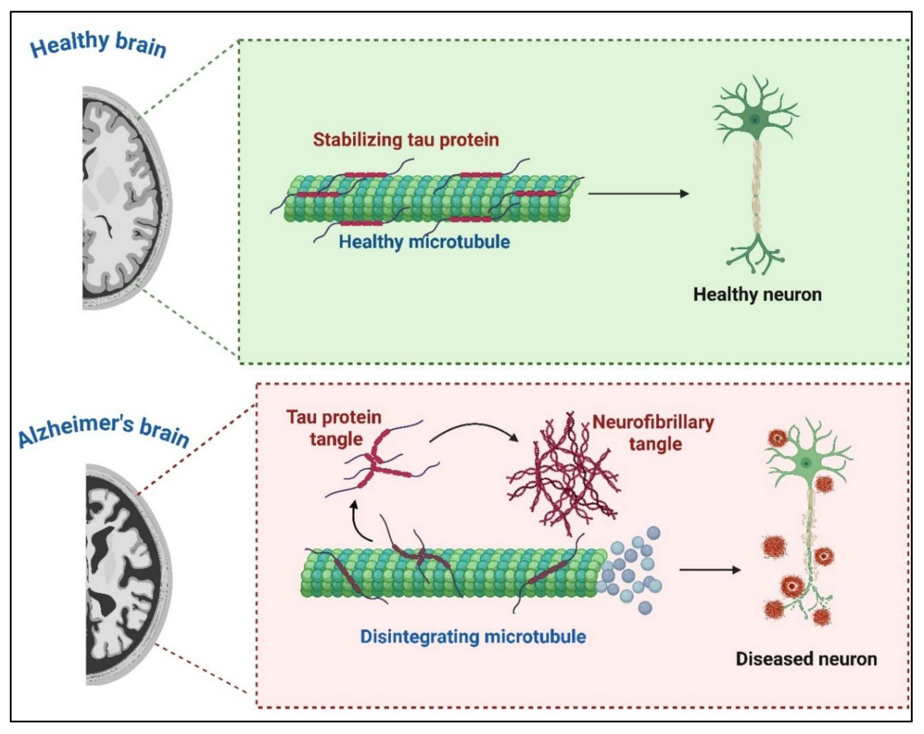
Figure 3.
Nb-based Drug Delivery Systems. This figure illustrates two primary approaches for Nb-based drug delivery. (A) Direct Conjugation: Therapeutic agents are directly linked to Nbs, enabling specific payload delivery via direct binding. (B) Nanocarrier Functionalization: Therapeutic agents are encapsulated within nanocarriers, which are then conjugated to Nbs for selective delivery to disease sites. Both strategies contribute to improved drug stability, bioavailability, controlled release, and the ability to overcome biological barriers, such as the blood-brain barrier, in CNS disorders (Created using Biorender).
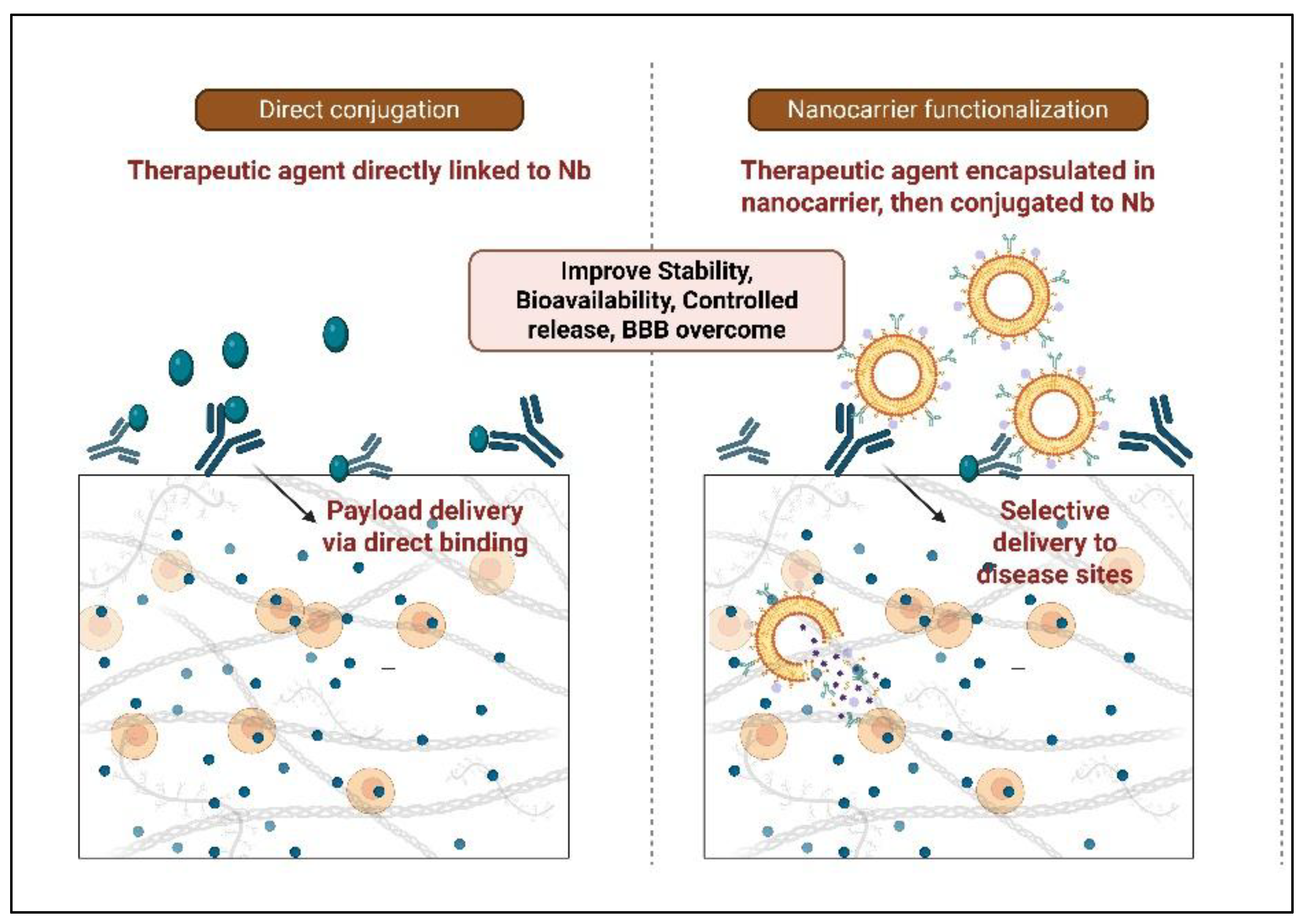
Figure 4.
Nanocarriers Functionalized with Nanobodies (Nbs) The figure displays various types of nanocarriers including Microbubbles, Nanospheres, Micelles, Nanocapsules, Dendrimers, Nanoparticles, and Liposomes that can be conjugated with Nanobodies. This conjugation strategy leverages the high specificity of Nbs to target these drug-loaded nanocarriers to specific disease sites, enhancing drug stability and selective delivery.
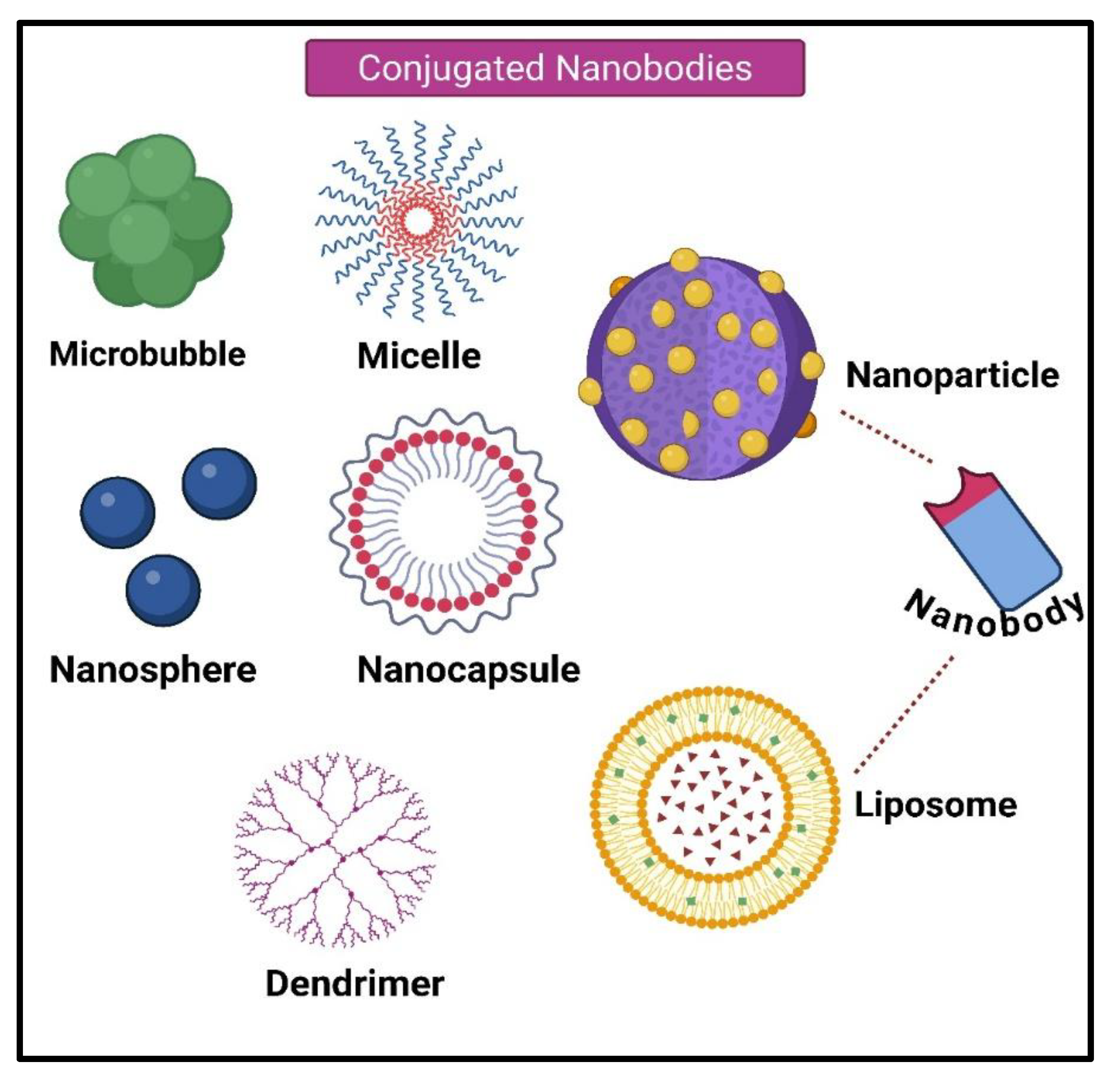
Disclaimer/Publisher’s Note: The statements, opinions and data contained in all publications are solely those of the individual author(s) and contributor(s) and not of MDPI and/or the editor(s). MDPI and/or the editor(s) disclaim responsibility for any injury to people or property resulting from any ideas, methods, instructions or products referred to in the content. |
© 2025 by the authors. Licensee MDPI, Basel, Switzerland. This article is an open access article distributed under the terms and conditions of the Creative Commons Attribution (CC BY) license (http://creativecommons.org/licenses/by/4.0/).
Copyright: This open access article is published under a Creative Commons CC BY 4.0 license, which permit the free download, distribution, and reuse, provided that the author and preprint are cited in any reuse.


