Submitted:
30 November 2023
Posted:
01 December 2023
You are already at the latest version
Abstract
Transitional vertebra has been reported along of the spine in many researches focused especially on carnivores. The current study had the objectives to identify the presence of transitional lumbosacral and sacrocaudal vertebra in domestic mammals, to realize a comparative analysis of the localization and conformation of this abnormal condition. For this purpose the researches included animals belonging to next species: cow – 29 specimens, sheep – 32 specimens, horse – 31 specimens, pig – 26 specimens, rabbit – 33 specimens, dog – 89 specimens and cat – 57 specimens. The most of the animals, respectively their spine were analysed post mortem, after the dissection performed in the anatomy laboratory, except the dogs and 48 cats that were examined radiologically. The investigations revealed the presence of transitional vertebra in all species as follow: in cows – 3 cases (8,7%), all being about the lumbarization of first sacral vertebra (S1); in sheep – 3 cases (9,37%), two lumbarization cases of S1 and one caudalisation of S4 (the last sacral vertebra); in horses – 4 cases (12,9%), all about the sacralisation of Cd1 (first caudal vertebra); in pigs – 3 cases (11,53%), two lumbarisation cases of S1 and one sacralisation of Cd1; in rabbits – 3 cases (9,09%), a lumbar supernumerary vertebra (L8) and two cases of caudalisation of S4; in dogs – 4 cases (4,49%), a lumbar supernumerary vertebra (L8) and 3 cases of sacralisation of last lumbar vertebra (L7 or L8); in cats – 3 cases (5,26%), two sacralisation cases of last lumbar vertebra and one caudalisation of last sacral vertebra (S3). A strong lumbarization process was observed in ruminants (especially in cows), then in pigs, the sacralisation being prevalent in carnivores. The sacrocaudal transitional vertebrae was predominant in horses, but occurred as an ossification process especially in old animals.
Keywords:
transitional vertebra
; lumbosacral and sacrocaudal junctions
; domestic mammals
; comparing morphology
1. Introduction
Knowledge of normal spinal morphology and the incidence of vertebral anomalies is important for distinction of pathologic change from functional anatomic variations [1]. Vertebral congenital anomalies were defined as any defect in vertebral body formation [2,3,4,5]. Among these, transitional vertebra is one of the most common anatomical abnormality of the spine. Transitional vertebra is considered a congenital anomaly, which occurs in various species of animals and in humans [2,6] and its presence can be clinically significant [7]. It’s supposed to exist a hereditary predisposition to transitional vertebra [8,9,10,11]. Transitional vertebrae can appear in any junction of the spine presenting common characteristics of both adjacent vertebral segments. Lumbosacral transitional vertebra (LTV) is considered to be the result of an incomplete homeotic transformation of a lumbar vertebra into a sacral vertebra (sacralisation) or a sacral vertebra into a lumbar vertebra (lumbarisation) [12]. The prevalence of sacralisation is higher than lumbarisation [13,14]. Lumbosacral transitional vertebra is described as a congenital anomaly which could have some effect on spinal biomechanics and consequently could affect degenerative processes in adjacent vertebra in mammals [12]. Its presence is identified having a higher prevalence in domesticated species compared to wild counterparts, as well as slower-moving species compared to fast and agile mammals [5].
The morphology of transitional vertebrae is variable, presenting modifications of one or both vertebral arch and body and can have right-to-left asymmetry or lateral cranial-to-caudal gradation in vertebral morphology [1,15,16].
In domestic mammals, the researches on transitional vertebrae are focused especially on dogs and less, in cats, in which, the congenital abnormalities of the spine are frequently identified radiographically [17]. The largest description of transitional vertebrae regarding the localization, the morphology, clinical significance and prevalence exist in dogs [8,18,19]. Such anomalies, as well as others (hemivertebrae, wedge vertebrae, block vertebrae, atlantoaxial malformations and spina bifida) and their prevalence have been described also in cats, ferrets [20,21,22,23]. In other domestic mammals the reports regarding the presence and type of transitional vertebrae are even less, for that we proposed to investigate the lumbosacrocaudal spine in as many species as possible, to have a comparative view, not only radiologically, but especially anatomical analysis. We consider it’s the first study in which are included all domestic species for this investigation. Anyway, we intended to highlight the particular morphology of transitional vertebrae in all the species by anatomical examination, not only radiologically as in the most existing researches on this subject.
2. Materials and Methods
The researches of this study were performed on a period of 14 years, between 2009-2023. The studies based on morphologic description realized directly on the bones and skeletons in the anatomy labs of Faculty of Veterinary Medicine from Iasi, Romania, and completed by radiologic exam. In case of some species – ruminants, horses, pigs, rabbits and cats, the specimens represented old or sick animals with various pathologies (metabolic, locomotor and others), but free of risk for humans, which died previously and the owners agreed to donate them for didactic process. After the valorisation of the animals for dissection, their bones were harvested to prepare the skeleton. In this way, along of the years, the bones collected were boiled and cleaned of debris mechanically and then chemically in a hydrogen peroxide solution 3%, a variable interval to obtain clean pieces. After that, the spine of animals was analysed anatomically, respectively the vertebrae were counted and studied their normal or modified characteristics and described in accordance with Nomina Anatomica Veterinaria [24].
There were valorised 29 specimens of cows, 32 specimens of sheep, 31 specimens of horses, 26 specimens of pigs and 33 specimens of rabbits (Table 1). In case of carnivores, for our purpose, it was chosen the x-ray exam as the method, being more facile. The animals were the patients of a veterinary clinic subjected to the x-ray exam of the spine. Totally, the studies involved 89 dogs and 48 cats, from different breeds, both males and females (Table 1).
The animals were brought to the clinic for various pathologies, presenting especially nervous and locomotor symptoms. The patients were clinically examined and then the x-ray exam of the spine was performed using latero-lateral and ventro-dorsal incidences. The radiographs obtained were analysed to diagnose the pathology, but also for the anatomical conformation of the spine, the number of the vertebra being also counted. Other 9 cats of common breed were examined post-mortem, as in the case of other species.
3. Results
The investigations performed revealed the presence of transitional and/or supernumerary vertebra in the lumbosacral and sacrococcygeal regions in all studied species (Table 2). The results are presented on species, as follows:
This section may be divided by subheadings. It should provide a concise and precise description of the experimental results, their interpretation, as well as the experimental conclusions that can be drawn.
3.1. Ruminants
In ruminants we identified the transitional vertebra in both, big and small ruminants (cows and sheep). In cows, the normal vertebral formula includes 6 lumbar vertebrae, 5 sacral vertebrae, the sacrum bone presenting a continuous median sacral crest and intermediate sacral crest (between the median one and lateral sacral crests) and 18-20 caudal vertebrae [25].
3.1.1.
In cows we consider it’s been found the most "spectacular" cases of lumbosacral transitional vertebrae, more exactly an evident and intense lumbarisation of the first sacral vertebra (S1). This aspect was found in three animals. In this way, can be remarked the first sacral vertebra (promontorium) is totally independent, having both cranial and caudal articular surfaces on the body. The transverse processes are separated from the wing of sacrum and tend to be similar with those of lumbar vertebrae. The two processes are asymmetric in width (Figure 1).
3.1.2.
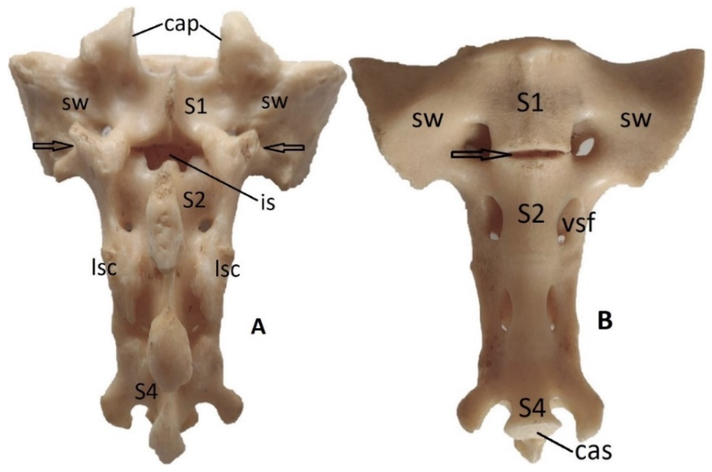
In sheep the normal vertebral formula includes 6 lumbar vertebrae, 4 sacral vertebrae, but the sacrum has an interrupted median sacral crest and also intermediate sacral crest (between the median one and lateral sacral crests) and 16-24 coccygeal vertebrae [25]. Our investigations in this species revealed the presence of transitional vertebrae in the same place, as in case of big ruminants, in two animals, but it’s been recorded only a tendency of lumbarisation of the first sacral vertebra. In this way, it was observed a partial separation of S1 and S2. Dorsally, in the first case (Figure 4A) the separation in more visible at the level of the arches, between the two vertebrae existing a large interarcuate space that normally it’s not present, in the second case not existing. Also, the tendency of separation, in both cases is visible on the sacral wings, where the transverse processes of S1 and S2 are not complete fused (Figure 4A - arrows). Ventrally, on both cases, can be remarked the incomplete fusion of the vertebral bodies of S1 and S2 and incomplete ossification of the intervertebral disc (Figure 4B – arrow). In a third case was found a sacrocaudal transitional vertebra consisting in the caudalisation of the last sacral vertebra that was completely detached (Figure 3).
Figure 2.
Cow, male, 4 years – the lumbarisation of the first sacral vertebra (S1 – promontorium) and the detachment by the second one (arrow), dorsal view: L6 – the sixth lumbar vertebra; tp – transverse processes of S1; cap – cranial articular processes; Iw – iliac wing; S2 – the second sacral vertebra; S5 – the fifth sacral vertebra.
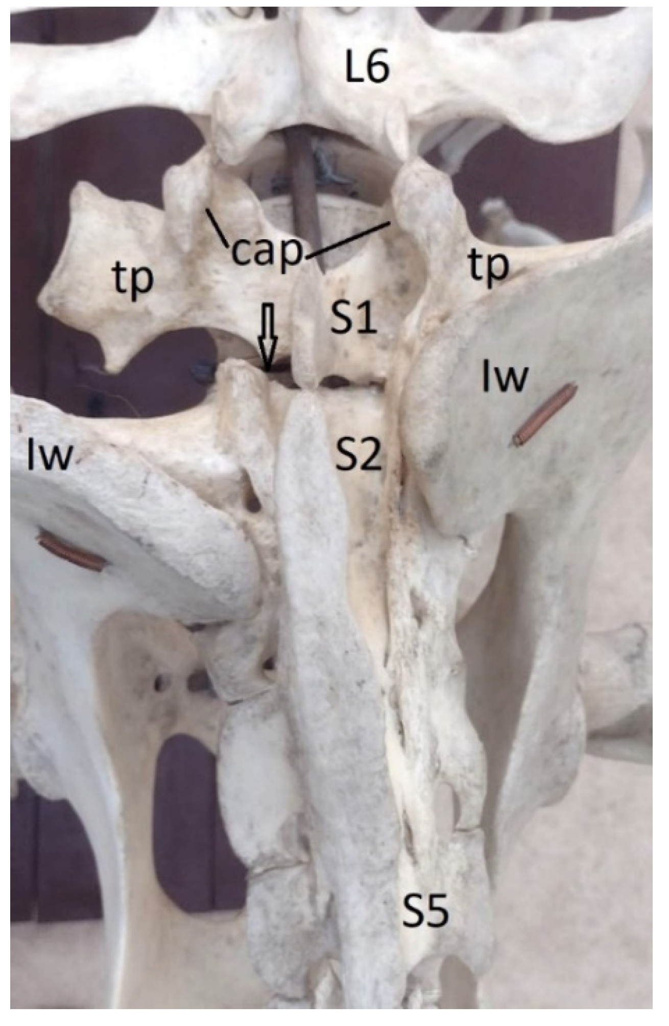
Figure 3.
Sheep, male, 4 years – the caudalisation of the last sacral vertebra (S4) and the detachment by the third one (arrows), dorsal view: L6 – the sixth lumbar vertebra; Iw – iliac wing; S1 – first sacral vertebra; S3 – the third sacral vertebra; f -femur.
Figure 3.
Sheep, male, 4 years – the caudalisation of the last sacral vertebra (S4) and the detachment by the third one (arrows), dorsal view: L6 – the sixth lumbar vertebra; Iw – iliac wing; S1 – first sacral vertebra; S3 – the third sacral vertebra; f -femur.
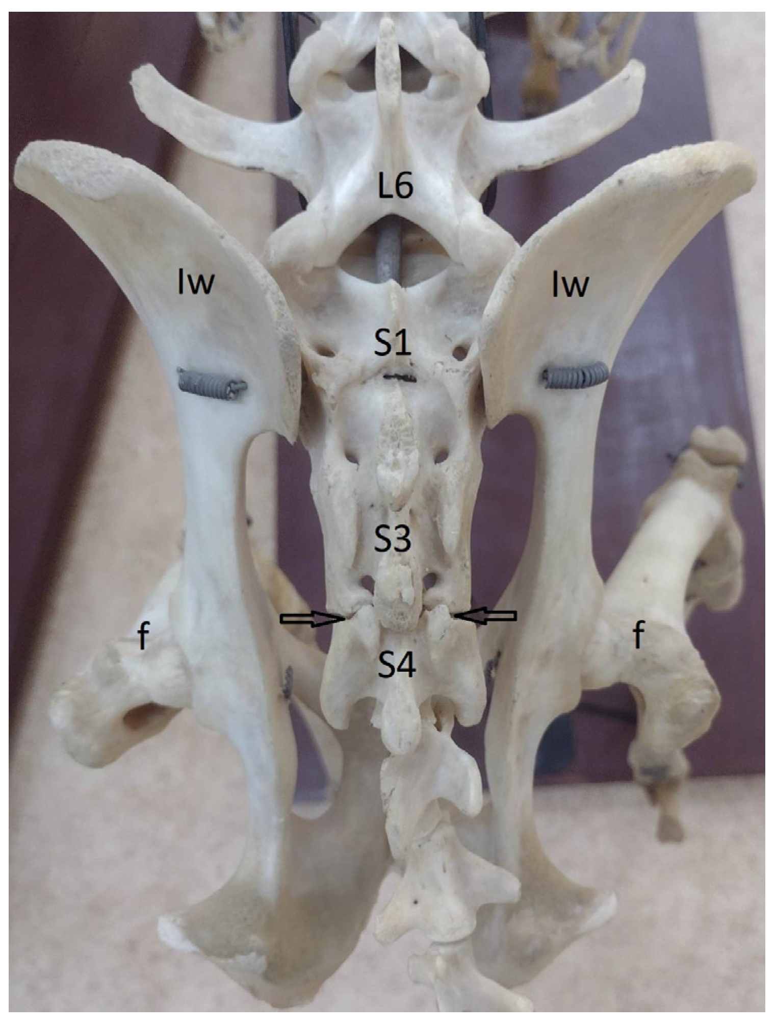
Figure 4.
Sheep, female, 3 years – the tendency of lumbarisation of the first sacral vertebra (S1 – promontorium); the separation line (arrows) is visible dorsally on the wings between the transverse processes of S1 and S2 together with the presence of the interarcuate space (is) and ventrally by the persistence of intervertebral disc between S1 and S2 (arrow). A – dorsal view, B – ventral view; cap – cranial articular processes; S2 – the second sacral vertebra; sw – sacral wing; lsc – lateral sacral crest; vsf – ventral sacral foramen; S4 – the forth sacral vertebra.
3.2. Horses
In horses, normally, the lumbar region includes 6 vertebrae, except the Thoroughed racehorses (Arabian and English) in which exist generally 5 vertebrae, the sacrum bone 5 vertebrae, with an interrupted median sacral crest, and the caudal region a variable number of vertebrae - 15-21 [25]. In our observations, transitional vertebra was found in four animals, in all cases in the sacrocaudal junction. It was noticed that the first caudal vertebra was fused with the last sacral vertebra (S5). As consequent, the sacrum bone counted 6 vertebrae. In two horses the vertebral bodies were totally fused due to the complete ossification of the intervertebral disc (Figure 5B). Also, the ossification process was present between the vertebral arches, and the interarcuate space became narrower (Figure 5A).
In the other two cases the fusion happened only between the vertebral bodies, and the intervertebral disc, even ossified, remained well visible (Figure 6). In all horses the transverse processes of the fifth sacral vertebra and first caudal one remained separated.
3.3. Pigs
In pigs the lumbar region counts 6 vertebrae, the sacrum bone includes 5 or 4 vertebrae and 20-23 caudal vertebrae. The sacrum bone has tick and vertical wings, no median crest and narrow interarcuate spaces [25]. In this species, we identified both types of transitional vertebrae, lumbosacral (2 cases) and sacrocaudal (2 cases).
The sacrocaudal transitional vertebra was identified in a pig, being observed the fusion of the first caudal vertebra with the sacrum bone (sacralisation). The merging produced both between the bodies and the arches and secondary, the interarcuate space became narrower (Figure 7).
In other 2 pigs, was noticed the detachment of the first sacral vertebra, respectively its lumbarization. The two vertebrae (S1 and S2) were completely separated, but the lumbarisation process was not so intense as in case of cows, the promontorium maintaining a more similar conformation as in case of a normal sacrum bone. Still, it could be remarked a tendency of elongation of the sacral wings, to turn on transverse processes (Figure 8 – circles).
3.4. Rabbits
In rabbits, lumbar region counts 7 vertebrae, the sacrum bone 4 vertebrae, with an interrupted median sacral crest, and the caudal region 14-16 vertebrae [25].
On our examined animals we identified a supernumerary lumbar vertebra (L8) in a rabbit, the lumbar region counting 8 lumbar vertebrae (Figure 9). The 8th lumbar vertebra had a proper conformation to that of normal lumbar vertebrae, not fused with adjacent vertebrae. It was noticed only its shortening compared with the others. It can be considered a lumbosacral transitional vertebra or a numerical variation of the lumbar vertebrae.
In other two rabbits, the observations revealed the incomplete fusion of the last sacral vertebra with sacrum bone and the persistence of the intervertebral disc, respectively a tendency of caudalisation of the last sacral vertebra – S4 (Figure 10).
3.5. Carnivores
In carnivores (dog and cat) exists 7 lumbar vertebrae, 3 sacral vertebrae, the median and intermediate sacral crests being interrupted and 20-23 caudal vertebrae [25,26].
3.5.1.
In dogs, among various pathologies, radiologically we identified transitional vertebrae in the lumbosacral junction, having the specific conformation of a lumbar vertebra (supernumerary vertebra) or fused more or less with the sacrum (sacralisation). Three animals had 8 lumbar vertebrae, the last one (L8) being generally shorter (Figure 11, Figure 12), two of them presenting also the sacralisation of L8. In the other dog has been observed the sacralisation of the 7th lumbar vertebra. In all cases the sacralisation was partial and asymmetric, a transverse process having a more appropriate shape to that of a normal one, the other being fused with the sacral wing and taking contact with the iliac wing (Figure 12).
3.5.2.
In cats, we found transitional vertebrae both in the lumbosacral region and sacrocaudal one. Two animals presented a large degree of sacralisation of the last lumbar vertebra. In a cat the transitional vertebra was associated with supernumerary vertebra, being counted 8 lumbar vertebrae, the last one being intense sacralised. The another cat presented sacralisation of L7. The sacralisation process was similar, being noticed that both transverse processes and the vertebral body were fused with those of the first sacral vertebra. It can be observed that the last lumbar vertebra (L7 or L8) is shorter, being almost entirely placed between the two iliac wings, and is no evident intervertebral disk between its body and the body of the first sacral vertebra. Also is visible that the transvers processes are shorter cranially and both are fused with the true sacral wings (the transverse processes of the first sacral vertebra) coming largely in contact with the iliac wings (Figure 13). There are present three sacral foramina on each lateral side of sacrum comparing with normal sacral bone in this species [25,26]. These aspects were observed at the x-ray exam.
Anatomically, in a cat, was identified a sacrum bone counting 2 vertebrae, the last one being independent and caudalised. The second sacral vertebrae presented caudal articular processes and caudal articular surface on the body (Figure 14).
4. Discussion
For ruminants and pigs exist very few descriptions of transitional vertebra. Unusual transitional vertebra is mentioned between the thoracic and lumbar vertebrae in sheep [27]. The post-mortem CT and MRI scanning of 41 sheep revealed the presence of the lumbosacral transitional vertebrae considered as an anatomic variation of the last lumbar vertebra due to the enlarged transverse process which come in contact with the sacrum or ilium. In this case the fusion was considered congenital due to the lack of other lesions [28]. In ruminants, our research revealed the lumbosacral transitional vertebrae, but only cases of lumbarisation, very evident in case of big ruminants, but not sacralisation, in contrast with previous investigations.
In pigs is mentioned the presence of the transitional vertebrae at the thoracolumbar junction. In a group of 37 pigs of Landrace and Duroc breeds, the transitional vertebra was identified in a rate of 22% - 8 pigs, four pigs having 5 lumbar vertebrae and the other four with 6 lumbar vertebrae. Transitional vertebrae only occurred at the thoracolumbar junction, represented thoracoisation of lumbar vertebrae/lumbar ribs and were counted with thoracic vertebrae. 7 transitional vertebrae were found in Landrace breed and a single one in Duroc Breed [29]. Thoracolumbar transitional vertebrae were not associated with abnormal curvature in the examined pigs. In humans, lumbosacral transitional vertebrae have been associated with lower back pain [30]. In addition to these observations, we identified transitional vertebrae in pigs, in both lumbosacral and sacrocaudal junction, as lumbarisation process of the first sacral vertebra and sacralisation of the first caudal vertebra. Also, in pigs we revealed the tendency of lumbarization of S1, but also the sacralization of Cd1, aspects that reflect the evident predisposition to transitional vertebra in this junctions.
In horses the data regarding the transitional vertebra are less, being difficult to realize such studies on live animals as CT scanning or tomography, other way it’s necessary to perform a post-mortem examination. The observations of Haussler et al., 1997 [1], revealed thoracholumbar transitional vertebrae on a rate of 22% (8 animals) of examined animals (36 Thoroughed racehorses), and sacrocaudal transitional vertebra on a rate of 36% (13 animals). In 61 % of animals the number of lumbar and sacral vertebrae was normal (6 and 5 vertebrae). Lumbosacral transitional vertebrae were not found, as in case of our research in this species. Other studies reported the sacralisation of L6, characterized by ankyloses and malformation of L6 and the sacrum in two horses with clinical sings of hind limb lameness and possible sacroiliac joint injury [31]. Regarding the sacrocaudal transitional vertebra has been suggested it appears often as a congenital development anomaly than as acquired ankyloses or pathologic changes. Also, it has been reported that an elongated sacrum is often seen in aged horses, in which the first and sometimes the second caudal vertebrae fuse with the sacrum [1]. In our findings only the first caudal vertebra was fused with the sacrum. Similarly to the study of Haussler et al., 1997 [1], this aspect was available in our investigations, by the presence of sacrocaudal transitional vertebrae in older specimens, in a higher rate.
In the descriptive post-mortem study of Spoormakers et al., 2021 [32], focused on anatomical variations in three breeds, the caudal cervical, thoracic, lumbar and sacral regions of the vertebral column of 30 Warmblood horses, 29 Shetland ponies and 18 Konik horses were examined using computed tomography. Transitional vertebrae were most frequently encountered in Shetland ponies. Lumbarization of the last thoracic vertebra was encountered in Shetland ponies in a rate of 7%. In one case was associated with a reduction to 17 rib pairs. In the other, the anomaly resulted in 18 ribs and 7 transverse processes on the left side, and 19 ribs with 6 transverse processes on the right side. Thoracoisation of the first lumbar vertebra was seen in one Shetland pony - 3% having 19 ribs bilaterally. Sacralisation of the last lumbar vertebra was seen in one Warmblood – 3%. Sacralization of the first caudal vertebra by fusion to sacrum was encountered in 33% of Shetland ponies, 29% of Warmblood horses and 6% of Konik horses. The incomplete sacralisation of the last lumbar vertebra was reported by Scilamati et al., 2023 [33], on a study on the last lumbar vertebrae of 40 horses, by CT scanning. In this case, the sacralisation on the last lumbar vertebra was associated with spondilolisthesis. These results, confirm again the higher rate of the sacrocaudal transitional vertebra comparing with others in this species [32]. The development of the imaging techniques led to the possibility to investigate the variability in the conformation and the abnormalities of the spine in this species.
In rabbits few studies reported that the transitional vertebrae, which are located at the junction of different segments of the spine, are also concerned. Malformations caused by transitional vertebrae were identified especially in the thoracolumbar junction, and less in the lumbosacral junction. Thoracolumbar transitional vertebra was observed in association with awith lordoscoliosis, wedge vertebrae with kyphoscoliosis, or lordoscoliosis with a twisted pelvis [34]. These aspects have been confirmed by Proks et al., 2018 [5], in their study, in which the thoracolumbar junction was the most common location of transitional vertebral anomalies, the lumbosacral junction being the second one. Proks et al., 2018 [5], on 330 rabbits, counted the vertebral formula, but also the presence of vertebral anomalies. There have been identified seven vertebral formulas with normal vertebral morphology in 84.8% of animals. The most common abnormalities were the transitional vertebrae found in 41 animals, in the thoracolumbar segment – 35 rabbits and lumbosacral one – 6 rabbits. In two rabbits the presence of thoracolumbar and lumbosacral transitional vertebrae has been associated to thoracic lordoscoliosis. No difference between sexes in the prevalence of anomalies was observed. Previous investigators have suggested that a vertebral formula with eight lumbar vertebrae could increase susceptibility to lumbar vertebral fractures in rabbits [35]; however, we observed this variant in only one rabbit. Few rabbits had a rudimentary intervertebral space between S3 and S4, similar to ferrets [22]. These findings correspond also with our two cases of incomplete fusion of the last sacral vertebrae
In dog, the anatomical variations and morphologic abnormalities along of the spine, including transitional vertebrae, have been investigated more than in other species, due to the possibility to use imagistic methods and techniques and being well known the predisposition of this species to these modifications. In dogs, the presence of a supernumerary vertebra (L8) is often reported and usually has no clinical importance [36]. Our observations showed that, in general the 8LV is shorter, as the last lumbar vertebra when the lumbar region counts 7 vertebrae. The most of the problems were reported when the supernumerary vertebra (L8) is associated with lumbosacral transitional vertebra (LTV). These variations are affected by gender, developmental factors and race, being observed that the males are affected more than the females [13]. Lumbosacral transitional vertebra (LTV) is a common congenital anomaly seen in several dog breeds: Pug, Jack Russell Terrier, French Bulldog, etc [37,38]. By correlation, our relatively low percentage of transitional vertebrae in dogs can be related to other breeds that have been included in the research and the lower number of animals investigated in this species.
Deepa and John, 2014 [13], reported that the sacralisation is more common in occurrence than lumbarization, in a rate of 2:1. Also, in our study we can confirm these aspects, transitional vertebrae being found in three males and a single female and no lumbarisation case has been found.
Regarding the shape of transverse processes [15,19], our investigations revealed only asymmetrical transitional vertebrae.
The presence of LTV is associated with degenerative lumbosacral stenosis (DLSS) [39] and can lead to premature degeneration of the lumbosacral junction and cauda equina syndrome, especially in German Shepherds [2,40].
In cats, little works have been published relating specifically to the incidence and types of congenital and non-congenital vertebral anomalies. In Manx cat were reported spina bifida and hemivertebrae in the caudal and sacral vertebrae [20], but the transitional vertebra is described as the most common abnormality at all level of the feline spine, the main site for transitional abnormalities being the sacrocaudal juntion [20,41]. This studies concluded that there is no evidence of association with clinical signs. Also in cats, as in dogs, the sacralisation is more prevalent than lumbarisation, all our cases being of strong sacralisation. Harris et al., 2018 [42], observed in their study that the lumbosacral transitional vertebrae were significantly more prevalent in cats with lumbosacral stenosis compared with the control feline population.
The investigations of Thanaboonnipat et al., 2021 [43], on 1365 cats, revealed as congenital anomalies, six lumbar vertebrae – 141 animals, sacralisation – 48 animals, lumbarisation - 16 animals, hemivertebrae – 1 animals, and five lumbar vertebrae – 1 animals. Even the number of animals was higher, the percentage of lumbosacral transitional vertebra (sacralisation+lumbarisation) in this study was appropriate to that of our findings. It has been concluded, that cats with abnormal lumbosacral vertebrae are predisposed to have more problems with the large bowel [43].
In all species investigated, we cannot establish a direct relation between the presence of transitional vertebra and other criteria, especially the breed, the most of the animals being of the common breed. Regarding the age, many cases were observed in relatively old animals, but we can affirm this at most in case of sacralisation of Cd1, where we consider to be an ossification process due to the age and not a congenital malformation. Further investigations can lead to more precise data. Also, for the gender it’s difficult to affirm the predisposition of males or females to transitional vertebra, in the most of the species being included a higher number of males, but the transitional vertebra was found both in males and females, except the dog, in which transitional vertebra occurred predominantly in males
5. Conclusions
Even if the most of the researches on transitional vertebra have been focused in carnivores, especially on dogs, we can affirm it occurs in all the species, at least in the lumbosacral and sacrocaudal junctions. Also, it has been found a large morphological variability of transitional vertebra between investigated species. In carnivores, in correlation with other researches, it’s obvious that predominates the sacralisation process and supernumerary vertebrae. An evident lumbarisation process in our study, occurs mostly in ruminants, both big and small, but also in pigs, these findings being among the first in this direction. The transitional sacrocaudal vertebrae seems to appear especially in horses, then in pigs as an ossification process, more or less intense, in case of old animals. Also in rabbit transitional vertebrae occur in both junctions, lumbosacral and sacrocaudal one, even not very spectacular in our findings.
Funding
This research received no external funding.
Institutional Review Board Statement
Ethical review and approval are waived for this study because euthanasia of the animals was unrelated to the study.
Informed Consent Statement
Informed owner consent was signed by the owner at the time of admission to the veterinary clinic.
Data Availability Statement
Suggested Data Availability Statements are available in section “MDPI Research Data Policies” at https://www.mdpi.com/ethics.
Conflicts of Interest
The authors declare no conflict of interest.
References
- Haussler, K.K.; Stover, S.M.; Willits, N.H. Developmental variation in lumbosacropelvic anatomy of Thoroughed racehorses. American Journal of Veterinary Research 1997, 58, 1083–1091. [Google Scholar] [CrossRef] [PubMed]
- Flückiger, M.; Geissbühler, U.; Lang, J. Lumbosacral transitional vertebra: how important are they for the health of affected dogs? Schweizer Archiv für Tierheilkunde 2009, 151, 133–135. [Google Scholar] [CrossRef] [PubMed]
- Westworth, D.R.; Struges, B.K. Congenital malformations in small animals. Veterinary Clinic of North America: Small Animal Practice 2010, 40, 951–981. [Google Scholar] [CrossRef] [PubMed]
- Gutierrez-Quintana, R.; Guevar, J.; Stalin, C.; Faller, K.; Yeamans, C.; Penderis, J. A proposed radiographic classification scheme for congenital thoracic vertebral malformations in brachycephalic “screw-tailed” dog breeds. Veterinary Radiology and Ultrasound 2014, 55, 585–591. [Google Scholar] [CrossRef] [PubMed]
- Proks, P.; Stehlik, L.; Nyvltova, I.; Necas, A.; Vignoli, M.; Jekl, J. Vertebral formula and congenital abnormalities of the vertebral column in rabbits. The Veterinary Journal 2018, 236, 80–88. [Google Scholar] [CrossRef]
- Kumar Jat, S.; Srivastava, A.; Malhotra, R.; Chadha, M.; Tandon, A.; Jain, A.K. Prevalence of lumbosacral transitional vertebra in patients with chronic low back pain: a descriptive cross-sectional study. Am J Neurodegener Dis. 2023, 12, 89–96. [Google Scholar]
- Bahadir Ulger, F.E.; Illeez, O.G. The effect of lumbosacral transitional vertebrae (LSTV) on paraspinal muscle volume in patients with low back pain. Acad Radiol. 2020, 27, 944–950. [Google Scholar] [CrossRef] [PubMed]
- Morgan, J.P.; Wind, A.; Davidson, A.P. Bone dysplasias in the labrador retriever: a radiographic study. Journal of the American Animal Hospital Association 1999, 35, 332–340. [Google Scholar] [CrossRef] [PubMed]
- Wigger, A.; Julier-Franz, C.; Tellhelm, B.; Kramer, M. Lumbosakraler Übergangswirbel beim Deutschen Schäferhund: Häufigkeit, Formen, Genetik und Korrelation zur Hüftgelenksdysplasie [Lumbosacral transitional vertebrae in the German shepherd dog: prevalence, classification, genetics, and association with canine hip dysplasia]. Tierärztl Prax K: Kleintiere/Heimtiere 2009, 37, 7–13. [Google Scholar]
- Fialová, I.; Paninárová, M.; Nečas, A.; Stehlík, L.; Proks, P. Prevalence of lumbosacral vertebrae in dogs in the Czech Republic. Acta Veterinaria BRNO. 2014, 83, 399–403. [Google Scholar] [CrossRef]
- Gluding, D.; Stock, K.F.; Tellhelm, B.; Kramer, M.; Eley, N. Genetic background of lumbosacral transitional vertebrae in German shepherd dogs. Journal of Small Animal Practice 2021, 62, 967–972. [Google Scholar] [CrossRef] [PubMed]
- Galis, F.; Carrier, D.R.; van Alphen, J.; van der Mije, S.D.; Van Dooren, T.J.M.; Metz, J.A.J.; ten Broek, C.M.A. Fast running restricts evolutionary change of the vertebral column in mammals. Proceedings of the National Academy of Sciences of the United States of America 2014, 111, 11401–11406. [Google Scholar] [CrossRef] [PubMed]
- Deepa, T.K.; John, M.K. A Study of Lumbarisation of First Sacral Vertebra Among the South Indians. International Journal of Medical Research and Health Sciences 2014, 3, 1–4. [Google Scholar] [CrossRef]
- Abutaher, C.P.; Avinash, P.R. Lumbosacral transitional vertebrae – occurrence and significance. International Journal of Current Research and Review 2019, 11, 1–4. [Google Scholar] [CrossRef]
- Breit, S.; Knaus, I.; Kunzel, W. Differentiation between lumbosacral transitional vertebrae, pseudolumbarisation, and lumbosacral osteophyte formation in ventrodorsal radiographs of the canine pelvis. Veterinary Journal 2003, 165, 36–42. [Google Scholar] [CrossRef] [PubMed]
- Konin, G.P.; Walz, D.M. Lumbosacral transitional vertebrae: classification, imaging findings, and clinical revalence. American Journal of Neuroradiology 2010, 31, 1778–1786. [Google Scholar] [CrossRef] [PubMed]
- Bailey, C.S.; Morgan, J.P. Congenital spinal malformations. Vet Clin North Am Small Anim Pract. 1992, 22, 985–1015. [Google Scholar] [CrossRef] [PubMed]
- Morgan, J.P. ; Transitional lumbosacral vertebral anomaly in the dog: a radiographic study. Journal of Small Animal Practice 1999, 40, 167–172. [Google Scholar] [CrossRef]
- Damur-Djuric, N.; Steffen, F.; Hässig, M.; Morgan, J.P.; Flückiger, M.A. Lumbosacral transitional vertebrae in dogs: classification, prevalence, and association with sacroiliac morphology. Veterinary Radiology & Ultrasound 2006, 47, 32–38. [Google Scholar]
- Newitt, A.; German, J.A.; Barr, J.F. Congenital abnormalities of the feline vertebral column. Veterinary Radiology & Ultrasound 2008, 49, 35–41. [Google Scholar]
- Westworth, D.R.; Struges, B.K. Congenital spinal malformations in small animals. Vet Clin North America Small Anim Pract. 2010, 40, 951–981. [Google Scholar] [CrossRef] [PubMed]
- Proks, P.; Stehlik, L.; Paninarova, M.; Irova, K.; Hauptman, K.; Jekl, V. Congenital abnormalities of the vertebral column in ferrets. Veterinary Radiology and Ultrasound 2015, 56, 117–123. [Google Scholar] [CrossRef] [PubMed]
- Gee Lee, E.; Yi Park, S.; Lee, K.; Jang, M.; Kim, T.J. Choi S, Park I. Radiographic evaluation of congenital vertebral anomalies in Korean raccoon dogs (Nyctereutes procyonoides koreensis). J Vet Sci. 2021, 22, e52. [Google Scholar] [CrossRef] [PubMed]
- I.C.V.G.A.N. Nomina Anatomica Veterinaria (N.A.V.), 6th ed.; Editorial Committee Hanover (Germany), Ghent (Belgium), Columbia, MO (USA), Rio de Janeiro (Brazil), 2017.
- Ferat-Postolache, A.; Covașă, C.T.; Munteanu, A. Anatomie Veterinară, Sistemul Locomotor. Ed. ʺIon Ionescu de la Bradʺ, Iași, 2021; pp. 97-123.
- König, H.E.; Liebich, H.-G. Veterinary Anatomy of Domestic Mammals, Textbook and Colour Atlas, 3rd ed.; Schattauer: Stuttgart, New York, 2007; pp. 75–85. [Google Scholar]
- Pugh, D.G; Baird, A.N.; Edmondson, M.A.; Passler, T. Sheep, Goat and Cervid Medicine, 3rd ed.; Elsevier: Edinburgh, New York, 2020. [Google Scholar]
- Nisolle, J.F.; Bihin, B.; Kirschvink, N.; Neveu, F.; Clegg, P.; Dugdale, A.; Wang, X.; Vandeweerd, J.M. Prevalence of Age-Related Changes in Ovine Lumbar Intervertebral Discs during Computed Tomography and Magnetic Resonance Imaging. Comaparative Medicine 2016, 66, 300–307. [Google Scholar]
- Olstad, K.; Aasmundstad, T.; Kongsro, J.; Grindfek, E. Osteochondrosis and other lesions in all intervertebral, articular process and rib joints from occiput to sacrum in pigs with poor back conformation, and relationship to juvenile kyphosis. BMC Veterinary Research 2022, 18. [Google Scholar] [CrossRef] [PubMed]
- Jancuska, J.M.; Spivak, J.M.; Bendo, J.A. A review of symptomatic lumbosacral transitional vertebrae: Bertolotti’s syndrome. Int J Spine Surg. 2015, 9. [Google Scholar] [CrossRef] [PubMed]
- Jeffcott, L.B. Radiographic appearance of equine lumb and pelvic abnormalities by linear tomography. Vet Radiol Ultr. 1983, 24, 201–213. [Google Scholar] [CrossRef]
- Spoormakers, T.J.P.; Veraa, S.; Graat, E.A.A.; van Weeren, P.R.; Brommer, H. A comparative study of breed differences in the anatomical configuration of the equine vertebral column. Journal of Anatomy 2021, 239, 829–838. [Google Scholar] [CrossRef]
- Scilimati, N.; Angeli, G.; Di Meo, A.; Dall’Aglio, C.; Pepe, M.; Beccati, F. Post-Mortem Computed Tomographic Features of the Most Caudal Lumbar Vertebrae, Anatomical Variations and Acquired Osseous Pathological Changes, in a Mixed Population of Horses. Animals 2023, 13, 743. [Google Scholar] [CrossRef]
- Chilson, K.; Gruhier, C.M.; van Praag, E. Deformities of the spine are also observed in rabbits. Medi Rabbit.com. 2018. [Google Scholar]
- Gibbs, C.; Hinton, M.H. Radiological examination of the rabbit. Journal of Small Animal Practice 1981, 22, 687–703. [Google Scholar] [CrossRef] [PubMed]
- Moeser, C.F.; Wade, C.M. Relationship between transitional lumbosacral vertebrae and eight lumbar vertebrae in a breeding colony of Labrador Retrievers and Labrador Crosses. Australian Veterinary Journal 2017, 95, 33–36. [Google Scholar] [CrossRef] [PubMed]
- Lappalainen, A.K.; Salomaa, R.; Junnila, J.; Snellman, M.; Laitinen-Vapaavuori, O. Alternative classification and screening protocol for transitional lumbosacral vertebra in German shepherd dogs. Acta Veterinaria Scandinavica 2012, 54, 27. [Google Scholar] [CrossRef] [PubMed]
- Gong, H.; Slunsky, P.; Klass, L.G.; Brunnberg, L. Prevalence of lumbosacral transitional vertebrae in dogs in Berlin. Polish Journal of Veterinary Sciences 2020, 23, 261–265. [Google Scholar] [CrossRef] [PubMed]
- Ondreka, N.; Amort, K.H.; Stock, K.F.; Tellhelm, B.; Klumpp, S.W.; Kramer, M.; Schmidt, M.J. Skeletal morphology and morphometry of the lumbosacral junction in German shepherd dogs and an evaluation of the possible genetic basis for radiographic findings. Veterinary Journal 2013, 196, 64–70. [Google Scholar] [CrossRef] [PubMed]
- Flückiger, M.A.; Damur-Djuric, N.; Hässig, M.; Morgan, J.P.; Steffen, F. A lumbosacral transitional vertebra in the dog predisposes to cauda equine syndrome. Veterinary Radiology & Ultrasound 2006, 47, 39–44. [Google Scholar]
- Newitt, L.M.A.; German, J.A.; Barr, J.F. Lumbosacral transitional vertebrae in cats and their effects on morphology of adjacent joints. Journal of Feline Medicine and Surgery 2009, 11, 941–947. [Google Scholar] [CrossRef] [PubMed]
- Harris, G.; Ball, J.; De Decker, S. Lumbosacral transitional vertebrae in cats and its relationship to lumbosacral vertebral canal stenosis. Journal of Feline Medicine and Surgery 2019, 21, 286–292. [Google Scholar] [CrossRef]
- Thanaboonnipat, C.; Kumjumroon, K.; Boonkwang, K.; Tangsutthichai, N.; Sukserm, W.; Choisunirachon, N. Radiographic lumbosacral vertebral abnormalities and constipation in cats. Veterinary World 2021, 14, 492–498. [Google Scholar] [CrossRef]
Figure 1.
Cow, female, 9 years – the lumbarization of the first sacral vertebra (S1 – promontorium). A – dorsal view, B – ventral view; tp – transverse processes of S1; cap – cranial articular processes; S2 – the second sacral vertebra; cas – caudal articular surface of S1; cras – cranial articular surface of S2; sw – sacral wing; lsc – lateral sacral crest; msc – median sacral crest; dsf – dorsal sacral foramen; vsf – ventral sacral foramen; S5 – the fifth (last) sacral vertebra.
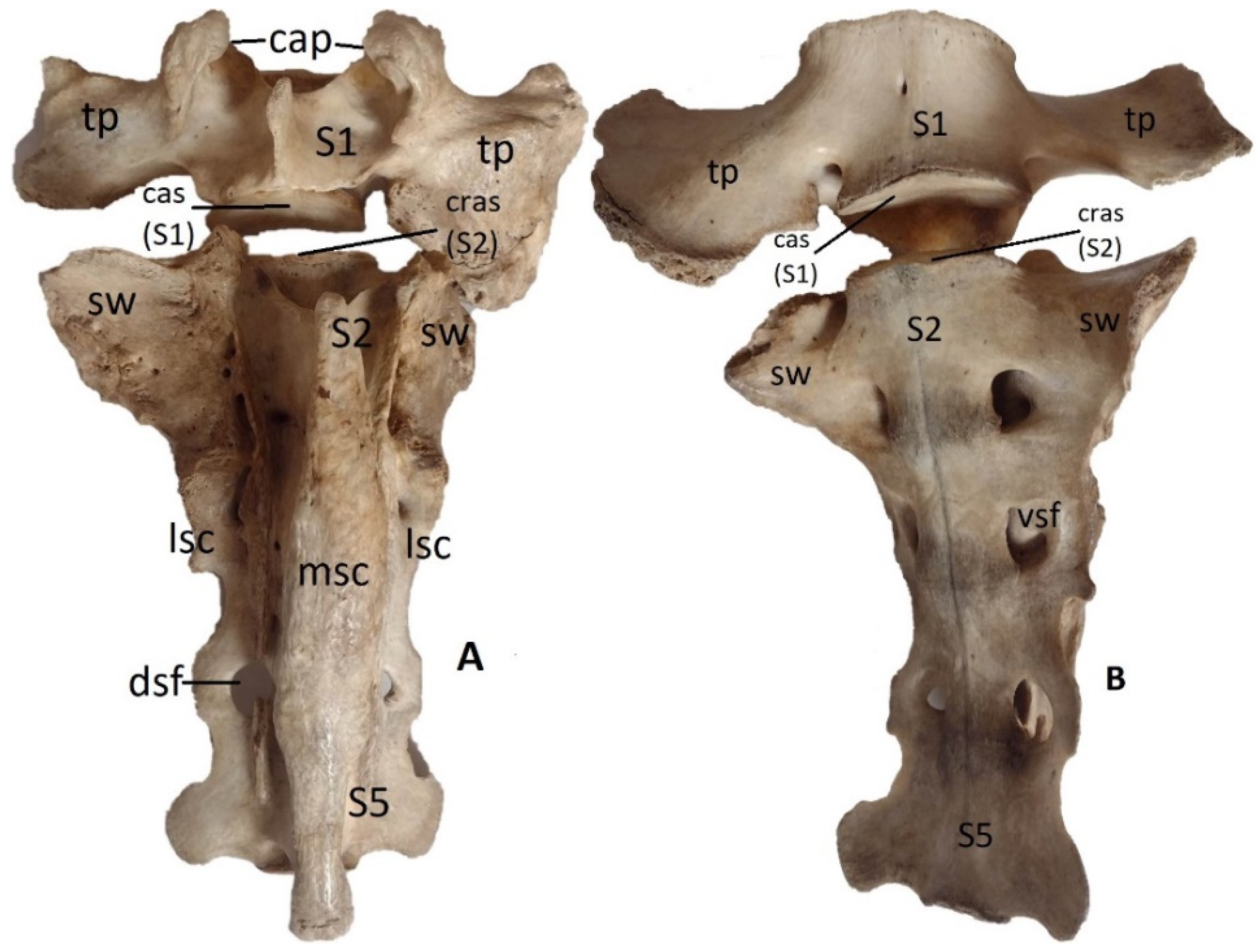
Figure 5.
Horse, male, 15 years, common breed – the fusion of the first caudal (Cd1) vertebra and the last sacral vertebra (S5). A – dorsal view: ossification process between the arches of Cd1 and S5 and the reduction of the interarcuate space between these vertebrae (arrows); sw – sacral wing, fa – facies auricularis (NAV), cap – cranial articular processes, dsf – dorsal sacral foramen, lsc – lateral sacral crest, sp – spinous process of Cd1. B – ventral view: complete fusion of the vertebral bodies of the first caudal vertebra (Cd1) and the last sacral vertebra (S5) – total ossification of the intervertebral disc (arrow); S1 – first sacral vertebra, sw – sacral wing, cas – cranial articular surface, vsf – ventral sacral foramen, lsc – lateral sacral crest.
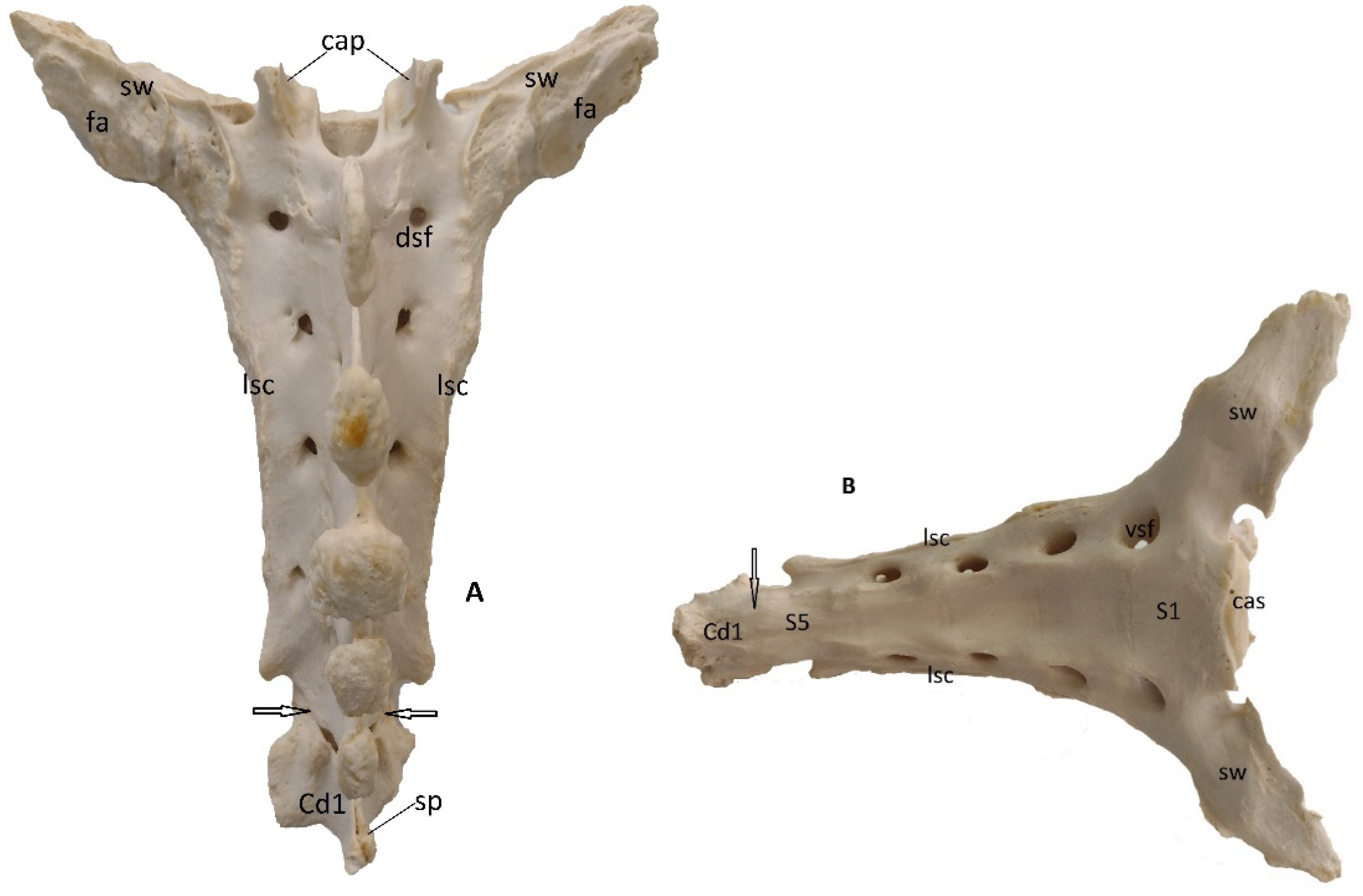
Figure 6.
Horse, male, 12 years – the fusion of the first caudal (Cd1) vertebra with the last sacral vertebra (S5), lateral view; the intervertebral disc is ossified but well visible; no contact between the vertebral arches of Cd1 and S5; sw – sacral wing, fa – facies auricularis; smc – sacral median crest (spinous processes).
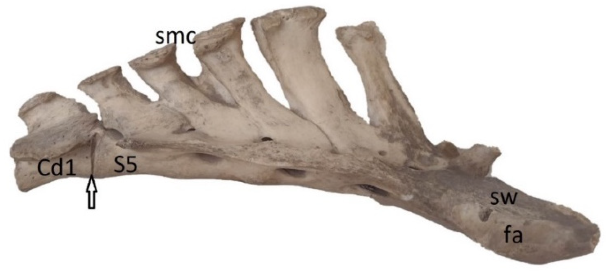
Figure 7.
Pig, male, 4 years – the sacralisation of the first caudal vertebra (Cd1), dorsal view: Cd1 is fused with S4 (last sacral vertebra) by their bodies and arches (arrow) and the interarcuate space (is) is narrower; S1 – first sacral vertebra; cap – cranial articular processes; sw – sacral wing; lsc – lateral sacral crest.
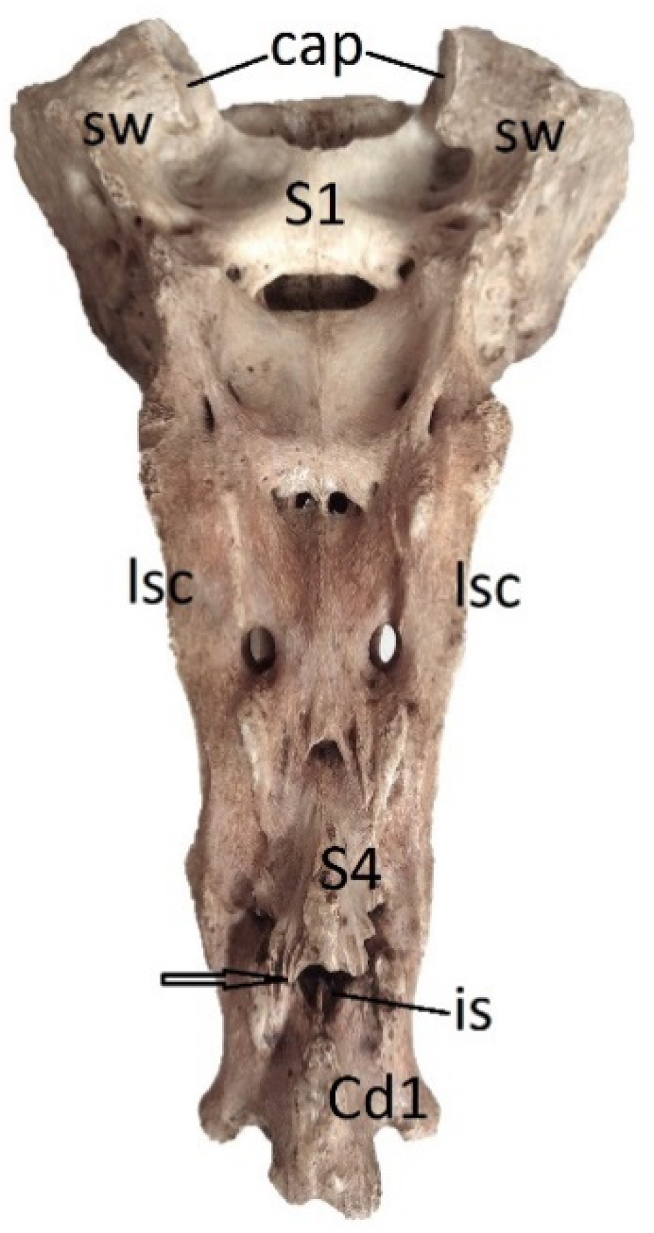
Figure 8.
Pig, F, 3 years, dorsal view – the lumbarisation of the first sacral vertebra (S1), and the detachment by the second one (arrows); the tendency of sacral wings to elongate as the transverse processes of lumbar vertebrae (circles): L6 – the sixth lumbar vertebra; S2 – second sacral vertebra; is – interarcuate space; il – ilium bone; S4 – the last sacral vertebra.
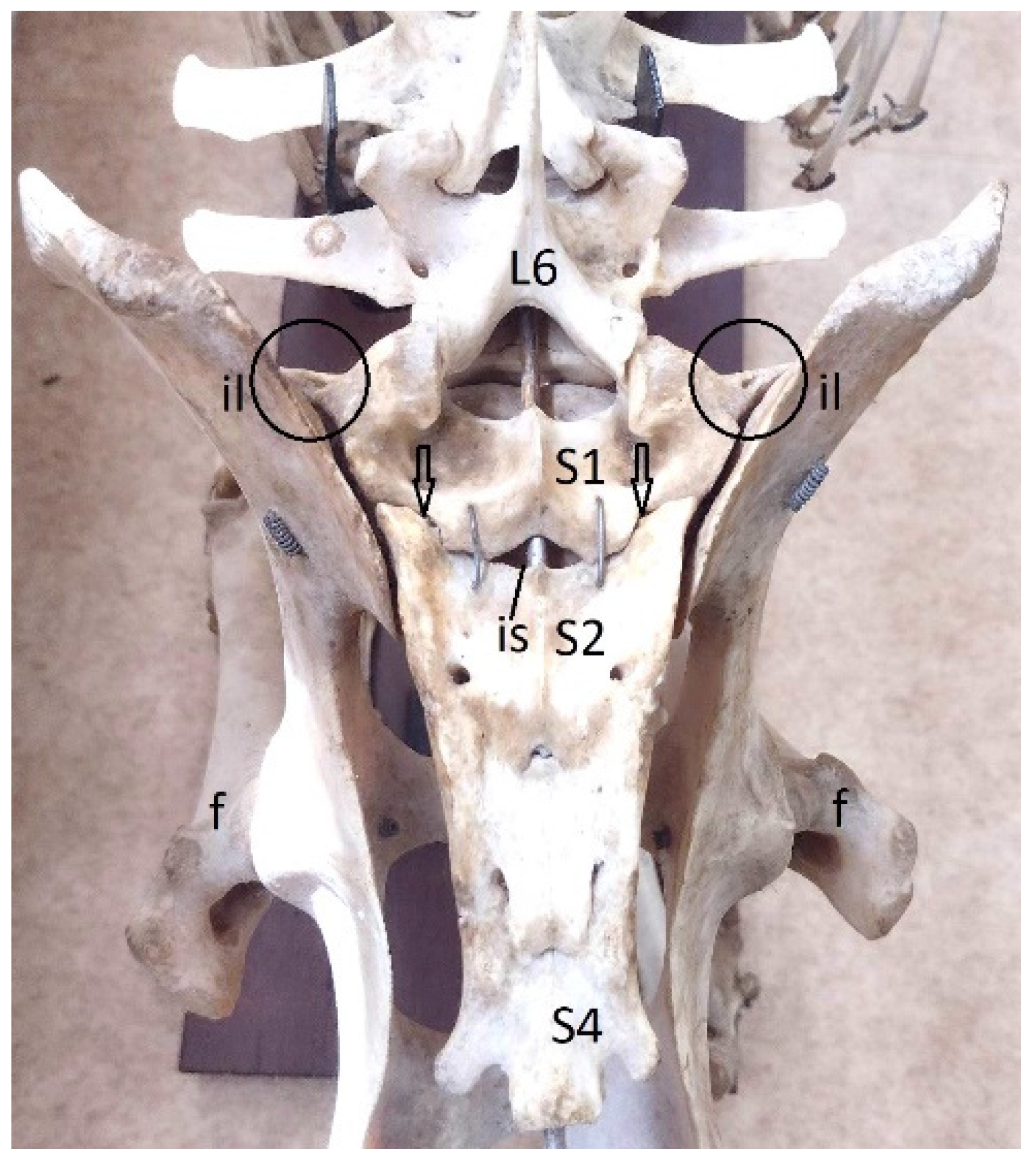
Figure 9.
Rabbit, ale, 5 years – lumbar spine and the presence of the 8th (L8) lumbar vertebra (supernumerary vertebra – lumbosacral transitional vertebra): L1-L7 – lumbar vertebrae 1-7; if – intervertebral foramina; vc – ventral crest; tp – transverse processes; sp – spinous processes; ap – articular processes; vb – vertebral body.
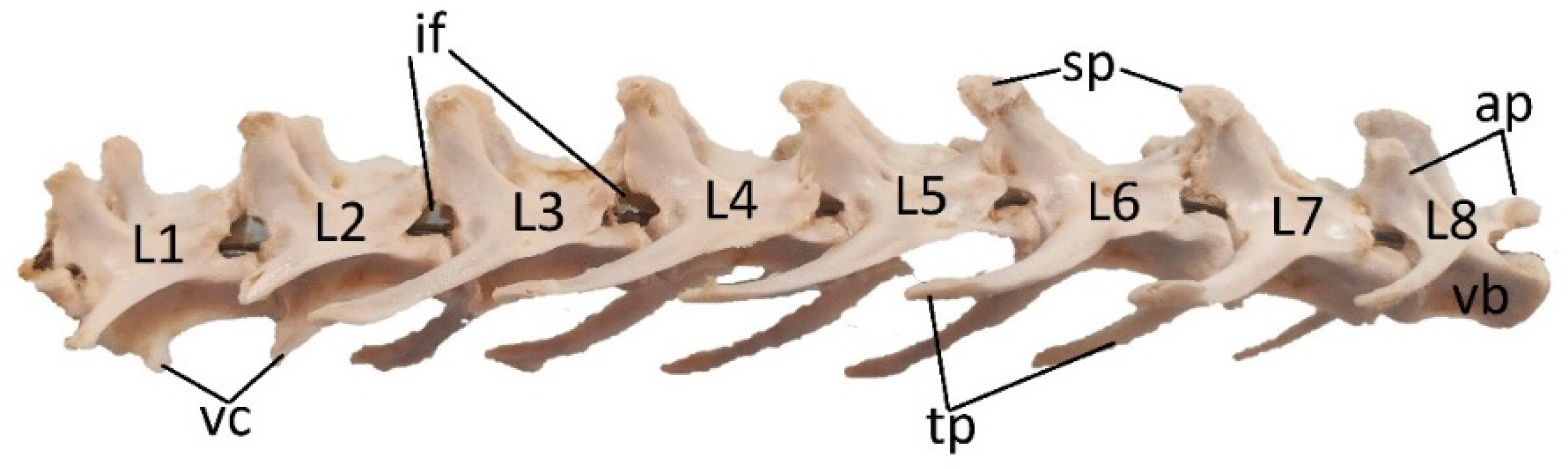
Figure 10.
Rabbit, male, 3 years – the tendency of caudalisation of the last sacral vertebra (S4) and the persistence of intervertebral disc (arrows); A - ventral view, B lateral view: L6 – the sixth lumbar vertebra; L7 – the seventh lumbar vertebra; S1 – first sacral vertebra; sw – sacral wing; fa – facies auricularis; tp – transverse processes; sp – spinous processes; S4 – the last sacral vertebra.
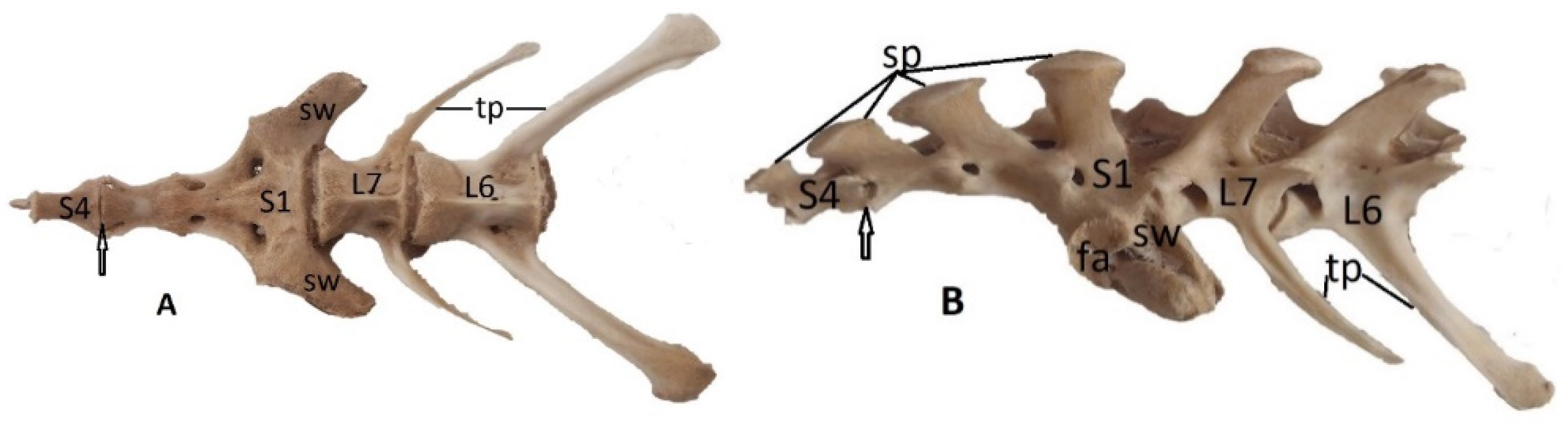
Figure 11.
Boxer dog, male, 6 years – supernumerary and transitional lumbo-sacral vertebra (L8), lateral view: shortening and sacralisation of L8; L1-L7 – lumbar vertebrae 1-7.
Figure 11.
Boxer dog, male, 6 years – supernumerary and transitional lumbo-sacral vertebra (L8), lateral view: shortening and sacralisation of L8; L1-L7 – lumbar vertebrae 1-7.
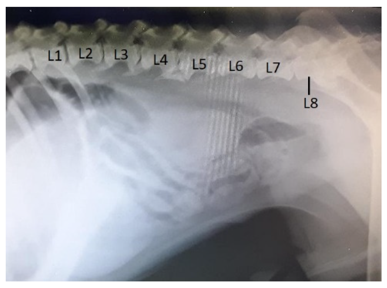
Figure 12.
Common breed dog, female, 14 years – supernumerary and asymmetric transitional lumbosacral vertebra (L8), ventro-dorsal view: shortening and sacralisation of L8; L1-L7 – lumbar vertebrae 1-7; the right transvers process (tp) of L8 is thin and not in contact with the iliac wing (iw), but the left one is fused with that of first sacral vertebra (S1) and takes contact with the iliac wing (arrow).
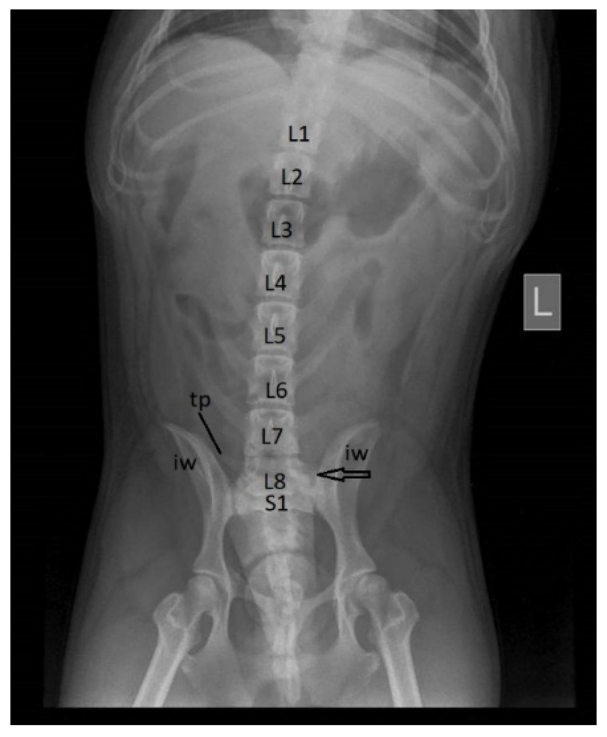
Figure 13.
Cat, female, 5 years – transitional lumbosacral vertebra-sacralization of L7 (last lumbar vertebra), ventro-dorsal view: L5 – the fifth lumbar vertebra; L6 – the sixth lumbar vertebra; S1 – the first sacral vertebra; tp – transverse processes of L7 – shortened and transformed in sacral wings; iw – iliac wing; vsf – ventral sacral foramina.
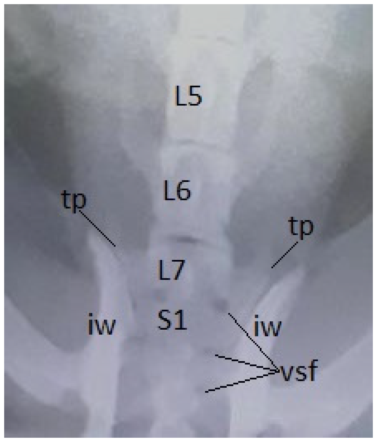
Figure 14.
Cat, female, 3 years – 2 sacral vertebrae-caudalisation of the last sacral vertebra (S3), dorsal view: S1 – first sacral vertebra; S2 – the second sacral vertebra; sw – sacral wing; cap – cranial articular processes; cas – caudal articular surface; cdap – caudal articular processes.
Figure 14.
Cat, female, 3 years – 2 sacral vertebrae-caudalisation of the last sacral vertebra (S3), dorsal view: S1 – first sacral vertebra; S2 – the second sacral vertebra; sw – sacral wing; cap – cranial articular processes; cas – caudal articular surface; cdap – caudal articular processes.
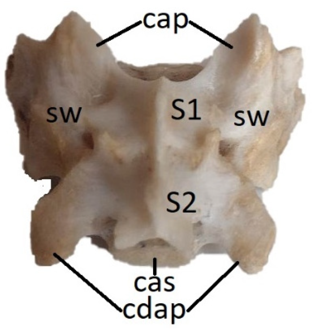

Table 1.
Data of animals of the present research.
| Species | Total number | Age(years) | Gender (number) |
Breed (number) | |
|---|---|---|---|---|---|
| ♀ | ♂ | ||||
| Cow | 29 | 3-17 | 16 | 13 | Common breed - 19 |
| Romanian Spotted Cattle – 6 | |||||
| Romanian Brown - 3 | |||||
| Sheep | 32 | 2-8 | 19 | 13 | Common breed – 20 |
| Tigaia – 7 | |||||
| Turcana – 5 | |||||
| Horse | 31 | 5-20 | 24 | 7 | Common breed - 21 |
| Romanian Saddle Horse – 6 | |||||
| Romanian Half Heavyweight Horse - 4 | |||||
| Pig | 26 | 2-7 | 18 | 8 | Common breed – 19 |
| Large White Pig - 7 | |||||
| Rabbit | 33 | 1-5 | 25 | 8 | Common breed - 22 |
| Flemish Giant - 11 | |||||
| Dog | 89 | 6 (months)-12 | 51 | 38 | Common breed - 39 |
| Pekingese – 15 | |||||
| French Bulldog – 13 | |||||
| Boxer - 11 | |||||
| Terrier breeds - 7 | |||||
| Caniche - 4 | |||||
| Cat | 57 | 1-7 | 27 | 30 | Common European Breed - 36 |
| Scottish Fold - 8 | |||||
| British Longhair - 7 | |||||
| Russian Blue - 6 | |||||
Table 2.
The prevalence (%) and the type of transitional vertebrae on species.
| Species | Transitional vertebrae -total number and % | Lumbosacral transitional vertebrae | Sacrocaudal transitional vertebrae | ||
|---|---|---|---|---|---|
| Sacralisation (L7, L8) | Lumbarisation (S1) | Sacralisation (Cd1) |
Caudalisation (last sacral vertebra) |
||
| Cow | 3 (8,7%) | - | 3 | - | - |
| Sheep | 3 (9,37%) | - | 2 | - | 1 |
| Horse | 4 (12,9%) | - | - | 4 | - |
| Pig | 3 (11,53%) | - | 2 | 1 | - |
| Rabbit | 3* (9,09%) | - | - | - | 2 |
| Dog | 4* (4,49%) | 3 | - | - | - |
| Cat | 3 (5,26%) | 2 | - | - | 1 |
*In rabbits and dogs, in a specimen was identified a supernumerary lumbar vertebra, but not sacralised.
Disclaimer/Publisher’s Note: The statements, opinions and data contained in all publications are solely those of the individual author(s) and contributor(s) and not of MDPI and/or the editor(s). MDPI and/or the editor(s) disclaim responsibility for any injury to people or property resulting from any ideas, methods, instructions or products referred to in the content. |
© 2023 by the authors. Licensee MDPI, Basel, Switzerland. This article is an open access article distributed under the terms and conditions of the Creative Commons Attribution (CC BY) license (http://creativecommons.org/licenses/by/4.0/).
Copyright: This open access article is published under a Creative Commons CC BY 4.0 license, which permit the free download, distribution, and reuse, provided that the author and preprint are cited in any reuse.


