Preprint
Review

The Anterolateral Ligament of the Knee in Paediatric Patients: What Do We Know? A Systematic Review

Altmetrics

Downloads

125

Views

29

Comments

0

A peer-reviewed article of this preprint also exists.

Submitted:

16 July 2023

Posted:

17 July 2023

You are already at the latest version

Alerts
Abstract
The knowledge on anatomy, function, biomechanics, and role on surgical procedures of the anterolateral ligament (ALL) of the knee is still controversial. Only a few papers have examined the ALL in children. The aim of this review is to analyse all the available literature about ALL in pediatric population. Following the PRISMA criteria, the literature was systematically reviewed, examining all the articles about ALL in pediatric patients. Eight articles were involved in the study. Five cadaveric studies, 2 diagnostic studies and 1 Cross-sectional study were found. The identification of the ALL is not always possible in diagnostic studies using the Magnetic Resonance (MRI) or in dissecting specimens. A high variability in the presence of the ligament, in its origin and insertion were found among the studies. In younger patients is more difficult to identify the ligament than in older children, suggesting that its presence may develop at some point during the growth. Further studies are needed for a detailed knowledge of the ALL.
Keywords: 
Subject: Medicine and Pharmacology  -   Orthopedics and Sports Medicine

1. Introduction

The anterolateral ligament (ALL) of the knee was originally described (but not named) in 1879 by the French surgeon Paul Segond [1], who stated the existence of a “fibrous band” in the lateral portion of the knee joint.
The name “anterolateral ligament” was first used only in 2012, after its identification, once again in France, in cadaveric specimens [2]. The authors described the anatomy of the ligament stating the origin on the lateral femoral condyle and the insertion on the lateral meniscus and tibial plateau, someplace posteriorly to Gerdy’s tubercle [2].
Subsequently, many authors tried to offer a full anatomical characterization of this apparent ligamentous structure. Some studies reported that the femoral origin of the ALL is variable [3,4,5] while the insertion was described (and afterward confirmed by different research papers) in the mid-lateral portion of the meniscus and the tibia, in the middle between Gerdy’s tubercle and the fibular head [6].
The function and biomechanics of the ALL are still controversial. Previously, Segond reported that this “pearly, resistant, fibrous band showed extreme amounts of tension during forced internal rotation”.
Since the 1970s, the idea that anterolateral rotatory instability was imputable not simply to ACL tears, but also to the structures of the anterolateral complex, started to get ahead.
Hughston et al, showing various patterns of rotational instability, illustrated a structure, the "mid-third lateral capsular ligament", that was closely united to the lateral meniscus and was implicated in rotational instability [7]. Just a few more studies mentioned this structure between 1970s and 1980s [8,9].
More recently, many studies confirmed that in knee flexion, the ALL resists internal rotation [10,11], while the role of the ALL in anterior stability is not fully understood [12,13].
The anterolateral knee keeps stability for anterior translation and anterolateral rotation of the knee. Some authors stated that is not a single structure that controls rotational stability, but rather that various structures work together: the ALL, the ITB, the capsulo-osseous layer, and the Kaplan fibers (connections between the iliotibial band and the distal femur) [14].
Many recent researches have detected the ALL, using diagnostic imaging (mainly MRI), cadaveric dissections and also surgically (through arthrotomy or arthroscopy) [15,16,17]. On the other hand, probably due to differences in anatomic dissection techniques, different authors denied the existence of this ligament, accentuating the relevance of other structures, like the deeper part of the iliotibial band (ITB) and the anterolateral capsule [18,19].
The identification of the ALL is sometimes very difficult, and detection of ALL tears is even more difficult.
Different authors thought that some ACL failures (re-rupture or clinical failure) may be due to ALL incompetence [23]. For this reason, the role of ALL reconstruction has become a subject of interest in many studies [22,24,25].
This ligament is not significant only in biomechanics but is also very important for his role in surgical procedures. In adults, combined ACL reconstruction with lateral extra-articular tenodesis (LET) or ALL reconstruction procedures are always more popular and show promising results [20,21,22]. Many reconstruction techniques have been proposed in the last decades, showing good outcomes in primary ACL injuries and for ACL revisions.
Whether compared to adults, the presence and the role of the ALL in the paediatric population is even more controversial and only a small number of authors have examined the ALL in children. In fact, because of the thinness and the poor identifiability of the ALL in children, its role in knee stability is uncertain. Furthermore, is not sure if an absence of the ALL may enhance the probability of an ACL tear in children who play sports [26].
The aim of this review is to analyse all the literature available about ALL in pediatric patients.

2. Materials and Methods

2.1. Search selection

This systematic review was directed following the recommendations of the Preferred Reporting Items for Systematic Reviews and Meta-Analyses (PRISMA) [27].
The aim was to find all the available literature about the ALL in children.
One medical electronic database (PubMed) was explored by a single author (LL) on May 25th, 2023. The research keywords used were ((paediatric) OR (pediatric) OR (children) OR (kids)) AND ((knee) AND (anterolateral ligament)).” MesSH terms were included. No temporal limitations were established. A total of n = 53 articles were found.
Studies of any level of evidence about the anterolateral ligament of the knee in paediatric patients were considered eligible for the study.

2.2. Inclusion and exclusion criteria

All the articles about other topics, with inadequate scientific methods, or without an available abstract were not considered. Only articles written in English were involved in the study. No duplicates were found.

2.3. Data extraction

The main parameters for each article were collected: aim of the study, sample, the presence of a control group, sex, side, mean age, results, and type of the study.

2.4. Quality assessment

Three authors (C.d.C., G.T. and F.d.M.) individually read the selected articles and evaluated their quality. All the authors discussed the quality of the articles selected. The senior author (V.P.) interfered in doubt cases.

3. Results

After the first preliminary screening, conducted by three authors (C.d.C., G.T. and F.d.M.), n = 12 articles were considered adequate for reading the full text. A precise full-text reading of the selected papers was completed by the same authors to verify their eligibility and n = 8 articles were selected, because met the inclusion criteria
The senior author (V.P.) intervened if the reviewer had any doubt about the inclusion of a study. The selected articles, their references and the ineligible studies were re-evaluated and debated by all the authors to reduce the risk of bias.
A PRISMA [27] flowchart is showed in Figure 1.
Reference lists from the chosen articles were examined. The selected articles [28,29,30,31,32,33,34,35] are summarized in Table 1.
The articles about ALL in children, included in the study, are sometimes heterogeneous but can easily be classified in 3 main categories (cross-sectional, cadaveric and diagnostic studies) as reported:
-
Survey - Cross-sectional studies
Madhan et al [29] aimed to verify the surgeon experiences about anterolateral ligament reconstruction (ALLR) as well as lateral extra-articular tenodesis (LET) in the paediatric patients.
-
Cadaveric/ descriptive anatomic studies
Radhawa et al [28] in 2022 evaluated 9 cadaveric specimens analyzing the paediatric anterolateral knee anatomy and in particular LCL origin, the popliteus origin and the insertion of the iliotibial band (ITB), their relationships to physeal structures and how this structural information might impact operating procedures for lateral extra-articular procedures.
Iseki et al [30] explored 15 pediatric knee anterolateral capsule from specimens (ALC) aged 6.3±3.3 years old and did not detect a clear ligament on histology, immunohistochemistry and gene expression evaluation.
Shea et al. [33] studied the incidence of the ALL in 14 preadolescent anatomic specimens with a mean age of 8 years old (range 7-11 years) and stated that the presence of this ligament in pediatric specimens is lower than in adult specimens.
Helito et al. [34] examined 20 fetal cadaveric specimens aged 28.64 ± 3.20 weeks and concluded that the ALL is present during fetal development.
Shea et al. [35] dissected 8 skeletally immature knee specimens of different ages (between 3 months and 10 years). They recognised the ALL only in 1 of 8 specimens. The authors stated that, considering this ligament is present in most adults specimens, (as reported in other studies [36]), probably the ALL mature afterwards in years.
-
Diagnostic studies
Helito et al, [32] observing 363 knee MRI, found the presence of the ALL in 69.4% of cases of all the knees of pediatric patients evaluated. The authors reported a lower visualization of the ligament in younger patients and a higher visualization in older patients (almost 18 years old). The ALL was not seen in female patients younger than 7 years and in male patients younger than 6 years.
The ligament was mostly seen in coronal sequences.
Liebenstein et al. [31] observed 61 MRI of teenagers with a mean age of 15 years (±2.3). The authors reported the presence of the femoral part of the ALL in 72.1% of the cases examined, while the meniscal part and the tibial part of the ALL were visible in 0% and 78.7% of the cases, respectively. The deep connections of the ITT to the distal femur were detected in 62.3% of the cases.

4. Discussion

4.1. History

The first citation of the ALL was dated 1879, when Segond described an avulsion fracture of the anterolateral margin of the tibia and described a "pearly band extending in an oblique fashion from the femur inserting into the avulsed tibial bone". In the 1970s, Hughston et al [7] illustrated a structure intimately connected to the lateral meniscus and apparently implicated in rotational instability. Between 1970s and 1980s other two authors (Johnson and Terry) [8,9] described the same ligament, however only Vieira in 2007 [37] stated that in the anterolateral portion of the knee, the capsule-osseus layer of the ITB works as an “anterolateral ligament”, terms used for the very first time by Vincent [2]. An accurate anatomic description of the ligament as a distinct structure was then done by Claes [6] in 2013.

4.2. Anatomy, Histology and Biomechanics

The anatomy of the ALL varies largely between individuals, especially regarding the femoral insertion [38]. The proximal origin of the ALL is usually located on the lateral femoral epicondyle, anteriorly to the fibular collateral ligament (FCL) insertion and proximally and posteriorly to the insertion of the popliteus tendon. ALL and FCL are considered together the "lateral collateral ligament complex. The insertion of the ALL is located posteriorly to Gerdy's tubercle and anteriorly to the fibular head [39]. Some fibers of the ligament continue into the lateral intermuscular septum of the thigh, some are united to the FCL and some are linked to the lateral meniscus. Some different ideas about the ligament anatomy are reported in the literature. Some authors [40] stated that 2 bundles of the ligament exist (one superficial and one deeper). Some others reported a close correlation (anatomical and functional) between ALL and ITB [41].
The histological examination helped in proving the presence of the ALL as a separate structure, individuating not only a capsular thickening but rather a well-organized connective tissue [34] made by type I collagen (90%) and fibroblasts [42]. The structure of the ALL is like the ACL regarding cellularity, characteristics, fibers orientation and nuclei’s form.
The function of the ALL in knee biomechanics is still controversial, probably due to differences in dissection techniques in different studies. Some authors stated that the ALL stabilize the knee in extension and internal rotation but others showed that it stabilize the joint only during flexion [10].

4.3. Identification of the ligament

Recognition of the ALL is not always simple, and its existence is not always accepted in the literature. This disagreement is essentially due to how the studies and the dissections were performed.
The ligament can be identified through diagnostic studies (mostly MRI) or arthroscopically. Identification of ALL tears is even more difficult.
MRI studies performed on ACL injuries showed an agreement of 40-80% in identifying ALL tears. These divergences are due to MRI quality and criteria of selection of patients [43]. The best way to identify the structure is using a 3 T MRI with 0.4-mm slice and fat-suppressed acquisition [44].
Zein reported that is possible to identify the ligament arthroscopically using a 30° scope through the anterolateral portal; in his study he gave precise instructions on how to see it [45].

4.4. Surgical techniques

Many procedures for ALL are reported in the literature. Sometimes ALL reconstruction (ALLR) is used as a synonym of lateral extra-articular tenodesis (LET), because both surgical techniques have the purpose of restore the anterolateral stability. However, the latter is only a functional and not an anatomical reconstruction, while the ALLR points to an anatomical reconstruction of the ALL.
LET was initially utilized as unique procedure in ACL injuries, but it showed high risk of failure. The combination of ACLR and LET is now again of interest and more than 10 different LET techniques are described in the literature without a superiority of one procedure over the others.
Indications for ALLR include but are not limited to high-grade pivot shift on examination (3+), young age (<20 years old), high-demand athlete, Segond fractures, revision ACLR, chronic ACL injuries (> 12 months).
One of the most used techniques for the ALLR consists of a reconstruction of ACL and ALL using a semitendinosus graft (used for ACLR) combined with gracilis tendon graft (used for ALLR) [22]
Many studies showed that, fixing the graft used in ALLR distally to Gerdy’s tubercle and proximally and posteriorly to the distal meta-epiphiseal junction of the femur leads to an adequate isometry, even if this placement is not anatomical. Some authors stated that the greatest agreement between anatomy and isometry is achieved when the ligament is fixed posteriorly and proximally to the lateral epicondyle in the femur and between the anatomical ALL insertion and Gerdy’s tubercle in the tibia. The ligament should be fixed in knee extension without tensioning the ligament in external rotation to prevent stiffness [14,22,46].
Post-operative rehabilitation protocols are usually the same used for isolated ACL reconstruction.
Even if not many studies were conducted about clinical outcomes, a good recovery is reported, and it seems that ACL revision reconstruction associated with ALL reconstruction reduces rotational laxity and shows a high rate of return to sport.

4.5. Overview on pediatric population

The presence of the ALL is largely studied in adult patients. However, a few studies have been conducted about the existence of the ALL in the pediatric population. Mainly all the studies can be distinguished in two types: diagnostic studies and cadaveric studies.
Mostly, the diagnostic studies about adults reported in the literature, used the MRI as tool for understanding the presence of the ligament. The existence of the ligament is commonly described in the literature, but the identification of the tear is not fully known.
Gossner [47] stated that the presence of anterolateral ligament can be described in the majority of patients undergoing standard MRI of the knee. The author suggests that with a decrease of cut depth, the view can improve and both orthopedic surgeons and radiologists must carefully look at this ligament studying MRI scans of the knee.
Similarly, Hartigan et al. [48] reported a high recognition of this structure (100% of all the 72 MRI evaluated from two radiologists. However, the ALL tears were noted in 26% by one radiologist and in 62% by the other physician. The authors stated using the standard 1.5-tesla MRI sequences is not sufficient to detect ALL tears and concluded that accurate imaging sequences may be fundamental to understand the presence of tears of this structure.
In according to other authors [43], an ALL tear can be detected in only one-third of the knee MRI of acute ACL injuries and the tear is usually in the proximal aspect of the ligament.
Cavaignac et al. [49] reported 100% sensitivity rate for detecting the presence of the ALL using ultrasonography, however they did not report the accuracy of detecting the ligament tear. Kandel et al. [50] stated that ultrasonography is a reliable method for evaluating the anterolateral ligament and can be used in the clinical practice. No studies have been conducted so far about the detection of the ALL using the ultrasonography in children.
On the other hand, all the diagnostic studies conducted in pediatric patients [31,32] were conducted using the MRI and only considered uninjured knees, reporting a presence of the ALL of around 70%. Liebenstein et al, [31] examining 61 MRI of teenagers with a mean age of 15 years (±2.3), did not find any correlation between age and presence of the ligament. Differently, Helito et al. [32] after an evaluation of 363 knee MRI, noticing that no female patients younger than 7 years and no male patients younger than 6 years had the ALL, stated that the ligament may develop at some point during the growth.
The presence of the ALL has been studied also in cadaveric specimens showing different results depending on the age of the children. Shea et al. [35] in their study conducted on 8 specimens found the ligament only in one case, however, as they stated, the restricted number of specimens and especially the age of most of the specimens (mean age of 3.4 years and a range 3 months – 10 years) could be an important limitation of the study.
After a few years, the same authors [33] dissected 14 specimens with a mean age of 14 years (range 7-11 years) and they reported the presence of the ligament in 9 out of 14 patients. The comparison of these two studies, considering the difference of the presence of the ligament in two different group ages, suggest that the ligament may become more distinct as it develops over time. The authors described that the ligament was very variable (sometimes very thin, sometimes sheet-like and in some cases well-defined ‘‘pearly band’’). Other than the structure itself, also the origin of the ligament varies largely, in according to the authors. Three anterolateral ligament origins were located distal and anterior from their respective lateral collateral ligament origins. Occasionally the anterolateral ligament and lateral collateral ligament shared the same origin location (2 cases), in some cases the ALL origin was proximal and posterior to the lateral collateral ligament origin, in one single case the ALL was anterior to the lateral collateral ligament, while in another one, the ALL was proximal and anterior to the lateral collateral ligament beginning.
Cadaveric studies were performed also in fetal specimens [51], as reported by Helito e al [34] who dissected and made a histological analysis of 20 specimens with age of 28.64 ± 3.20 weeks. After applying an internal rotation of the tibia during knee flexion, the authors clearly identified the ligament in 20 out of 20 patients, nearly to the LCL, just underneath the iliotibial tract. Also in this circumstance, a large variety of the origin of the ligament was reported among the different cases. The histological analysis of the ligament exhibited dense, well organized collagenous (type I) fibers with elongated fibroblasts with an increased cell concentration compared with the adult ALL. This last study differs in a significant way from the study of Shea et al. [33,35] who suggested that the ALL is an inconstant structure in children and that it only increases after the anterolateral capsule of the knee is subjected to physiological loads. Diversely, Helito et al. [34] stated that the ALL previously appear in fetuses with a mean age of 28.64 weeks and develops similarly to other ligaments of the knee region.
More recently, Iseki et al. [30] aimed to clearly identify a distinct ligament in the anterolateral capsule (ALC) of the knee. They dissected 15 ALC specimens (aged 6.3±3.3 years), 5 lateral collateral ligament (LCLs) (aged 3.4±1.3 years) and 5 quadriceps tendon QTs (aged 2.0±1.2 years). The authors made a RNA isolation and gene expression analysis, a histology and immunohistochemistry analysis and a cell morphology analysis, showing that, using those tools, a clear distinct ligament could not be observed in the ALC.
Contemplating the high variability of the anterolateral knee anatomy, Randhawa et al. [28] in 2022 examined 9 paediatric cadaveric knee specimens (aged 4.2 years: range 2 months–10 years) with the aim to fully understand the anatomy of this area. The authors mainly focused on LCL, Popliteus and ileo-tibial band (ITB), while the ALL was only marginal in their study.
Considering the importance of the aforementioned ligament also in surgical procedures, Madhan et al. [29] wanted to understand the knowledge and preferences of surgeons, in the clinical practice in performing ACL reconstruction in children and adolescents together with ALL reconstruction or Lateral Extra-articular Tenodesis (LET). The authors reported that on a sample of 63 surgeons engaged through a survey, around 50% of pediatric sports surgeons occasionally perform ALL augmentation with primary ACLR and 79% with revision ACLR. The authors noticed that doctors with sports medicine fellowship were more likely to execute these procedures. Almost all the surgeons who do these techniques explicit curiosity in investigating them prospectively or to randomize patients.

4.6. Limitations

The present study has some limitations. First, not many studies about ALL in children were reported in the literature. Furthermore, the way many studies were conducted was heterogeneous and sometimes they lead to unreliable conclusions.

5. Conclusions

The existence, the anatomy, the function, and the biomechanics of the ALL are still not well-known.
The ALL is a rotational stabilizer of the knee, but its anatomy varies largely between individuals of different ages. This ligament needs to be further explored in adults and in children. Only a few articles studied the ALL in the pediatric population. Further studies on diagnostic imaging, specimens, and especially in vivo patients are necessary for a detailed knowledge of the ALL.

Author Contributions

Conceptualization, L.L. and V.P.; methodology, C.d.C., A.R. and M.M.; software, M.M.; validation, G.T. and M.S.; formal analysis, M.M. and M.S.; investigation, F.D.M.; resources, F.D.M and C.d.C.; data curation, L.L., A.R. and F.D.M.; writing—original draft preparation, L.L. and C.d.C.; writing—review and editing, L.L. and V.P.; visualization, M.S.; supervision, G.T, A.R. and V.P.; project administration, L.L. and M.S.; funding acquisition, G.T. All authors have read and agreed to the published version of the manuscript.

Institutional Review Board Statement

Not applicable.

Informed Consent Statement

Not applicable.

Data Availability Statement

Not applicable.

Conflicts of Interest

The authors declare no conflict of interest.

References

  1. Segond, P. Recherches Cliniques et Experi- ´ Mentales Sur Les Epanchements Sanguins Du ´ Genou Par Entorse 1879.
  2. Vincent, J.-P.; Magnussen, R.A.; Gezmez, F.; Uguen, A.; Jacobi, M.; Weppe, F.; Al-Saati, M.F.; Lustig, S.; Demey, G.; Servien, E.; et al. The Anterolateral Ligament of the Human Knee: An Anatomic and Histologic Study. Knee Surg Sports Traumatol Arthrosc 2012, 20, 147–152. [Google Scholar] [CrossRef] [PubMed]
  3. Rustagi, S.M.; Gopal, P.; Ahuja, M.S.; Arora, N.C.; Sood, N. The Anterolateral Ligament of the Knee: Descriptive Anatomy and Clinical Correlation. IJOO 2019, 53, 89–93. [Google Scholar] [CrossRef] [PubMed]
  4. Kennedy, M.I.; Claes, S.; Fuso, F.A.F.; Williams, B.T.; Goldsmith, M.T.; Turnbull, T.L.; Wijdicks, C.A.; LaPrade, R.F. The Anterolateral Ligament: An Anatomic, Radiographic, and Biomechanical Analysis. Am J Sports Med 2015, 43, 1606–1615. [Google Scholar] [CrossRef] [PubMed]
  5. Daggett, M.; Ockuly, A.C.; Cullen, M.; Busch, K.; Lutz, C.; Imbert, P.; Sonnery-Cottet, B. Femoral Origin of the Anterolateral Ligament: An Anatomic Analysis. Arthroscopy: The Journal of Arthroscopic & Related Surgery 2016, 32, 835–841. [Google Scholar] [CrossRef]
  6. Claes, S.; Vereecke, E.; Maes, M.; Victor, J.; Verdonk, P.; Bellemans, J. Anatomy of the Anterolateral Ligament of the Knee. J. Anat. 2013, 223, 321–328. [Google Scholar] [CrossRef]
  7. Hughston, J.C.; Andrews, J.R.; Cross, M.J.; Moschi, A. Classification of Knee Ligament Instabilities. Part II. The Lateral Compartment. J Bone Joint Surg Am 1976, 58, 173–179. [Google Scholar] [CrossRef]
  8. Johnson, L.L. Lateral Capsular Ligament Complex: Anatomical and Surgical Considerations. Am J Sports Med 1979, 7, 156–160. [Google Scholar] [CrossRef] [PubMed]
  9. Terry, G.C.; Hughston, J.C.; Norwood, L.A. The Anatomy of the Iliopatellar Band and Iliotibial Tract. Am J Sports Med 1986, 14, 39–45. [Google Scholar] [CrossRef]
  10. Parsons, E.M.; Gee, A.O.; Spiekerman, C.; Cavanagh, P.R. The Biomechanical Function of the Anterolateral Ligament of the Knee. Am J Sports Med 2015, 43, 669–674. [Google Scholar] [CrossRef]
  11. Lording, T.; Corbo, G.; Bryant, D.; Burkhart, T.A.; Getgood, A. Rotational Laxity Control by the Anterolateral Ligament and the Lateral Meniscus Is Dependent on Knee Flexion Angle: A Cadaveric Biomechanical Study. Clinical Orthopaedics & Related Research 2017, 475, 2401–2408. [Google Scholar] [CrossRef]
  12. Thein, R.; Boorman-Padgett, J.; Stone, K.; Wickiewicz, T.L.; Imhauser, C.W.; Pearle, A.D. Biomechanical Assessment of the Anterolateral Ligament of the Knee: A Secondary Restraint in Simulated Tests of the Pivot Shift and of Anterior Stability. The Journal of Bone and Joint Surgery 2016, 98, 937–943. [Google Scholar] [CrossRef] [PubMed]
  13. Drews, B.H.; Kessler, O.; Franz, W.; Dürselen, L.; Freutel, M. Function and Strain of the Anterolateral Ligament Part I: Biomechanical Analysis. Knee Surg Sports Traumatol Arthrosc 2017, 25, 1132–1139. [Google Scholar] [CrossRef] [PubMed]
  14. Sabatini, L.; Capella, M.; Vezza, D.; Barberis, L.; Camazzola, D.; Risitano, S.; Drocco, L.; Massè, A. Anterolateral Complex of the Knee: State of the Art. WJO 2022, 13, 679–692. [Google Scholar] [CrossRef] [PubMed]
  15. Cho, H.-J.; Kwak, D.-S. Anatomical Consideration of the Anterolateral Ligament of the Knee. BioMed Research International 2019, 2019, 1–6. [Google Scholar] [CrossRef] [PubMed]
  16. Herbst, E.; Albers, M.; Burnham, J.M.; Fu, F.H.; Musahl, V. The Anterolateral Complex of the Knee. Orthopaedic Journal of Sports Medicine 2017, 5, 232596711773080. [Google Scholar] [CrossRef] [PubMed]
  17. Ariel De Lima, D.; Helito, C.P.; Lacerda De Lima, L.; De Castro Silva, D.; Costa Cavalcante, M.L.; Dias Leite, J.A. Anatomy of the Anterolateral Ligament of the Knee: A Systematic Review. Arthroscopy: The Journal of Arthroscopic & Related Surgery 2019, 35, 670–681. [Google Scholar] [CrossRef]
  18. Sheean, A.J.; Shin, J.; Patel, N.K.; Lian, J.; Guenther, D.; Musahl, V. The Anterolateral Ligament Is Not the Whole Story: Reconsidering the Form and Function of the Anterolateral Knee and Its Contribution to Rotatory Knee Instability. Techniques in Orthopaedics 2018, 33, 219–224. [Google Scholar] [CrossRef]
  19. Urban, S.; Pretterklieber, B.; Pretterklieber, M.L. The Anterolateral Ligament of the Knee and the Lateral Meniscotibial Ligament – Anatomical Phantom versus Constant Structure within the Anterolateral Complex. Annals of Anatomy - Anatomischer Anzeiger 2019, 226, 64–72. [Google Scholar] [CrossRef]
  20. Stability Group; Getgood, A. ; Bryant, D.; Firth, A. The Stability Study: A Protocol for a Multicenter Randomized Clinical Trial Comparing Anterior Cruciate Ligament Reconstruction with and without Lateral Extra-Articular Tenodesis in Individuals Who Are at High Risk of Graft Failure. BMC Musculoskelet Disord 2019, 20, 216. [Google Scholar] [CrossRef]
  21. Hewison, C.E.; Tran, M.N.; Kaniki, N.; Remtulla, A.; Bryant, D.; Getgood, A.M. Lateral Extra-Articular Tenodesis Reduces Rotational Laxity When Combined With Anterior Cruciate Ligament Reconstruction: A Systematic Review of the Literature. Arthroscopy: The Journal of Arthroscopic & Related Surgery 2015, 31, 2022–2034. [Google Scholar] [CrossRef]
  22. Sonnery-Cottet, B.; Thaunat, M.; Freychet, B.; Pupim, B.H.B.; Murphy, C.G.; Claes, S. Outcome of a Combined Anterior Cruciate Ligament and Anterolateral Ligament Reconstruction Technique With a Minimum 2-Year Follow-Up. Am J Sports Med 2015, 43, 1598–1605. [Google Scholar] [CrossRef] [PubMed]
  23. Chen, J.; Xu, C.; Cho, E.; Huangfu, X.; Zhao, J. Reconstruction for Chronic ACL Tears with or without Anterolateral Structure Augmentation in Patients at High Risk for Clinical Failure: A Randomized Clinical Trial. Journal of Bone and Joint Surgery 2021, 103, 1482–1490. [Google Scholar] [CrossRef] [PubMed]
  24. Getgood, A.M.J.; Bryant, D.M.; Litchfield, R.; Heard, M.; McCormack, R.G.; Rezansoff, A.; Peterson, D.; Bardana, D.; MacDonald, P.B.; Verdonk, P.C.M.; et al. Lateral Extra-Articular Tenodesis Reduces Failure of Hamstring Tendon Autograft Anterior Cruciate Ligament Reconstruction: 2-Year Outcomes From the STABILITY Study Randomized Clinical Trial. Am J Sports Med 2020, 48, 285–297. [Google Scholar] [CrossRef] [PubMed]
  25. Sonnery-Cottet, B.; Haidar, I.; Rayes, J.; Fradin, T.; Ngbilo, C.; Vieira, T.D.; Freychet, B.; Ouanezar, H.; Saithna, A. Long-Term Graft Rupture Rates After Combined ACL and Anterolateral Ligament Reconstruction Versus Isolated ACL Reconstruction: A Matched-Pair Analysis From the SANTI Study Group. Am J Sports Med 2021, 49, 2889–2897. [Google Scholar] [CrossRef] [PubMed]
  26. Madhan, A.S.; Patel, N.M. The Anterolateral Ligament of the Knee. JBJS Reviews 2020, 8, e0136–e0136. [Google Scholar] [CrossRef] [PubMed]
  27. Page, M.J.; McKenzie, J.E.; Bossuyt, P.M.; Boutron, I.; Hoffmann, T.C.; Mulrow, C.D.; Shamseer, L.; Tetzlaff, J.M.; Akl, E.A.; Brennan, S.E.; et al. The PRISMA 2020 Statement: An Updated Guideline for Reporting Systematic Reviews. BMJ 2021, n71. [Google Scholar] [CrossRef]
  28. Randhawa, S.; Stavinoha, T.J.; Trivedi, S.; Ganley, T.J.; Tompkins, M.; Ellis, H.; Wilson, P.; Green, D.W.; Fabricant, P.D.; Musahl, V.; et al. Pediatric Reference Anatomy for ACL Reconstruction and Secondary Anteroalteral Ligament or Lateral Extra-Articular Tenodesis Procedures. Journal of ISAKOS 2022, S2059775422000517. [Google Scholar] [CrossRef]
  29. Madhan, A.S.; Ganley, T.J.; McKay, S.D.; Pandya, N.K.; Patel, N.M. Trends in Anterolateral Ligament Reconstruction and Lateral Extra-Articular Tenodesis With ACL Reconstruction in Children and Adolescents. Orthopaedic Journal of Sports Medicine 2022, 10, 232596712210880. [Google Scholar] [CrossRef]
  30. Iseki, T.; Rothrauff, B.B.; Kihara, S.; Novaretti, J.V.; Shea, K.G.; Tuan, R.S.; Fu, F.H.; Alexander, P.G.; Musahl, V. Paediatric Knee Anterolateral Capsule Does Not Contain a Distinct Ligament: Analysis of Histology, Immunohistochemistry and Gene Expression. Journal of ISAKOS 2021, 6, 82–87. [Google Scholar] [CrossRef]
  31. Liebensteiner, M.C.; Henninger, B.; Kittl, C.; Attal, R.; Giesinger, J.M.; Kranewitter, C. The Anterolateral Ligament and the Deep Structures of the Iliotibial Tract: MRI Visibility in the Paediatric Patient. Injury 2019, 50, 602–606. [Google Scholar] [CrossRef]
  32. Helito, C.P.; Helito, P.V.P.; Leão, R.V.; Louza, I.C.F.; Bordalo-Rodrigues, M.; Cerri, G.G. Magnetic Resonance Imaging Assessment of the Normal Knee Anterolateral Ligament in Children and Adolescents. Skeletal Radiol 2018, 47, 1263–1268. [Google Scholar] [CrossRef] [PubMed]
  33. Shea, K.G.; Milewski, M.D.; Cannamela, P.C.; Ganley, T.J.; Fabricant, P.D.; Terhune, E.B.; Styhl, A.C.; Anderson, A.F.; Polousky, J.D. Anterolateral Ligament of the Knee Shows Variable Anatomy in Pediatric Specimens. Clinical Orthopaedics & Related Research 2017, 475, 1583–1591. [Google Scholar] [CrossRef]
  34. Helito, C.P.; do Prado Torres, J.A.; Bonadio, M.B.; Aragão, J.A.; de Oliveira, L.N.; Natalino, R.J.M.; Pécora, J.R.; Camanho, G.L.; Demange, M.K. Anterolateral Ligament of the Fetal Knee: An Anatomic and Histological Study. Am J Sports Med 2017, 45, 91–96. [Google Scholar] [CrossRef] [PubMed]
  35. Shea, K.G.; Polousky, J.D.; Jacobs, J.C.; Yen, Y.-M.; Ganley, T.J. The Anterolateral Ligament of the Knee: An Inconsistent Finding in Pediatric Cadaveric Specimens. Journal of Pediatric Orthopaedics 2016, 36, e51–e54. [Google Scholar] [CrossRef]
  36. Pomajzl, R.; Maerz, T.; Shams, C.; Guettler, J.; Bicos, J. A Review of the Anterolateral Ligament of the Knee: Current Knowledge Regarding Its Incidence, Anatomy, Biomechanics, and Surgical Dissection. Arthroscopy: The Journal of Arthroscopic & Related Surgery 2015, 31, 583–591. [Google Scholar] [CrossRef]
  37. Cruells Vieira, E.L.; Vieira, E.Á.; Teixeira Da Silva, R.; Dos Santos Berlfein, P.A.; Abdalla, R.J.; Cohen, M. An Anatomic Study of the Iliotibial Tract. Arthroscopy: The Journal of Arthroscopic & Related Surgery 2007, 23, 269–274. [Google Scholar] [CrossRef]
  38. Sonnery-Cottet, B.; Daggett, M.; Fayard, J.-M.; Ferretti, A.; Helito, C.P.; Lind, M.; Monaco, E.; De Pádua, V.B.C.; Thaunat, M.; Wilson, A.; et al. Anterolateral Ligament Expert Group Consensus Paper on the Management of Internal Rotation and Instability of the Anterior Cruciate Ligament - Deficient Knee. J Orthop Traumatol 2017, 18, 91–106. [Google Scholar] [CrossRef]
  39. Patel, R.M.; Brophy, R.H. Anterolateral Ligament of the Knee: Anatomy, Function, Imaging, and Treatment. Am J Sports Med 2018, 46, 217–223. [Google Scholar] [CrossRef]
  40. Helito, C.P.; Do Amaral, C.; Nakamichi, Y.D.C.; Gobbi, R.G.; Bonadio, M.B.; Natalino, R.J.M.; Pécora, J.R.; Cardoso, T.P.; Camanho, G.L.; Demange, M.K. Why Do Authors Differ With Regard to the Femoral and Meniscal Anatomic Parameters of the Knee Anterolateral Ligament?: Dissection by Layers and a Description of Its Superficial and Deep Layers. Orthopaedic Journal of Sports Medicine 2016, 4, 232596711667560. [Google Scholar] [CrossRef]
  41. Mansour, R.; Yoong, P.; McKean, D.; Teh, J.L. The Iliotibial Band in Acute Knee Trauma: Patterns of Injury on MR Imaging. Skeletal Radiol 2014, 43, 1369–1375. [Google Scholar] [CrossRef]
  42. Helito, C.P.; Demange, M.K.; Bonadio, M.B.; Tírico, L.E.P.; Gobbi, R.G.; Pécora, J.R.; Camanho, G.L. Anatomy and Histology of the Knee Anterolateral Ligament. Orthopaedic Journal of Sports Medicine 2013, 1, 232596711351354. [Google Scholar] [CrossRef] [PubMed]
  43. Helito, C.P.; Helito, P.V.P.; Costa, H.P.; Demange, M.K.; Bordalo-Rodrigues, M. Assessment of the Anterolateral Ligament of the Knee by Magnetic Resonance Imaging in Acute Injuries of the Anterior Cruciate Ligament. Arthroscopy: The Journal of Arthroscopic & Related Surgery 2017, 33, 140–146. [Google Scholar] [CrossRef]
  44. Caterine, S.; Litchfield, R.; Johnson, M.; Chronik, B.; Getgood, A. A Cadaveric Study of the Anterolateral Ligament: Re-Introducing the Lateral Capsular Ligament. Knee Surg Sports Traumatol Arthrosc 2015, 23, 3186–3195. [Google Scholar] [CrossRef] [PubMed]
  45. Zein, A. “Mohamed N.E. Step-by-Step Arthroscopic Assessment of the Anterolateral Ligament of the Knee Using Anatomic Landmarks. Arthroscopy Techniques 2015, 4, e825–e831. [Google Scholar] [CrossRef] [PubMed]
  46. Saithna, A.; Thaunat, M.; Delaloye, J.R.; Ouanezar, H.; Fayard, J.M.; Sonnery-Cottet, B. Combined ACL and Anterolateral Ligament Reconstruction. JBJS Essential Surgical Techniques 2018, 8, e2. [Google Scholar] [CrossRef]
  47. Gossner, J. The Anterolateral Ligament of the Knee – Visibility on Magnetic Resonance Imaging. Revista Brasileira de Ortopedia (English Edition) 2014, 49, 98–99. [Google Scholar] [CrossRef]
  48. Hartigan, D.E.; Carroll, K.W.; Kosarek, F.J.; Piasecki, D.P.; Fleischli, J.F.; D’Alessandro, D.F. Visibility of Anterolateral Ligament Tears in Anterior Cruciate Ligament–Deficient Knees With Standard 1.5-Tesla Magnetic Resonance Imaging. Arthroscopy: The Journal of Arthroscopic & Related Surgery 2016, 32, 2061–2065. [Google Scholar] [CrossRef]
  49. Cavaignac, E.; Wytrykowski, K.; Reina, N.; Pailhé, R.; Murgier, J.; Faruch, M.; Chiron, P. Ultrasonographic Identification of the Anterolateral Ligament of the Knee. Arthroscopy: The Journal of Arthroscopic & Related Surgery 2016, 32, 120–126. [Google Scholar] [CrossRef]
  50. Physical Therapy Practice, Oberriet, Switzerland; Kandel, M. ; Cattrysse, E.; Department of Experimental Anatomy, Vrije Universiteit Brussel, Brussels, Belgium; De Maeseneer, M.; Department of Radiology, Universitair Ziekenhuis Brussel, Brussels, Belgium; Lenchik, L.; Department of Radiology, Wake Forest School of Medicine, Winston-Salem, NC, USA; Paantjens, M.; Ministry of Defence, the Netherlands; et al. Inter-Rater Reliability of an Ultrasound Protocol to Evaluate the Anterolateral Ligament of the Knee. J Ultrason 2019, 19, 181–186. [Google Scholar] [CrossRef]
  51. Ferretti, M.; Levicoff, E.A.; Macpherson, T.A.; Moreland, M.S.; Cohen, M.; Fu, F.H. The Fetal Anterior Cruciate Ligament: An Anatomic and Histologic Study. Arthroscopy: The Journal of Arthroscopic & Related Surgery 2007, 23, 278–283. [Google Scholar] [CrossRef]
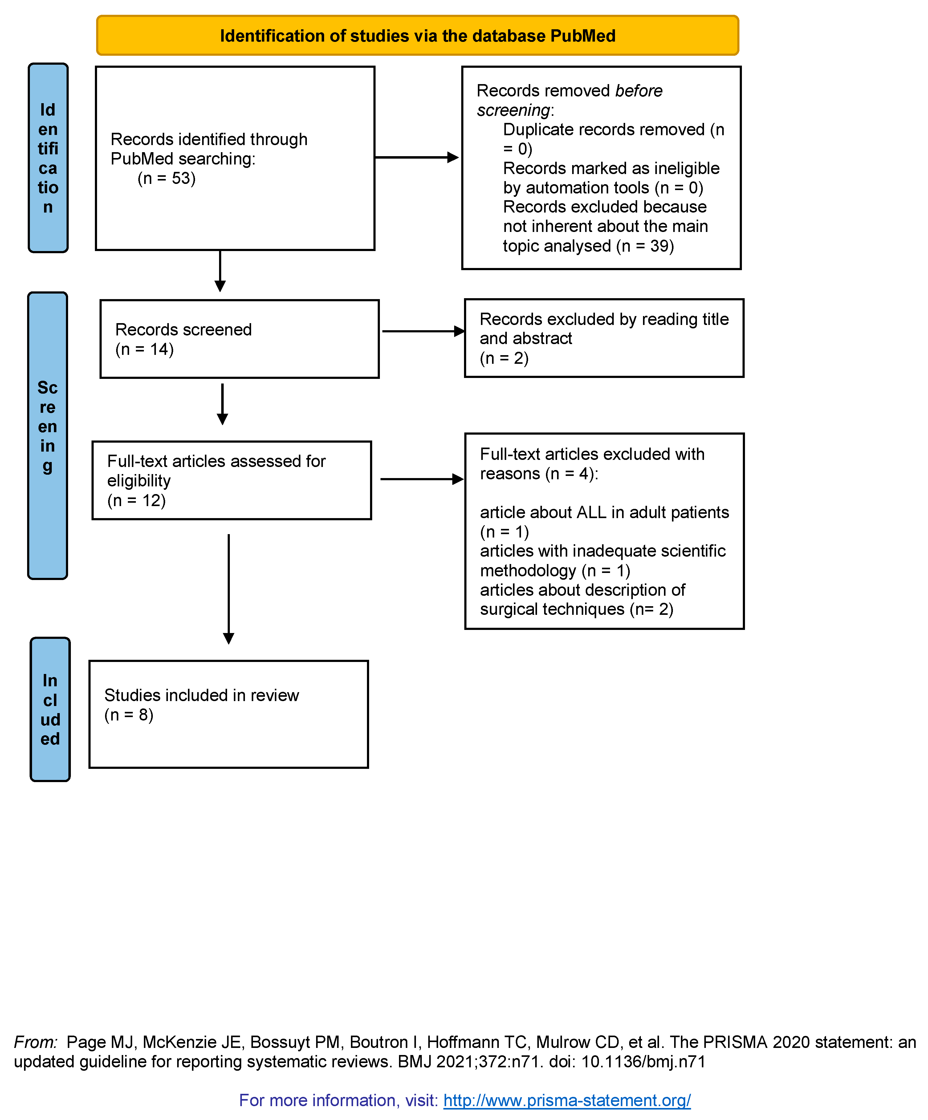
Figure 1. Identification of studies.
Preprints 79624 g001
Table 1. Main results of the eight selected articles.
Author Year Article Aim of the study Sample Control group Female Male Right Left Mean age Results Type of study
Randhawa [28] 2022 Pediatric reference anatomy for ACL reconstruction and secondary Anterolateral ligament or lateral extra-articular tenodesis procedures. To evaluate the structure of the knee joint physes, lateral collateral ligament (LCL) origin, popliteus origin, and ITB attachment 9 cadaveric specimens - 7 2 5 4 4.2 (range 10 months – 10 years) It explains positions of the femoral lateral collateral ligament (LCL), popliteus origins and tibial iliotibial band (ITB) attachment and their own physeal relations. Cadaveric/ descriptive anatomic study
Madhan [29] 2022 Trends in Anterolateral Ligament Reconstruction and Lateral Extra-articular Tenodesis with ACL Reconstruction in Children and Adolescents. To measure physician methods about Anterolateral ligament reconstruction (ALLR) and lateral extra-articular tenodesis (LET) in the paediatric patients 63 surgeons - - - - - More than 50% of pediatric sports surgeons occasionally perform ALL augmentation with primary ACLR and 79% with revision ACLR. Doctors with sports medicine fellowship were more likely to execute these procedures. Survey - Cross-sectional study.
Iseki [30] 2021 Paediatric knee anterolateral capsule does not contain a distinct ligament: analysis of histology, immunohistochemistry and gene expression. to explore the
existence of the ligament phenotype in pediatric knee anterolateral capsule (ALC)
15 cadaveric specimens 5 pediatric LCLs (age 3.4±1.3 years)
5 pediatric quadriceps tendon QTs (age 2.0±1.2 years)
6.3±3.3 years A clear ligament could not be detected in the ALC established on histology, immunohistochemistry and gene expression evaluation Controlled laboratory study

Liebensteiner [31] 2019 The anterolateral ligament and the deep structures of the iliotibial tract: MRI visibility in the paediatric patient To evaluate the view of ALL and the deep structures of the
iliotibial tract using the MRI in paediatric patients.
61 patients - 36 25 15 years (±2.3) ALL (and the deep structures of the ITT) can be detected by MRI Diagnostic study - Level 3.
Helito [32] 2018 Magnetic resonance imaging assessment of the normal knee
anterolateral ligament in children and adolescents
To describe the ALL in healthy knees of pediatric patients by MRI. 363 patients 163 200 Vision of the ALL improve with age
Diagnostic study - Level 3
Shea [33] 2017 Anterolateral Ligament of the Knee Shows Variable Anatomy in Pediatric Specimens To explore the existence of the ALL in preadolescent anatomic specimens 14 cadaveric specimens 2 12 7 7 8 years (range 7-11 years) The occurrence of the ALL
in pediatric specimens is lower than
in adult specimens.
Cadaveric Study
Helito [34] 2017 Anterolateral Ligament of the Fetal Knee: An Anatomic and Histological Study. To assess the ALL in human fetuses to establish its existence 20 fetal cadaveric specimens 10 10 10 10 28.64 ± 3.20 weeks. The ALL is present during fetal development Descriptive laboratory study
Shea [35] 2016 The Anterolateral Ligament of the Knee: An Inconsistent Finding in Pediatric Cadaveric Specimens To estimate whether the ALL might be detected on paediatric cadaveric knee specimens 8 cadaveric specimens 5 3 7 1 3.4 years (range 3 months – 10 years) The ALL could grow late in life Cadaveric Study
Disclaimer/Publisher’s Note: The statements, opinions and data contained in all publications are solely those of the individual author(s) and contributor(s) and not of MDPI and/or the editor(s). MDPI and/or the editor(s) disclaim responsibility for any injury to people or property resulting from any ideas, methods, instructions or products referred to in the content.
Copyright: This open access article is published under a Creative Commons CC BY 4.0 license, which permit the free download, distribution, and reuse, provided that the author and preprint are cited in any reuse.
Prerpints.org logo

Preprints.org is a free preprint server supported by MDPI in Basel, Switzerland.

Subscribe

© 2024 MDPI (Basel, Switzerland) unless otherwise stated