
Submitted:
01 November 2026
Posted:
04 November 2026
You are already at the latest version
Abstract
Aptamers, short single-stranded oligonucleotides selected to bind a chosen target, have for three decades been presented as programmable substitutes for antibodies. The past five years have changed the terms of that comparison. Selection can now be performed inside living cells, generative models propose binders that were never present in the starting library, and electrochemical aptamer switches report drug concentrations in human interstitial fluid with minute resolution over a day. In parallel, population-scale aptamer proteomics has made the aptamer a workhorse of biomarker discovery rather than a curiosity of the assay laboratory. Clinical adoption nevertheless remains uneven, and the reasons are mechanistic rather than merely engineering-related. An aptamer is a conformational ensemble whose binding equilibrium is re-weighted by ions, competing proteins and surface chemistry, so an affinity measured in selection buffer is a poor predictor of behaviour in blood. Here we examine the molecular principles that govern aptamer recognition, the device architectures that convert binding into a clinical readout, and the clinical evidence accumulated since 2021. We argue that the rate-limiting step has moved from molecular discovery to the construction of evidence that regulators, payers and clinicians can audit, and we propose a framework for generating it.
Keywords:
broiler
; sunflower concentrate
; soybean meal
; amino acids
; digestibility
1. Introduction
Recognition is the first step of every diagnostic test. For half a century the dominant recognition element has been the immunoglobulin, and the analytical performance of clinical immunoassays is correspondingly mature. Antibodies, however, carry structural constraints that no amount of process development removes: they require an animal or a mammalian expression system, they are heterogeneous in glycosylation and charge, they denature irreversibly at the temperatures and interfaces encountered outside the cold chain, and they cannot be raised against small molecules or against epitopes that are not immunogenic. Aptamers - single-stranded DNA or RNA oligonucleotides, typically 25-80 nucleotides long, selected in vitro for binding to a chosen target - were introduced in 1990 as a solution to precisely these problems.1,2 Because they are made by chemical synthesis, an aptamer is a single molecular species with a defined mass, a defined sequence and a defined conjugation chemistry. Because they are selected in a tube, the selection conditions can be chosen to suit the intended application, including the ionic strength, the temperature and the presence of competitor molecules.
The field has not lacked enthusiasm, and it has not lacked critics. Between 2004, when pegaptanib became the first aptamer approved as a drug,3 and 2023, when avacincaptad pegol was approved for geographic atrophy, the therapeutic pipeline advanced slowly and largely outside oncology, the area where the promise had been greatest.4 Diagnostic development followed a different and, for a long time, less visible trajectory: thousands of aptasensors were published, almost all of them demonstrated in buffer or in diluted serum, and very few were tested against a clinical decision. The resulting literature is large, heterogeneous and difficult to interpret, and it has produced a well-justified scepticism among clinical chemists.5
The period covered by this Review, 2021 to the present, is the period in which that trajectory began to change. Four developments are responsible. First, selection itself was re-engineered: microfluidic and hydrogel-based partitioning reduced the number of rounds, intracellular screens made binding measurable inside a living cell, and deep generative models made it possible to propose sequences that no library contained.6-8 Second, the mechanistic understanding of aptamer binding improved to the point where affinity could be discussed together with thermodynamics, kinetics and specificity rather than as a single number.9,10 Third, the analytical chemistry of the interface matured: antifouling architectures, ratiometric and kinetic-differential readouts, and microneedle formats moved electrochemical aptamer sensors from minutes in buffer to a full day in living tissue.11,12 Fourth, and most consequentially for clinical practice, aptamer proteomics reached population scale, so that aptamers became the enabling reagent for measuring thousands of plasma proteins in tens of thousands of people.13,14
This Review is organised around the proposition that these four developments are linked by a single physical constraint. An aptamer is not a rigid lock; it is a flexible polymer that samples an ensemble of conformations, of which only a subset binds. Everything that follows - the design of a selection, the choice of a transduction scheme, the behaviour of a sensor in plasma, and the credibility of a clinical claim - is a consequence of that fact. We first describe the molecular basis of recognition (Section 2), then the methods by which binders are now produced and engineered (Section 3). We then examine how recognition is converted into a clinical result (Section 4), review the clinical evidence base assembled since 2021 (Section 5), and analyse why devices that perform well in buffer often underperform in patients (Section 6). We close with a translational framework (Section 7) and an outlook (Section 8).
2. How Aptamers Recognise Their Targets
2.1. The Conformational Ensemble
The earliest mental model of aptamer binding was a lock-and-key complementarity, inherited from enzymology and reinforced by the early crystal structures of RNA aptamer-ligand complexes. That model is misleading in a way that matters for diagnostics, because it implies that an aptamer has one structure that either fits a target or does not. Solution measurements tell a different story. Isothermal titration calorimetry, circular dichroism and NMR applied to a dopamine-binding DNA aptamer showed that the free aptamer populates a largely unfolded terminal stem, and that ligand binding is enthalpically driven and entropically penalised - the signature of a disorder-to-order transition rather than of rigid surface complementarity.15 Shortening the terminal stem weakened binding progressively, and a functional electrochemical sensor was obtained only when the region that folds on binding was positioned close enough to the electrode for the conformational change to modulate electron transfer.15 The practical corollary, often ignored, is that the same aptamer can be an excellent binder and a useless sensor: the structural element that generates the signal is not necessarily the element that generates affinity.
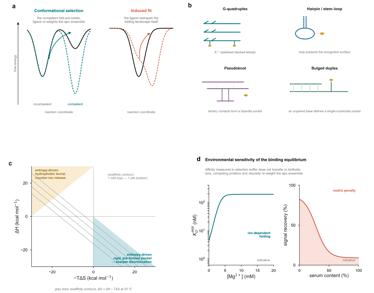
Two limiting mechanisms describe how the ensemble responds to a ligand (Fig. 1a). In conformational selection, the competent fold is present at low population in the apo state, and the ligand increases its occupancy by mass action. In induced fit, the ligand engages a loosely formed encounter complex and actively reshapes the folding landscape. The distinction is not semantic: conformational selection predicts that binding affinity and the magnitude of the conformational change can be tuned independently, which is exactly what the sensor engineer requires, whereas induced fit couples them. Solution biophysical methods - NMR imino-proton exchange, single-molecule FRET, temperature-jump relaxation and, most recently, nanoscale field-effect transistors capable of resolving single binding events - can now distinguish the two regimes for individual aptamer-ligand pairs.10,16 Carbon-nanotube transistors functionalised with a single aptamer resolved association and dissociation of individual ligands, exposing heterogeneity that ensemble measurements average away.10 Nanopipette and nanopore geometries provide a complementary route, converting the conformational change itself into a measurable current blockade.17,18
2.2. The Thermodynamic Grammar of Specificity
Affinity is reported as a dissociation constant, but a dissociation constant is a single number derived from two independent thermodynamic quantities, and the same KD can arise from very different physical situations (Fig. 1c). Because ΔG = ΔH − TΔS, binders that share a KD lie on a common isoaffinity contour but can differ profoundly in the enthalpy and entropy that produce it. A systematic analysis of aptamer binding thermodynamics across targets found that affinity and specificity are not interchangeable and that the two can move in opposite directions as selection stringency is increased.9 Binders whose affinity is dominated by a large, favourable enthalpy - hydrogen bonding and shape complementarity within a pre-organised pocket - typically discriminate more sharply against close analogues than binders of equal affinity whose binding is entropy-driven, because the latter rely on the burial of hydrophobic surface and the release of counter-ions, processes that are comparatively insensitive to the identity of the ligand.9 This has an immediate design consequence. Selection protocols that enrich on the basis of slow dissociation, the standard route to high affinity, do not necessarily enrich for specificity, and a selection that reports only KD is under-determined.
The same analysis explains a recurring frustration in the aptamer literature: aptamers selected against a protein frequently cross-react with its homologues, and aptamers selected against a small molecule frequently bind chemically unrelated molecules of similar hydrophobicity. Where the binding epitope has been mapped, specificity tracks the epitope rather than the whole molecule. Aptamers that recognise distinct surfaces of the same protein can differ by orders of magnitude in their ability to discriminate a pathological conformer, a phenomenon now documented for several diagnostic targets.19 Epitope-level characterisation, by hydrogen-deuterium exchange, limited proteolysis or competitive binding, is therefore not a refinement but a prerequisite for any claim of clinical specificity.
2.3. Structural Motifs and the Geometry of the Binding Site
Aptamer folds are drawn from a small vocabulary - hairpins and internal loops, G-quadruplexes, three-way junctions, pseudoknots and, in RNA, a variety of riboswitch-like architectures - but the binding sites they present are structurally diverse (Fig. 1b). G-quadruplexes, stabilised by monovalent cations, stack planar guanine tetrads and present a compact, roughly planar surface suited to small aromatic ligands and to protein loops; the cation dependence of tetrad formation makes the affinity of such aptamers sensitive to potassium and sodium in a way that has direct consequences for sensor performance in serum.20 Hairpins present a loop that can be engineered to expose a defined set of bases, and are the workhorse of structure-switching designs. Pseudoknots assemble tertiary contacts that generate bipartite pockets not predictable from secondary structure alone. High-resolution structures remain scarce relative to the number of reported aptamers, but recent entries - including a class V GTP-binding RNA aptamer solved in complex with its ligand - show that ligand recognition can be achieved by topological presentation of a base rather than by a conventional binding cleft, which explains why sequence-based alignments often fail to identify functional motifs.21 Computational modelling of protein-RNA aptamer recognition has improved correspondingly, although predicted interfaces remain insufficiently accurate to substitute for structural measurement.22
Fluorogenic aptamers illustrate the same principle from the opposite direction. Squash, Pepper and their relatives bind and activate small fluorophores, and their structures revealed that they achieve this by repurposing folds that evolved for other purposes - the adenine riboswitch fold in the case of Squash.23,24 For diagnostics, such light-up aptamers provide a genetically encodable readout and underpin several amplification schemes.25 A distinct class couples binding to a change in the redox potential of a bound cofactor, effectively converting recognition into an electrical signal without a label.26
2.4. Kinetics as the Missing Axis
A binding equilibrium is a ratio of rates, and for diagnostic purposes the rates matter as much as the ratio. A binder with a dissociation constant of 1 nM achieved through a very slow off-rate reports the integral of exposure rather than the instantaneous concentration, which is acceptable for a single-time-point laboratory test but disqualifying for continuous monitoring (Fig. 5d). Conversely, a binder that equilibrates in seconds sacrifices affinity but gains the ability to follow a concentration that changes on the timescale of minutes. The two regimes are now routinely separated experimentally: surface plasmon resonance and microscale thermophoresis give ensemble rates, single-molecule field-effect measurements give distributions of rates, and nanopore recordings give the dwell-time distribution of individual binding events.10,27,28 What has changed since 2021 is that kinetic information is beginning to be produced at selection scale rather than one aptamer at a time. Platforms that couple binding to a sequencing readout now report apparent rate constants for thousands of sequences in a single experiment, turning the selection from a ranking of affinities into a map of the kinetic landscape.8,29
Figure 1.
The molecular basis of aptamer recognition. a, Two limiting mechanisms. In conformational selection the competent fold is populated at low occupancy in the apo ensemble and the ligand re-weights the ensemble; in induced fit the ligand reshapes the folding landscape itself. b, Structural motifs that present binding surfaces: G-quadruplex, hairpin, pseudoknot and bulged duplex, each with a bound ligand shown schematically. c, Thermodynamic phase diagram. Binders sharing a dissociation constant lie on a common isoaffinity contour (ΔG = ΔH − TΔS at 25 °C) but differ in mechanism; enthalpy-driven, rigid-pocket binders generally discriminate more sharply than entropy-driven binders of equal affinity. d, Environmental sensitivity of the binding equilibrium: apparent affinity as a function of divalent-cation concentration, and signal recovery as a function of serum content. Both panels are indicative and illustrate the direction and magnitude of the effects discussed in Section 2.
Figure 1.
The molecular basis of aptamer recognition. a, Two limiting mechanisms. In conformational selection the competent fold is populated at low occupancy in the apo ensemble and the ligand re-weights the ensemble; in induced fit the ligand reshapes the folding landscape itself. b, Structural motifs that present binding surfaces: G-quadruplex, hairpin, pseudoknot and bulged duplex, each with a bound ligand shown schematically. c, Thermodynamic phase diagram. Binders sharing a dissociation constant lie on a common isoaffinity contour (ΔG = ΔH − TΔS at 25 °C) but differ in mechanism; enthalpy-driven, rigid-pocket binders generally discriminate more sharply than entropy-driven binders of equal affinity. d, Environmental sensitivity of the binding equilibrium: apparent affinity as a function of divalent-cation concentration, and signal recovery as a function of serum content. Both panels are indicative and illustrate the direction and magnitude of the effects discussed in Section 2.
3. Discovery and Engineering
3.1. From 1015 to One
Systematic evolution of ligands by exponential enrichment (SELEX) remains the conceptual template of the field: a random library is exposed to the target, non-binders are partitioned away, the retained species are amplified, and the cycle is repeated until the library is dominated by a small number of sequences (Fig. 2a). The practical problem has always been the partition step, which determines both the enrichment achieved per round and the artefacts that accumulate. Solid-phase supports and nitrocellulose filters - the classical choices - introduce nonspecific adsorption and favour binders to the support. The most consequential recent advance is a partition matrix that does not adsorb protein: a non-fouling porous hydrogel allowed high-affinity aptamers to be obtained in a single step, bypassing the amplification cycles in which artefacts are generated.6 Microfluidic implementations achieve a comparable compression of the workflow by automating the fluidic steps and by using the chip geometry to exclude weakly retained species, and automated systems have now been applied to tissue samples as well as to purified proteins.30,31
The choice of selection format is determined by what the assay must recognise. Protein-SELEX against a recombinant ectodomain yields binders to an epitope that may be absent or masked on the native protein. Cell-SELEX, in which whole cells are the target, biases selection towards epitopes presented in their native context and requires no knowledge of the target identity; it has produced binders to poorly characterised tumour cell surfaces and, in a recent example, identified a membrane protein that mediates aptamer-dependent growth inhibition.32 Tissue-SELEX extends the principle to intact tissue architecture and is increasingly used where the diagnostic question concerns a histological state rather than a single molecule.33,34 Capture-SELEX inverts the geometry for small molecules, immobilising the library and eluting with the free target, which avoids the need to conjugate a small molecule to a solid support.35
3.2. Selection Inside a Cell, and Selection for Function
A persistent limitation of test-tube selection is that it optimises binding in a medium that resembles no biological compartment. Intracellular selection addresses this directly. By coupling aptamer binding to a CRISPR-based transcriptional reporter, the CRISmers system converts an intracellular binding event into a selectable phenotype, so that sequences are enriched on the basis of performance inside a living cell rather than in buffer. The approach was subsequently generalised: a bacterial CRISPR-hybrid system coupled to fluorescence-activated cell sorting evolved orthogonal aptamer-protein pairs, and a combination of intracellular selection with a nucleic-acid language model produced, in a single round, RNA aptamers that outperformed those obtained from six to sixteen conventional rounds against CD3ε, the SARS-CoV-2 receptor-binding domain and the intracellular transcription factor c-Myc.7 The commentary that has accompanied these data-driven selection methods is explicit that they represent a change in kind rather than in degree: the experimental step generates a small, biologically relevant dataset, and the model performs the extrapolation that iterative selection used to perform physically.36
A parallel development makes selection itself massively parallel. SPARK-seq couples aptamer binding to a sequencing readout in a format that reports both identity and apparent kinetics for very large numbers of sequences simultaneously, and has been used to profile aptamer-target pairs at a scale incomparable with plate-based characterisation.8,29,36 The strategic significance is that selection is being converted from a screen that returns a handful of sequences into a measurement that returns a dataset - a change with direct consequences for how the field should report its results.
3.3. Generative Design
The third route to a binder does not begin with a library at all. Generative models trained on high-throughput selection data learn the sequence-structure-affinity relationship and propose new sequences, which are then made and tested. Variational autoencoders established the principle; diffusion models improved it. AptaDiff, a conditional discrete diffusion model with motif-dependent latent embeddings, generated aptamers outside the space covered by the sequencing data on which it was trained and, with Bayesian affinity-guided optimisation, produced binders whose dissociation constants were 2.4- to 3.6-fold lower than the best experimentally selected candidates for the same targets.37 Structure-conditioned inverse-folding models extend the idea to tertiary geometry, generating sequences expected to adopt a specified three-dimensional fold and thereby allowing the design of a binding site rather than the discovery of one.38 Comprehensive benchmarking of protein-aptamer complex prediction has begun to appear alongside these methods, and the early results are sobering: predicted structures are frequently plausible but not accurate at the level required to rank candidate binders, and the benchmarks themselves are contested.39,40
Two caveats deserve emphasis because they bear directly on clinical credibility. First, generative models are trained largely on successful selections and therefore learn what binders look like without learning what non-binders look like; the negative data that would penalise cross-reactivity are absent from essentially all public training sets. Second, a generated sequence has no evolutionary audit trail. A conventionally selected aptamer can be traced round by round, which matters to a regulator assessing reproducibility; a de novo design cannot, and the field has not yet agreed on the documentation that would substitute.38,41 Both caveats argue for the same practical rule: a designed aptamer should be validated by the same experimental pipeline as a selected one, and the design step should be reported as a hypothesis rather than as a result.
3.4. Post-Selection Chemistry
Between a selected sequence and a usable reagent lies a chemical step that is frequently under-reported and frequently decisive (Fig. 2d). Unmodified oligonucleotides are degraded by serum nucleases with half-lives of minutes, and the modifications that prevent this - 2′-fluoro and 2′-O-methyl ribose, locked nucleic acid bridges, phosphorothioate backbones, 3′-inverted termini and terminal caps - each carry a penalty in affinity, synthesis cost or both.42,43 The penalties are structure-specific and not predictable from sequence: a systematic study of the thrombin-binding aptamer showed that the same modification can stabilise or destabilise the fold depending on where it is placed, and that improvements in nuclease resistance are frequently offset by losses in binding.44 Guanine-to-inosine substitution provides a more graded route to tuning affinity and specificity, and circularisation improves both nuclease resistance and transdermal delivery.45,46 Conjugation to a target protein confers partial protection, an effect large enough to confound comparisons of free and bound aptamer stability.47 Light-up aptamers have benefited from dedicated engineering of nuclease-resistant variants, which has extended their usefulness in living cells.48
Valency is the second lever. A monovalent aptamer binds one epitope with the affinity that selection delivered; a multivalent construct binds several epitopes on the same particle or cell and gains avidity. DNA frameworks make the spacing and orientation of the ligands programmable, and the gain can be large: a tetrahedral framework presenting three matched aptamers showed a roughly twenty-fold improvement in dissociation constant in serum relative to the monomer, and the improvement was attributable to fluid stability rather than to intrinsic affinity.49 Dendritic and DNA-scaffolded multimers achieve similar effects for therapeutic and imaging applications.50-52 The design rule is that multivalency helps when the target is multivalent - a cell surface, a virion, an exosome - and does little when it is not.
3.5. The Validity of the Underlying Data
A selection is only as good as the sequencing that reads it out, and the sequencing step has its own error structure. Determining the precision of high-throughput sequencing in this context, and the extent to which that precision limits the ability to distinguish a genuine enrichment from a technical one, is now recognised as a prerequisite for interpreting selection data.53 Unique molecular identifiers remove part of the ambiguity by allowing amplification duplicates to be collapsed, and are increasingly standard in RNA selections.54 Cost-effective strategies for evaluating candidate aptamers after selection have been proposed to reduce the gap between the number of sequences reported and the number actually tested.55 The broader point is that the field has historically reported the output of a selection rather than the performance of a validated binder, and that this substitution is the origin of much of the reproducibility problem discussed in Section 6. Methods that characterise hundreds of candidate sequences in parallel by measuring their resistance to nuclease digestion offer one route to closing this gap without a dedicated biophysical instrument for every candidate.56
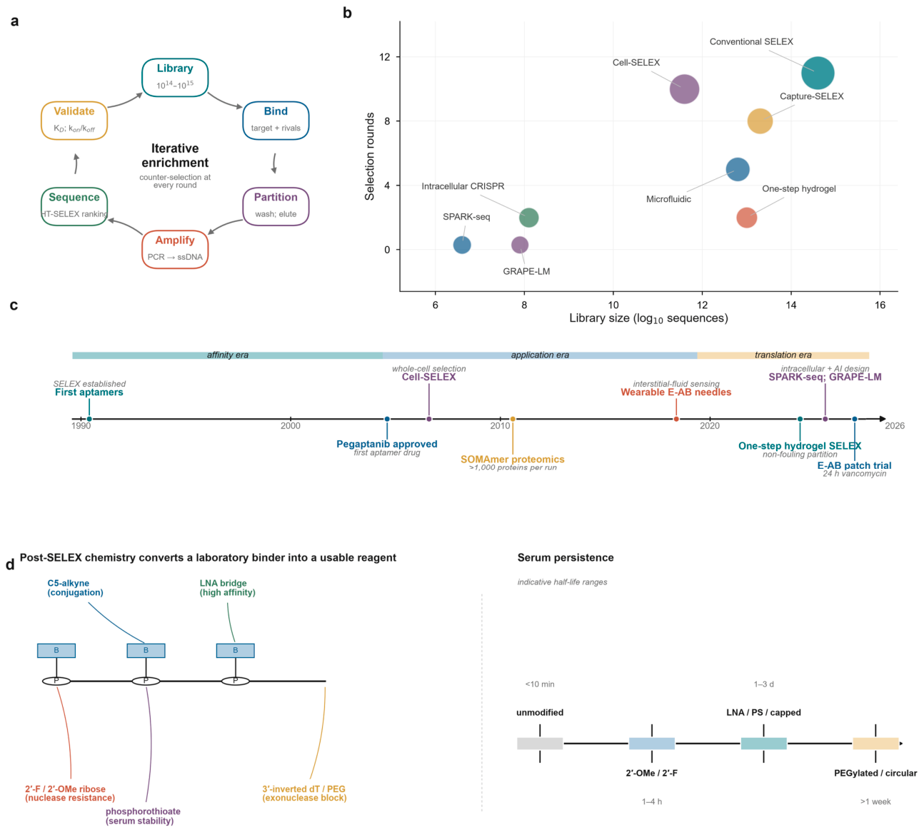
Figure 2.
The aptamer discovery pipeline, from library to validated reagent. a, The selection cycle. A combinatorial library of 1014-1015 sequences is exposed to the target, partitioned, amplified and re-annealed; each round includes counter-selection against off-targets. b, Selection methods plotted by library size and number of rounds. Intracellular CRISPR-based screens and generative models operate on libraries four to seven orders of magnitude smaller than classical SELEX and require a single round. c, Milestones from the first aptamers (1990) to the pilot clinical trial of a wearable electrochemical aptamer patch (2026), divided into an affinity era, an application era and a translation era. d, Post-selection chemistry. Sugar, phosphate and base modifications, and terminal caps, convert a laboratory binder into a reagent with useful serum persistence; ranges are indicative and compiled from the literature cited in Section 3.4.
Figure 2.
The aptamer discovery pipeline, from library to validated reagent. a, The selection cycle. A combinatorial library of 1014-1015 sequences is exposed to the target, partitioned, amplified and re-annealed; each round includes counter-selection against off-targets. b, Selection methods plotted by library size and number of rounds. Intracellular CRISPR-based screens and generative models operate on libraries four to seven orders of magnitude smaller than classical SELEX and require a single round. c, Milestones from the first aptamers (1990) to the pilot clinical trial of a wearable electrochemical aptamer patch (2026), divided into an affinity era, an application era and a translation era. d, Post-selection chemistry. Sugar, phosphate and base modifications, and terminal caps, convert a laboratory binder into a reagent with useful serum persistence; ranges are indicative and compiled from the literature cited in Section 3.4.
| Box 1 Terms that are routinely conflated KD The equilibrium dissociation constant. A ratio of rates, not a measure of signal. An aptamer with a 1 nM KD may be unusable in a sensor if binding produces no conformational change. koff The dissociation rate constant. Determines whether a sensor reports an instantaneous concentration or a time-averaged exposure; values below ~10-4 s-1 are incompatible with continuous monitoring over minutes. Specificity versus selectivity Specificity is the ability to discriminate the intended target from defined interferents; selectivity is the ability to do so in a complex mixture. The first can be demonstrated in buffer, the second cannot. Limit of detection versus limit of blank The limit of blank is the highest apparent signal expected from a sample containing no analyte; the limit of detection is the lowest concentration reliably distinguished from it. Reporting the first as the second is the most common analytical overstatement in the field. Analytical versus clinical sensitivity Analytical sensitivity is the slope of the calibration curve; clinical sensitivity is the proportion of diseased patients correctly identified. The two are related only through prevalence and the decision threshold. Regeneration versus reversibility Reversibility is a thermodynamic property of the binding equilibrium; regeneration is a process by which the sensor is returned to its baseline. A fully reversible aptamer may still require chemical regeneration if the interface has aged. |
4. From Recognition to Readout
4.1. Electrochemical Transduction
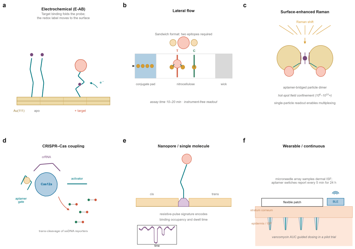
The electrochemical aptamer-based (E-AB) sensor is the most thoroughly mechanistically characterised architecture in the field (Fig. 3a). An aptamer is tethered to a gold electrode through a thiol and carries a redox reporter at its distal end; binding changes the mean distance between reporter and surface, and therefore the electron-transfer rate, which is read by square-wave or alternating-current voltammetry. Because the readout is a change in kinetics rather than a change in mass or charge, the sensor is comparatively insensitive to the total amount of adsorbed material, which is why it works in undiluted blood at all. The mechanism is also its weakness: the signal depends on a conformational change of a specific magnitude, so an aptamer that binds tightly without changing shape generates no signal, and an aptamer that changes shape for reasons unrelated to binding generates a false one.15,16
Engineering of the interface has driven most of the recent progress. Nanoporous gold electrodes increase the surface area available for probe attachment while excluding large fouling species, and have allowed continuous measurement of anthracycline pharmacokinetics in the blood of living animals with nanomolar precision and second-scale resolution.12,57 Tetrahedral DNA nanostructures act as a spacer layer that controls probe orientation and density, improving both signal amplitude and reproducibility.20 Field-effect geometries provide a lower-power alternative for point-of-care drug measurement, and extended-gate transistor devices functionalised with an aptamer have achieved clinically relevant sensitivity for small-molecule drugs in serum.58 Matrix-activated architectures, in which the sensor is deliberately designed to exploit rather than exclude the adsorbed protein layer, produced stable responses in undiluted human serum where conventional monolayers failed.59 The recurring design constraint is probe density: a dense monolayer maximises the number of recognition elements but sterically prevents the folding that generates signal (Fig. 5e).
4.2. Optical Transduction
Optical formats trade the simplicity of an electrode for the sensitivity of photon counting or the specificity of a spectral signature. Fluorescence anisotropy, FRET and structure-switching fluorophore-quencher pairs all convert binding into a change in emission, and the underlying structure-switching principle, introduced two decades ago,60 remains the most portable design in the field. Photonic crystal and liquid-crystal formats convert binding into a visible colour change without instrumentation, and structure-switching aptamers have been engineered specifically to suppress the background that limits their sensitivity. Systematic screening of switchable beacon probes has turned the design of such switches from an ad hoc exercise into a selection problem, and pipelines now exist for generating switchable beacons against a chosen protein target.61,62 Programmable fluorescent aptamer switches have extended the principle to the discrimination of single nucleotide variants, a capability of direct relevance to somatic mutation detection.63 A switchable aptamer selected for the respiratory syncytial virus G protein lowered the limit of detection of a photonic crystal sensor by three orders of magnitude relative to its parent aptamer, and reduced background noise in serum, saliva and nasal swabs.64
Surface-enhanced Raman scattering (SERS) offers the highest multiplexing potential of the optical methods, because each reporter has a narrow vibrational fingerprint (Fig. 3c). The gain comes from electromagnetic hot spots between coupled nanoparticles, which are also the source of irreproducibility: the enhancement factor depends exponentially on interparticle distance. Aptamers address this by acting as the spacer that defines the gap. A droplet microfluidic platform using salt-induced aggregation of gold nanoparticles with a HER2 aptamer achieved a limit of detection of 4.5 log10 particles per mL for HER2-positive exosomes in five minutes per sample, and distinguished HER2-positive from HER2-negative breast cancer patients in clinical samples.65 Dual-modal readouts - SERS with fluorescence, or colorimetry with photothermal signal - are now common precisely because they allow one channel to correct the other.20
4.3. Amplification-Coupled Formats
The third family couples aptamer recognition to an enzymatic amplification step, most often a CRISPR-Cas effector (Fig. 3d). The architecture is attractive because it separates recognition from amplification: the aptamer provides specificity in a format that can be tuned to any target, and the Cas enzyme provides gain. In the common implementation, binding of the target displaces or releases an activator strand that switches on Cas12a, whose collateral cleavage of single-stranded reporters generates fluorescence. Bivalent aptamers accelerate the kinetics of this activation and improve the limit of detection for small molecules,66 and the approach has been applied to viral antigens, cardiac markers and protein biomarkers in serum.67,68
The performance of these assays is frequently reported as a limit of detection that is not achievable in a clinical matrix, and a critical assessment of aptamer-CRISPR biosensors for small molecules concluded that many published limits are derived from buffer and that the specificity of the Cas step is often conflated with the specificity of the whole assay.5 The distinction matters: Cas12a requires a protospacer-adjacent motif on its DNA activator, and the sequence constraints this imposes, together with the possibility of background activation by contaminating nucleic acids, mean that the analytical specificity of an aptamer-CRISPR assay is not simply the product of its parts.20
4.4. Single-Molecule and Nanopore Readout
Nanopore sensing converts binding into a change in ionic current as a molecule traverses, or is held within, a nanoscale aperture (Fig. 3e). For aptamers the appeal is that a single binding event is observable, so that the distribution of dwell times reports the kinetics of the interaction rather than an ensemble average. Protein nanopores and solid-state nanopores have both been used, and aptamer-functionalised pores have detected small molecules and toxins at concentrations below those accessible to ensemble methods.27,28 Nanopipettes functionalised with a conformational-switch aptamer achieved serotonin sensing at physiologically relevant concentrations.17 The limitation is throughput: a single pore measures one molecule at a time, and although multiplexed arrays are in development, the sample volumes and times required remain incompatible with routine clinical use. The realistic near-term role of nanopore readout is therefore mechanistic - as a tool for validating that a candidate aptamer actually switches - rather than diagnostic.
4.5. Wearable and Continuous Monitoring
The application for which aptamers are structurally best suited is continuous measurement, because a binding equilibrium that reverses within minutes can track a concentration that changes within minutes, and because an oligonucleotide can be regenerated chemically in situ in a way that an antibody cannot. Microneedle arrays functionalised with aptamers sample dermal interstitial fluid, which tracks plasma free drug concentrations with a lag of minutes, and several groups have demonstrated continuous monitoring of antibiotics and chemotherapeutics in animals.69-71 The decisive step was clinical. A pilot-phase trial of a wearable electrochemical aptamer-based patch in six healthy participants captured vancomycin concentrations in dermal interstitial fluid with five-minute resolution over 24 hours, was well tolerated and near-painless, and yielded pharmacokinetic parameters that sparse blood sampling cannot resolve.11 Two limitations were explicit in the report: signal degradation meant that the informative window was the first 12 hours, and the measurements were not yet transmitted wirelessly in real time.
A parallel line of work targets hormones and small molecules at home. A wearable aptamer nanobiosensor for female hormone monitoring demonstrated that the same architecture can follow an endogenous analyte whose concentration changes over a menstrual cycle rather than over an infusion.72 The engineering problems that remain are shared across these devices: drift, fouling, calibration-free operation and the absence of an accepted regulatory pathway for a sensor whose output is a continuous curve rather than a discrete result (Fig. 4c).
Figure 3.
Transduction architectures. a, Electrochemical aptamer-based sensing. Target binding folds the probe and moves a redox reporter towards the electrode, changing the electron-transfer rate. b, Lateral flow. A sandwich format requires two epitopes; a control line captures unbound conjugate. c, Surface-enhanced Raman scattering. An aptamer defines the gap between coupled nanoparticles, and the confined field amplifies the reporter spectrum. d, CRISPR-Cas coupling. Target binding releases an activator that switches on Cas12a, whose collateral cleavage of single-stranded reporters generates gain. e, Nanopore readout. An aptamer-captured analyte produces a characteristic resistive pulse whose dwell time reports binding kinetics. f, Wearable format. A microneedle array samples dermal interstitial fluid and reports continuously over a dosing interval.
Figure 3.
Transduction architectures. a, Electrochemical aptamer-based sensing. Target binding folds the probe and moves a redox reporter towards the electrode, changing the electron-transfer rate. b, Lateral flow. A sandwich format requires two epitopes; a control line captures unbound conjugate. c, Surface-enhanced Raman scattering. An aptamer defines the gap between coupled nanoparticles, and the confined field amplifies the reporter spectrum. d, CRISPR-Cas coupling. Target binding releases an activator that switches on Cas12a, whose collateral cleavage of single-stranded reporters generates gain. e, Nanopore readout. An aptamer-captured analyte produces a characteristic resistive pulse whose dwell time reports binding kinetics. f, Wearable format. A microneedle array samples dermal interstitial fluid and reports continuously over a dosing interval.
5. The Clinical Evidence Base
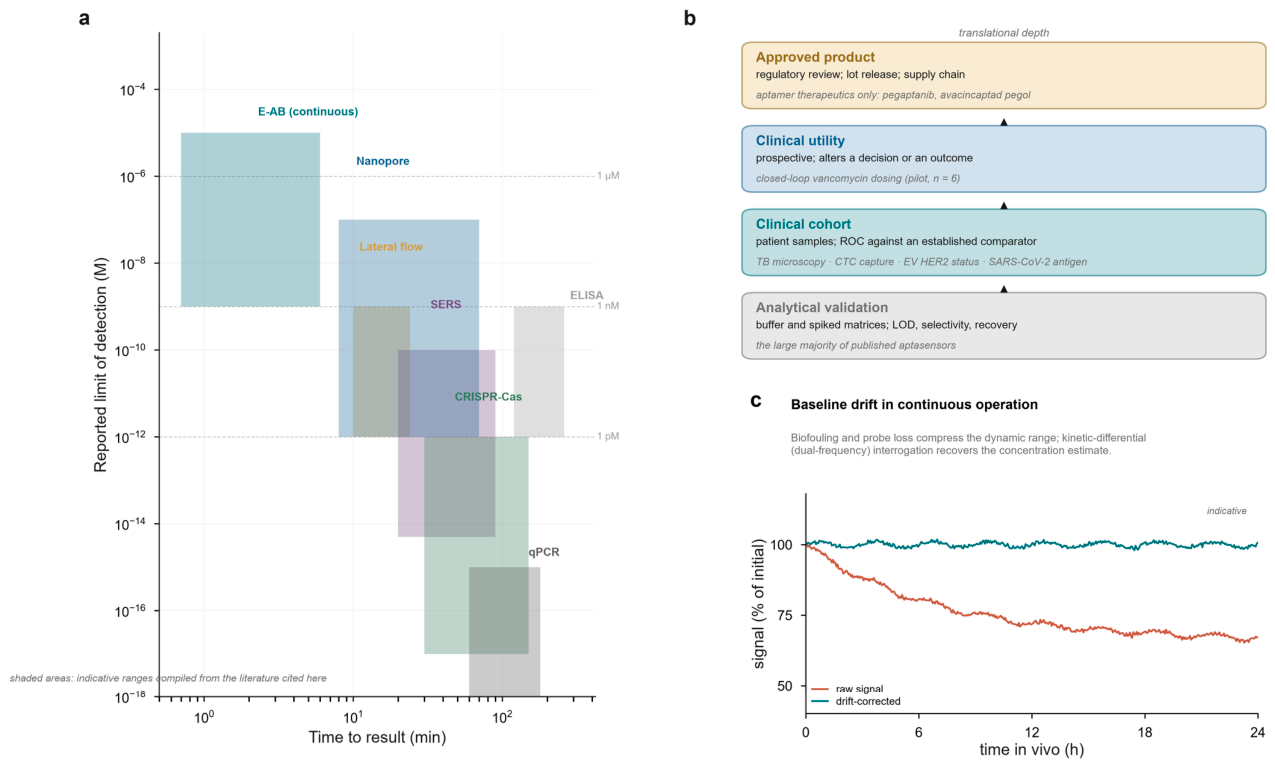
The literature on aptamers in clinical samples is large but unevenly distributed across the evidential hierarchy summarised in Fig. 4b. Most reports establish analytical performance in spiked buffer; a smaller number test patient samples against an established comparator; very few are prospective; and, at the time of writing, no aptamer-based in vitro diagnostic has completed a pivotal premarket review as a standalone device, although two aptamer therapeutics have been approved.3,4
5.1. Infectious Disease and Sepsis
Infectious disease is the area in which aptamer assays have come closest to clinical use, because the analytical requirement - detect a abundant antigen in a respiratory swab or a bacterial surface marker in blood - is well matched to the achievable sensitivity of a lateral flow or electrochemical device. Aptamer lateral flow strips for SARS-CoV-2 achieve limits of detection in the low picomolar range for nucleocapsid protein and, more importantly, retain specificity against seasonal coronaviruses.73,74 The most informative recent development is not a new transducer but a new recognition strategy: a lateral flow platform using several aptamers directed at distinct sites on the same viral protein, combined with quantum-dot labels, detected nucleocapsid at 1.4 pg mL-1 and inactivated virus at 1,643 U mL-1, and in a set of 30 positive and 20 negative patient samples raised the positive detection rate from 55% to 87% relative to comparable commercial products.75 The gain came from avidity and from averaging over several epitopes, not from a better fluorophore - a general lesson for antigen detection.
Sepsis illustrates both the opportunity and the difficulty. The clinical requirement is a result within an hour that distinguishes bacterial infection from sterile inflammation and guides antibiotic choice. Heparin-binding protein, a granule protein released by activated neutrophils, is an attractive target, and DNA aptamers with dissociation constants of 1-3 nM have been selected against it and incorporated into an amplification-coupled fluorescent assay with a limit of detection of 5.6 pM.76 Point-of-care aptasensors for C-reactive protein, procalcitonin and interleukin-6 have been reported in considerable number,71,77 and a recent systematic review of electrochemical aptamer-based sepsis sensors concluded that the field is limited less by sensitivity than by inconsistent validation protocols and by the scarcity of testing in undiluted clinical matrices.78 Bloodstream infection adds a sample-preparation problem that no aptamer can solve on its own: the organism must first be separated from blood cells and plasma, and microfluidic isolation coupled to aptamer detection has been proposed as an integrated workflow.79
Tuberculosis deserves separate mention because it is the application in which the evidence base is deepest. Aptamers against the mannose-capped lipoarabinomannan of Mycobacterium tuberculosis have been applied to lesion tissue and to urine, and two systematic reviews - one with meta-analysis - have pooled the diagnostic accuracy of aptamer-based tuberculosis tests, finding sensitivity and specificity that are competitive with existing antigen tests in some strata but with substantial heterogeneity between studies.80-82 Malaria provides a comparable case: aptamers against Plasmodium falciparum histidine-rich protein II and lactate dehydrogenase have been incorporated into electrochemical and lateral flow formats with limits of detection in the low picomolar range.83 An aptamer-based electrical biosensor for Zika virus detected the virus in human serum without amplification, illustrating that the limiting factor for such assays is usually the interface rather than the recognition element.84 The common thread in these neglected-disease applications is that the value proposition is robustness and cost rather than sensitivity, and that the field has been slower than it should be to demonstrate either under field conditions.
5.2. Cardiovascular Disease
Cardiac troponin is the reference standard for myocardial infarction, and any new recognition element for it is measured against a highly optimised immunoassay. Aptamers against cardiac troponin T with micromolar dissociation constants have been used in an electrochemiluminescence lateral flow format with a limit of detection of 143 pM85 - analytically respectable but three orders of magnitude short of the high-sensitivity immunoassays that define clinical practice, and the gap illustrates why troponin is unlikely to be the first cardiovascular win. The more promising targets are those for which no good antibody-based point-of-care test exists. Soluble suppression of tumourigenicity 2 (sST2) is a prognostic marker in heart failure; aptamers selected in the presence of serum matrix achieved dissociation constants of 8-46 nM and, coupled to Cas12a amplification, detected sST2 at 0.8 ng mL-1 in patient serum with a correlation of R2 = 0.98 against ELISA.68 N-terminal pro-B-type natriuretic peptide has been measured with a wearable microneedle aptamer sensor,70 and aptamer-based plasma proteomics in population cohorts has begun to nominate cardiovascular risk proteins de novo.13,86 The pattern is consistent: aptamers are most useful where the clinical question is about a panel or a trend rather than about a single decision threshold.
5.3. Oncology and Liquid Biopsy
Oncology is where the largest number of aptamer assays has been reported and where the gap between publication and practice is widest. Circulating tumour cells, tumour-derived extracellular vesicles and exosomal proteins are all attractive because they are scarce, phenotypically informative and accessible from blood. Aptasensors based on AS1411, a nucleolin-binding aptamer, detected circulating tumour cells at 2 cells per mL without enrichment;87 a dual-target electrochemical assay for MUC1-expressing lung cancer cells reached 1 cell per mL with an area under the curve of 0.96 in 39 clinical samples.71 Multivalent presentation improves capture under flow: tetrahedral DNA frameworks presenting three matched EpCAM aptamers captured circulating tumour cells with sufficient reproducibility to classify liver cancer patients as responders or non-responders with 83% accuracy.49
Extracellular vesicles present a harder recognition problem because the target is a heterogeneous population of membrane fragments rather than a molecule with a fixed stoichiometry. Dual-aptamer proximity ligation coupled to hybridisation chain reaction and CRISPR readout achieved a limit of detection of 330 particles per microlitre and reported complete separation of cancer from non-cancer plasma in a small cohort;88 aptamer-guided fluorescence polarisation has been developed specifically for vesicle-based liquid biopsy.89 The recurring methodological weakness is the absence of an independent, phenotype-defined reference standard: vesicle counts are not standardised between laboratories, and a classification accuracy of 100% in a small convenience cohort should be read as a hypothesis rather than as a validated performance characteristic.
5.4. Neurodegeneration
Blood-based biomarkers for Alzheimer disease have moved rapidly into clinical use, and aptamers have been applied to tau, amyloid-β and α-synuclein with mixed success. The analytical challenge is conformational rather than merely quantitative: the pathogenic species are oligomers and specific phospho-isoforms present at femtomolar concentrations against a large background of the same protein in its physiological form. Aptamers selected against oligomeric tau have been used both diagnostically and, in a recent therapeutic application, to direct monocyte-mediated clearance of extracellular tau and reduce neuroinflammation in mice.90 Aptamer-based electrochemical assays for amyloid-β and for α-synuclein have been reported with limits of detection in the picomolar range.20 Here too, population proteomics has been more informative than any single assay: aptamer panels measured in large cohorts have produced protein signatures that predict cognitive decline as strongly as phosphorylated tau.14
5.5. Therapeutic Drug Monitoring
Therapeutic drug monitoring is the clinical application in which the aptamer's physical advantages - reversibility, chemical definability, tolerance of a non-physiological interface - translate most directly into a clinical benefit (Box 3). Drugs with narrow therapeutic indices, notably vancomycin, aminoglycosides and busulfan, are currently monitored by sparse blood sampling and population pharmacokinetic models, which cannot capture the exposure of an individual patient during a dosing interval. Electrochemical aptamer sensors have now been demonstrated for vancomycin, tobramycin, daunorubicin, irinotecan and methotrexate, in buffer, in serum and in living animals.11,12,57,91 The pilot clinical trial of a microneedle patch described above is, to our knowledge, the first demonstration that the approach yields interpretable individual pharmacokinetics in humans.11 A comprehensive review of aptamer-based point-of-care sensors for drug monitoring catalogues both the progress and the outstanding problem: the absence of a metrological framework for validating a continuous sensor against the chromatographic reference method it is intended to replace.92
5.6. Population-Scale Proteomics
The largest clinical impact of aptamers to date has nothing to do with a point-of-care device. Slow off-rate modified aptamers (SOMAmers) bearing hydrophobic base modifications bind plasma proteins with antibody-like affinity, and arrays of them measure thousands of proteins in a single small sample. The platform has been deployed at population scale: the UK Biobank Pharma Proteomics Project characterised the plasma proteomes of 54,219 participants and mapped 14,287 primary protein quantitative trait loci, most of them previously undescribed.13 A parallel comparison of the two dominant affinity platforms - aptamer-based and antibody-based - found only modest correlation between them, with concordance varying by protein and by genomic support for the assay, a result that should temper any claim that either platform measures the proteome rather than a particular projection of it.86 Used as a discovery engine, however, the approach has been extraordinarily productive: proteomic signatures based on 5-20 proteins predicted the ten-year incidence of 52 diseases better than conventional risk factors,14 and proteomic profiling of a randomised trial population revealed the systemic effects of a GLP-1 receptor agonist across 495 protein targets.93 For the diagnostic community the lesson is that the aptamer has already succeeded as a reagent; what it has not yet done is succeed as a device.
5.7. Aptamers as Mechanistic Probes and as Therapeutic Leads
The same properties that make an aptamer useful in a detection device make it useful as a research reagent, and the two applications increasingly inform one another. Because an aptamer can be selected against a defined conformational state and can be produced as a single molecular species, it is well suited to interrogating protein function in cells. The most developed example is targeted protein degradation. Bispecific aptamer chimeras that bind a membrane protein on one arm and a lysosome-targeting receptor on the other recruit the protein to degradation, and the approach has been extended to light-triggered and circular variants with improved pharmacokinetics.94-96 Aptamer 'glues' that mechanically cluster a target on the membrane achieve degradation at concentrations well below those required by occupancy-driven degraders,97,98 and the underlying mechanistic principle - that clustering, not occupancy, can be the degrading signal - was itself established using aptamers as the clustering agent.99 The same logic explains why multivalent aptamers behave differently from their monomeric parents in capture and imaging applications (Section 3.4).
Therapeutics that have reached patients remain few, but the set is growing and is instructive about which molecular formats survive translation. Pegaptanib, an RNA aptamer against vascular endothelial growth factor, was approved in 2004;3 avacincaptad pegol, a PEGylated RNA aptamer against complement component C5, was approved in 2023 for geographic atrophy;4 and an L-nucleotide aptamer against C5a has progressed to a phase 2 trial in complement-mediated disease.100 Aptamers against fibroblast growth factor 2 and against nucleolin have been in clinical development for oncology and ophthalmology respectively.101 Imaging is a nearer-term application: radiolabelled aptamers against EpCAM and against protein tyrosine kinase 7 have been used for tumour-specific positron emission tomography in preclinical models, and a first-in-human study of a PTK7-targeting aptamer tracer has now completed a first-in-human study in bladder cancer.102,103 These developments matter for diagnostics for a structural reason: a tracer that has passed through toxicology and human imaging has demonstrated the pharmacokinetics, the stability in blood and the absence of off-target binding that a diagnostic reagent also requires, and the two pipelines therefore share most of their evidentiary burden.
Figure 4.
Analytical performance, evidential depth and operational stability. a, Reported limits of detection against time to result for the principal transduction families. Shaded areas indicate the ranges typically reported in the literature cited here, not the performance of any individual device; the overlap between families is the central analytical fact of the field. b, The evidential hierarchy. Most published aptasensors reach the first tier only; the second tier is populated by a small number of infectious-disease, oncology and cardiac studies; the third by a single pilot clinical trial; the fourth by aptamer therapeutics rather than diagnostics. c, Baseline drift in continuous operation and its correction by kinetic-differential interrogation.
Figure 4.
Analytical performance, evidential depth and operational stability. a, Reported limits of detection against time to result for the principal transduction families. Shaded areas indicate the ranges typically reported in the literature cited here, not the performance of any individual device; the overlap between families is the central analytical fact of the field. b, The evidential hierarchy. Most published aptasensors reach the first tier only; the second tier is populated by a small number of infectious-disease, oncology and cardiac studies; the third by a single pilot clinical trial; the fourth by aptamer therapeutics rather than diagnostics. c, Baseline drift in continuous operation and its correction by kinetic-differential interrogation.
6. Why Aptasensors Underperform in Patients
A recurring pattern in the literature is a device that reports a femtomolar limit of detection in buffer and fails to discriminate patients from controls in serum. The gap is not a single failure but the superposition of six mechanisms, each of which is now sufficiently well characterised to be designed around (Fig. 5).
6.1. The Protein Corona and Nonspecific Adsorption
Within seconds of immersion in plasma a surface acquires a layer of adsorbed protein that is dominated by albumin, immunoglobulin and apolipoproteins. For an aptamer sensor this layer has three effects: it occludes the binding interface, it adds a steric barrier that damps the conformational change on which signal depends, and it can generate a signal of its own if the adsorbed protein interacts with the redox reporter. The consequence is a compression of the calibration curve and a loss of dynamic range that is not recoverable by simply increasing gain. Systematic measurements in blood plasma have shown that the responses of several E-AB sensors are compressed by nonspecific interactions and that removal of the major protein and lipid fractions restores much of the sensitivity observed in buffer.20,104 Two design responses are effective: antifouling architectures that present a zwitterionic or polyethylene-glycol brush to the sample while keeping the aptamer accessible, and selection or counter-selection performed in the intended matrix so that matrix-tolerant sequences are enriched rather than discovered late.59,68
6.2. Nuclease Hydrolysis
Unmodified oligonucleotides have serum half-lives measured in minutes, and the degradation products are themselves a source of false signal in assays that rely on strand displacement. The chemistry described in Section 3.4 mitigates but does not eliminate the problem, and the protection is not uniform: single-stranded loops, which are frequently the binding element, are the most exposed. For continuous sensors the issue is compounded by the need for the probe to remain functional for days at 37 °C in a compartment that contains both nucleases and cells. Encapsulation in nanoporous supports, conjugation to a bulky carrier that sterically excludes nucleases, and the use of mirror-image L-nucleotides for non-natural targets each address part of the problem.42,57
6.3. Target Heterogeneity and Epitope Masking
An aptamer selected against a recombinant protein recognises an epitope defined by that molecule's sequence and fold. In blood, the same protein may be glycosylated, cleaved, bound to a carrier protein or assembled into a complex, and any of these can bury the epitope (Fig. 5c). This is a principal reason for the discrepancy between the affinity measured on recombinant material and the sensitivity achieved in a clinical sample, and it is the strongest argument for cell-SELEX or tissue-SELEX when the diagnostic question concerns a native state.33,34 Where the epitope is known, the effect can be turned to advantage: aptamers that discriminate a specific post-translational state are more informative than pan-protein binders, and this is the basis of the most promising aptamer assays for neurodegenerative disease.90
6.4. Interface Ageing, Drift and Regeneration
A sensor that is calibrated once and used repeatedly must remain stable, and aptamer interfaces do not. Probe desorption, thiol oxidation, aggregation of the redox reporter and gradual accumulation of adsorbed material all produce a monotonic drift that is easily mistaken for a change in analyte concentration (Fig. 4c). Kinetic-differential interrogation, in which the sensor is driven at two frequencies and the ratio of responses is used rather than either response alone, removes much of the common-mode drift and allows calibration-free operation; the approach is now standard in continuous E-AB measurement.57,105 Regeneration by chaotropic wash, alkaline or acidic pulses, or photochemical release extends the usable life of a sensor and is a prerequisite for any reusable format.20
6.5. Sequence-Level Irreproducibility
The most cited criticism of the aptamer literature is that many published sequences do not bind their stated targets when tested independently. The causes are traceable and largely avoidable: selection without counter-selection against the support and against homologous proteins; enrichment judged by pool binding rather than by individual sequence characterisation; sequencing depth insufficient to distinguish enrichment from noise; and reporting of a single high-affinity sequence from a pool that was never clonally validated.53,55 The remedies are unglamorous: negative selection against immobilisation matrices and abundant serum proteins, per-sequence affinity determination by an independent method, and deposition of the raw sequencing data. Where these steps have been taken, the reported affinities have generally held up; where they have not, the failure rate is high enough to have damaged the credibility of the field as a whole.5
6.6. Reporting and the Evidence Gap
The final mechanism is sociological rather than physical. A device paper typically reports a limit of detection, a linear range and a selectivity panel against two or three unrelated proteins; it rarely reports the number needed to diagnose, the confidence interval on sensitivity, the effect of sample haemolysis, the lot-to-lot variability of the electrode, or the performance of the assay on the same samples as the incumbent method. These omissions make meta-analysis impossible and make it difficult for a clinical laboratory to judge whether a new assay would improve on what it already has. The reporting standards proposed for biosensors in general apply with particular force to aptamers, and their adoption is a precondition for the field's next stage.5,106
Figure 5.
Six mechanisms that separate buffer performance from clinical performance. a, Protein corona formation occludes the interface and compresses the calibration curve. b, Nuclease hydrolysis degrades the unpaired regions that form the binding pocket. c, Epitope masking by glycosylation, cleavage or partner binding invalidates affinity measured on recombinant protein. d, Slow dissociation converts an instantaneous measurement into a time-averaged one. e, Interfacial crowding at high probe density sterically prevents the conformational change that generates signal. f, Synthesis, conjugation and surface chemistry introduce sensor-to-sensor gain differences that are indistinguishable from biological variation unless the readout is ratiometric or kinetic.
Figure 5.
Six mechanisms that separate buffer performance from clinical performance. a, Protein corona formation occludes the interface and compresses the calibration curve. b, Nuclease hydrolysis degrades the unpaired regions that form the binding pocket. c, Epitope masking by glycosylation, cleavage or partner binding invalidates affinity measured on recombinant protein. d, Slow dissociation converts an instantaneous measurement into a time-averaged one. e, Interfacial crowding at high probe density sterically prevents the conformational change that generates signal. f, Synthesis, conjugation and surface chemistry introduce sensor-to-sensor gain differences that are indistinguishable from biological variation unless the readout is ratiometric or kinetic.
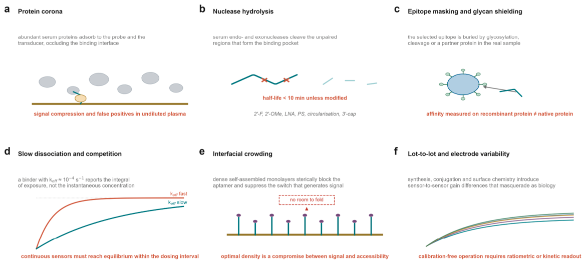

Table 1.
Representative aptamer-based detection formats with reported analytical performance and stage of validation.
Table 1.
Representative aptamer-based detection formats with reported analytical performance and stage of validation.
| Target (matrix) | Recognition format | Transduction | Reported LOD | Validation stage | Ref. |
|---|---|---|---|---|---|
| SARS-CoV-2 nucleocapsid (nasopharyngeal) | Multiple aptamers, distinct epitopes | Quantum-dot lateral flow | 1.4 pg·mL-1 | Patient samples (n = 50) | 75 |
| Cardiac troponin T (serum) | Sandwich pair from centrifugal SELEX | Electrochemiluminescence lateral flow | 143 pM | Buffer and spiked serum | 85 |
| Soluble ST2 (serum) | Single aptamer, serum-matrix selection | Cas12a fluorescence | 0.8 ng·mL-1 | Patient samples versus ELISA | 68 |
| Heparin-binding protein (serum) | Single aptamer, KD 1.0 nM | Rolling-circle amplification | 5.6 pM | Buffer and spiked serum | 76 |
| HER2-positive exosomes (plasma) | Single aptamer | SERS in droplets | 4.5 log10 particles ·mL-1 | Patient samples | 65 |
| Tumour-derived extracellular vesicles (plasma) | Dual-aptamer proximity ligation | CRISPR-HCR | 330 particles· µL-1 | Patient plasma (small cohort) | 88 |
| Circulating tumour cells (blood) | AS1411, multivalent nanovehicle | Fluorescence turn-on | 2 cells ·mL-1 | Patient blood | 87 |
| Circulating tumour cells (blood) | Tetrahedral framework, trivalent EpCAM | Magnetic capture, imaging | n.d. | Treatment-response monitoring | 49 |
| Vancomycin (interstitial fluid) | Single aptamer, microneedle array | Electrochemical (E-AB) | µM range | Pilot clinical trial (n = 6) | 11 |
| Daunorubicin (whole blood) | Single aptamer, nanoporous gold | Electrochemical (E-AB) | nM, seconds | Rodent pharmacokinetics | 12 |
| Plasma proteome (plasma) | SOMAmer arrays | Hybridisation-based fluorescence | ~10-12 M | Population cohort (n = 54,219) | 13 |
n.d., not determined; HCR, hybridisation chain reaction.
7. A Translational Framework
If the rate-limiting step is evidence rather than discovery, then the field needs an explicit account of what evidence would be sufficient. We propose a four-tier framework, aligned with the analytical and clinical validation pathways used for in vitro diagnostics, in which each tier has a defined deliverable and a defined failure mode (Fig. 4b).
Tier 1, analytical validation, establishes that the assay measures what it claims in the matrix in which it will be used. The minimum content is a calibration curve in the intended matrix rather than in buffer, a limit of blank and a limit of detection derived from replicate measurements, a selectivity panel that includes the closest available homologues and the most abundant interferents, a recovery experiment, and an explicit statement of the dynamic range over which the response is monotonic. The characteristic failure at this tier is a limit of detection extrapolated from a three-sigma rule applied to a calibration curve whose lowest point is already ten times the clinical decision threshold.
Tier 2, clinical validation, establishes that the assay discriminates a clinically defined population. The essential design is a case-control or cohort study in which the assay is run blinded on samples from patients classified by an independent reference standard, with sensitivity and specificity reported together with confidence intervals at a prespecified threshold, and with the comparator measured on the same samples. Reporting an area under the receiver-operating-characteristic curve without a threshold, or a threshold chosen after seeing the data, is not clinical validation. The characteristic failure at this tier is spectrum bias: samples drawn from a convenience set of severely affected patients and healthy volunteers, which overstates performance relative to the intended-use population.
Tier 3, clinical utility, establishes that using the assay changes a decision and improves an outcome. This requires a prospective design, and for a continuous monitor it requires a different kind of endpoint altogether - time in therapeutic range, proportion of doses correctly adjusted, or incidence of nephrotoxicity - rather than a correlation coefficient against a reference method. The pilot vancomycin study sits at the boundary between tiers 2 and 3: it demonstrated that the sensor produces pharmacokinetic parameters in humans, but the cohort was too small and too short to show that acting on those parameters improves anything.11
Tier 4, approval and post-market surveillance, requires manufacturing consistency at a level that the academic literature rarely addresses. For an oligonucleotide reagent this means a defined synthesis and purification specification, a validated conjugation process, stability data under the conditions of shipping and use, and a batch-release assay that is sensitive to the failure modes that matter - truncation, depurination, incomplete conjugation and, for surface-immobilised formats, probe density. The two approved aptamer therapeutics demonstrate that this tier is achievable for oligonucleotides;3,4 no aptamer-based diagnostic has yet completed it.
Three cross-cutting requirements apply at every tier. The first is mechanistic verification that the reported signal originates in the intended binding event, established by a binding-site mutant, a competitive ligand or a scrambled-sequence control, rather than by correlation alone. The second is open deposition of the sequences and the raw selection data, without which independent replication is impossible.53 The third is reporting of the matrix: an assay validated in diluted serum has not been validated in plasma, and one validated in plasma has not been validated in whole blood.
| Box 2 A minimum reporting set for an aptamer-based diagnostic Selection Library design and diversity; number of rounds; counter-selection targets; the matrix used during selection; raw high-throughput sequencing data deposited with accession. Sequence Full sequence with any truncation and modification stated position by position; the predicted and, where available, experimentally determined secondary structure. Affinity Affinity determined by an independent method (surface plasmon resonance, isothermal titration calorimetry or microscale thermophoresis), with association and dissociation rate constants, in buffer and in the intended matrix. Mechanism Evidence that the reported signal originates in the intended binding event: scrambled sequence, binding-site mutant or competitive inhibition. Analytical performance Limit of blank, limit of detection and limit of quantification in the intended matrix; dynamic range; recovery; interference panel including close homologues and the most abundant matrix proteins. Clinical performance Cohort description and reference standard; sensitivity and specificity with confidence intervals at a prespecified threshold; the comparator measured on the same samples. Stability Reagent shelf life; sensor-to-sensor and lot-to-lot variability; drift over the intended operating period. Reproducibility Independent replication in a second laboratory, reported whether or not it succeeded. |
| Box 3 Case study: closed-loop dosing of vancomycin Vancomycin is a glycopeptide antibiotic with a narrow therapeutic window and substantial inter-patient variability in exposure. Standard practice is to measure a trough concentration before the fourth dose and to adjust thereafter using a population pharmacokinetic model. The approach is acknowledged to be a poor description of individual exposure, and it is particularly poor in critically ill patients, in whom volume of distribution and clearance change over days. Aptamer-based electrochemical sensing offers an alternative because the recognition element can be regenerated in situ and because the binding equilibrium of a small-molecule aptamer reverses within seconds. A microneedle array placed on the skin samples dermal interstitial fluid, in which free vancomycin concentration tracks the plasma free concentration with a short lag. In the pilot-phase trial, six healthy participants wore patches for 24 hours and the sensors reported interstitial concentrations every five minutes.11 Fitting the interstitial and plasma concentrations to a two-compartment model resolved distribution and clearance phases that the sparse sampling schedule cannot, and patches placed at different anatomical sites gave consistent results within and between participants. Three obstacles separate this from practice. The signal degraded after roughly 12 hours, which is incompatible with a five-day course; the data were not transmitted in real time, so the loop was not closed; and the study was conducted in healthy volunteers rather than in the patients whose pharmacokinetics are most variable. Each is an engineering and clinical-study problem rather than a molecular one - which is precisely why the case is instructive. The molecular recognition problem was solved years ago; what remained was the interface, the metrology and the trial design. |
8. Outlook
Four trajectories seem likely to define the next five years, and they differ in how close they are to clinical consequence.
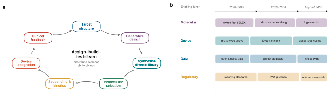
The first is the industrialisation of design (Fig. 6a). The combination of intracellular functional selection, sequencing-scale kinetic profiling and generative models has collapsed the discovery cycle from months to weeks and, more importantly, has changed what a selection produces: a dataset with which a model can be interrogated, rather than a shortlist of sequences.7,8,37 The immediate task is to make that dataset trustworthy. Training sets that contain only successful binders cannot teach a model to avoid cross-reactivity, and the absence of a standard benchmark - including negative sequences and clinically relevant interferents - is the principal obstacle to predictive design. The benchmarking efforts that have begun to appear are therefore as important as the models themselves.39
The second is the migration of the sensing interface from the laboratory to the body (Fig. 6b). Continuous molecular monitoring is the application for which aptamers have a structural advantage that antibodies cannot match, and the first human data are now in hand.11 What stands between the pilot and practice is not molecular recognition but metrology: a continuous sensor needs a calibration strategy that survives weeks in tissue, a drift model that is defensible to a regulator, and a reference method against which its output can be judged. The pharmacokinetic modelling in the pilot study, which resolved distribution and clearance phases invisible to sparse sampling, indicates what the clinical value would be if those problems were solved.11
The third is the consolidation of aptamer proteomics as a discovery platform and its extension into routine laboratory medicine. The demonstration that 5-20 proteins can predict the ten-year incidence of dozens of diseases is a result about the proteome rather than about aptamers, but it depended on an aptamer array to be obtainable at population scale.14 Two developments would convert this into clinical laboratory practice: a reduction in the cost and complexity of the array to the point where it can be run on individual samples rather than on cohorts, and an agreed approach to the discordance between affinity platforms, which at present means that a protein's measured concentration depends on the assay used to measure it.86
The fourth is a change in what the field claims. The most useful recent contributions have been critical rather than promotional: assessments that separate what an assay can do in buffer from what it does in blood, meta-analyses that expose the heterogeneity of clinical validation, and comparisons that treat aptamers as one recognition element among several rather than as a universal replacement.5,81,107 A field that reports its failures with the same care as its successes will be believed when it reports a success.
There is a temptation, in a field with this history, to predict that the next few years will bring the first aptamer-based diagnostic to routine use. It may. But the more defensible prediction is narrower and more interesting: that the aptamer will stop being evaluated as a substitute for the antibody and start being evaluated as a reagent with its own specification - reversible, sequence-defined, chemically programmable, and therefore suited to measurement problems that antibodies were never able to address. Continuous monitoring, multiplexed panels on a single interface, and assays in which the recognition element must be regenerated in situ are all problems of that kind. On that reading, the clinical future of the aptamer lies less in replacing the immunoassay than in measuring what the immunoassay cannot.
Figure 6.
A closed-loop design cycle and the translational horizon. a, Design-build-test-learn cycle in which target structure, generative design, synthesis, intracellular selection, sequencing and device integration are connected by a feedback path from clinical performance. A single round replaces the six to sixteen rounds of classical selection. b, Enabling layers and indicative horizons for the molecular, device, data and regulatory developments that would be required for routine clinical use.
Figure 6.
A closed-loop design cycle and the translational horizon. a, Design-build-test-learn cycle in which target structure, generative design, synthesis, intracellular selection, sequencing and device integration are connected by a feedback path from clinical performance. A single round replaces the six to sixteen rounds of classical selection. b, Enabling layers and indicative horizons for the molecular, device, data and regulatory developments that would be required for routine clinical use.
Author Contributions
Yunwang Zhao: Writing-original draft, Hongxian Cheng: Writing-review & editing, Shiyi Zhou: Visualization, Lei He and Chenlin Xiu: Methodology, Formal analysis, Baihai Huang: Conceptualization. Xiongxiong Lu: Writing-review & editing, Writing-original draft, Methodology, Conceptualization.
Competing Interests: The authors declare no competing interests.
References
- Tuerk, C.; Gold, L. Systematic Evolution of Ligands by Exponential Enrichment: RNA Ligands to Bacteriophage T4 DNA Polymerase. Science 1990, 249, 505–510. [Google Scholar] [CrossRef] [PubMed]
- Ellington, A.D.; Szostak, J.W. In vitro selection of RNA molecules that bind specific ligands. Nature 1990, 346, 818–822. [Google Scholar] [CrossRef] [PubMed]
- Ng, E.W.M.; et al. Pegaptanib, a targeted anti-VEGF aptamer for ocular vascular disease. Nat. Rev. Drug Discov. 2006, 5, 123–132. [Google Scholar] [CrossRef]
- Du, Z.; et al. Molecular bioengineering of functional nucleic acids. Nat. Rev. Bioeng. 2025, 4, 153–170. [Google Scholar] [CrossRef]
- Brandenberg, O.F.; Janssen, E.M.L.; Schubert, O.T. A critical assessment of aptamer and CRISPR-Cas12a-based biosensors for small molecule detection. npj Biosens. 3 2026, 28. [Google Scholar] [CrossRef]
- Singh, N.K.; et al. High-affinity one-step aptamer selection using a non-fouling porous hydrogel. Nat. Biotechnol. 2024, 42, 1224–1231. [Google Scholar] [CrossRef]
- Zhang, J.; et al. Single-round evolution of RNA aptamers with GRAPE-LM. In Nat. Biotechnol.; 2026. [Google Scholar] [CrossRef]
- Luo, G.; et al. SPARK-seq: A high-throughput platform for aptamer discovery and kinetic profiling. Science 2026, 391, eadv6127. [Google Scholar] [CrossRef]
- Alkhamis, O.; et al. Exploring the relationship between aptamer binding thermodynamics, affinity, and specificity. Nucleic Acids Res. 2025, 53, gkaf219. [Google Scholar] [CrossRef]
- Lee, Y.; et al. Carbon-nanotube field-effect transistors for resolving single-molecule aptamer-ligand binding kinetics. Nat. Nanotechnol. 2024, 19, 660–667. [Google Scholar] [CrossRef]
- Booth, M.A.; et al. Pilot phase clinical trial of a wearable, electrochemical aptamer-based patch for continuous drug concentration measurement. Nat. Biotechnol. 2026. [Google Scholar] [CrossRef]
- Son, K.; et al. Continuous, Week-Long, Seconds-Resolved In Vivo Drug Measurements Performed with a Xenonucleic Acid-Employing Electrochemical, Aptamer-Based Sensor. J. Am. Chem. Soc. 2026, 148, 10016–10025. [Google Scholar] [CrossRef]
- Sun, B.B.; et al. Plasma proteomic associations with genetics and health in the UK Biobank. Nature 2023, 622, 329–338. [Google Scholar] [CrossRef] [PubMed]
- Carrasco-Zanini, J.; et al. Proteomic signatures improve risk prediction for common and rare diseases. Nat. Med. 2024, 30, 2489–2498. [Google Scholar] [CrossRef]
- Kaiyum, Y.A.; et al. Ligand-Induced Folding in a Dopamine-Binding DNA Aptamer. ChemBioChem 2024, 25, e202400493. [Google Scholar] [CrossRef]
- Eisen, S.R.; Dauphin-Ducharme, P.; Johnson, P.E. Solution-based biophysical characterization of conformation change in structure-switching aptamers. Q. Rev. Biophys. 2024, 57, e9. [Google Scholar] [CrossRef]
- Nakatsuka, N.; et al. Aptamer Conformational Change Enables Serotonin Biosensing with Nanopipettes. Anal. Chem. 2021, 93, 4033–4041. [Google Scholar] [CrossRef]
- Cao, M.; Zhang, L.; Tang, H.; Qiu, X.; Li, Y. Single-Molecule Investigation of the Protein-Aptamer Interactions and Sensing Application Inside the Single Glass Nanopore. Anal. Chem. 2022, 94, 17405–17412. [Google Scholar] [CrossRef]
- Liu, X.; et al. The specificity and selectivity of aptamer depends on the binding epitope: insights from aptamer Hislxl-2a. Anal. Chim. Acta 2026, 1404, 345431. [Google Scholar] [CrossRef]
- Chen, Q.; Xue, C.; Hu, Y.; Hong, Q.; Liu, Z. From Molecular Recognition to Clinical Readout: Design Principles for Functional Nucleic Acid-Material Biosensors in Medical Diagnostics. Molecules 2026, 31, 3358. [Google Scholar] [CrossRef]
- Stafflinger, H.; et al. Crystal structure of the class V GTP-binding RNA aptamer bound to its ligand: GTP recognition by a topologically complex intermolecular G-quadruplex. Nucleic Acids Res. 2025, 53, gkaf1315. [Google Scholar] [CrossRef]
- Qiu, L.; Zou, X. Advances in Protein-RNA aptamer recognition and modeling: Current trends and future perspectives. Curr. Opin. Struct. Biol. 2025, 94, 103133. [Google Scholar] [CrossRef]
- Truong, L.; et al. The fluorescent aptamer Squash extensively repurposes the adenine riboswitch fold. Nat. Chem. Biol. 2022, 18, 191–198. [Google Scholar] [CrossRef]
- Huang, K.; et al. Structure-based investigation of fluorogenic Pepper aptamer. Nat. Chem. Biol. 2021, 17, 1289–1295. [Google Scholar] [CrossRef]
- Sunbul, M.; et al. Super-resolution RNA imaging using a rhodamine-binding aptamer with fast exchange kinetics. Nat. Biotechnol. 2021, 39, 686–690. [Google Scholar] [CrossRef] [PubMed]
- Samuelian, J.S.; et al. An RNA aptamer that shifts the reduction potential of metabolic cofactors. Nat. Chem. Biol. 2022, 18, 1263–1269. [Google Scholar] [CrossRef]
- Lv, P.; et al. Recent Advances in Aptamer-Based Nanopore Sensing at Single-Molecule Resolution. Chem. Asian J. 2022, 17, e202200364. [Google Scholar] [CrossRef]
- Sett, A. Threading Precision: Progress and Emerging Trends in Aptamer-Based Nanopore Sensing. Biosensors 2026, 16, 401. [Google Scholar] [CrossRef] [PubMed]
- Wu, Q. High-throughput identification of aptamer-target pairs with SPARK-seq. Nat. Rev. Genet. 2026, 27, 267–267. [Google Scholar] [CrossRef]
- Chung, Y.D.; Tsai, Y.C.; Wang, C.H.; Lee, G.B. Aptamer selection via versatile microfluidic platforms and their diverse applications. Lab Chip 2025, 25, 1047–1080. [Google Scholar] [CrossRef]
- Lin, C.S.; Tsai, Y.C.; Hsu, K.F.; Lee, G.B. Optimization of aptamer selection on an automated microfluidic system with cancer tissues. Lab Chip 2021, 21, 725–734. [Google Scholar] [CrossRef]
- Cui, W.; et al. Identifying a cancer therapeutic target: Cell-SELEX identifies a membrane protein for aptamer-mediated growth suppression. Proc. Natl. Acad. Sci. USA 2026, 123, e2514681123. [Google Scholar] [CrossRef]
- Li, L.; et al. Identification of a New DNA Aptamer by Tissue-SELEX for Cancer Recognition and Imaging. Anal. Chem. 2021, 93, 7369–7377. [Google Scholar] [CrossRef]
- Wong, K.Y.; Wong, M.S.; Lee, J.H.; Liu, J. From cell-SELEX to tissue-SELEX for targeted drug delivery and aptamer nanomedicine. Adv. Drug Deliv. Rev. 2025, 224, 115646. [Google Scholar] [CrossRef] [PubMed]
- Yu, Y.; Li, Y.; Liu, J. A high-affinity dopamine aptamer: implications of library diversity and negative selection. Chem. Sci. 2026, 17, 9997–10006. [Google Scholar] [CrossRef]
- Mayer, G. Lighting the spark of aptamer data science. Science 2026, 391, 24–25. [Google Scholar] [CrossRef]
- Wang, Z.; et al. AptaDiff: de novo design and optimization of aptamers based on diffusion models. Brief. Bioinform. 2024, 25, bbae517. [Google Scholar] [CrossRef] [PubMed]
- Ji, D.K.; et al. Nucleic acid aptamers: new methods for selection, target validation, molecular diagnostics and therapeutics. Signal Transduct. Target. Ther. 2026, 11, 369. [Google Scholar] [CrossRef] [PubMed]
- Zhao, J.; Tram, K.; Yan, H.; Li, Y. Comprehensive evaluation of artificial intelligence-empowered approaches for protein-aptamer complex prediction. Brief. Bioinform. 2026, 27, bbag206. [Google Scholar] [CrossRef]
- Dolu, K.O. Letter to the editor: methodological considerations in the benchmarking of AI-based protein-aptamer complex prediction. Brief. Bioinform. 2026, 27, bbag308. [Google Scholar] [CrossRef]
- Kar, R.K. High-throughput and computational techniques for aptamer design. Expert Opin. Drug Discov. 2024, 19, 1457–1469. [Google Scholar] [CrossRef]
- Pan, Z.; et al. Stability modification of therapeutic aptamers: from biostability bottlenecks to nuclease-resistant construct design. RSC Chem. Biol. 2026, 7, 964–982. [Google Scholar] [CrossRef] [PubMed]
- Mili, M.; Bachu, V.; Kuri, P.R.; Singh, N.K.; Goswami, P. Improving synthesis and binding affinities of nucleic acid aptamers and their therapeutics and diagnostic applications. Biophys. Chem. 2024, 309, 107218. [Google Scholar] [CrossRef]
- Murray, M.T.; Wetmore, S.D. Unlocking precision in aptamer engineering: a case study of the thrombin binding aptamer illustrates why modification size, quantity, and position matter. Nucleic Acids Res. 2024, 52, 10823–10835. [Google Scholar] [CrossRef] [PubMed]
- Manuel, B.A.; Sterling, S.A.; Sanford, A.A.; Heemstra, J.M. Systematically Modulating Aptamer Affinity and Specificity by Guanosine-to-Inosine Substitution. Anal. Chem. 2022, 94, 6436–6440. [Google Scholar] [CrossRef]
- Liu, Z.; et al. Circularization of Aptamer Nanomedicine Improves Nuclease Resistance, Targeted Recognition, and Transdermal Effect for Topical Psoriasis Therapy. Nano Lett. 2025, 25, 9139–9148. [Google Scholar] [CrossRef] [PubMed]
- Tabuchi, Y.; Yang, J.; Taki, M. Relative Nuclease Resistance of a DNA Aptamer Covalently Conjugated to a Target Protein. Int. J. Mol. Sci. 2022, 23, 7778. [Google Scholar] [CrossRef]
- Kehrli, J.; Cubi, R.; Gupta, T.; Klymchenko, A.; Ryckelynck, M. oShark: A Modified Nuclease-Resistant Light-Up Aptamer. J. Am. Chem. Soc. 2025, 147, 46174–46186. [Google Scholar] [CrossRef] [PubMed]
- Chen, Y.; et al. DNA Framework-Ensembled Aptamers Enhance Fluid Stability in Circulating Tumor Cells Capture for Tumor Treatment Evaluation. Angew. Chem. Int. Ed. 2025, 64, e202425252. [Google Scholar] [CrossRef]
- Kawamoto, Y.; et al. Multivalent dendritic DNA aptamer molecules for the enhancement of therapeutic effects. Chem. Commun. 2024, 60, 6256–6259. [Google Scholar] [CrossRef]
- Tang, L.; et al. De Novo Evolution of an Antibody-Mimicking Multivalent Aptamer via a DNA Framework. Small Methods 2023, 7, 2300327. [Google Scholar] [CrossRef]
- Jia, L.; et al. Engineering Biomimetic Biosensor Using Dual-Targeting Multivalent Aptamer Regulated 3D DNA Walker Enables High-Performance Detection of Heterogeneous Circulating Tumor Cells. Small 2023, 19, 2302542. [Google Scholar] [CrossRef]
- Canoura, J.; et al. Determining the Precision of High-Throughput Sequencing and Its Influence on Aptamer Selection. Anal. Chem. 2024, 96, 17720–17729. [Google Scholar] [CrossRef]
- Zhang, D.; et al. Streamlining RNA Aptamer Selection via Unique Molecular Identifiers and High-Throughput Sequencing. Anal. Chem. 2024, 96, 16686–16694. [Google Scholar] [CrossRef]
- Wen, K.; Meng, X.; Lara, K.; Lin, Q. Cost-effective evaluation of Aptamer candidates in SELEX-based Aptamer isolation. Talanta 2024, 275, 126103. [Google Scholar] [CrossRef] [PubMed]
- Byrd, C.; et al. High-Throughput Aptamer Characterization via Real-Time Nuclease Digestion. J. Am. Chem. Soc. 2026, 148, 10421–10436. [Google Scholar] [CrossRef] [PubMed]
- Liu, S.; Li, X.; Ouyang, W. Continuous Molecular Monitoring Using Electrochemical Aptamer-Based Sensors: Remaining Challenges for Long-Term In Vivo Deployment. Adv. Sci. 2026, 13, e77896. [Google Scholar] [CrossRef]
- Zhang, M.; He, J.; Zheng, Y.; Shi, G.; Zhang, M. Aptamer-engineered extended-gate field-effect transistor device for point-of-care therapeutic drug monitoring. Analyst 2022, 147, 4413–4416. [Google Scholar] [CrossRef]
- Barbaroux, A.; et al. Matrix-Activated Aptamer Sensors for Human Serum Analysis. Angew. Chem. Int. Ed. 2026, 65, e3409471. [Google Scholar] [CrossRef]
- Nutiu, R.; Li, Y. Structure-Switching Signaling Aptamers: Transducing Molecular Recognition into Fluorescence Signaling. Chem. Eur. J. 2004, 10, 1868–1876. [Google Scholar] [CrossRef]
- Berezovski, M.V. A selection pipeline for switchable aptamer beacons in broad-spectrum protein detection. Nat. Biomed. Eng. 2025, 10, 848–849. [Google Scholar] [CrossRef]
- Cheng, X.; et al. Systematic functional screening of switchable aptamer beacon probes. Nat. Biomed. Eng. 2025, 10, 980–995. [Google Scholar] [CrossRef]
- Yan, Z.; et al. Programmable fluorescent aptamer-based RNA switches for rapid identification of point mutations. Nat. Chem. 2025, 17, 1826–1838. [Google Scholar] [CrossRef]
- Kalsoom, I.; et al. Structure switching aptamer enhance sensitivity and specificity of photonic crystal-based sensors for RSV-G protein detection. Biosens. Bioelectron. 2025, 273, 117091. [Google Scholar] [CrossRef]
- Ho, K.H.W.; et al. SERS-Based Droplet Microfluidic Platform for Sensitive and High-Throughput Detection of Cancer Exosomes. ACS Sens. 2024, 9, 4860–4869. [Google Scholar] [CrossRef]
- Li, X.; Chen, X.; Mao, M.; Peng, C.; Wang, Z. Accelerated CRISPR/Cas12a-based small molecule detection using bivalent aptamer. Biosens. Bioelectron. 2022, 217, 114725. [Google Scholar] [CrossRef]
- Liu, N.; Liu, R.; Zhang, J. CRISPR-Cas12a-mediated label-free electrochemical aptamer-based sensor for SARS-CoV-2 antigen detection. Bioelectrochemistry 2022, 146, 108105. [Google Scholar] [CrossRef]
- Chen, Z.; et al. Aptamer-based CRISPR-Cas12a fluorescent biosensors for serum biomarker detection. Analyst 2026, 151, 4516–4534. [Google Scholar] [CrossRef] [PubMed]
- Garg, M.; et al. Integrated hydrogel microneedle aptamer sensor for wearable insulin monitoring. Nat. Rev. Bioeng. 2025, 4, 107–109. [Google Scholar] [CrossRef]
- Chen, T.; et al. A Wearable Microneedle-Based Electrochemical Aptamer Sensor: Enabling Real-Time Dynamic NT-proBNP Monitoring for Enhanced Heart Failure Management. ACS Sens. 2026, 11, 5780–5792. [Google Scholar] [CrossRef]
- Li, X.; et al. Aptamers-based wearable electrochemical sensors for continuous monitoring of biomarkers in vivo. Microsyst. Nanoeng. 2025, 11, 241. [Google Scholar] [CrossRef] [PubMed]
- Ye, C.; et al. A wearable aptamer nanobiosensor for non-invasive female hormone monitoring. Nat. Nanotechnol. 2024, 19, 330–337. [Google Scholar] [CrossRef]
- Gupta, A.; et al. A novel G-quadruplex aptamer-based spike trimeric antigen test for the detection of SARS-CoV-2. Mol. Ther. Nucleic Acids 2021, 26, 321–332. [Google Scholar] [CrossRef]
- Abrego-Martinez, J.C.; et al. Aptamer-based electrochemical biosensor for rapid detection of SARS-CoV-2: Nanoscale electrode-aptamer-SARS-CoV-2 imaging by photo-induced force microscopy. Biosens. Bioelectron. 2022, 195, 113595. [Google Scholar] [CrossRef]
- Li, H.; et al. Multiple aptamer recognition-based quantum dot lateral flow platform: ultrasensitive point-of-care testing of respiratory infectious diseases. J. Mater. Chem. B 2025, 13, 1681–1689. [Google Scholar] [CrossRef]
- Zhou, X.; et al. Screening and Application of DNA Aptamers for Heparin-Binding Protein. Molecules 2024, 29, 1717. [Google Scholar] [CrossRef]
- Kuttoth, H.; Pathak, T.; Sandhyarani, N. A Point-of-Care Aptasensor for the Real-Time Detection of Sepsis Biomarker. Langmuir 2025, 41, 16822–16831. [Google Scholar] [CrossRef]
- Fan, Y.; et al. Aptamer- and DNAzyme-based biosensors for pathogen detection: diagnostic strategies and clinical applications. Sens. Diagn. 2026, 5, 761–782. [Google Scholar] [CrossRef]
- Wu, C.; Gao, W.; Yu, S.; Wang, Y. Streamlining diagnosis of bloodstream infections with high-throughput microfluidic isolation and aptamer-based detection strategies. Talanta 2026, 296, 128507. [Google Scholar] [CrossRef]
- Zheng, R.; et al. Evaluation of Aptamer Fluorescence Microscopy in the Diagnosis of Pulmonary Tuberculosis. Microbiol. Spectr. 2022, 10, e02602-21. [Google Scholar] [CrossRef]
- Yadevan, S.; Zaidi, N.F.M.; Yunus, M.H.; Selvam, K.; Mustaffa, K.M.F. Aptamer technology in latent tuberculosis diagnosis: A systematic review and meta-analysis. Asian Pac. J. Trop. Biomed. 2025, 15, 305–312. [Google Scholar] [CrossRef]
- Isaei, E.; Sobhanipoor, M.H.; Rahimlou, M.; Firouzeh, N. The application of aptamer in tuberculosis diagnosis: a systematic review. Trop. Dis. Travel Med. Vaccines 2024, 10, 25. [Google Scholar] [CrossRef]
- Lo, Y.; et al. An electrochemical aptamer-based biosensor targeting Plasmodium falciparum histidine-rich protein II for malaria diagnosis. Biosens. Bioelectron. 2021, 192, 113472. [Google Scholar] [CrossRef]
- Park, G.; et al. Selection of DNA aptamer and its application as an electrical biosensor for Zika virus detection in human serum. Nano Converg. 2022, 9, 41. [Google Scholar] [CrossRef]
- Bachu, V.; Mili, M.; Dutta, A.; Thungon, P.D.; Goswami, P. Electrochemiluminescence-Based Lateral Flow Assay for Detection of Cardiac Troponin T Using Aptamers Developed through a Modified SELEX Technique. ACS Appl. Opt. Mater. 2024, 2, 1688–1698. [Google Scholar] [CrossRef]
- Eldjarn, G.H.; et al. Large-scale plasma proteomics comparisons through genetics and disease associations. Nature 2023, 622, 348–358. [Google Scholar] [CrossRef]
- Kou, Q.; et al. Ultrasensitive detection of circulating tumor cells in clinical blood samples by a three-dimensional network nanovehicle-based aptasensor platform. Sens. Actuators B Chem. 2024, 404, 135172. [Google Scholar] [CrossRef]
- Song, S.F.; et al. CRISPR-based dual-aptamer proximity ligation coupled hybridization chain reaction for precise detection of tumor extracellular vesicles and cancer diagnosis. Talanta 2024, 280, 126780. [Google Scholar] [CrossRef]
- Pham, C.V.; et al. An aptamer-guided fluorescence polarisation platform for extracellular vesicle liquid biopsy. J. Extracell. Vesicles 2024, 13, e12502. [Google Scholar] [CrossRef]
- Zhuo, Y.; et al. Targeted clearance of extracellular Tau using aptamer-armed monocytes alleviates neuroinflammation in mice with Alzheimer’s disease. Nat. Biomed. Eng. 2025, 10, 1343–1357. [Google Scholar] [CrossRef]
- He, J.; Wang, J.; Zhang, M.; Shi, G. Selection of a Structure-Switching Aptamer for the Specific Methotrexate Detection. ACS Sens. 2021, 6, 2436–2441. [Google Scholar] [CrossRef]
- Shi, C.; et al. Comprehensive Review of the Application of Aptamer-Based Point-Of-Care Sensors for Therapeutic Drug Monitoring. Ther. Drug Monit. 2026, 48, 27–41. [Google Scholar] [CrossRef]
- Maretty, L.; et al. Proteomic changes upon treatment with semaglutide in individuals with obesity. Nat. Med. 2025, 31, 267–277. [Google Scholar] [CrossRef]
- Miao, Y.; et al. Bispecific Aptamer Chimeras Enable Targeted Protein Degradation on Cell Membranes. Angew. Chem. Int. Ed. 2021, 60, 11267–11271. [Google Scholar] [CrossRef]
- Zhang, R.; et al. Phototriggered LYTAC: Photoactive Bispecific Aptamer Chimera Enhances Targeted Degradation of Membrane Protein through Regulating Cell Autophagy. J. Am. Chem. Soc. 2025, 147, 20989–21002. [Google Scholar] [CrossRef]
- Hamada, K.; et al. Development of a bispecific DNA-aptamer-based lysosome-targeting chimera for HER2 protein degradation. Cell Rep. Phys. Sci. 2023, 4, 101296. [Google Scholar] [CrossRef]
- Zhang, G.R.; Zhang, C.; Fu, T.; Tan, W.; Wang, X.Q. An Aptamer Glue Enables Hyperefficient Targeted Membrane Protein Degradation. JACS Au 2024, 4, 2907–2914. [Google Scholar] [CrossRef]
- Liu, Y.; et al. Aptamer-Based Targeted Protein Degradation. ACS Nano 2023, 17, 6150–6164. [Google Scholar] [CrossRef]
- Malik, A.; Son, S.; Lee, J.; Park, K.S. Aptamer-Based Targeted Protein Degradation: Mechanistic Principles, Delivery Strategies, and Translational Opportunities. Med. Res. Rev. med. 2026, 70110. [Google Scholar] [CrossRef]
- Hoehlig, K.; Perez, A.; Sell, S.; Klussmann, S.; Vater, A. AON-D21 - Complement C5a-neutralizing human/mouse cross-reactive L-aptamer from bench to phase 2 clinical trial. Immunobiology 2025, 230, 153013. [Google Scholar] [CrossRef]
- Nakamura, Y. Multiple Therapeutic Applications of RBM-007, an Anti-FGF2 Aptamer. Cells 2021, 10, 1617. [Google Scholar] [CrossRef]
- Li, F.; Zeng, Z.; Hamilton, D.; Zu, Y.; Li, Z. EpCAM-Targeting Aptamer Radiotracer for Tumor-Specific PET Imaging. Bioconjug. Chem. 2021, 32, 1139–1145. [Google Scholar] [CrossRef]
- Ding, D.; et al. Aptamer-based Positron Emission Tomography Imaging Allows Specific Detection of Residual Bladder Cancer: A First-in-Human Study. Eur. Urol. 2026, 89, 93–95. [Google Scholar] [CrossRef]
- Liu, Y.; Pandey, R.; McCarthy, M.J.; Raymond, O. Electrochemical Aptamer-Based Biosensors for Cocaine Detection in Human Saliva: Exploring Matrix Interference. Anal. Chem. 2025, 97, 1097–1106. [Google Scholar] [CrossRef]
- Downs, A.M.; Plaxco, K.W. Real-Time, In Vivo Molecular Monitoring Using Electrochemical Aptamer Based Sensors: Opportunities and Challenges. ACS Sens. 2022, 7, 2823–2832. [Google Scholar] [CrossRef]
- Díaz-Fernández, A. Aptamer-Based Biosensors for Point-of-Care Diagnostics. Biosensors 2025, 16, 11. [Google Scholar] [CrossRef]
- Ahirwar, R.; Khan, N.; Kumar, S. Aptamer-based sensing of breast cancer biomarkers: a comprehensive review of analytical figures of merit. Expert Rev. Mol. Diagn. 2021, 21, 703–721. [Google Scholar] [CrossRef]
Disclaimer/Publisher’s Note: The statements, opinions and data contained in all publications are solely those of the individual author(s) and contributor(s) and not of MDPI and/or the editor(s). MDPI and/or the editor(s) disclaim responsibility for any injury to people or property resulting from any ideas, methods, instructions or products referred to in the content. |
© 2026 by the authors. Licensee MDPI, Basel, Switzerland. This article is an open access article distributed under the terms and conditions of the Creative Commons Attribution (CC BY) license (http://creativecommons.org/licenses/by/4.0/).
Copyright: This open access article is published under a Creative Commons CC BY 4.0 license, which permit the free download, distribution, and reuse, provided that the author and preprint are cited in any reuse.