

Submitted:
27 October 2026
Posted:
28 October 2026
You are already at the latest version
Abstract
Mitochondrial dysfunction, oxidative stress, neuroinflammation, and impaired protein homeostasis form interconnected pathological networks in acute central nervous system injury and chronic neurodegenerative disease. Glucagon-like peptide-1 receptor agonists (GLP-1RAs), originally developed for metabolic disorders, exhibit neuroprotective activity in experimental models through effects on neuronal survival, metabolic adaptation, and inflammatory signaling. Melatonin complements these actions through mitochondrial redox protection, preservation of bioenergetics, and circadian regulation. This narrative review examines the pharmacological diversity and central nervous system accessibility of GLP-1RAs, including emerging multi-receptor agonists, and their convergence with melatonin on AMPK, sirtuin–PGC-1α, and mitochondrial quality-control networks. Evidence from stroke, traumatic brain injury, Alzheimer’s disease, Parkinson’s disease, and type 2 diabetes-associated cognitive decline highlights opportunities to translate these mechanisms into neuroprotective strategies. Experimental efficacy, exploratory cognitive findings, and existing clinical experience provide a foundation for further investigation, while heterogeneous clinical outcomes help define appropriate populations, endpoints, and treatment conditions. A mitochondrial-support framework is proposed in which melatonin may preserve the cellular environment needed for GLP-1RA-mediated metabolic and survival responses. Together, these complementary mechanisms provide a promising foundation for developing combined GLP-1RA–melatonin strategies to preserve mitochondrial function, attenuate neuroinflammation, and support cognitive and functional recovery.
Keywords:
glucagon-like peptide-1 receptor agonists
; melatonin
; mitochondrial dysfunction
; neuroinflam-mation
; oxidative stress
; AMPK
; SIRT1–PGC-1α-SIRT3 signaling
; stroke
; type 2 diabetes mellitus
1. Introduction
Glucagon-like peptide-1 receptor agonists (GLP-1RAs) and melatonin have attracted growing interest as multifunctional agents whose actions extend beyond glycemic control and circadian regulation. Their effects on metabolic signaling, mitochondrial function, oxidative stress, and inflammatory responses provide a promising foundation for investigating their neuroprotective strategies. GLP-1RAs engage pathways associated with neuronal survival and metabolic adaptation, while melatonin supports mitochondrial redox balance and bioenergetic preservation. These complementary properties are particularly relevant to neurological conditions in which mitochondrial impairment and neuroinflammation reinforce cellular injury. This review examines their individual actions and convergence on mitochondrial regulatory networks, with emphasis on the experimental evidence, emerging clinical findings, and translational opportunities [1,2,3,4].
1.1. The Global Burden of Acute CNS Injury and Chronic Neurodegenerative Disease
Acute and chronic neurological disorders are neural manifestations leading to progressive neuronal dysfunction, disability, cognitive decline, and premature mortality, placing significant burden on patients, families, and healthcare systems. Acute neurodegenerative conditions, such as ischemic or hemorrhagic stroke, traumatic brain injury (TBI), and spinal cord injury (SCI) are featured by rapid neuronal damage following an acute insult and can initiate secondary processes related to progressive neurodegeneration. In contrast, chronic neurodegenerative diseases (NDs) including Alzheimer’s disease (AD), Parkinson’s disease (PD), Amyotrophic lateral sclerosis (ALS), Huntington’s disease (HD), and Friedrich ataxia (FA) are progressive and incurable disorders defined by the selective degeneration and eventual loss of neuronal populations (Figure 1). Their growing prevalence, particularly in the context of global population aging, represents a major public health problem. Despite substantial research efforts, the development of effective disease-modifying therapies remains limited [5]. Consequently, attention has been directed toward identification of novel therapeutic strategies and effective compounds capable of preventing or slowing neurodegenerative processes [6,7].
Both acute and chronic conditions share several pathological mechanisms, including oxidative stress, neuroinflammation, mitochondrial dysfunction, and disruption of cellular homeostasis (Figure 1). TBI provides a clear example of how interconnected mechanisms can emerge following an acute insult and potentially contribute to long-term neurodegenerative processes. Following TBI, axonal damage and mitochondrial impairment can promote oxidative stress, while sustained activation of glial and peripheral immune cells contributes to chronic neuroinflammation. These alterations may disrupt neuronal homeostasis and facilitate the accumulation of altered proteins, thereby enhancing the risk of subsequent ND [8]
1.2. Shared Pathological Hallmarks: The "Fatal Quartet"
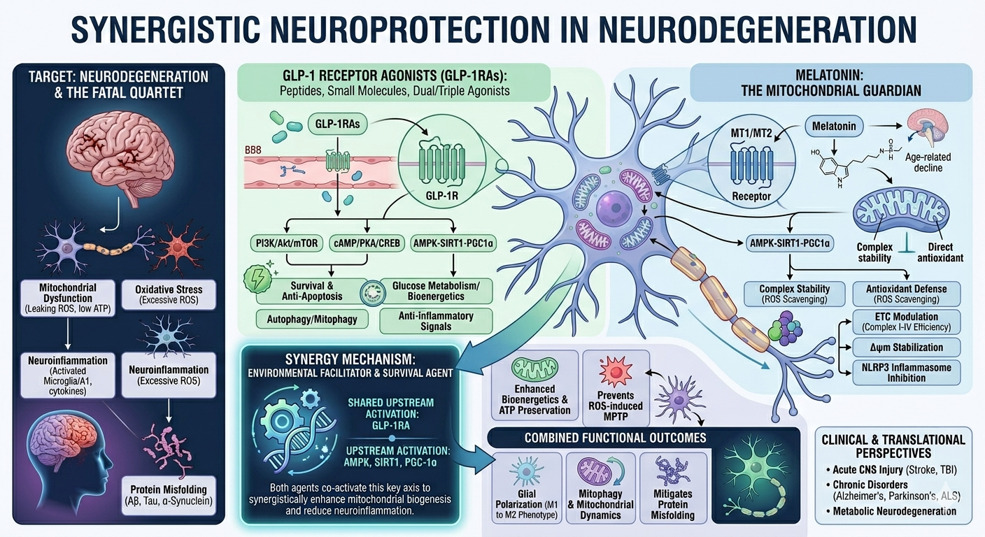
Neurodegenerative diseases share several common pathological mechanisms despite their distinct clinical manifestation. In the molecular context, four interconnected bioprocesses — mitochondrial dysfunction, oxidative stress, neuroinflammation, and protein misfold/unfold — are central drivers of neuronal damage and disease progression (Figure 1). Mitochondrial are essential organelles for neuronal homeostasis, supplying the energy required for several physiological mechanisms including neuronal plasticity, synaptic activity, and cellular survival. Consequently, mitochondrial dysfunction disrupts bioenergetics balance and neuronal function, resulting in neurodegeneration.
Changes in mitochondrial energy metabolism are linked to the pathophysiology of acute brain injury including trauma, neurotoxins, and ischemia, and even chronic NDs. The mitochondrial dysfunction-related mechanisms in neural metabolism are oxidative phosphorylation (OXPHOS), disruption of mitochondrial structure, failure in intracellular trafficking, and depletion of mitochondrial nicotinamide adenine dinucleotide (NAD); these altered cellular bioenergetics result in activation of apoptotic signaling pathways (Figure 1), ultimately affecting the balance between apoptosis and necrotic cell death [9]. Mitochondria are the principal source of intracellular reactive oxygen species (ROS), and under physiological conditions, antioxidant defenses maintain redox homeostasis. Excessive ROS generation leads to oxidative damage of DNA, proteins, and lipids while impairing mitochondrial function. This self-perpetuating cycle compromises neuronal survival, synaptic functioning and integrity, and cognitive function, thereby favoring neurodegenerative processes [10]. A recent review by Wen and colleagues [11] demonstrated that mitochondrial dysfunction and oxidative stress are interconnected mechanisms that contribute to neuronal damage and ND progression. Alterations in redox balance, mitochondrial bioenergetics, calcium homeostasis, and cell-death signaling pathways can create a self-reinforcing cycle of cellular dysfunction (Figure 1).
A landmark study published in 2006 highlighted mitochondrial dysfunction as a central and early event in the development of NDs [12]. The authors demonstrated that impaired mitochondrial bioenergetics, oxidative stress, mitochondrial DNA damage, and dysregulated cell death pathways collectively contribute to neuronal degeneration, making mitochondria as a major therapeutic element for slowing disease progression [12]. It has been documented that in AD, metabolic reprogramming in microglia has been linked to epigenetic alterations, including increased H4K12 acetylation, which enhances the expression of glycolytic genes and promotes a shift from OXPHOS toward glycolysis [13]. This metabolic adaptation may contribute to amyloid-β (Aβ) plaque accumulation while impairing astrocytic fatty acid utilization, potentially disturbing cerebral lipid homeostasis and causing disease progression [14]. Together, these findings highlight the importance of altered mitochondrial bioenergetics and lipid metabolism in the pathophysiology of NDs.
Neuroinflammation is increasingly recognized as a central player in both acute and chronic neurodegenerative disorders, often preceding the onset of clinical manifestation. A recent study reported the involvement of the EP2 receptor, CCR2 signaling, and the JAK/STAT signaling in regulating immune cell activation, cytokine production, and inflammation, thereby contributing to the progression of neuroinflammatory diseases and representing potential therapeutic candidates (Figure 1) [15]. In a recent review study, Shafi and colleagues [16] documented the relationship between activated glial cells and infiltrating immune cells during neuroinflammation and revealed the diversity of interconnected signaling networks orchestrating inflammatory responses in the central nervous system (CNS). For instance, molecules participating in the regulation of neuroinflammation included NF-κB, MAPK, and the NLRP3 inflammasome. Microglia and mast cells are innate immune cells that coordinate neuroinflammation in the CNS. While intercellular communication contributes to tissue protection and repair following injury, persistent activation promotes the release of pro-inflammatory mediators, amplifies neuroinflammation, and accelerates neural dysfunction and neurodegeneration [17].
NDs are strongly associated with the aggregation of misfolded proteins. Abnormal accumulation of specific molecules is characteristic of many diseases, including α-synuclein in PD, hyperphosphorylated tau and β-amyloid in AD, mutant huntingtin in HD, misfolded príon proteins in príon disorders [18]. Under pathological conditions, normally soluble proteins may adopt abnormal conformations and assemble into oligomers, amyloid fibrils, intracellular inclusions or extracellular plaques, impairing their physiological functions and potentially exerting direct neurotoxic effects (Figure 1). Cells normally maintain protein homeostasis through a highly coordinated quality-control mechanisms, including the ubiquitin–proteasome system, chaperone-mediated autophagy, and macroautophagy. These pathways are useful to recognize, refold, or eliminate damaged and misfolded proteins. Importantly, when protein production, folding, trafficking, or degradation becomes dysregulated, these protective functions become insufficient, resulting in accumulation and aggregation of abnormal proteins, which may progressively disrupt neuronal function. Overall, the accumulation of misfolded proteins can compromise essential cellular processes, trigger oxidative stress and mitochondrial dysfunction, impair intracellular communication, and ultimately induce neuronal death [19].
2. The GLP-1 Receptor Agonist Landscape: Pharmacology and Neuroprotective Mechanisms
Researchers transformed a gut hormone, Glucagon-like peptide-1 (GLP-1), discovered in the 1980s that stimulates insulin into groundbreaking diabetes and obesity medications by the early 2000s through the development of stable GLP-1 receptor agonists (GLP-1RAs). Because natural GLP-1 is swiftly degraded by enzymes such as dipeptidyl peptidase-4 (DPP-4) and neprilysin, pharmacologically stable GLP-1RAs were developed to bypass this pharmacokinetic limitation [1]. GLP-1RAs differ in their molecular structure and pharmacokinetic profiles, contributing to variability in their effects on glycemic control and weight loss. These differences may also produce cardiovascular benefits, as demonstrated by clinical outcome trials showing cardioprotective effects for some agents. Beyond type 2 diabetes mellitus (T2DM) management, the metabolic actions of GLP-1RAs have expanded their potential applications to prediabetes, obesity, and metabolic liver diseases [20].
Beyond their established metabolic effects in T2DM and obesity, GLP-1RAs have emerged as potential candidates for the treatment of NDs, as suggested by preclinical and early clinical evidence showing promotion of neuronal survival and counteracting pathological processes observed in PD and AD. However, the therapeutic potential of these agents in the central nervous system (CNS) may depend on their ability to cross the blood-brain barrier (BBB), a property that only a subset of currently available GLP-1RAs have demonstrated. Recently, a comprehensive synthesis of 14 studies showed preclinical evidence that several GLP-1RAs, particularly liraglutide, semaglutide, and exenatide, can cross the BBB, potentially exerting direct effects within the CNS [21]. Human studies further suggest that these agents may influence brain connectivity. In addition, GLP-1 signaling has been associated with improved metabolic homeostasis, attenuation of neuroinflammation and oxidative stress, and preservation of neuronal plasticity; these pleiotropic actions have gained interest in repurposing GLP-1RAs as potential therapeutic agents for NDs, although experimental and clinical studies are needed to ensure their efficacy and underlying mechanisms [22].
Metabolic dysfunction, oxidative stress, inflammation, mitochondrial impairment, and neurovascular damage share biological mechanisms that finally contribute to neurodegeneration, altered brain metabolism, and cerebrovascular injury. It is well-known that GLP-1RAs are expressed in glial cells, neurons, and in the neurovascular unit. Although clinical trials are still inconclusive on the role of GLP-1 signaling in neuroprotection, preclinical studies evidenced that GLP-1RAs are capable of attenuating neuroinflammation and oxidative stress while maintaining mitochondrial function and the BBB integrity (Figure 2) [23].
Importantly, a large genome-wide association study investigated why individuals respond differently to GLP-1RAs used for obesity treatment. Analysis of 27,885 treated individuals revealed genetic variants in GLP1R associated with greater weight loss, while variation in GLP1R and GIPR was linked to treatment-related nausea and vomiting, particularly among tirzepatide users [24]. Therefore, genetic differences in drug targets can influence both therapeutic efficacy and adverse effects, supporting the development of personalized approaches for obesity treatment.
2.1. Blood-Brain Barrier and Neuropharmacology of Peptides and Small Molecules
Unlike large peptides, GLP-1RAs, including exenatide, liraglutide, and semaglutide, which have structural barriers, high plasma-protein binding, and restricted entry into brain parenchyma, newly developed oral small molecules display favorable lipophilicity and low molecular weight (usually under 500 Da) that allow them to freely cross the BBB via passive diffusion. Small molecules, non-peptidyl GLP-1 and dual GLP-1/GIP receptor agonists (e.g., orforglipron, danuglipron, CT-996) represent a commercially scalable, orally bioavailable alternative to peptide-based metabolic therapies, though their clinical translation depends on target sensitivity while mitigating off-target toxicities [25]. Holler and colleagues documented modern methodologies engineered to overcome the low oral bioavailability of GLP-1RAs in metabolic disease therapy, categorizing advances into chemical absorption promoters, nano- and micro-formulations, targeted gene vectors, gut microbiome platforms while addressing safety profiles and potential off-label use.
GLP-1 signaling is intrinsically present in the brain, where preproglucagon neurons and GLP-1R on neurons and glial cells form a widespread regulatory network involved in metabolic and neurobiological functions. These receptors are mainly located in the cortex, hippocampus, and hypothalamus, and basal ganglia [26]. Although GLP-1RAs can potentially access the CNS, their BBB penetration varies substantially according to pharmacokinetic properties, molecular size, and albumin binding, with some compounds presenting limited direct brain exposure; consequently, the majority of neuroprotective effects may be a result of both central and peripheral mechanisms, including vagal and vascular pathways [27]. Notably, GLP-1 receptor activation may also strengthen BBB integrity, potentially reducing vascular dysfunction associated with aging and neurodegeneration [28]. However, whether greater BBB penetration necessarily translates into superior clinical effectiveness remains unsolved.
Recent trials have investigated whether GLP-1 receptor activation can slow AD progression and provide neuroprotective effects beyond its metabolic functions. The EVOKE and EVOKE+ phase 3 trials examined the potential disease-modifying effects of oral semaglutide in patients with early-stage symptomatic AD. Approximately 1,840 amyloid-confirmed patients were randomized to semaglutide or placebo for 156 weeks. Based on plasma biomarkers and cerebrospinal fluid analysis, semaglutide induced central effects, including possible BBB-related mechanisms, vascular protection, and attenuation of neuroinflammation [29]. Regarding liraglutide, early-phase clinical trials have supported their central effect showing maintenance of brain glucose metabolism and slower cortical decline [30]. A recent cohort study of 60,860 patients revealed that semaglutide and tirzepatide may be associated with reduced risks of dementia and stroke in people with T2DM and obesity, potentially reflecting beneficial effects on metabolic dysfunction, vascular health, neuroinflammation, long-term cognitive and survival outcomes [31]. Despite these observational findings, no direct neuroprotective or disease-modifying effect can be anticipated.
2.2. Dual and Triple Agonists: The Next Frontier in Neurodegeneration
GLP-1RAs are agents that mimic the function of GLP-1. The class includes a few compounds with specific pharmacological profiles such as liraglutide, semaglutide, exenatide, dulaglutide, lixisenatide, and the dual GIP/GLP-1 receptor agonist tirzepatide (Figure 2) [25]. Triple GLP-1R/GIPR/GCGR agonists are emerging as promising agents for the treatment of metabolic disorders, particularly T2DM, obesity, and metabolic dysfunction-associated steatohepatitis (MASH). For instance, Retatrutide has progressed to phase 3 clinical development, with ongoing trials focused on its efficacy in obesity and related conditions, while efocipegtrutide has advanced to phase 2 studies for MASH. These multi-receptor agonists may provide broader metabolic benefits than single- or dual-receptor therapies, although long-term safety and clinical efficacy remain under investigation.
Increasing evidence associates impaired insulin signaling in the pathophysiology of AD and PD, linking insulin resistance to oxidative stress, neuroinflammation, protein aggregation, and neuronal dysfunction. Preclinical studies reveal that dual GLP-1/GIP agonists can achieve greater brain exposure and BBB penetration than conventional GLP-1 receptor agonists, resulting in enhanced neuroprotection. Furthermore, triple receptor agonists provide even broader protection by simultaneously targeting multiple metabolic and neurodegenerative pathways [32]. These incretin-based therapies tend to be more effective when treatment is initiated at early stages before irreversible neuronal damage occurs. Further studies showed that GLP-1/GIP receptor agonists are capable of restoring impaired insulin signaling, glucose utilization, mitochondrial function, and neuronal energy metabolism while reducing chronic inflammation and proinflammatory cytokines; importantly, dual agonists with enhanced BBB penetration may provide stronger protection against mitochondrial dysfunction, inflammatory damage, and neuronal loss in AD and PD [2].
In an MPTP-induced mouse model of PD, the dual GLP-1/GIP receptor agonist DA3-CH produced superior neuroprotective effects than liraglutide, thereby improving motor performance, restoring tyrosine hydroxylase and dopaminergic signaling in the substantia nigra and striatum, and markedly augmenting glial cell line-derived neurotrophic factor (GDNF) expression [33]. Of significant value, both treatments reduced reactive microglia and astrocytes, supporting attenuation of neuroinflammation. Recently, Tian and colleagues [34], using a subacute MPTP-induced PD model, showed that tirzepatide showed mitochondrial neuroprotective effects in dopaminergic neurons preserving tyrosine hydroxylase expression. in the substantia nigra. The mechanistic effects of the dual agonist included improvements in mitochondrial ultrastructure and ATP production, suppression of excessive Drp1-mediated mitochondrial fission and restoration of mitophagy regulators (PINK1, Parkin, and p62). Tirzepatide treatment produced several neuroprotective and metabolic effects despite not improving cognitive and anxiety-related behaviors in a APP/PS1 mice model [35]. The treatment reduced cortical amyloid plaque burden, astrocyte activation (GFAP) and Aβ-induced neuronal apoptosis. Importantly, the dual GLP-1/GIP receptor activation enhanced brain glucose metabolism by increasing the expression. of GLUT1 and key glycolytic and pentose phosphate pathway enzymes. It also attenuated Aβ-induced mitochondrial dysfunction by regulating ROS generation, preserving mitochondrial membrane potential, and improving ATP availability in astrocytes via GLP-1R signaling.
2.3. Neuroprotective Signaling Mechanisms of GLP-1
Evidence from preclinical models suggests that GLP-1ARs may exert neuroprotective effects through multiple interconnected mechanisms. These are mediated by activation of cAMP/PKA/CREB and PI3K/Akt signaling, enhancing neurotrophic support, inhibition of tau hyperphosphorylation, inhibition of the microglial NLRP3 inflammasome, and improving mitochondrial biogenesis and autophagy (Figure 2) [36]. Notably, these effects have been associated with reduced protein aggregation, neuronal loss, and functional impairment in experimental models of AD and PD, stroke, HD, multiple sclerosis, and ALS, among other neurological conditions. Particular attention should be paid to the differences in the ability of individual GLP-1RAs to cross the BBB and induce variable effects when considering their potential use in NDs.
In T2DM, GLP-1R activation initiates cAMP-dependent signaling through PKA and EPAC, leading to CREB activation and enhanced insulin production and secretion. In parallel, PI3K/Akt signaling promotes β-cell survival and proliferation by limiting apoptosis and regulating key metabolic factors, including FoxO1 and GLUT2, thereby supporting glucose sensing and maintaining pancreatic β-cell function [37]. GLP-1RAs may have a mechanistic convergence to further protect against neurodegeneration via metabolic signaling, mitochondrial function, inflammation, and protein homeostasis control. Activation of PI3K/Akt inhibits GSK-3β and finally reduces pathological tau phosphorylation, while cAMP/PKA/CREB signaling enhances neurotrophic support through BDNF (Figure 2) [37]. These agents promote mitochondrial biogenesis and energy production, reduce microglial activation and inflammasome-driven neuroinflammation, and induce autophagy and lysosomal clearance of damaged molecules. Wong and colleagues [38] demonstrated experimentally that GLP-1 receptor activation exerts a potent systemic anti-inflammatory effect through CNS-dependent mechanisms. In mice challenged with different toll-like receptor agonists, exendin-4 significantly suppressed the increase in circulating TNF-α levels, highlighting a GLP-1R signaling-mediated neuroimmune pathway in the CNS.
2.4. GLP-1R Signaling Under Mitochondrial Stress: The Potential Role of Melatonin
Although GLP-1RAs exert multiple neuroprotective actions, their capacity to restore neuronal homeostasis may be constrained when mitochondrial damage is severe or persistent. It is well-documented that GLP-1R signaling can improve mitochondrial bioenergetics by enhancing insulin/PI3K-Akt and cAMP-dependent pathways, promoting ATP production, mitochondrial quality control, and mitophagy, while simultaneously reducing oxidative stress and neuroinflammation. However, these effects may depend on the residual functional capacity of the mitochondrial network; extensive oxidative damage, impaired electron transport, loss of membrane potential, or defective mitochondria could limit the ability of GLP-1R activation alone to fully restore mitochondrial function. In this context, melatonin (N-acetyl-5-methoxytryptamine) may provide a complementary mitochondrial-stabilizing strategy because of its distinctive ability to accumulate within mitochondria and directly regulate the organelle’s redox and bioenergetic environment. There are a vast number of studies showing in different contexts that melatonin can reduce mitochondrial ROS/RNS production, preserve mitochondrial potential, support antioxidant defenses through SIRT3/Nrf2, regulate mitochondrial dynamics involving DRP1 and OPA1, and promote PINK1/Parkin-dependent mitophagy (Figure 2). Therefore, a combined GLP-1R-melatonin approach could target complementary levels of neuronal dysfunction; while GLP-1R activation would primarily improve metabolic signaling, melatonin would provide stabilization of mitochondrial redox balance, membrane integrity, dynamics, etc. This co-participation may be relevant under conditions of sustained mitochondrial stress, in which metabolic signaling alone is insufficient to overcome accumulated mitochondrial damage. Obviously, additional experimental studies are required to determine whether this combination produces additive or synergistic neuroprotection (Figure 2).
3. Melatonin: The Mitochondrial Guardian
Melatonin has a major role in preserving the optimal function of mitochondria. It accomplishes this by defending the cell’s energy centers against oxidative stress and metabolic strain. Beyond its circadian role, it stabilizes electron transport, preserves membrane integrity, and supports bioenergetic resilience. By buffering reactive oxygen species and reinforcing mitochondrial signaling, melatonin helps maintain ATP synthesis and cellular vitality across diverse physiological challenges.
3.1. Beyond Chronobiology: Mitochondrial Redox Protection
Melatonin (N-acetyl-5-methoxytryptamine) is traditionally associated with the regulation of circadian rhythms, yet this represents only one facet of its multifaceted biological activities. Accumulating evidence positions mitochondria as a major target and potentially a site of synthesis and action for melatonin, placing this molecule at the intersection of mitochondrial redox regulation and homeostasis [4,39]. This emerging perspective holds relevance for dysfunctional cell conditions, including neurodegeneration, in which mitochondrial impairment and oxidative stress (OS) are closely intertwined.
Mitochondria occupy a paradoxical position in cellular redox biology. As the primary site of oxidative phosphorylation, they continuously generate reactive oxygen species (ROS) while simultaneously remaining highly vulnerable to ROS-mediated damage. Under physiological conditions, mitochondrial ROS contribute to signaling and adaptive responses. However, when ROS production exceeds antioxidant capacity, oxidative damage accumulates within mitochondrial DNA (mtDNA), membrane lipids, and respiratory chain proteins, compromising mitochondrial function and cellular viability [40]. In neurons, which possess exceptionally high energetic demands and rely heavily on oxidative phosphorylation, this imbalance can be particularly detrimental.
Melatonin is uniquely positioned to counteract this redox imbalance for several reasons. Its amphiphilic nature enables it to readily cross biological membranes, including the blood–brain barrier and mitochondrial membranes, allowing it to reach sites where oxidative damage is generated [4,41]. Within mitochondria, melatonin provides multilayered protection rather than acting through a single antioxidant mechanism. Melatonin can directly neutralize highly reactive species, including hydroxyl radicals, superoxide-derived species, peroxynitrite, and other reactive nitrogen species [4,42]. Moreover, the antioxidant activity of its metabolites, cyclic 3-hydroxymelatonin, N1-acetyl-N2-formyl-5-methoxykynuramine (AFMK), and N1-acetyl-5-methoxykynuramine (AMK), extends its redox-protective effects, contributing to the so-called "antioxidant cascade" [43]. Melatonin also influences the cellular antioxidant network, acting not merely as a chemical antioxidant but also as a regulator of the cellular redox environment. Besides scavenging ROS after they have formed, melatonin can strengthen endogenous defense systems by modulating superoxide dismutase (SOD), glutathione peroxidase (GPx), catalase, and glutathione-related pathways [44]. Concurrently, inhibition of pro-oxidant pathways, including inducible nitric oxide synthase (iNOS), may limit the accumulation of reactive nitrogen species and the transition from oxidative to nitrosative stress [45].
An additional layer of complexity has emerged from the observation that mitochondria may themselves contribute to melatonin synthesis [46,47]. This possibility has shifted the conceptualization of melatonin from a hormone acting primarily through extracellular or nuclear signaling toward a locally available mitochondrial defense molecule. Such a mechanism could be particularly relevant in neurons, where the constant energetic demand imposed by oxidative phosphorylation places mitochondria under persistent metabolic and oxidative pressure.
Taken together, these findings support a broader view of melatonin as a mitochondrial guardian. By combining direct radical scavenging, reinforcement of endogenous antioxidant defenses, suppression of pro-oxidant pathways, and potentially local mitochondrial synthesis, melatonin may intervene at multiple levels of the oxidative stress–mitochondrial dysfunction axis. This integrated mode of action provides a mechanistic basis for its ability to preserve mitochondrial integrity and neuronal viability under metabolic and inflammatory stress [48].
3.2. Mitochondrial Bioenergetics: Preservation of Respiratory Function and Energy Homeostasis
In addition to its antioxidant actions, melatonin directly influences mitochondrial bioenergetics. Dysfunction of the electron transport chain (ETC), particularly complexes I and III, is a common feature of aging and neurodegenerative diseases [49]. ETC impairment promotes electron leakage and ROS generation, establishing a vicious cycle that progressively damages mitochondrial components [49].
Melatonin improves mitochondrial respiration by stabilizing ETC complexes and optimizing electron flow. Experimental evidence indicates that melatonin enhances the activity of complexes I and IV, reduces electron leakage, and improves coupling efficiency between respiration and ATP synthesis [50,51]. By preserving mitochondrial membrane integrity and cardiolipin stability, melatonin maintains the structural organization of respiratory complexes required for efficient oxidative phosphorylation [52,53].
Preserving ATP production is particularly relevant in neurons, given their high, continuous metabolic demand to maintain ion gradients, membrane potential, and synaptic function. ATP depletion contributes to synaptic dysfunction, impaired axonal transport, calcium dysregulation, and ultimately neuronal death [54]. In this context, melatonin appears to protect neuronal energy metabolism by preserving mitochondrial function, including maintenance of mitochondrial membrane potential (Δψm), oxygen utilization, ETC protein integrity [55].
Melatonin also protects mitochondria against calcium overload, thereby limiting the opening of the mitochondrial permeability transition pore (mPTP), a key event that promotes mitochondrial membrane potential dissipation, ATP depletion, and the activation of cell death pathways [56]. By preserving mitochondrial integrity under conditions of calcium stress, melatonin may help maintain cellular homeostasis and prevent the progression from mitochondrial dysfunction to cell death [55].
3.3. Molecular Pathways Underlying Melatonin-Mediated Mitochondrial Protection
Melatonin engages multiple molecular pathways that contribute to mitochondrial homeostasis. These include receptor-dependent signaling through MT1 and MT2 receptors and receptor-independent actions mediated by direct interactions with mitochondrial and redox-sensitive targets [57]
The membrane receptors MT1 and MT2, both G-protein-coupled receptors, are widely expressed throughout the central nervous system, including in the hippocampus, cortex, substantia nigra, and hypothalamus [58,59]. Their activation modulates cAMP-dependent signaling, protein kinase cascades, and transcriptional programs involved in neuronal resilience, synaptic plasticity, inflammatory responses, and mitochondrial homeostasis [60,61]. Through these pathways, receptor-mediated signaling can influence the same mitochondrial processes described above, documenting an important link between melatonin signaling and the maintenance of cellular energy and redox balance.
Among these pathways, the SIRT1–PGC-1α axis is a well-characterized link between melatonin signaling and mitochondrial adaptation. SIRT1, a NAD⁺-dependent deacetylase, regulates mitochondrial homeostasis in part by activating PGC-1α, a major transcriptional regulator of mitochondrial biogenesis. Melatonin-induced SIRT1 activation has been associated with enhanced mitochondrial turnover, antioxidant defense, and mtDNA integrity [62,63,64,65,66]. Receptor-mediated signaling may act upstream of this axis, linking MT1/MT2 activation to mitochondrial regulation [67].
Not all mitochondrial effects of melatonin, however, depend on membrane receptor activation. Because of its physicochemical properties and its ability to accumulate within mitochondria, melatonin can exert receptor-independent actions through direct interactions with ROS, mitochondrial membranes, and redox-sensitive molecular targets [4,68]. These mechanisms complement receptor-mediated signaling by providing a more immediate layer of mitochondrial protection. This distinction may be particularly relevant under pathological conditions in which receptor expression or downstream signaling is impaired.
The coexistence of these two modes of action—signaling through MT1/MT2 receptors and direct mitochondrial activity—provides a mechanistic framework for understanding the broad protective profile of melatonin. Receptor-dependent pathways can regulate longer-term processes such as mitochondrial biogenesis, antioxidant gene expression, and cellular stress adaptation, whereas receptor-independent mechanisms can directly counteract oxidative damage and preserve mitochondrial function. Their convergence may therefore integrate redox protection and bioenergetic preservation with mitochondrial quality control, ultimately contributing to neuronal resilience and survival [3].
4. Convergence of GLP-1R and Melatonin Signaling on Mitochondrial Networks
Growing evidence suggests that GLP-1RAs and melatonin substantially converge on common mitochondrial regulatory networks. A potential point of convergence involves the interconnected metabolic regulators AMP-activated protein kinase (AMPK), sirtuins, and PGC-1α, which collectively coordinate cellular energy sensing, mitochondrial biogenesis, and metabolic adaptation [69].
4.1. AMPK: Energy Sensing and Mitochondrial Metabolic Adaptation
AMPK functions as a central cellular energy sensor activated in response to an increased AMP/ATP or ADP/ATP ratio, coordinating adaptive responses to metabolic and energetic stress [70]. In neurons, where energy demand is particularly high and energy reserves are limited, AMPK helps regulate glucose and lipid metabolism, mitochondrial homeostasis, and cellular survival [71,72].
GLP-1RAs may influence this metabolic network by activating GLP-1 receptors expressed in the nervous system, which are coupled primarily to Gs/cAMP-dependent signaling. GLP-1R stimulation activates downstream protein kinase pathways, including PKA and PI3K/Akt-related signaling, thereby promoting trophic and pro-survival responses and improving neuronal resistance to metabolic, oxidative, and excitotoxic stress [73,74,75]. Indeed, GLP-1R stimulation by GLP-1 or exendin-4 has been shown to protect primary cortical neurons against hypoxic injury and dopaminergic neurons against neurotoxic insults, including 6-hydroxydopamine-induced toxicity [76]. Importantly, these protective effects were abolished by pharmacological GLP-1R antagonism and were absent in neurons derived from GLP-1R knockout mice, demonstrating their dependence on functional GLP-1R signaling [76].
Beyond its canonical cAMP-dependent signaling, GLP-1R activation has also been linked to modulation of AMPK signaling in the central nervous system. Recent experimental evidence indicates that GLP-1 receptor agonists can promote CaMKK2-dependent AMPK phosphorylation in brain models of Alzheimer’s disease, contributing to metabolic and anti-inflammatory responses [77]. The relationship between GLP-1R signaling and AMPK, however, appears to be context- and cell-type-dependent, as GLP-1R activation has also been reported to suppress AMPK phosphorylation in specific hindbrain neuronal populations [78].
Melatonin, in turn, may regulate cellular metabolic adaptation through AMPK-dependent signaling. Rather than functioning as an isolated downstream target, AMPK may serve as a central metabolic hub that integrates melatonin’s energetic and redox effects with mitochondrial adaptation. Experimental evidence indicates that melatonin activates the AMPK/PGC-1α pathway, promoting mitochondrial biogenesis and restoring mitochondrial oxidative capacity under conditions of metabolic stress [79]. This pathway may be further linked to SIRT3-dependent mitochondrial regulation, as melatonin-induced activation of AMPK–PGC-1α–SIRT3 signaling has been associated with increased expression of mitochondrial biogenesis and antioxidant regulators, including NRF1, TFAM, and SOD2 [80]. AMPK also plays a key metabolic role by promoting fatty-acid utilization. Upon activation, AMPK phosphorylates and inhibits acetyl-CoA carboxylase (ACC), thereby reducing malonyl-CoA levels and the consequent inhibition of carnitine palmitoyltransferase-1 (CPT1). This facilitates the mitochondrial uptake of long-chain fatty acids and their subsequent oxidation through β-oxidation [81]. In this context, melatonin-induced AMPK phosphorylation has been associated with increased expression of genes involved in fatty-acid oxidation, including PPARα and CPT1α, suggesting that AMPK activation may contribute to the promotion of fatty-acid oxidation by melatonin [82]. AMPK also provides a mechanistic link between metabolic signaling and mitochondrial quality control by promoting autophagy through the activation of Unc-51-like kinase 1 (ULK1), a key initiator of autophagy, and by reducing mTOR-dependent inhibition of autophagy [83]. Activation of this pathway promotes the removal of dysfunctional mitochondria through mitophagy, thereby limiting the accumulation of damaged, ROS-generating organelles [84,85]. In this context, melatonin enhances AMPK activation, thereby promoting mitophagy and contributing to mitochondrial quality control [86,87].
4.2. SIRT1–PGC-1α–SIRT3: Redox–Metabolic Regulation and Mitochondrial Adaptation
SIRT1 is a NAD⁺-dependent deacetylase that links cellular metabolic and redox status to transcriptional regulation. Unlike AMPK, which primarily acts as an energy-sensing kinase, SIRT1 regulates mitochondrial adaptation through post-translational modification of metabolic transcriptional regulators, most notably PGC-1α. AMPK and SIRT1 are therefore functionally interconnected rather than organized exclusively in a linear upstream–downstream relationship, with both pathways contributing to the regulation of PGC-1α-dependent mitochondrial programs [88]. PGC-1α represents a major transcriptional convergence point between these metabolic pathways: AMPK can promote PGC-1α activity through phosphorylation, whereas SIRT1 enhances its transcriptional activity through NAD⁺-dependent deacetylation [79,89].
Preclinical evidence suggests that GLP-1 receptor activation may enhance SIRT1 activity and PGC-1α signaling, potentially helping maintain mitochondrial homeostasis under metabolic stress. In a peripheral (non-CNS) model of human skeletal muscle myotubes exposed to palmitate-induced insulin resistance, GLP-1 signaling enhanced SIRT1 activity and PGC-1α deacetylation, indicating that this link is not restricted to neurons. Consistent with this, studies in rodent and in vitro models suggest beneficial effects of GLP-1 receptor agonists on selected aspects of mitochondrial content and morphology [90,91]. Through SIRT1-dependent deacetylation, PGC-1α becomes transcriptionally more active, promoting the expression of genes involved in mitochondrial biogenesis, oxidative phosphorylation, and fatty-acid oxidation, while also contributing to antioxidant defense [89,92].
Melatonin similarly engages the SIRT1–PGC-1α pathway, although through distinct upstream mechanisms related to its regulation of cellular redox and metabolic homeostasis. Experimental evidence indicates that melatonin activates the SIRT1/PGC-1α/SIRT3 signaling axis [62,93]), promoting mitochondrial biogenesis and strengthening mitochondrial antioxidant defenses. Furthermore, melatonin-induced activation of this pathway has also been associated with increased PGC-1α activity and upregulation of NRF1 and TFAM, together with increased SIRT3 and SOD2 expression [80]. SIRT3 additionally provides a mitochondrial regulatory layer by deacetylating proteins involved in oxidative metabolism and antioxidant defense, thereby supporting mitochondrial efficiency and cellular resistance to oxidative stress [94].
The convergence of GLP-1RAs and melatonin on this regulatory network is therefore particularly relevant to mitochondrial resilience. GLP-1RAs can engage metabolic and pro-survival signaling pathways that intersect with AMPK, SIRT1, and PGC-1α, whereas melatonin can activate AMPK- and SIRT1-dependent pathways associated with PGC-1α and SIRT3-mediated mitochondrial adaptation. Although the upstream mechanisms differ, both interventions may ultimately enhance PGC-1α-dependent transcriptional programs governing mitochondrial biogenesis, oxidative metabolism, and antioxidant protection.
4.3. GLP-1RAs and Melatonin in Mitochondrial Quality Control and Inflammation
Mitochondrial homeostasis depends on the continuous balance between the generation of new organelles, the removal of dysfunctional mitochondria, and the remodeling of the mitochondrial network [95]. This balance is particularly important in neurons, in which high energetic requirements and limited regenerative capacity make the accumulation of damaged mitochondria especially detrimental [96]. GLP-1RAs and melatonin converge functionally on several interconnected components of this balance, including mitochondrial biogenesis, mitophagy and organelle dynamics, and NLRP3 inflammasome-driven inflammation [97]. Mitochondrial biogenesis is governed by transcriptional programs involving nuclear respiratory factors NRF1 and NRF2 and the mitochondrial transcription factor TFAM, which drive mitochondrial replication, transcription, oxidative phosphorylation, and antioxidant defense; this program provides a mechanism for replacing dysfunctional organelles and sustaining oxidative metabolic capacity [98,99]. Melatonin increases the expression of NRF1, TFAM, and SOD2, with consequent increases in mitochondrial biogenesis and antioxidant capacity [80]. GLP-1R activation has been reported to promote mitochondrial biogenesis and PGC-1α-associated responses in several preclinical models. However, these effects appear to be context- and tissue-dependent, and the available evidence remains heterogeneous [100].
Enhanced mitochondrial biogenesis is tightly coupled with the selective clearance of damaged organelles. Mitophagy represents a major component of mitochondrial quality control by selectively targeting dysfunctional mitochondria for autophagic degradation [96,101]. As noted in Section 4.1, AMPK-dependent activation of ULK1 provides one route into this process [84,85]. However, downstream selective recognition of damaged mitochondria principally involves the PINK1/Parkin pathway, in which loss of mitochondrial membrane potential promotes PINK1 accumulation on the outer mitochondrial membrane and recruitment and activation of Parkin, facilitating ubiquitination of mitochondrial proteins and subsequent autophagic removal [102]. By eliminating dysfunctional, ROS-generating mitochondria, mitophagy prevents the progressive accumulation of damaged organelles that would otherwise amplify oxidative and inflammatory signaling [103].
This relationship between mitochondrial biogenesis and mitophagy is particularly relevant to the proposed co-targeting model. Melatonin promotes mitochondrial turnover and the clearance of damaged mitochondria through PINK1/Parkin-dependent mitophagy under conditions of mitochondrial stress [104,105]. GLP-1 receptor signaling has also been linked to the regulation of autophagic and mitochondrial quality-control mechanisms, supporting the concept that GLP-1RAs may facilitate the removal of damaged organelles while improving metabolic resilience [106]. In diabetic and obese db/db mice, the GLP-1RA semaglutide improved cardiac function and mitochondrial morphology while increasing AMPK and ULK1 phosphorylation, mitochondrial respiratory capacity, and markers of mitophagy, including Parkin and LC3 [34]. Similarly, in AC16 human cardiomyocytes subjected to hypoxia/reoxygenation, semaglutide increased autophagic activity and modulated the PINK1/Parkin pathway, with concomitant reductions in oxidative stress and mitochondrial injury [31]. Recent evidence in mice exposed to chronic ethanol also showed that semaglutide restored mitochondrial fission/fusion balance and increased PINK1/Parkin and LC3-related markers, consistent with improved mitophagy and autophagic clearance [107].
Mitochondrial quality control also depends on the proper regulation of mitochondrial dynamics. Fusion, mediated by MFN1/MFN2 and OPA1, and fission, mediated by DRP1, allow the network to adapt to energetic demand and to segregate damaged mitochondrial regions for subsequent mitophagic removal, so that an appropriate fusion–fission balance is essential for mitochondrial homeostasis [108]. Melatonin has been shown to counteract excessive DRP1-mediated fission and to support MFN2/OPA1-dependent fusion under conditions of mitochondrial stress, thereby limiting network fragmentation [41,93]. GLP-1RAs act on the same axis: in a murine model of chronic alcohol-induced neuronal stress, semaglutide restored the fission/fusion balance in prefrontal cortex neurons [107]; in a rat model of ischemic stroke, semaglutide suppressed DRP1-dependent fission and upregulated MFN2, restoring ATP production and reducing ROS generation via the GLP-1R/FoXO1 axis [109].
A further consequence of improved mitochondrial quality is attenuated mitochondria-driven inflammatory signaling. Dysfunctional mitochondria release ROS, membrane-derived signals, and damage-associated molecular patterns that converge on inflammasome activation, promoting caspase-1-dependent processing of IL-1β and IL-18 and amplifying neuroinflammatory responses [40]. Among these inflammatory platforms, the NLRP3 inflammasome is particularly relevant to neurodegenerative pathology [110]. Its activation promotes caspase-1 maturation and subsequent processing of pro-inflammatory cytokines, including IL-1β and IL-18, thereby amplifying inflammatory responses within the central nervous system [111,112].
Both melatonin and GLP-1RAs suppress NLRP3 inflammasome activation across multiple experimental models of mitochondrial and oxidative stress injury. Melatonin attenuated NLRP3-driven neuroinflammation in MPP⁺-stimulated BV2 microglia and in an MPTP-induced mouse model of Parkinson’s disease [113] and suppressed NLRP3 assembly and IL-1β maturation in a murine model of glucolipotoxic cardiomyopathy [114]. GLP-1RAs similarly reduced NLRP3 expression in cortical and thalamic microglia in a rat model of diabetic neuropathic pain [115]and suppressed NLRP3, caspase-1, and IL-18 in a model of pressure overload-induced cardiac injury through enhanced mitophagy [116]. Critically, these effects are not restricted to direct modulation of inflammatory pathways but are also mechanistically anchored to their mitochondrial actions: by preserving membrane potential, reducing ROS output, and promoting clearance of dysfunctional organelles, both agents remove the upstream mitochondrial signals that sustain inflammasome engagement.
5. Clinical and Translational Perspectives
There is a plethora of evidence that compromised mitochondrial physiology is a major contributor to cognitive deterioration caused by a variety of insults and diseases [117,118,119]. This is predicated on the fact that neurons have extremely high requirements for oxygen and glucose, with even a modest depression in the availability of either constituent being meaningful in terms of promoting mitochondrial dyshomeostasis and neuronal injury due to an elevation in the production of toxic ROS [120]. These damaging agents indiscriminately degrade lipids, proteins, and DNA [121]. An estimated 80% of the ATP neurons produced is a result of mitochondrial oxidative phosphorylation (OXPHOS) so hypoxia is always consequential relative to energy availability and tissue recovery from injury. It is documented that neuronal mitochondria constitutively synthesize melatonin that may presumably be a critical factor in the maintenance of day-to-day brain health [47]. Moreover, mitochondrial melatonin production may be inducible under conditions where these organelles develop a highly oxidizing environment [122]. Evidence from both animals and humans shows that endogenous melatonin production diminishes with age and, as a consequence, its availability as a protective antioxidant similarly declines [123,124]. This is important since stroke (and most neurodegenerative disorders) are conditions that occur most often in the elderly [125].
Neuroinflammation is also a major feature of many neural pathologies (See Section 5 above) [126]. Mitochondria sit at the crossroads of energy production and innate immune signaling, making them essential drivers of neuroinflammation when they malfunction. During stress, injury, or aging, impaired OXPHOS increases the oxidizing environment of the mitochondria, which destroys lipids and proteins and destabilizes mitochondrial membranes. This instability promotes the release of mitochondrial DNA, cardiolipin, and other mitochondrial danger signals into the cytosol, where they activate inflammatory pathways such as cGAS-STING, NLRP3 inflammasomes, and TRL9 [127,128]. These pathways amplify microglial activation and trigger production of cytokines including IL-1β, TNF-α, and IL-6. This creates a feed-forward loop of oxidative stress and inflammation [129]. Concurrently, disrupted mitochondrial dynamics, i.e., excessive fission, impaired fusion, and defective mitophagy, lead to the accumulation of dysfunctional mitochondria that further fuel inflammatory signaling. Via these interconnected mechanisms, mitochondrial dysfunction transforms metabolic stress into persistent neuroinflammatory responses that contribute to neural pathologies.
5.1. Evidence from Acute Disorders: Stroke and TBI
In the case of stroke or acute traumatic brain injury (TBI), hypoxia typically has a sudden onset and is unpredictable as to when it will happen. As a result, some neural damage will always occur, with a therapeutic goal being to find effective therapies to mitigate the damage and reestablish more normal mitochondrial and cellular function, particularly in the penumbral area of the lesion.
During ischemic stroke, mitochondrial dysfunction is a central propeller of neuronal injury, where the abrupt loss of cerebral blood flow collapses oxidative phosphorylation, depletes ATP, and triggers excessive reactive oxygen species (ROS) generation (see Section 3 above) [130]. Impaired electron transport chain (ETC) activity leads to leakage of electrons, with the chemical reduction of ground-state oxygen causing the formation of the superoxide anion radical (O₂•⁻), the precursor of even more toxic ROS including hydrogen peroxide (H₂O₂), the hydroxyl radical (•OH), and the peroxynitrite anion (ONOO⁻) [131]. These reactions accelerate oxidative damage to lipids, proteins, and mitochondrial DNA. The bioenergetic failure promotes calcium overload, opening of the mitochondrial permeability transition pore, and activation of apoptotic pathways, all of which amplify neuronal death and blood–brain barrier disruption [132]. These processes are consistently identified as core mechanisms of post-ischemic injury.
Most experimental stroke studies have been performed using rodents and middle cerebral artery occlusion (frequently with short-term occlusion) [133]. Melatonin exerted broad protective effects against the mitochondrial derangements that occurred in these stroke models, which translated into morphological and molecular neuroprotection as well as improvements in neuroplasticity and neurocognitive outcomes. The neuronal and glial preservation was most frequently attributed in major part to direct radical scavenging and indirect stimulation of antioxidative enzymes in addition to limiting glutamate excitotoxicity [134]. As a potent antioxidant and free radical scavenger, melatonin directly reduces ROS levels and limits lipid peroxidation, thereby preserving mitochondrial membrane integrity [135]. It also stabilizes electron transport chain function, maintains ATP production, and prevents mitochondrial depolarization. In experimental stroke models, melatonin reduces infarct volume, edema, and neuronal apoptosis while supporting mitochondrial homeostasis and improving functional recovery [136]. Its anti-inflammatory actions further mitigate microglial activation and cytokine driven injury, complementing its mitochondrial protection. Clinical observations also link melatonin levels with oxidative stress severity in stroke patients, suggesting translational relevance.
Mitochondrial dysfunction clearly represents a pivotal pathological event in stroke, and melatonin’s multifaceted actions, i.e., antioxidant, anti-apoptotic, anti-inflammatory, and mitochondrial stabilizing, etc., position it as a promising neuroprotective candidate for limiting ischemic brain damage. Examination of clinical literature shows that melatonin has been evaluated, in a limited manner, as a neuroprotective adjunct against ischemic/reperfusion injury in humans. The earliest studies in this series were initiated because of the preclinical data showing reduced oxidative stress, preserved blood–brain barrier integrity, and smaller infarct volumes with the use of several different antioxidants [137]. These early studies were performed before mitochondria were seriously considered as site of action of melatonin in protecting against ischemia/reperfusion injury. The studies did reveal that melatonin had a high safety profile and tolerability in humans. Later investigations centralized mitochondria more directly as a likely site at which melatonin acts to reduce stroke damage and it has been used either as a standalone treatment or given in conjunction with other potential protective agents [138]. In a double-blind randomized controlled study in which patients received 6 mg/day for three days before and after carotid endarterectomy, a process that produces transient ischemic/reperfusion injury, the authors reported that the treatment lowered circulating levels of inflammatory cytokines suggesting that melatonin probably limited neuroinflammation which occurred as a result of the interrupt neural blood supply; this report was discussed in the review by Zhang and colleagues [134], but it was not uncovered in a search of PubMed. In a pilot study [139], melatonin was used in patients beginning 24 hours after acute stroke onset (20 mg daily for 5 days). At 30- and 90-days post treatment, the subjects were examined using the Modified Rankin Score (mRS), which evaluates overall disability rather than specific neurological or cognitive deficiencies. Since the mRS improved in the patients who used melatonin, the authors suggested that the indoleamine had beneficial effects in limiting neural disability.
Despite the large number of studies documenting the high efficacy of melatonin in reducing neurological pathophysiology resulting from experimental stroke, its use in humans for this purpose is obviously scant [140]. As in animals, melatonin use in human stroke would only serve in preserving mitochondrial function and bioenergetic stability in the penumbral region that surrounds the ischemic core. In animal studies melatonin was typically given in advance of stroke induction. This is impractical for real-time human investigations, since the timing of a stroke is unpredictable and, additionally, patients often do not arrive in the emergency unit within the timeframe of the therapeutic window [141]. Brain ischemia/reperfusion issues, caused either by interrupted brain blood supply or cardiac arrest, predominantly affects the elderly. Epidemiological data indicate that over 70% of ischemic strokes occur in individuals aged 65 and above, and older patients are more likely to experience worse functional recovery and higher mortality rates [142]. For experimental investigations, the use of young animals was the norm. This leaves clinicians with little information relative to dose, etc., when melatonin might be considered even as a post stroke treatment in elderly patients. While melatonin remains a promising therapeutic candidate for human stroke, larger, well controlled Phase 3 clinical trials are still needed to clarify optimal dosing, timing, safety, and whether melatonin can meaningfully improve functional outcomes in stroke patients.
Traumatic brain injury (TBI; also known as closed head injury or concussion injury), is a major source of disability worldwide and it contributes to neural damage which involves molecular perturbations much like those that lead to pathophysiological impairments propagated during ischemic stroke [143]. In addition to head injuries, these lesions often follow crushing injuries of the spinal cord. The magnitude of the pathological changes that follow TBI vary widely depending on the severity of the insult and can involve both the gray and white matter [144,145]. The interconnected molecular pathways related to TBI can amplify neuronal and glial damage long after the initial mechanical insult.
As with stroke, central to the damage caused by TBI is obviously associated with mitochondrial disturbances [146]. Similar to other conditions that disrupt neurophysiology and cognitive functions, the changes in mitochondria lead to excessive ROS generation, compromised ATP synthesis by interference with OXPHOS, and calcium overload [147]. Also contributing to neural pathology of concussion injury is the neuroinflammatory response initiated by activation of microglia, astrocytes, and pericytes [148]. These signaling events sustain a self-amplifying inflammatory loop that leads to BBB damage and the infiltration of immune cells [149]. Specific innate immune pathways such as TLR4 signaling are quickly upregulated which advances MMP-9 activity, interfering with synaptic communication, and leading to impairments in cognition and memory. Additionally, the cGAS-STING pathway, a released DNA sensing innate immune process, is upregulated which contributes to the neural inflammatory response [150].
Clinical data relative to the role of melatonin in TBI is scarce and the reported results are variable but, nevertheless, the findings are encouraging. Interestingly, CSF concentrations of melatonin are reportedly elevated in humans following TBI. A possible explanation for this is that the increased neural oxidative stress that follows traumatic brain damage leads to a compensatory rise in the endogenous antioxidant melatonin production in an attempt to reduce the pending neural damage. In this scheme, melatonin is inducible during elevated ROS-mediated cellular damage [151], The idea that local melatonin synthesis is inducible during oxidative stress is not novel and is certainly consistent with melatonin production in plants [152,153]. In both children and adults, melatonin has been successfully used to ameliorate post concussive neurological symptoms. In these studies, melatonin was given at doses of 2, 3, or 10 mg per day, and for each age group sleep quality and duration were improved [154,155]. Additional outcome measures on adults revealed recovered neural functional connectivity as evidence by MRI analysis [155]. In general, the quality of life was also better in the melatonin-treated individuals. Considering the huge activation of pathological pathways, e.g., ROS generation, that can accompany TBI, the authors suggest the use of larger doses of melatonin in subsequent human studies. Allometric calculations based on animal studies predict a dose range of 1.0-1.5 mg per kg body weight would be pragmatic [156].
5.2. Evidence from Chronic Disorders: Alzheimer’s and Parkinson’s Diseases
Alzheimer’s disease (AD) is an insidious age-related neurodegenerative condition the begins with initially undetectable subtle molecular changes, e.g., impaired clearance of extracellular amyloid β (Aβ) peptides, early synaptic dysfunction, and rising oxidative stress due to mitochondrial damage that silently disrupts vulnerable brain physiology often beginning in the hippocampus [157]. As Aβ plaques accumulate, microglia and astrocytes become chronically activated, promoting inflammation and releasing cytokines which stimulate ROS formation that further injures neurons. Intraneuronal tau proteins become hyperphosphorylated, forming neurofibrillary tangles that disrupt axonal transport and hasten neuronal death. Eventually, these interacting processes spread through cortical networks, causing progressive memory loss, executive dysfunction, language impairment, and ultimately widespread cognitive decline that typifies the final stages of AD.
Mitochondrial dysfunction in neurons precedes the appearance of overt AD disease. In affected neurons, mitochondria show impaired OXOHOS, reduced Complex I and IV activities, and diminished ATP generation; these changes leave neurons energetically fragile [158,159]. The subsequent excessive production of ROS further damage lipids, proteins, and mitochondrial DNA, which self amplifies the degree of oxidative stress. Aβ and hyperphosphorylated tau directly disrupt mitochondrial dynamics, blocking axonal transport and fragmenting the mitochondrial network. These defects impair synaptic function, weaken calcium buffering, and promote neuronal vulnerability, making mitochondrial failure a core pathological feature of Alzheimer’s progression.
The rise in mitochondrial oxidative stress and neuroinflammation forms a tightly interwoven pathological axis in AD, driving progressive synaptic failure and neuronal loss through a cascade of molecular disruptions that amplify one another. In early AD, mitochondrial dysfunction, particularly impaired complex I and IV activity reduce ATP production and elevate superoxide and hydrogen peroxide levels, overwhelming antioxidant defenses [160]. Excess ROS oxidize lipids, generating toxic aldehydes, which form adducts with tau, APP (amyloid processing proteins) related enzymes, and mitochondrial proteins, altering their structure and function [161]. Oxidized mitochondrial DNA and cardiolipin leak into the cytosol, activating cGAS–STING and NLRP3 inflammasomes, which drive IL 1β and IL 18 maturation. These cytokines, along with ROS induced activation of NF-κB, shift microglia from homeostatic states toward pro inflammatory M1 phenotype [162]. Moreover, oxidative modifications of BACE1 and γ secretase enhance amyloidogenic Aβ42 production; Aβ oligomers in turn localize to mitochondria, inhibit cytochrome c oxidase, and promote further ROS generation, creating a vicious feed forward loop. ROS also promote tau hyperphosphorylation by activating kinases such as GSK 3β and CDK5, while inflammatory mediators impair phosphatases, which stabilize pathogenic tau species that disrupt axonal transport and mitochondrial trafficking [163]. Astrocytes contribute additional inflammatory pressure through JAK–STAT3 activation and reduced glutamate uptake, heightening excitotoxic stress that further burdens neuronal mitochondria. Over time, sustained oxidative and inflammatory signaling allows dysfunctional mitochondria and protein aggregates to accumulate [160]. The convergence of these molecular events transforms oxidative stress and neuroinflammation from secondary responses into primary impellers of neurodegeneration, linking the intertwined neuronal networks into a self-perpetuating cycle of metabolic failure, immune activation, and structural breakdown that progressively erodes cognitive function.
In experimental disease models, melatonin inhibits key molecular disruptors related to AD by targeting oxidative stress, amyloid and tau pathology, mitochondrial dysfunction, and impaired neurogenesis [164]. As a potent antioxidant, melatonin directly scavenges reactive oxygen and nitrogen species and upregulates endogenous antioxidant enzymes, reducing the oxidizing environment that accelerates amyloid β (Aβ) aggregation and neuronal injury. Due to its multiple actions in reducing oxidative damage, melatonin functions as an antioxidant force multiplier in resisting molecular damage. Published evidence documents that melatonin counteracts multiple pathological processes in AD, including the oxidative imbalance and protein misfolding. Melatonin also interferes with Aβ generation by modulating secretase activities and preventing the conformational changes that promote plaque formation [165]. In addition to suppressing Aβ accumulation, melatonin stabilizes microtubules and reduces tau hyperphosphorylation, thereby limiting neurofibrillary tangle formation. Mitochondrial protection is also a major mechanism: melatonin preserves membrane potential, reduces mitochondrial ROS production, and prevents permeability transition–driven apoptosis [3,166]. Finally, melatonin supports impaired adult neurogenesis, a significant contributor to cognitive decline by promoting precursor cell proliferation, migration, and differentiation. Through this combination of antioxidant, anti-amyloid, anti tau, mitochondrial, and pro neurogenic actions, melatonin exerts broad molecular inhibition of AD related pathology [167]. The ameliorative actions of melatonin in AD likely involve both receptor-mediated signaling pathways as well as actions that may not rely on receptors; melatonin receptors are widely distributed in the brain.
Several small but informative translational human studies focused on modulating AD–related symptoms and biomarkers with melatonin have been published. In the trial reports, the primary endpoints typically have been sleep-regulation, circadian stabilization, and early cognitive or biomarker behavioral changes, reflecting melatonin’s potential mechanistic relevance to AD pathology [168,169]. The most recent translational studies have involved preclinical to clinical bridging by examining whether melatonin can influence AD related biomarkers in older adults with or without mild cognitive impairment. A scheduled NIH funded trial is using 5 mg melatonin nightly for nine months to evaluate effects on cognitive performance and blood-based AD proteins, while also tracking circadian activity via actigraphy (ClinicalTrials.gov: ID NCT03954899). This study is organized as a translational design that links mechanistic hypotheses, i.e., oxidative stress reduction, amyloid modulation, and circadian restoration to measurable human outcomes. It is the opinion of some scientists as well as the present authors that the 5 mg melatonin dose is far too low and 9 months is too short a duration to provide any serious evidence that melatonin has clinically beneficial effects against AD [156,170]. This precaution has been repeatedly stressed by the group that has provided some of the best clinical evidence that melatonin, given at appropriate doses, would be of value as a countermeasure to AD progression [156,168]. The results of the studies published to date show that melatonin is safe, biologically plausible as a treatment, and mechanistically aligned with AD pathology. Hopefully, more optimally designed trials will soon be forthcoming to establish the role of melatonin, if any, in managing AD.
Parkinson’s disease (PD) is a progressive neurological disorder which becomes overtly apparent when the number of dopamine producing neurons in the mesencephalic substantia nigra pars compacta become severely depleted [171]. Dopamine is essential extrapyramidal neurotransmitter that functions in coordinating smooth, controlled muscle movements, with its loss causing disabling motor symptoms such as tremors, muscle rigidity, slowed movement, and balance difficulties [172]. While PD most often appears in older adults, it less commonly occurs in people earlier in life. The condition advances slowly and is often undetected, and symptoms because of genetic influences vary widely from person to person. While there is currently no cure, a combination of medications, lifestyle therapies, and in some cases including surgical lesioning can significantly improve quality of life and help individuals to maintain independence.
As with AD, mitochondrial dysfunction is a central feature of PD, which determines many of the molecular changes that lead to dopaminergic neuron loss in the substantia nigra. A primary defect is reduced Complex I activity in the ETC thereby limiting ATP production and increasing electron leak, generating excessive ROS that randomly damage lipids, proteins, and mitochondrial DNA [173]. The resulting oxidative stress disrupts mitochondrial dynamics, causing mitochondrial fragmentation, impaired fusion–fission balance, and defective axonal transport. Mutations in PINK1 and Parkin further compromise mitophagy, allowing damaged mitochondria to accumulate and amplify cellular stress [174]. Additionally, α synuclein, a protein located in neurons, misfolds and interferes with mitochondrial membranes, inhibits respiratory enzymes, and induces calcium overload which influences opening of the mPTP, triggering apoptotic pathways [175]. Neuroinflammation is also upregulated resulting in the release of cytokines and nitric oxide derivatives that further impair mitochondrial respiration. Collectively, these interconnected molecular disturbances create an energy deficient, pro-oxidant environment that accelerates neuronal degeneration and hastens the progression of PD.
There are several experimental models of PD that are used to test the ability of drugs to modify the progression of this disease [176]. In these models, melatonin overcomes mitochondrial deficits by coordinating a multi-tiered defense system that stabilizes mitochondrial membrane potential (∆Ψm) and prevents oxidative destruction of dopaminergic neurons [177]. In the paraquat PD model, the toxin exposure destabilizes ∆Ψm by damaging membrane integrity, disturbing ion transport, and damaging respiratory chain activity, ultimately accelerating the loss of tyrosine hydroxylase–positive neurons in the substantia nigra [178]. Melatonin reverses these changes by activating the PI3K/Akt/Nrf2 antioxidant axis, promoting the upregulation of HO-1 and NQO1 to neutralize excess ROS while concurrently enhancing expression of mitochondrial complex I and II subunits NDUFS3 and SDHA, which results in the restoration of ETC efficiency and ∆Ψm homeostasis [179]. Additionally, melatonin also recruits SIRT1 as an upstream regulator of the PDK1/Akt pathway. Silencing SIRT1 abolishes this signaling, documenting its essential role in melatonin-mediated cytoprotection. Melatonin further strengthens mitochondrial resilience by upregulating SIRT3 and PGC 1α, a change that boosts SOD2 activity, promotes mitochondrial biogenesis, and maintains the function of mtDNA-encoded complexes I, III, and IV [122]. By integrating antioxidant signaling and ensuring mitochondrial quality control, melatonin contributes to a network-level protective framework that stabilizes ∆Ψm and shields dopaminergic neurons from PD-related degeneration. These findings describe the actions of administered melatonin in all experimental models of PD. There are currently many novel melatonin delivery platforms that will likely increase the efficacy of this drug in altering the course of PD and other neurodegenerative disorders [180]. Finally, the circadian synchronizing actions of melatonin in attenuating the severity of neurological disorders should not be overlooked.
5.3. Metabolic Neurodegeneration: T2DM-Associated Cognitive Decline
Type 2 diabetes mellitus (T2DM) provides a particularly relevant context for investigating GLP-1 receptor agonists (GLP-1RAs) and melatonin because metabolic dysfunction intersects with cerebrovascular injury, neuroinflammation, and impaired neuronal energy metabolism. Diabetes-associated cognitive impairment encompasses deficits in memory, processing speed, and executive function and increases susceptibility to both Alzheimer’s disease and vascular dementia. It is not, however, synonymous with Alzheimer’s pathology. Hyperglycemia, insulin resistance, vascular disease, and hypoglycemia contribute through partly distinct mechanisms; consequently, improved glycemic control represents an important therapeutic opportunity, although it may not by itself reverse established cognitive impairment [181,182,183].
The mechanistic rationale extends beyond circulating glucose. Impaired insulin signaling can compromise neuronal stress adaptation and synaptic function, while sustained metabolic stress promotes oxidative injury, inflammatory signaling, and disturbed mitochondrial quality control. Experimental literature links these abnormalities to impaired hippocampal function and altered amyloid and tau processing. GLP-1RAs may address peripheral metabolic and vascular risk while influencing neuronal signaling; melatonin offers a complementary rationale involving redox protection, mitochondrial preservation, and circadian regulation. These overlapping actions provide a strong rationale for further investigation of combination approaches, particularly in metabolically enriched populations [181,182].
The strongest large-scale randomized evidence comes from the exploratory cognitive analysis of REWIND [184]. Among 8,828 participants with baseline and follow-up cognitive assessments, dulaglutide was evaluated over a median of 5.4 years using the Montreal Cognitive Assessment and Digit Symbol Substitution Test. The initial comparison did not show a statistically significant reduction in substantive cognitive impairment (hazard ratio [HR], 0.93; 95% confidence interval [CI], 0.85–1.02). After post hoc adjustment for baseline cognitive scores, the HR was 0.86 (95% CI, 0.79–0.95), suggesting a modest protective signal. This distinction is important: REWIND provides encouraging support for further investigation, although additional dedicated trials are needed before dulaglutide can be considered a dementia-preventive treatment [184].
Complementary evidence emerged from pooled LEADER, SUSTAIN-6, and PIONEER 6 data involving 15,820 participants [182]. Dementia was reported in 15 GLP-1RA-treated participants and 32 placebo recipients during a median of 3.61 years, corresponding to an HR of 0.47 (95% CI, 0.25–0.86). A parallel Danish cohort of 120,054 patients showed lower dementia incidence with increasing GLP-1RA exposure. Although dementia was not a prespecified endpoint in the trials and was identified through adverse-event reporting, with only 47 events, these findings generate a compelling hypothesis that merits testing in dedicated dementia-prevention trials [182].
Smaller mechanistic trials provide more direct cognitive assessments. Cheng and colleagues randomized 36 metformin-treated patients to liraglutide, dapagliflozin, or acarbose for 16 weeks [185]. Liraglutide improved delayed memory, attention, and executive function and enhanced odor-induced hippocampal activation. The study provides an important early link between cognitive changes and functional imaging. Its small sample, open-label design, and short duration indicate the need for larger, longer-term cognition-focused trials, while also supporting the promise of GLP-1RA-based approaches [185].
Large observational studies have strengthened interest in this area. Wang and colleagues emulated seven target trials within a population of 1,094,761 eligible patients with T2DM [186]. Semaglutide was associated with fewer first-time Alzheimer’s diagnoses, with HRs ranging from 0.33 versus insulin to 0.59 versus other GLP-1RAs. A subsequent analysis involving 1,710,995 eligible patients extended the association to Alzheimer’s disease-related dementias [187]. Although diagnostic coding, treatment selection, disease severity, healthcare access, and residual confounding require consideration, these large-scale findings provide substantial support for prospective evaluation of whether GLP-1RAs can modify clinically meaningful dementia outcomes.
Active-comparator findings add valuable nuance. In a UK cohort of adults aged ≥60 years, GLP-1RA initiation was not associated with lower dementia incidence than DPP-4 inhibitor initiation in the intention-to-treat analysis (HR, 0.95; 95% CI, 0.87–1.04), although continuous-use analysis suggested an association with reduced risk [183]. Dementia incidence was also comparable between GLP-1RA and SGLT2 inhibitor initiators. These findings highlight the importance of comparator selection, treatment persistence, and exposure definitions, and they reinforce the value of designing trials capable of distinguishing class-level effects from treatment-specific effects [183].
Melatonin’s diabetes-related neuroprotective evidence remains predominantly experimental but is biologically coherent and increasingly supportive. Studies summarized in the mechanistic literature report improved learning and memory alongside reduced hippocampal oxidative stress, inflammatory mediator expression, and insulin-signaling abnormalities [188,189,190,191]. Relevant models include high-fat feeding, combined high-fat diet/streptozotocin exposure, and experimentally induced brain insulin resistance. These models reproduce complementary components of metabolic disease, and their findings collectively support melatonin’s potential to protect hippocampal function under metabolic stress. Intracerebral streptozotocin models of Alzheimer-like dysfunction should be interpreted separately from systemic diabetes models, but they may provide additional insight into Alzheimer-relevant pathways. Overall, the experimental findings provide strong biological plausibility and a clear basis for carefully designed translational studies [188,189,190,191].
Human findings are promising and warrant further study. Zhang and colleagues reported an association between lower serum melatonin and mild cognitive impairment in T2DM, providing an initial signal that endogenous melatonin status may be relevant to cognitive health [192]. More recently, Pervaiz and colleagues studied 154 patients receiving 5 mg nightly melatonin or placebo for 12 weeks and reported improved cognitive scores [193]. Although the quasi-experimental design, allocation terminology, statistical reporting, and limited circadian characterization indicate that independent replication is needed, the results provide an encouraging preliminary clinical signal and support larger randomized trials [192,193].
Metabolic safety is an important consideration when evaluating co-administration [188,194]. Although a meta-analysis reported improvements in selected glycemic measures, a randomized crossover trial in 17 men with T2DM found that 10 mg nightly melatonin for three months reduced clamp-measured insulin sensitivity by approximately 12% [194,195]. This finding underscores the value of optimizing dose and timing rather than assuming that greater melatonin exposure will necessarily produce greater benefit [188,194]. Future combination trials should therefore incorporate dose and timing studies, cognitive and functional endpoints, sleep assessment, and glycemic monitoring. The current evidence supports investigating GLP-1RA–melatonin combinations in metabolically enriched populations under appropriately monitored conditions [190].
5.4. Potential of Combined Therapies
Naturally occurring melatonin and synthetic GLP-1 receptor agonists (GLP-1RAs) reduce the likelihood of stroke or aid in recovery from neural ischemia and hypoxia using overlapping mechanisms. Melatonin and its metabolites possess multiple means to diminish oxidative/nitrosative stress, thereby protecting the blood-brain barrier (BBB) from damage and reducing atherosclerotic vascular lesions that contribute to stroke [196,197]. Additionally, melatonin prevents opening of the mitochondrial permeability transition pore (mPTP), improves the efficiency of the electron transport chain (ETC), which ensures sufficient ATP production, and limits NMDA overactivation by reducing the glutamate surge normally associated with stroke [197]. GLP-1RAs also employ multiple mechanisms to lower stroke incidence and assist in recovery of damaged cells, especially in the penumbra [198]. These drugs stabilize the BBB, reduce blood pressure and excitotoxicity, quell microglial activation and cytokine release, and lessen cellular loss due to apoptosis [199,200].
The proven efficacies of melatonin and GLP-1RAs in reducing neural damage in a host of experimental conditions, along with their high therapeutic safety profiles, should encourage the use of these multifunctional molecules in clinical trials as protection against brain impairment due to stroke, trauma, and other disease processes [3,156].
With mechanisms similar to those of melatonin, GLP 1RAs have demonstrated usefulness as an AD therapy [201]. Like melatonin, experimental findings routinely confirm that GlP-1RAs target oxidative stress, metabolic and inflammatory pathways shared by AD and type 2 diabetes. AD has been referred to as type 3 diabetes since the neurons exhibit insulin resistance. Synthetic GLP-1RAs limit Aβ accumulation, decrease tau hyperphosphorylation, improve synaptic plasticity, and enhance neuronal survival, suggesting wide neuroprotective effects. These benefits appear to stem from improved insulin signaling in the brain, reduced oxidative stress, and mitigation of chronic neuroinflammation, all of which are central to AD pathogenesis and similar to the actions of melatonin [202]. Also, like melatonin, GLP-1RAs rectify mitochondrial dysfunction, which enhances the rationale for repurposing GLP-1RAs for neurodegeneration.
Animal studies have provided robust evidence of cognitive improvement in AD models, and the initial clinical trials support the potential utility of GLP-1RAs as a therapy for this debilitating neurodegenerative condition [203]. Considering the similar actions of naturally occurring melatonin and synthetic GLP-1RAs in relieving the burden of AD, trials that utilize these agents in combination may prove beneficial and should be considered as a novel approach for arresting the progression of AD. Moreover, in consideration of their overlapping actions in cardiovascular disease, combined melatonin plus GLP-1RAs may also be favorable for these neurological disorders [3,204,205]. Finally, the severity of the side effects, e.g., the gastrointestinal disturbances, of GLP-1RAs would likely be attenuated if used with melatonin.
The neuroprotective actions of GLP-1 RAs also have been observed in animal PD models in reference to the preservation of the dopaminergic neurons and partial maintenance of normal motor function. Again, these actions are reminiscent of those caused by melatonin in the same PD models [169]. Thus, in a Phase II clinical trial where exenatide and liraglutide were tested, movement disorders were significantly normalized. Overall, however, the outcomes of clinical trials that examined PD patients after GLP-1RAs treatment have yielded mixed results, and outcomes varied according to the specific GLP-1RA tested [201]. For example, in a Phase 2 trial, exenatide reportedly improved motor function as assessed using the Movement Disorder Society Unified Parkinson’s Disease Rating Scale Part III (MDS UPDRS Part III) with the benefits persisting for the 12-week washout period. In a Phase 3 trial using the same drug, however, this outcome was not duplicated, with no improvement over that of placebo-treated subjects [206]. In a Phase 2 trial, lixisenatide, a GLP-1RA which is known to cross the BBB, retarded motor progression in early PD. Despite the mixed results, the findings suggest that GLP-1RAs may have a biological rationale for treating PD. To help standardize outcomes, subsequent trials should carefully consider metabolic status, especially insulin resistance, and prioritize drugs known to penetrate the BBB [182,207]. As with the other neurological disorders discussed, there is some justification for examining the potential benefits of combined GLP-1RAs and melatonin in subsequent clinical trials with special emphasis on patient selection, clearly defined biomarkers, and the inclusion of GLP-1RAs that have proven efficacy in entering the brain from the circulation.
If orally administered melatonin and GLP-1RAs in combination are to be used as safeguards against destructive processes or disorders of the CNS, it is essential that the therapeutic agents access the neural parenchyma from the circulation. This requires transfer through the BBB. A defining pharmacokinetic property of melatonin is the ease with which it crosses the BBB. The features that allow this to occur include its small size, it is only loosely bound to protein in the blood, and its lipophilicity, so it likely diffuses into the brain primarily by passive diffusion [208]. GLP-1RAs may cross the BBB, but the degree to which they do so depends on the specific drug [209]. They may enter by passive diffusion, utilizing specific transport systems, or via the circumventricular organs, which lack a BBB. The newer GLP-1RAs are being engineered to improve their uptake by the brain. Both melatonin and GLP-1RAs are available as intranasal formulations; once administered via this route, they are absorbed by the olfactory nerves and transferred to the brain, thereby circumventing the BBB and likely improving their efficacy.
6. Conclusions
Mitochondrial dysfunction and neuroinflammation provide a unifying framework for understanding the functions of GLP-1RAs and melatonin beyond conventional metabolic and chronobiological roles. Experimental findings demonstrate that GLP-1RAs support survival and metabolic adaptation, while melatonin preserves redox balance, bioenergetics, and resilience [2,4,37,153]. Their convergence on AMPK, sirtuin–PGC-1α, and mitochondrial quality-control networks provides a compelling rationale for complementary intervention (Figure 3).
The proposed mitochondrial-support framework positions melatonin as a contributor to the cellular environment in which GLP-1RA-mediated responses may operate. Experimental findings and emerging clinical signals, particularly in metabolically vulnerable populations, support further development [182,184,185]. Differences among clinical outcomes offer opportunities to refine selection, exposure, timing, and assessment.
Studies comparing the agents individually and together can determine how complementary mechanisms translate into benefit. GLP-1RAs and melatonin represent a promising research direction for preserving mitochondrial function, attenuating neuroinflammation, and supporting cognitive and functional recovery across acute injury and chronic disease.
Author Contributions
Conceptualization, W.M.; methodology, W.M.; software, R.S.; validation, W.M., R.S., R.J.R..; data curation, R.S..; writing—original draft preparation, W.M., R.S., V.A.S., L.G.A.C., C.G., S.C., W.B., R.J.R.; writing—review and editing, W.M., R.S., V.A.S., L.G.A.C., C.G., S.C., W.B., R.J.R; visualization, W.M.; supervision, W.M.; project administration, W.M., R.S.; funding acquisition, W.M. All authors have read and agreed to the published version of the manuscript.
Funding
This work was supported by grants from the National Agency for the Promotion of Research, Technological Development and Innovation (ANPCyT FONCyT, Argentina; Grant PICT 2000 Serie A 4000), the National Scientific and Technical Research Council (PIP 0985 CONICET), and the Research and Technology Council of Cuyo University (SIIP, Mendoza, Argentina).
Institutional Review Board Statement
Not applicable. This review reports no newly conducted human or animal research.
Informed Consent Statement
Not applicable.
Data Availability Statement
No new data were created or analyzed in this study. Data sharing is not applicable to this article.
Conflicts of Interest
The authors declare no conflicts of interest.
Abbreviations
The following abbreviations are used in this manuscript:
| Δψm | Mitochondrial membrane potential |
| 6-OHDA | 6-Hydroxydopamine |
| Aβ | Amyloid-β |
| Aβ42 | Amyloid-β peptide comprising 42 amino acids |
| AC16 | Human ventricular cardiomyocyte cell line designation |
| ACC | Acetyl-CoA carboxylase |
| AD | Alzheimer’s disease |
| ADP | Adenosine diphosphate |
| AFMK | N1-acetyl-N2-formyl-5-methoxykynuramine |
| Akt | Protein kinase B |
| ALS | Amyotrophic lateral sclerosis |
| AMK | N1-acetyl-5-methoxykynuramine |
| AMP | Adenosine monophosphate |
| AMPK | AMP-activated protein kinase |
| APP | Amyloid precursor protein |
| APP/PS1 | Amyloid precursor protein/presenilin 1 transgenic model |
| ATP | Adenosine triphosphate |
| BACE1 | β-Site amyloid precursor protein-cleaving enzyme 1 |
| BBB | Blood–brain barrier |
| BDNF | Brain-derived neurotrophic factor |
| BV2 | Murine microglial cell line designation |
| cAMP | Cyclic adenosine monophosphate |
| CaMKK2 | Calcium/calmodulin-dependent protein kinase kinase 2 |
| CCR2 | C-C chemokine receptor 2 |
| CDK5 | Cyclin-dependent kinase 5 |
| cGAS | Cyclic GMP–AMP synthase |
| CMA | Chaperone-mediated autophagy |
| CNS | Central nervous system |
| CPT1 | Carnitine palmitoyltransferase 1 |
| CPT1α | Carnitine palmitoyltransferase 1A, liver isoform |
| CREB | cAMP response element-binding protein |
| CSF | Cerebrospinal fluid |
| DA3-CH | Experimental dual GLP-1/GIP receptor agonist designation |
| db/db | Leptin receptor-deficient mouse model designation |
| DNA | Deoxyribonucleic acid |
| DPP-4 | Dipeptidyl peptidase-4 |
| DRP1 | Dynamin-related protein 1 |
| EP2 / EP2R | Prostaglandin E2 receptor 2 |
| EPAC | Exchange protein directly activated by cAMP |
| ETC | Electron transport chain |
| FA | Friedreich ataxia |
| FoxO1 | Forkhead box O1 |
| GCGR | Glucagon receptor |
| GDNF | Glial cell line-derived neurotrophic factor |
| GFAP | Glial fibrillary acidic protein |
| GIP | Glucose-dependent insulinotropic polypeptide |
| GIPR | Glucose-dependent insulinotropic polypeptide receptor |
| GLP-1 | Glucagon-like peptide-1 |
| GLP-1R | Glucagon-like peptide-1 receptor |
| GLP-1RAs | Glucagon-like peptide-1 receptor agonists |
| GLUT1 | Glucose transporter 1 |
| GLUT2 | Glucose transporter 2 |
| GPCR | G-protein-coupled receptor |
| GPx | Glutathione peroxidase |
| Gs | Stimulatory heterotrimeric G protein |
| GSK-3β | Glycogen synthase kinase-3β |
| H₂O₂ | Hydrogen peroxide |
| H4K12 | Histone H4 lysine 12 |
| HD | Huntington’s disease |
| HO-1 | Heme oxygenase 1 |
| IL-1β | Interleukin-1β |
| IL-6 | Interleukin-6 |
| IL-18 | Interleukin-18 |
| iNOS | Inducible nitric oxide synthase |
| JAK | Janus kinase |
| LC3 | Microtubule-associated protein 1 light chain 3 |
| M1 / M2 | Pro-inflammatory / alternatively activated macrophage or microglial phenotype designations |
| MAPK | Mitogen-activated protein kinase |
| MASH | Metabolic dysfunction-associated steatohepatitis |
| MDS-UPDRS | Movement Disorder Society–Unified Parkinson’s Disease Rating Scale |
| MFN1 | Mitofusin 1 |
| MFN2 | Mitofusin 2 |
| MMP-9 | Matrix metalloproteinase 9 |
| MPP⁺ | 1-Methyl-4-phenylpyridinium |
| mPTP | Mitochondrial permeability transition pore |
| MPTP | 1-Methyl-4-phenyl-1,2,3,6-tetrahydropyridine |
| MRI | Magnetic resonance imaging |
| mRS | Modified Rankin Scale |
| MT1 | Melatonin receptor 1 |
| MT2 | Melatonin receptor 2 |
| mtDNA | Mitochondrial DNA |
| mTOR | Mechanistic target of rapamycin |
| NAD | Nicotinamide adenine dinucleotide |
| NAD⁺ | Oxidized nicotinamide adenine dinucleotide |
| ND / NDs | Neurodegenerative disease / diseases |
| NDUFS3 | NADH:ubiquinone oxidoreductase core subunit S3 |
| NF-κB | Nuclear factor κB |
| NIH | National Institutes of Health |
| NLRP3 | NLR family pyrin domain-containing 3 |
| NMDA | N-Methyl-D-aspartate |
| NQO1 | NAD(P)H quinone dehydrogenase 1 |
| NRF1 | Nuclear respiratory factor 1 |
| NRF2 | Nuclear respiratory factor 2; also known as GA-binding protein |
| Nrf2 | Nuclear factor erythroid 2-related factor 2 |
| O₂- ⁻ | Superoxide anion radical |
| - OH | Hydroxyl radical |
| ONOO⁻ | Peroxynitrite anion |
| OPA1 | Optic atrophy 1 |
| OS | Oxidative stress |
| OXPHOS | Oxidative phosphorylation |
| p62 | Sequestosome 1 |
| PD | Parkinson’s disease |
| PDK1 | 3-Phosphoinositide-dependent protein kinase 1 |
| PGC-1α | Peroxisome proliferator-activated receptor-γ coactivator 1α |
| PI3K | Phosphoinositide 3-kinase |
| PINK1 | PTEN-induced kinase 1 |
| PKA | Protein kinase A |
| PPARα | Peroxisome proliferator-activated receptor α |
| RNS | Reactive nitrogen species |
| ROS | Reactive oxygen species |
| SCI | Spinal cord injury |
| SDHA | Succinate dehydrogenase complex flavoprotein subunit A |
| SIRT1 | Sirtuin 1 |
| SIRT3 | Sirtuin 3 |
| SOD | Superoxide dismutase |
| SOD2 | Mitochondrial superoxide dismutase 2 |
| STAT | Signal transducer and activator of transcription |
| STAT3 | Signal transducer and activator of transcription 3 |
| STING | Stimulator of interferon genes |
| T2D / T2DM | Type 2 diabetes / type 2 diabetes mellitus |
| TBI | Traumatic brain injury |
| TFAM | Mitochondrial transcription factor A |
| TLR4 | Toll-like receptor 4 |
| TLR9 | Toll-like receptor 9 |
| TNF-α | Tumor necrosis factor α |
| ULK1 | Unc-51-like autophagy activating kinase 1 |
| UPS | Ubiquitin–proteasome system |
References
- Drucker, D.J. Mechanisms of Action and Therapeutic Application of Glucagon-like Peptide-1. Cell Metab. 2018, 27, 740–756. [Google Scholar] [CrossRef] [PubMed]
- Holscher, C. Incretin Hormones GLP-1 and GIP Normalize Energy Utilization and Reduce Inflammation in the Brain in Alzheimer’s Disease and Parkinson’s Disease: From Repurposed GLP-1 Receptor Agonists to Novel Dual GLP-1/GIP Receptor Agonists as Potential Disease-Modifying Therapies. CNS Drugs 2025, 39, 1201–1220. [Google Scholar] [CrossRef] [PubMed]
- Reiter, R.J.; Sharma, R.N.; Manucha, W.; Rosales-Corral, S.; Almieda Chuffa, L.G.; Loh, D.; et al. Dysfunctional mitochondria in age-related neurodegeneration: Utility of melatonin as an antioxidant treatment. Ageing Res. Rev. 2024, 101, 102480. [Google Scholar] [CrossRef] [PubMed]
- Reiter, R.J.; Tan, D.X.; Rosales-Corral, S.; Galano, A.; Zhou, X.J.; Xu, B. Mitochondria: Central Organelles for Melatonin’s Antioxidant and Anti-Aging Actions. Molecules 2018, 23. [Google Scholar] [CrossRef] [PubMed]
- Bhat, M.A.; Dhaneshwar, S. Neurodegenerative Diseases: New Hopes and Perspectives. Curr. Mol. Med. 2024, 24, 1004–1032. [Google Scholar] [CrossRef] [PubMed]
- Benatar, M.; Wuu, J.; McHutchison, C.; Postuma, R.B.; Boeve, B.F.; Petersen, R.; et al. Preventing amyotrophic lateral sclerosis: Insights from pre-symptomatic neurodegenerative diseases. Brain 2022, 145, 27–44. [Google Scholar] [CrossRef] [PubMed]
- Paul, S.; Bravo Vazquez, L.A.; Perez Uribe, S.; Roxana Reyes-Perez, P.; Sharma, A. Current Status of microRNA-Based Therapeutic Approaches in Neurodegenerative Disorders. Cells 2020, 9. [Google Scholar] [CrossRef] [PubMed]
- Brett, B.L.; Gardner, R.C.; Godbout, J.; Dams-O’Connor, K.; Keene, C.D. Traumatic Brain Injury and Risk of Neurodegenerative Disorder. Biol. Psychiatry 2022, 91, 498–507. [Google Scholar] [CrossRef] [PubMed]
- Soane, L.; Kahraman, S.; Kristian, T.; Fiskum, G. Mechanisms of impaired mitochondrial energy metabolism in acute and chronic neurodegenerative disorders. J. Neurosci. Res. 2007, 85, 3407–3415. [Google Scholar] [CrossRef] [PubMed]
- Martinez-Cue, C.; Rueda, N. Cellular Senescence in Neurodegenerative Diseases. Front Cell Neurosci. 2020, 14, 16. [Google Scholar] [CrossRef] [PubMed]
- Wen, P.; Sun, Z.; Gou, F.; Wang, J.; Fan, Q.; Zhao, D.; et al. Oxidative stress and mitochondrial impairment: Key drivers in neurodegenerative disorders. Ageing Res. Rev. 2025, 104, 102667. [Google Scholar] [CrossRef] [PubMed]
- Lin, M.T.; Beal, M.F. Mitochondrial dysfunction and oxidative stress in neurodegenerative diseases. Nature 2006, 443, 787–795. [Google Scholar] [CrossRef] [PubMed]
- Pan, R.Y.; He, L.; Zhang, J.; Liu, X.; Liao, Y.; Gao, J.; et al. Positive feedback regulation of microglial glucose metabolism by histone H4 lysine 12 lactylation in Alzheimer’s disease. Cell Metab. 2022, 34, 634–48 e6. [Google Scholar] [CrossRef] [PubMed]
- Tuyt, L.M.; Dokter, W.H.; Birkenkamp, K.; Koopmans, S.B.; Lummen, C.; Kruijer, W.; et al. Extracellular-regulated kinase 1/2, Jun N-terminal kinase, and c-Jun are involved in NF-kappa B-dependent IL-6 expression in human monocytes. J. Immunol. 1999, 162, 4893–4902. [Google Scholar] [CrossRef]
- Hoffman, O.; Varvel, N.H.; Roopra, A.S.; Dingledine, R. Neuroinflammation and Disease: Pathways and Opportunities. Annu Rev. Pharmacol. Toxicol. 2026, 66, 541–563. [Google Scholar] [CrossRef] [PubMed]
- Shafi, A.; Akmal, M.; Sethi, A.; Chauhdary, Z. A mechanistic insight of neuro-inflammation signaling pathways and implication in neurodegenerative disorders. Inflammopharmacology 2026, 34, 309–318. [Google Scholar] [CrossRef] [PubMed]
- Sandhu, J.K.; Kulka, M. Decoding Mast Cell-Microglia Communication in Neurodegenerative Diseases. Int. J. Mol. Sci. 2021, 22. [Google Scholar] [CrossRef] [PubMed]
- Ciechanover, A.; Kwon, Y.T. Degradation of misfolded proteins in neurodegenerative diseases: Therapeutic targets and strategies. Exp. Mol. Med. 2015, 47, e147. [Google Scholar] [CrossRef] [PubMed]
- Khanam, H.; Ali, A.; Asif, M.; Shamsuzzaman. Neurodegenerative diseases linked to misfolded proteins and their therapeutic approaches: A review. Eur. J. Med. Chem. 2016, 124, 1121–1141. [Google Scholar] [CrossRef] [PubMed]
- Andersen, A.; Lund, A.; Knop, F.K.; Vilsboll, T. Glucagon-like peptide 1 in health and disease. Nat. Rev. Endocrinol. 2018, 14, 390–403. [Google Scholar] [CrossRef] [PubMed]
- West, J.; Li, M.; Wong, S.; Le, G.H.; Teopiz, K.M.; Valentino, K.; et al. Are Glucagon-Like Peptide-1 (GLP-1) Receptor Agonists Central Nervous System (CNS) Penetrant: A Narrative Review. Neurol. Ther. 2025, 14, 1157–1166. [Google Scholar] [CrossRef] [PubMed]
- Gulyaeva, N.V. Glucagon-Like Peptide-1 Receptor Agonists Are Promising for the Treatment of Brain Diseases: An Outlook from the Perspective of Integrative Physiology. J. Evol. Biochem. Physiol. 2025, 61, 1326–1348. [Google Scholar] [CrossRef]
- Yang, M.; Liang, Z. GLP-1 receptor agonists in neurological diseases: Mechanisms and therapeutic prospects from metabolism to neuroprotection. Front Immunol. 2026, 17, 1839620. [Google Scholar] [CrossRef] [PubMed]
- Su, Q.J.; Ashenhurst, J.R.; Xu, W.; Tran, V.; Ryanne Wu, R.; Weldon, C.H.; et al. Genetic predictors of GLP1 receptor agonist weight loss and side effects. Nature 2026, 653, 770–775. [Google Scholar] [CrossRef] [PubMed]
- Holler, N.; Ruseska, I.; Schachner-Nedherer, A.L.; Zimmer, A.; Petschacher, C. Oral Treatment of Obesity by GLP-1 and Its Analogs. Pharmaceutics 2025, 17. [Google Scholar] [CrossRef] [PubMed]
- Hamilton, A.; Holscher, C. Receptors for the incretin glucagon-like peptide-1 are expressed on neurons in the central nervous system. Neuroreport 2009, 20, 1161–1166. [Google Scholar] [CrossRef] [PubMed]
- Gabery, S.; Salinas, C.G.; Paulsen, S.J.; Ahnfelt-Ronne, J.; Alanentalo, T.; Baquero, A.F.; et al. Semaglutide lowers body weight in rodents via distributed neural pathways. JCI Insight 2020, 5. [Google Scholar] [CrossRef] [PubMed]
- Gandhi, A.; Parhizgar, A. GLP-1 receptor agonists in Alzheimer’s and Parkinson’s disease: Endocrine pathways, clinical evidence, and future directions. Front Endocrinol. 2025, 16, 1708565. [Google Scholar] [CrossRef] [PubMed]
- Cummings, J.L.; Atri, A.; Feldman, H.H.; Hansson, O.; Sano, M.; Knop, F.K.; et al. Evoke and evoke+: Design of two large-scale, double-blind, placebo-controlled, phase 3 studies evaluating efficacy, safety, and tolerability of semaglutide in early-stage symptomatic Alzheimer’s disease. Alzheimers Res. Ther. 2025, 17, 14. [Google Scholar] [CrossRef] [PubMed]
- Kellar, D.; Craft, S. Brain insulin resistance in Alzheimer’s disease and related disorders: Mechanisms and therapeutic approaches. Lancet Neurol. 2020, 19, 758–766. [Google Scholar] [CrossRef] [PubMed]
- Li, L.; Jin, L.; Tian, Y.; Wang, J. Semaglutide enhances PINK1/Parkin-dependent mitophagy in hypoxia/reoxygenation-induced cardiomyocyte injury. Mol. Med. Rep. 2025, 31. [Google Scholar] [CrossRef] [PubMed]
- Mohammed, O.; Kelemu, T. Incretin-Based Therapies in Alzheimer’s and Parkinson’s Disease: Advancing Neuroprotection With Dual and Triple Agonists-A Review. Health Sci. Rep. 2025, 8, e71065. [Google Scholar] [CrossRef] [PubMed]
- Yuan, Z.; Li, D.; Feng, P.; Xue, G.; Ji, C.; Li, G.; et al. A novel GLP-1/GIP dual agonist is more effective than liraglutide in reducing inflammation and enhancing GDNF release in the MPTP mouse model of Parkinson’s disease. Eur. J. Pharmacol. 2017, 812, 82–90. [Google Scholar] [CrossRef] [PubMed]
- Tian, R.; Liu, K.; Lai, H.; Liao, C.; Li, J.; Tu, H. GLP-1/GIP dual agonist tirzepatide alleviates mice model of Parkinson’s disease by promoting mitochondrial homeostasis. Int. Immunopharmacol. 2025, 165, 115443. [Google Scholar] [CrossRef] [PubMed]
- Yang, S.; Zhao, X.; Zhang, Y.; Tang, Q.; Li, Y.; Du, Y.; et al. Tirzepatide shows neuroprotective effects via regulating brain glucose metabolism in APP/PS1 mice. Peptides 2024, 179, 171271. [Google Scholar] [CrossRef] [PubMed]
- Li, Q.; Wang, J.; Qin, X.; Zhong, B.; Wei, Q.; Zeng, C.; et al. Energy metabolic dysregulation in heat stroke: From mitochondrial dysfunction to multi-organ failure mechanisms and targeted intervention. Front Pharmacol. 2026, 17, 1813630. [Google Scholar] [CrossRef] [PubMed]
- Zheng, Z.; Zong, Y.; Ma, Y.; Tian, Y.; Pang, Y.; Zhang, C.; et al. Glucagon-like peptide-1 receptor: Mechanisms and advances in therapy. Signal Transduct. Target Ther. 2024, 9, 234. [Google Scholar] [CrossRef] [PubMed]
- Wong, C.K.; McLean, B.A.; Baggio, L.L.; Koehler, J.A.; Hammoud, R.; Rittig, N.; et al. Central glucagon-like peptide 1 receptor activation inhibits Toll-like receptor agonist-induced inflammation. Cell Metab. 2024, 36, 130–43 e5. [Google Scholar] [CrossRef] [PubMed]
- Acuna Castroviejo, D.; Lopez, L.C.; Escames, G.; Lopez, A.; Garcia, J.A.; Reiter, R.J. Melatonin-mitochondria interplay in health and disease. Curr. Top. Med. Chem. 2011, 11, 221–240. [Google Scholar] [CrossRef] [PubMed]
- Xu, X.; Pang, Y.; Fan, X. Mitochondria in oxidative stress, inflammation and aging: From mechanisms to therapeutic advances. Signal Transduct. Target Ther. 2025, 10, 190. [Google Scholar] [CrossRef] [PubMed]
- Carloni, S.; Nasoni, M.-G.; Perrone, S.; Bargagni, E.; Gentile, C.; Manucha, W.; et al. Melatonin and Mitochondrial Function: Insights into Bioenergetics, Dynamics, and Gene Regulation. BIOCELL 2026, 50, 1--22. [Google Scholar] [CrossRef]
- Kolodziejska, R.; Wozniak, A.; Bilski, R.; Wesolowski, R.; Kupczyk, D.; Porzych, M.; et al. Melatonin-A Powerful Antioxidant in Neurodegenerative Diseases. Antioxidants 2025, 14. [Google Scholar] [CrossRef] [PubMed]
- Tan, D.-X.; Manchester, L.C.; Terron, M.P.; Flores, L.J.; Reiter, R.J. One molecule, many derivatives: A never-ending interaction of melatonin with reactive oxygen and nitrogen species? J. Pineal Res. 2007, 42, 28–42. [Google Scholar] [CrossRef]
- Rodriguez, C.; Mayo, J.C.; Sainz, R.M.; Antolin, I.; Herrera, F.; Martin, V.; et al. Regulation of antioxidant enzymes: A significant role for melatonin. J. Pineal Res. 2004, 36, 1–9. [Google Scholar] [CrossRef] [PubMed]
- Blanco, S.; Munoz-Gallardo, M.D.M.; Hernandez, R.; Peinado, M.A. The Interplay Between Melatonin and Nitric Oxide: Mechanisms and Implications in Stroke Pathophysiology. Antioxidants 2025, 14. [Google Scholar] [CrossRef] [PubMed]
- Reiter, R.J.; Ma, Q.; Sharma, R. Melatonin in Mitochondria: Mitigating Clear and Present Dangers. Physiology 2020, 35, 86–95. [Google Scholar] [CrossRef] [PubMed]
- Suofu, Y.; Li, W.; Jean-Alphonse, F.G.; Jia, J.; Khattar, N.K.; Li, J.; et al. Dual role of mitochondria in producing melatonin and driving GPCR signaling to block cytochrome c release. Proc. Natl. Acad. Sci. U S A 2017, 114, E7997–E8006. [Google Scholar] [CrossRef] [PubMed]
- Bao, Y.; Miao, G.; He, N.; Bao, X.; Shi, Z.; Hu, C.; et al. Melatonin as a Guardian of Mitochondria: Mechanisms and Therapeutic Potential in Neurodegenerative Diseases. Biology 2026, 15. [Google Scholar] [CrossRef] [PubMed]
- Yang, H.M. Mitochondrial Dysfunction in Neurodegenerative Diseases. Cells 2025, 14. [Google Scholar] [CrossRef] [PubMed]
- Carloni, S.; Nasoni, M.G.; Casabianca, A.; Orlandi, C.; Capobianco, L.; Iaconisi, G.N.; et al. Melatonin Reduces Mito-Inflammation in Ischaemic Hippocampal HT22 Cells and Modulates the cGAS-STING Cytosolic DNA Sensing Pathway and FGF21 Release. J. Cell Mol. Med. 2024, 28, e70285. [Google Scholar] [CrossRef] [PubMed]
- Hardeland, R. Melatonin and the electron transport chain. Cell Mol. Life Sci. 2017, 74, 3883–3896. [Google Scholar] [CrossRef] [PubMed]
- Paradies, G.; Paradies, V.; Ruggiero, F.M.; Petrosillo, G. Oxidative stress, cardiolipin and mitochondrial dysfunction in nonalcoholic fatty liver disease. World J. Gastroenterol. 2014, 20, 14205–14218. [Google Scholar] [CrossRef] [PubMed]
- Paradies, G.; Paradies, V.; Ruggiero, F.M.; Petrosillo, G. Role of Cardiolipin in Mitochondrial Function and Dynamics in Health and Disease: Molecular and Pharmacological Aspects. Cells 2019, 8. [Google Scholar] [CrossRef] [PubMed]
- Tao, H.; Fujisawa, K. Energy Homeostasis Disruption in Neurological Disorders: Mitochondrial Dysfunction, High-Energy Phosphate Transfer, and Extracellular ATP-Dependent Purinergic Dysregulation. Int. J. Mol. Sci. 2026, 27. [Google Scholar] [CrossRef] [PubMed]
- Lopez, A.; Garcia, J.A.; Escames, G.; Venegas, C.; Ortiz, F.; Lopez, L.C.; et al. Melatonin protects the mitochondria from oxidative damage reducing oxygen consumption, membrane potential, and superoxide anion production. J. Pineal Res. 2009, 46, 188–198. [Google Scholar] [CrossRef] [PubMed]
- Andrabi, S.A.; Sayeed, I.; Siemen, D.; Wolf, G.; Horn, T.F. Direct inhibition of the mitochondrial permeability transition pore: A possible mechanism responsible for anti-apoptotic effects of melatonin. FASEB J. 2004, 18, 869–871. [Google Scholar] [CrossRef] [PubMed]
- Kopustinskiene, D.M.; Bernatoniene, J. Molecular Mechanisms of Melatonin-Mediated Cell Protection and Signaling in Health and Disease. Pharmaceutics 2021, 13. [Google Scholar] [CrossRef] [PubMed]
- Dubocovich, M.L.; Markowska, M. Functional MT1 and MT2 melatonin receptors in mammals. Endocrine 2005, 27, 101–110. [Google Scholar] [CrossRef] [PubMed]
- Lacoste, B.; Angeloni, D.; Dominguez-Lopez, S.; Calderoni, S.; Mauro, A.; Fraschini, F.; et al. Anatomical and cellular localization of melatonin MT1 and MT2 receptors in the adult rat brain. J. Pineal Res. 2015, 58, 397–417. [Google Scholar] [CrossRef] [PubMed]
- Liu, J.; Clough, S.J.; Hutchinson, A.J.; Adamah-Biassi, E.B.; Popovska-Gorevski, M.; Dubocovich, M.L. MT1 and MT2 Melatonin Receptors: A Therapeutic Perspective. Annu Rev. Pharmacol. Toxicol. 2016, 56, 361–383. [Google Scholar] [CrossRef] [PubMed]
- Slominski, R.M.; Reiter, R.J.; Schlabritz-Loutsevitch, N.; Ostrom, R.S.; Slominski, A.T. Melatonin membrane receptors in peripheral tissues: Distribution and functions. Mol. Cell Endocrinol. 2012, 351, 152–166. [Google Scholar] [CrossRef] [PubMed]
- Carloni, S.; Riparini, G.; Buonocore, G.; Balduini, W. Rapid modulation of the silent information regulator 1 by melatonin after hypoxia-ischemia in the neonatal rat brain. J. Pineal Res. 2017, 63. [Google Scholar] [CrossRef] [PubMed]
- Li, P.; Liu, Y.; Burns, N.; Zhao, K.S.; Song, R. SIRT1 is required for mitochondrial biogenesis reprogramming in hypoxic human pulmonary arteriolar smooth muscle cells. Int. J. Mol. Med. 2017, 39, 1127–1136. [Google Scholar] [CrossRef] [PubMed]
- Li, X. SIRT1 and energy metabolism. Acta Biochim Biophys. Sin. 2013, 45, 51–60. [Google Scholar] [CrossRef] [PubMed]
- Olmos, Y.; Sanchez-Gomez, F.J.; Wild, B.; Garcia-Quintans, N.; Cabezudo, S.; Lamas, S.; et al. SirT1 regulation of antioxidant genes is dependent on the formation of a FoxO3a/PGC-1alpha complex. Antioxid. Redox Signal. 2013, 19, 1507–1521. [Google Scholar] [CrossRef] [PubMed]
- Perrone, S.; Carloni, S.; Benedetti, S.; Weiss, M.; Marseglia, L.; Dell’Orto, V.; et al. Antioxidant and Analgesic Effect of Melatonin Involving Sirtuin 1: A Randomised Pilot Clinical Study. J. Cell. Mol. Med. 2025, 29, e70605. [Google Scholar] [CrossRef]
- Cecon, E.; Oishi, A.; Jockers, R. Melatonin receptors: Molecular pharmacology and signalling in the context of system bias. Br. J. Pharmacol. 2018, 175, 3263–3280. [Google Scholar] [CrossRef] [PubMed]
- Leon, J.; Acuna-Castroviejo, D.; Escames, G.; Tan, D.X.; Reiter, R.J. Melatonin mitigates mitochondrial malfunction. J. Pineal Res. 2005, 38, 1–9. [Google Scholar] [CrossRef] [PubMed]
- Chen, J.; Liu, B.; Yao, X.; Yang, X.; Sun, J.; Yi, J.; et al. AMPK/SIRT1/PGC-1alpha Signaling Pathway: Molecular Mechanisms and Targeted Strategies From Energy Homeostasis Regulation to Disease Therapy. CNS Neurosci. Ther. 2025, 31, e70657. [Google Scholar] [CrossRef] [PubMed]
- Herzig, S.; Shaw, R.J. AMPK: Guardian of metabolism and mitochondrial homeostasis. Nat. Rev. Mol. Cell Biol. 2018, 19, 121–135. [Google Scholar] [CrossRef] [PubMed]
- Muraleedharan, R.; Dasgupta, B. AMPK in the brain: Its roles in glucose and neural metabolism. FEBS J. 2022, 289, 2247–2262. [Google Scholar] [CrossRef] [PubMed]
- Shaw, R.J.; Ganley, I.G.; Prudent, J.; Hardie, D.G. AMPK: A master regulator of mitochondrial quality and quantity. Trends Cell Biol. 2026, 36, 889–901. [Google Scholar] [CrossRef] [PubMed]
- Li, Y.; Tweedie, D.; Mattson, M.P.; Holloway, H.W.; Greig, N.H. Enhancing the GLP-1 receptor signaling pathway leads to proliferation and neuroprotection in human neuroblastoma cells. J. Neurochem. 2010, 113, 1621–1631. [Google Scholar] [CrossRef] [PubMed]
- Reich, N.; Holscher, C. The neuroprotective effects of glucagon-like peptide 1 in Alzheimer’s and Parkinson’s disease: An in-depth review. Front Neurosci. 2022, 16, 970925. [Google Scholar] [CrossRef] [PubMed]
- Zhang, L.; Zhang, W.; Tian, X. The pleiotropic of GLP-1/GLP-1R axis in central nervous system diseases. Int. J. Neurosci. 2023, 133, 473–491. [Google Scholar] [CrossRef] [PubMed]
- Li, Y.; Perry, T.; Kindy, M.S.; Harvey, B.K.; Tweedie, D.; Holloway, H.W.; et al. GLP-1 receptor stimulation preserves primary cortical and dopaminergic neurons in cellular and rodent models of stroke and Parkinsonism. Proc. Natl. Acad. Sci. U S A 2009, 106, 1285–1290. [Google Scholar] [CrossRef] [PubMed]
- Zhang, Y.; Chen, H.; Feng, Y.; Liu, M.; Lu, Z.; Hu, B.; et al. Activation of AMPK by GLP-1R agonists mitigates Alzheimer-related phenotypes in transgenic mice. Nat. Aging 2025, 5, 1097–1113. [Google Scholar] [CrossRef] [PubMed]
- Hayes, M.R.; Leichner, T.M.; Zhao, S.; Lee, G.S.; Chowansky, A.; Zimmer, D.; et al. Intracellular signals mediating the food intake-suppressive effects of hindbrain glucagon-like peptide-1 receptor activation. Cell Metab. 2011, 13, 320–330. [Google Scholar] [CrossRef] [PubMed]
- Qi, X.; Wang, J. Melatonin improves mitochondrial biogenesis through the AMPK/PGC1alpha pathway to attenuate ischemia/reperfusion-induced myocardial damage. Aging 2020, 12, 7299–7312. [Google Scholar] [CrossRef] [PubMed]
- Yu, L.; Gong, B.; Duan, W.; Fan, C.; Zhang, J.; Li, Z.; et al. Melatonin ameliorates myocardial ischemia/reperfusion injury in type 1 diabetic rats by preserving mitochondrial function: Role of AMPK-PGC-1alpha-SIRT3 signaling. Sci. Rep. 2017, 7, 41337. [Google Scholar] [CrossRef] [PubMed]
- Grahame Hardie, D. AMP-activated protein kinase: A key regulator of energy balance with many roles in human disease. J. Intern Med. 2014, 276, 543–559. [Google Scholar] [CrossRef] [PubMed]
- Xu, L.; Li, D.; Li, H.; Zhang, O.; Huang, Y.; Shao, H.; et al. Suppression of obesity by melatonin through increasing energy expenditure and accelerating lipolysis in mice fed a high-fat diet. Nutr. Diabetes 2022, 12, 42. [Google Scholar] [CrossRef] [PubMed]
- Kim, J.; Kundu, M.; Viollet, B.; Guan, K.L. AMPK and mTOR regulate autophagy through direct phosphorylation of Ulk1. Nat. Cell Biol. 2011, 13, 132–141. [Google Scholar] [CrossRef] [PubMed]
- Egan, D.F.; Shackelford, D.B.; Mihaylova, M.M.; Gelino, S.; Kohnz, R.A.; Mair, W.; et al. Phosphorylation of ULK1 (hATG1) by AMP-activated protein kinase connects energy sensing to mitophagy. Science 2011, 331, 456–461. [Google Scholar] [CrossRef] [PubMed]
- Longo, M.; Bishnu, A.; Risiglione, P.; Montava-Garriga, L.; Cuenco, J.; Sakamoto, K.; et al. Opposing roles for AMPK in regulating distinct mitophagy pathways. Mol. Cell 2024, 84, 4350–67 e9. [Google Scholar] [CrossRef] [PubMed]
- Dong, L.; Sun, Q.; Qiu, H.; Yang, K.; Xiao, B.; Xia, T.; et al. Melatonin protects against developmental PBDE-47 neurotoxicity by targeting the AMPK/mitophagy axis. J. Pineal Res. 2023, 75, e12871. [Google Scholar] [CrossRef] [PubMed]
- Tang, H.; Yang, M.; Liu, Y.; Zhu, X.; Liu, S.; Liu, H.; et al. Melatonin alleviates renal injury by activating mitophagy in diabetic nephropathy. Front Endocrinol. 2022, 13, 889729. [Google Scholar] [CrossRef] [PubMed]
- Canto, C.; Auwerx, J. PGC-1alpha, SIRT1 and AMPK, an energy sensing network that controls energy expenditure. Curr. Opin. Lipidol. 2009, 20, 98–105. [Google Scholar] [CrossRef] [PubMed]
- Gerhart-Hines, Z.; Rodgers, J.T.; Bare, O.; Lerin, C.; Kim, S.H.; Mostoslavsky, R.; et al. Metabolic control of muscle mitochondrial function and fatty acid oxidation through SIRT1/PGC-1alpha. EMBO J. 2007, 26, 1913–1923. [Google Scholar] [CrossRef] [PubMed]
- Jeon, J.Y.; Choi, S.E.; Ha, E.S.; Lee, H.B.; Kim, T.H.; Han, S.J.; et al. GLP-1 improves palmitate-induced insulin resistance in human skeletal muscle via SIRT1 activity. Int. J. Mol. Med. 2019, 44, 1161–1171. [Google Scholar] [CrossRef] [PubMed]
- Old, V.J.; Davies, M.J.; Papamargaritis, D.; Choudhary, P.; Watson, E.L. The Effects of Glucagon-Like Peptide-1 Receptor Agonists on Mitochondrial Function Within Skeletal Muscle: A Systematic Review. J. Cachexia Sarcopenia Muscle 2025, 16, e13677. [Google Scholar] [CrossRef] [PubMed]
- Brenmoehl, J.; Hoeflich, A. Dual control of mitochondrial biogenesis by sirtuin 1 and sirtuin 3. Mitochondrion 2013, 13, 755–761. [Google Scholar] [CrossRef] [PubMed]
- Nasoni, M.G.; Carloni, S.; Canonico, B.; Burattini, S.; Cesarini, E.; Papa, S.; et al. Melatonin reshapes the mitochondrial network and promotes intercellular mitochondrial transfer via tunneling nanotubes after ischemic-like injury in hippocampal HT22 cells. J. Pineal Res. 2021, 71, e12747. [Google Scholar] [CrossRef] [PubMed]
- Trinh, D.; Al Halabi, L.; Brar, H.; Kametani, M.; Nash, J.E. The role of SIRT3 in homeostasis and cellular health. Front Cell Neurosci. 2024, 18, 1434459. [Google Scholar] [CrossRef] [PubMed]
- Palikaras, K.; Tavernarakis, N. Mitochondrial homeostasis: The interplay between mitophagy and mitochondrial biogenesis. Exp. Gerontol. 2014, 56, 182–188. [Google Scholar] [CrossRef] [PubMed]
- Wang, Y.; Liu, N.; Lu, B. Mechanisms and roles of mitophagy in neurodegenerative diseases. CNS Neurosci. Ther. 2019, 25, 859–875. [Google Scholar] [CrossRef] [PubMed]
- Ye, L.; Fu, X.; Li, Q. Mitochondrial Quality Control in Health and Disease. MedComm (2020) 2025, 6, e70319. [Google Scholar] [CrossRef] [PubMed]
- Gureev, A.P.; Shaforostova, E.A.; Popov, V.N. Regulation of Mitochondrial Biogenesis as a Way for Active Longevity: Interaction Between the Nrf2 and PGC-1alpha Signaling Pathways. Front Genet. 2019, 10, 435. [Google Scholar] [CrossRef] [PubMed]
- Kelly, D.P.; Scarpulla, R.C. Transcriptional regulatory circuits controlling mitochondrial biogenesis and function. Genes Dev. 2004, 18, 357–368. [Google Scholar] [CrossRef] [PubMed]
- Greenblatt, Z.L.; Tork, A.; Cruz, I.; Fausak, E.D.; Tran, H.A.; Krovidy, M.; et al. Direct effects of glucagon-like Peptide-1 receptor agonists on mitochondrial function in human-derived in vitro models: A systematic review and meta-analysis. Metab. Open 2026, 31, 100484. [Google Scholar] [CrossRef] [PubMed]
- Pickles, S.; Vigie, P.; Youle, R.J. Mitophagy and Quality Control Mechanisms in Mitochondrial Maintenance. Curr. Biol. 2018, 28, R170–R85. [Google Scholar] [CrossRef] [PubMed]
- Narendra, D.P.; Youle, R.J. The role of PINK1-Parkin in mitochondrial quality control. Nat. Cell Biol. 2024, 26, 1639–1651. [Google Scholar] [CrossRef] [PubMed]
- Wang, Q.; Sun, Y.; Li, T.Y.; Auwerx, J. Mitophagy in the pathogenesis and management of disease. Cell Res. 2026, 36, 11–37. [Google Scholar] [CrossRef] [PubMed]
- Wang, S.; Zhao, Z.; Feng, X.; Cheng, Z.; Xiong, Z.; Wang, T.; et al. Melatonin activates Parkin translocation and rescues the impaired mitophagy activity of diabetic cardiomyopathy through Mst1 inhibition. J. Cell Mol. Med. 2018, 22, 5132–5144. [Google Scholar] [CrossRef] [PubMed]
- Zhang, L.; Ding, F.; Wu, X.; Wang, R.; Wan, Y.; Hu, J.; et al. Melatonin ameliorates glyphosate- and hard water-induced renal tubular epithelial cell senescence via PINK1-Parkin-dependent mitophagy. Ecotoxicol. Environ. Saf. 2023, 255, 114719. [Google Scholar] [CrossRef] [PubMed]
- Chang, R.; Tsai, A.P.; Wang, B.; Li, C.J. The GLP-1-Mitochondria Axis in Metabolic Aging. Aging Cell 2026, 25, e70676. [Google Scholar] [CrossRef] [PubMed]
- Wang, Z.; Zhao, W.; Chen, X.; Zhao, S.; Li, X.; Yang, Q.; et al. Activation of GLP-1R ameliorates alcohol withdrawal induced anxiety-like behavior by regulating neuronal mitochondrial quality control. Front Pharmacol. 2026, 17, 1820128. [Google Scholar] [CrossRef] [PubMed]
- Tabara, L.C.; Segawa, M.; Prudent, J. Molecular mechanisms of mitochondrial dynamics. Nat. Rev. Mol. Cell Biol. 2025, 26, 123–146. [Google Scholar] [CrossRef] [PubMed]
- Wang, W.; Pang, M.; Zhang, Y.; Hou, S.; Xia, Y.; Shi, Y.; et al. Semaglutide Mitigates Ischemic Brain Injury by Inhibiting Ferroptosis via Modulation of FoXO1 and DRP1 Pathways. Mol. Neurobiol. 2025, 62, 16168–16188. [Google Scholar] [CrossRef] [PubMed]
- Litwiniuk, A.; Baranowska-Bik, A.; Domanska, A.; Kalisz, M.; Bik, W. Contribution of Mitochondrial Dysfunction Combined with NLRP3 Inflammasome Activation in Selected Neurodegenerative Diseases. Pharmaceuticals 2021, 14. [Google Scholar] [CrossRef] [PubMed]
- Gan, Q.; Fu, X.; Zhou, T.; Fan, N.; Nan, N.; Wang, Y.; et al. Mitochondrial DNA drives NLRP3-IL-1beta axis activation in microglia by binding to NLRP3, leading to neurodegeneration in Parkinson’s disease models. Cell Death Dis. 2026, 17, 213. [Google Scholar] [CrossRef] [PubMed]
- Heneka, M.T.; McManus, R.M.; Latz, E. Inflammasome signalling in brain function and neurodegenerative disease. Nat. Rev. Neurosci. 2018, 19, 610–621. [Google Scholar] [CrossRef] [PubMed]
- Zheng, R.; Ruan, Y.; Yan, Y.; Lin, Z.; Xue, N.; Yan, Y.; et al. Melatonin Attenuates Neuroinflammation by Down-Regulating NLRP3 Inflammasome via a SIRT1-Dependent Pathway in MPTP-Induced Models of Parkinson’s Disease. J. Inflamm. Res. 2021, 14, 3063–3075. [Google Scholar] [CrossRef] [PubMed]
- Lin, H.T.; Tsai, Y.F.; Liao, P.L.; Wei, J.C. Neurodegeneration and Stroke After Semaglutide and Tirzepatide in Patients With Diabetes and Obesity. JAMA Netw. Open 2025, 8, e2521016. [Google Scholar] [CrossRef] [PubMed]
- Zhang, Q.; Li, Q.; Liu, S.; Zheng, H.; Ji, L.; Yi, N.; et al. Glucagon-like peptide-1 receptor agonist attenuates diabetic neuropathic pain via inhibition of NOD-like receptor protein 3 inflammasome in brain microglia. Diabetes Res. Clin. Pract. 2022, 186, 109806. [Google Scholar] [CrossRef] [PubMed]
- He, W.; Wei, J.; Liu, X.; Zhang, Z.; Huang, R.; Jiang, Z. Semaglutide ameliorates pressure overload-induced cardiac hypertrophy by improving cardiac mitophagy to suppress the activation of NLRP3 inflammasome. Sci. Rep. 2024, 14, 11824. [Google Scholar] [CrossRef] [PubMed]
- Abbasi, I.N.; Amin, N.; Xu, Q.; Hussain, A.B.; Wu, F.; Yuan, X.; et al. SIRT1/AMPK/PGC1alpha pathway in ischemic stroke: Elucidating neuroprotective strategies (Review). Mol. Med. Rep. 2026, 34. [Google Scholar] [CrossRef] [PubMed]
- Bisht, J.; Rawat, P.; Shin, A.C.; Hegde, V. Mitochondrial Dysfunction in Alzheimer’s Disease and Mitochondria-Targeted Therapeutics. Cells 2026, 15. [Google Scholar] [CrossRef] [PubMed]
- Kuwar, O.K.; Khan, S.; Maloo, A.; Singh, C.; Sharma, M.; Maparu, K.; et al. Mitochondrial Dysfunction as a Driver of Neurodegeneration in Parkinson’s and Huntington’s Disease: Molecular Insights and Emerging Interventions. Mol. Neurobiol. 2026, 63. [Google Scholar] [CrossRef] [PubMed]
- Hasberg, L.; Lauterbach, V.K.; Ochsenreiter, T.; Riemer, J. Redox signals and oxidative stress in the control of mitochondrial protein import. Protein Sci. 2026, 35, e70665. [Google Scholar] [CrossRef] [PubMed]
- Atasever, E.; Turgut, S.; Andican, G.; Cakatay, U. Redox bioenergetics of neuronal senescence in Alzheimer’s disease. Int. Rev. Neurobiol. 2026, 186, 49–74. [Google Scholar] [CrossRef] [PubMed]
- Reiter, R.J.; Sharma, R.; Bai, Y.; Chuffa, L.G.A.; Loh, D.; Fan, L.; et al. Function of intramitochondrial melatonin and its association with Warburg metabolism. Cell Signal. 2025, 131, 111754. [Google Scholar] [CrossRef] [PubMed]
- Reiter, R.J. The ageing pineal gland and its physiological consequences. Bioessays 1992, 14, 169–175. [Google Scholar] [CrossRef] [PubMed]
- Sack, R.L.; Lewy, A.J.; Erb, D.L.; Vollmer, W.M.; Singer, C.M. Human melatonin production decreases with age. J. Pineal Res. 1986, 3, 379–388. [Google Scholar] [CrossRef] [PubMed]
- Yousufuddin, M.; Young, N. Aging and ischemic stroke. Aging 2019, 11, 2542–2544. [Google Scholar] [CrossRef] [PubMed]
- Kodi, T.; Nandakumar, K.; Kishore, A. AMPK-NLRP3 Inflammasome Crosstalk: Structural Insights, Molecular Mechanisms, and Therapeutic Implications for Inflammation and Neuroinflammation. Mol. Neurobiol. 2026, 63. [Google Scholar] [CrossRef] [PubMed]
- Bradshaw, P.C.; Aldridge, J.L.; Jamerson, L.E.; McNeal, C.; Pearson, A.C.; Frasier, C.R. The Role of Cardiolipin in Brain Bioenergetics, Neuroinflammation, and Neurodegeneration. Mol. Neurobiol. 2025, 62, 7022–7040. [Google Scholar] [CrossRef] [PubMed]
- Deus, C.M.; Tavares, H.; Beatriz, M.; Mota, S.; Lopes, C. Mitochondrial Damage-Associated Molecular Patterns Content in Extracellular Vesicles Promotes Early Inflammation in Neurodegenerative Disorders. Cells 2022, 11. [Google Scholar] [CrossRef] [PubMed]
- Abdelaziz, A.M. The cGAS-STING-Glymphatic-gut Axis in Parkinson’s disease: A proposed self-amplifying triad of Neuroinflammation and therapeutic opportunity. Int. Immunopharmacol. 2026, 179, 116628. [Google Scholar] [CrossRef] [PubMed]
- Gao, Z.; Li, J.; Wang, L.; Jiang, D. Oxidative stress and inflammatory signaling in hypertensive stroke: Mechanistic integration in vascular and neurovascular dysfunction. Mitochondrion 2026, 91, 102176. [Google Scholar] [CrossRef] [PubMed]
- Halliwell, B. Reactive oxygen species (ROS), oxygen radicals and antioxidants: Where are we now, where is the field going and where should we go? Biochem Biophys. Res. Commun. 2022, 633, 17–19. [Google Scholar] [CrossRef] [PubMed]
- Park, J.H. Neurochemical Mechanisms Underlying Tanshinone-Mediated Neuroprotection and Formulation Strategies in Cerebral Ischemia/Reperfusion Injury. Neurochem Res. 2026, 51. [Google Scholar] [CrossRef] [PubMed]
- Boonmag, S.; Reiter, R.J.; Govitrapong, P. Unveiling melatonin’s multifaceted actions against ferroptotic neurotoxicity in ischemic stroke. Toxicol. Rep. 2026, 16, 102219. [Google Scholar] [CrossRef] [PubMed]
- Zhang, C.; Ma, Y.; Zhao, Y.; Guo, N.; Han, C.; Wu, Q.; et al. Systematic review of melatonin in cerebral ischemia-reperfusion injury: Critical role and therapeutic opportunities. Front Pharmacol. 2024, 15, 1356112. [Google Scholar] [CrossRef] [PubMed]
- Volpe, O.A.J.; Zuccari, D.; Chuffa, L.G.A.; Reiter, R.J. Melatonin and Lipid Peroxidation: Antioxidant Shield and Therapeutic Potential. Front Biosci. (Landmark Ed) 2025, 30, 45321. [Google Scholar] [CrossRef] [PubMed]
- Liu, L.; Cao, Q.; Gao, W.; Li, B.; Xia, Z.; Zhao, B. Melatonin protects against focal cerebral ischemia-reperfusion injury in diabetic mice by ameliorating mitochondrial impairments: Involvement of the Akt-SIRT3-SOD2 signaling pathway. Aging 2021, 13, 16105–16123. [Google Scholar] [CrossRef] [PubMed]
- Shinozuka, K.; Staples, M.; Borlongan, C.V. Melatonin-based therapeutics for neuroprotection in stroke. Int. J. Mol. Sci. 2013, 14, 8924–8947. [Google Scholar] [CrossRef] [PubMed]
- Shukla, M.; Boonmag, S.; Boontem, P.; Govitrapong, P. Melatonin as a Neuroprotective Agent in Ischemic Stroke: Mechanistic Insights Centralizing Mitochondria as a Potential Therapeutic Target. BIOCELL 2026, 50, --. [Google Scholar] [CrossRef]
- Mehrpooya, M.; Mazdeh, M.; Rahmani, E.; Khazaie, M.; Ahmadimoghaddam, D. Melatonin supplementation may benefit patients with acute ischemic stroke not eligible for reperfusion therapies: Results of a pilot study. J. Clin. Neurosci. 2022, 106, 66–75. [Google Scholar] [CrossRef] [PubMed]
- Pawluk, H.; Kolodziejska, R.; Grzesk, G.; Wozniak, A.; Kozakiewicz, M.; Kosinska, A.; et al. Increased Oxidative Stress Markers in Acute Ischemic Stroke Patients Treated with Thrombolytics. Int. J. Mol. Sci. 2022, 23. [Google Scholar] [CrossRef] [PubMed]
- Ortiz-Martinez, G.; Ortega-Varela, L.F.; Olvera-Cortes, M.E.; Russi-Hernandez, M.; Anzures-Gutierrez, S.A.; Ramirez-Medina, S.; et al. Melatonin as a Pleiotropic Modulator of Mitochondrial Function and Cellular Signaling in Ischemic Brain Injury. Cells 2026, 15. [Google Scholar] [CrossRef] [PubMed]
- Jones, N.; Binte, S.; Jadavji, N.M. Highlighting the mechanisms through which increased dietary folic acid and choline impact regeneration in the brain after damage. Neural Regen. Res. 2026. [Google Scholar] [CrossRef] [PubMed]
- Agoston, D.V.; Meabon, J.S. Primary Blast-Induced Traumatic Brain Injury as a Risk Factor for (Cerebro)vascular Disorder: Clinical Manifestations, Blast Physics, Biomechanics, Pathobiology, and Critical Gaps. Int. J. Mol. Sci. 2026, 27. [Google Scholar] [CrossRef] [PubMed]
- Khambadkone, S.G.; Piantino, J.A. Concussion pathophysiology: From biomechanical insult to clinical phenotype - or is injury truly the beginning? Semin Pediatr. Neurol. 2026, 58, 101266. [Google Scholar] [CrossRef] [PubMed]
- Lambez, B.; Vakil, E. Traumatic Brain Injury-Induced White Matter Disruption and Its Impact on Information Processing Speed-Theoretical and Clinical Implications: A Selective Review. J. Clin. Med. 2026, 15. [Google Scholar] [CrossRef] [PubMed]
- Wang, S.; Shi, L.; Tian, Y.; Wu, J.; Guo, F.; Yu, A. Research progress on the relationship between mitochondrial dysfunction and inflammatory response in brain cells following traumatic brain injury: A review. Exp. Neurol. 2026, 401, 115752. [Google Scholar] [CrossRef] [PubMed]
- Thapak, P.; Gomez-Pinilla, F. The bioenergetics of traumatic brain injury and its long-term impact for brain plasticity and function. Pharmacol. Res. 2024, 208, 107389. [Google Scholar] [CrossRef] [PubMed]
- Maclean, F.L.; Horne, M.K.; Williams, R.J.; Nisbet, D.R. Review: Biomaterial systems to resolve brain inflammation after traumatic injury. APL Bioeng. 2018, 2, 021502. [Google Scholar] [CrossRef] [PubMed]
- Visser, K.; Koggel, M.; Blaauw, J.; van der Horn, H.J.; Jacobs, B.; van der Naalt, J. Blood-based biomarkers of inflammation in mild traumatic brain injury: A systematic review. Neurosci. Biobehav Rev. 2022, 132, 154–168. [Google Scholar] [CrossRef] [PubMed]
- He, X.; Yin, X.; Chen, Q.; Gao, H.; Zhao, F.; Wang, G. The Role of the cGAS-STING Pathway in Central Nervous System Diseases. CNS Neurol. Disord. Drug Targets 2026. [Google Scholar] [CrossRef] [PubMed]
- Lorente, L.; Martin, M.M.; Ruiz, C.; Abreu-Gonzalez, P.; Ramos-Gomez, L.; Argueso, M.; et al. Serum melatonin levels in predicting mortality in patients with severe traumatic brain injury. Anaesth. Crit. Care Pain Med. 2021, 40, 100966. [Google Scholar] [CrossRef] [PubMed]
- Corpas, F.J.; Taboada, J.; Rivero, R.M.; Reiter, R.J.; Palma, J.M. Functions of Endogenously Produced and Exogenously Applied Melatonin in Higher Plants. Antioxid. Redox Signal. 2025, 43, 151–188. [Google Scholar] [CrossRef] [PubMed]
- Reiter, R.J.; Sharma, R.; Loh, D.; Chuffa, L.G.A.; Bai, Y.; Zuccari, D.; et al. Melatonin Targets Mitochondrial Redox Homeostasis: Optimizing the Intracellular Microenvironment. Int. J. Mol. Sci. 2026, 27. [Google Scholar] [CrossRef] [PubMed]
- Barlow, K.M.; Kirk, V.; Brooks, B.; Esser, M.J.; Yeates, K.O.; Zemek, R.; et al. Efficacy of Melatonin for Sleep Disturbance in Children with Persistent Post-Concussion Symptoms: Secondary Analysis of a Randomized Controlled Trial. J. Neurotrauma 2021, 38, 950–959. [Google Scholar] [CrossRef] [PubMed]
- Grima, N.A.; Rajaratnam, S.M.W.; Mansfield, D.; Sletten, T.L.; Spitz, G.; Ponsford, J.L. Efficacy of melatonin for sleep disturbance following traumatic brain injury: A randomised controlled trial. BMC Med. 2018, 16, 8. [Google Scholar] [CrossRef] [PubMed]
- Cardinali, D.P. Melatonin as a chronobiotic/cytoprotective agent in bone. Doses involved. J. Pineal Res. 2024, 76, e12931. [Google Scholar] [CrossRef] [PubMed]
- Radman, G.; Denning, A.E.; Ravikumar, S.; Spotorno, N.; Ittyerah, R.; Levorse, L.M.; et al. Characterizing the MRI signature of hippocampal sclerosis of aging. Alzheimers Res. Ther. 2026, 18. [Google Scholar] [CrossRef] [PubMed]
- Akhter, F.; Samanta, S.; Sosunov, A.A.; Wu, H.; Tieu, E.K.; Yan, S.F.; et al. Mitochondrial oxidative stress promotes the accumulation of advanced glycation end products. PLoS ONE 2026, 21, e0352355. [Google Scholar] [CrossRef] [PubMed]
- Mandal, P.; Trushina, E.; Arnold, M.; Kaddurah-Daouk, R.; Baloni, P. Mitochondrial bioenergetic signatures differentiate asymptomatic from symptomatic Alzheimer’s disease. Commun. Biol. 2026, 9. [Google Scholar] [CrossRef] [PubMed]
- Khan, Y.; Rekha, A.; Ballal, S.; Maharana, L.; Maqbool, M.; Goyal, K.; et al. Omics-driven strategies for identifying biomarkers in Alzheimer’s disease. Metab. Brain Dis. 2026, 41. [Google Scholar] [CrossRef] [PubMed]
- Malvankar, S.R.; Wolfe, M.S. The gamma-secretase complex: From discovery to a therapeutic target. RSC Chem. Biol. 2026, 7, 1456–1481. [Google Scholar] [CrossRef] [PubMed]
- Zeng, Y.; Qian, S.; Cao, Y.; Xiao, W. Unravelling the complex interplay of cuproptosis, lncRNAs, and immune infiltration in Alzheimer’s disease: A step towards novel therapeutic targets. Ann. Hum. Biol. 2024, 51, 2342531. [Google Scholar] [CrossRef] [PubMed]
- Wang, Y.H.; Ding, P.L.; Qin, H.; Zhang, K.X.; Ge, J.W.; Wu, D.H.; et al. Glycogen Synthase Kinase-3beta Inhibition Ameliorates Synaptic and Mitochondrial Dysfunction in a Sporadic Alzheimer’s Disease-Like Model. Mol. Neurobiol. 2026, 63. [Google Scholar] [CrossRef] [PubMed]
- Bellaver, B.; Povala, G.; De Bastiani, M.A.; Pola, I.; Ferreira, P.C.L.; Bauer-Negrini, G.; et al. Abeta- and tau-associated neuroinflammatory signatures in Alzheimer’s disease. Alzheimers Dement (N Y) 2026, 12, e70275. [Google Scholar] [CrossRef] [PubMed]
- Wongchitrat, P.; Pakpian, N.; Kitidee, K.; Phopin, K.; Dharmasaroja, P.A.; Govitrapong, P. Alterations in the Expression of Amyloid Precursor Protein Cleaving Enzymes mRNA in Alzheimer Peripheral Blood. Curr. Alzheimer Res. 2019, 16, 29–38. [Google Scholar] [CrossRef] [PubMed]
- Cardinali, D.P.; Pagano, E.S.; Scacchi Bernasconi, P.A.; Reynoso, R.; Scacchi, P. Melatonin and mitochondrial dysfunction in the central nervous system. Horm. Behav. 2013, 63, 322–330. [Google Scholar] [CrossRef] [PubMed]
- Panmanee, J.; Phanchana, M.; Pearngam, P.; Petchyam, N.; Promthep, K.; Wisomka, P.; et al. A Proteomics Profiling Reveals the Neuroprotective Effects of Melatonin on Exogenous beta-amyloid-42 Induced Mitochondrial Impairment, Intracellular beta-amyloid Accumulation and Tau Hyperphosphorylation in Human SH-SY5Y Cells. Cell Biol. Int. 2025, 49, 659–673. [Google Scholar] [CrossRef] [PubMed]
- Cardinali, D.P.; Vigo, D.E.; Olivar, N.; Vidal, M.F.; Brusco, L.I. Melatonin Therapy in Patients with Alzheimer’s Disease. Antioxidants 2014, 3, 245–277. [Google Scholar] [CrossRef] [PubMed]
- Unal, O.; Akgun-Unal, N.; Baltaci, A.K. Unveiling mysteries of aging: The potential of melatonin in preventing neurodegenerative diseases in older adults. Biogerontology 2025, 26, 125. [Google Scholar] [CrossRef] [PubMed]
- Cardinali, D.P. Melatonin: Clinical Perspectives in Neurodegeneration. Front Endocrinol. 2019, 10, 480. [Google Scholar] [CrossRef] [PubMed]
- Kalinderi, K.; Papaliagkas, V.; Goula, O.; Fidani, L.; Chatzidimitriou, M. Pathways and Genetic Determinants of Impulse Control Disorders in Parkinson’s Disease. Life 2026, 16. [Google Scholar] [CrossRef] [PubMed]
- Ukasi, S.; Hajra, S.; Kim, H.J.; Sriphan, S.; Pongampai, S.; Charoonsuk, T.; et al. Flexible and Self-Powered Wearable Sensors for Tremor Monitoring in Parkinson’S Disease: Recent Advances in Materials and Device Architectures. Adv. Healthc. Mater. 2026, 15, e71380. [Google Scholar] [CrossRef] [PubMed]
- Bosnjak, M.; Misirkic Marjanovic, M.; Kosic, M.; Mandic, M.; Vucicevic, L.; Paunovic, V.; et al. The role of energy deficit in autophagy failure in Parkinson’s disease. Front Aging Neurosci. 2026, 18, 1846307. [Google Scholar] [CrossRef] [PubMed]
- Olagunju, A.S.; Ahammad, F.; Alagbe, A.A.; Otenaike, T.A.; Teibo, J.O.; Mohammad, F.; et al. Mitochondrial dysfunction: A notable contributor to the progression of Alzheimer’s and Parkinson’s disease. Heliyon 2023, 9, e14387. [Google Scholar] [CrossRef] [PubMed]
- Chen, X.; Chen, M.; Wolynes, P.G. Exploring the Interplay between Disordered and Ordered Oligomer Channels on the Aggregation Energy Landscapes of alpha-Synuclein. J. Phys. Chem. B 2022, 126, 5250–5261. [Google Scholar] [CrossRef] [PubMed]
- Lal, R.; Singh, A.; Watts, S.; Chopra, K. Experimental models of Parkinson’s disease: Challenges and Opportunities. Eur. J. Pharmacol. 2024, 980, 176819. [Google Scholar] [CrossRef] [PubMed]
- Tamtaji, O.R.; Reiter, R.J.; Alipoor, R.; Dadgostar, E.; Kouchaki, E.; Asemi, Z. Melatonin and Parkinson Disease: Current Status and Future Perspectives for Molecular Mechanisms. Cell Mol. Neurobiol. 2020, 40, 15–23. [Google Scholar] [CrossRef] [PubMed]
- Wang, Y.; Zhang, X.; Zhang, W.; Han, M.; Jia, Q.; Bo, C.; et al. Melatonin protects dopaminergic neurons in paraquat-induced Parkinson in rats through PI3K/AKT/Nrf2 pathway. Food Chem. Toxicol. 2025, 203, 115600. [Google Scholar] [CrossRef] [PubMed]
- Jomova, K.; Raptova, R.; Alomar, S.Y.; Alwasel, S.H.; Nepovimova, E.; Kuca, K.; et al. Reactive oxygen species, toxicity, oxidative stress, and antioxidants: Chronic diseases and aging. Arch. Toxicol. 2023, 97, 2499–2574. [Google Scholar] [CrossRef] [PubMed]
- Yang, Z.; Chen, Y.; Li, S.; Lei, X.; Wu, X.; Wang, Y.; et al. Overcoming Biological Barriers: A Comprehensive Review of Advanced Melatonin Delivery Systems for Therapeutic Applications. Int. J. Med. Sci. 2026, 23, 2462–2485. [Google Scholar] [CrossRef] [PubMed]
- Muriach, M.; Flores-Bellver, M.; Romero, F.J.; Barcia, J.M. Diabetes and the brain: Oxidative stress, inflammation, and autophagy. Oxid. Med. Cell Longev. 2014, 2014, 102158. [Google Scholar] [CrossRef] [PubMed]
- Norgaard, C.H.; Friedrich, S.; Hansen, C.T.; Gerds, T.; Ballard, C.; Moller, D.V.; et al. Treatment with glucagon-like peptide-1 receptor agonists and incidence of dementia: Data from pooled double-blind randomized controlled trials and nationwide disease and prescription registers. Alzheimers Dement (N Y) 2022, 8, e12268. [Google Scholar] [CrossRef] [PubMed]
- Wu, C.Y.; Alkabbani, W.; Shah, B.R.; Kapral, M.K.; Edwards, J.D.; Maxwell, C.J.; et al. Comparative dementia risk with GLP1 receptor agonists, SGLT2 inhibitors, or DPP4 inhibitors: A population-based cohort study. Alzheimers Res. Ther. 2025, 17, 269. [Google Scholar] [CrossRef] [PubMed]
- Cukierman-Yaffe, T.; Gerstein, H.C.; Colhoun, H.M.; Diaz, R.; Garcia-Perez, L.E.; Lakshmanan, M.; et al. Effect of dulaglutide on cognitive impairment in type 2 diabetes: An exploratory analysis of the REWIND trial. Lancet Neurol. 2020, 19, 582–590. [Google Scholar] [CrossRef] [PubMed]
- Cheng, H.; Zhang, Z.; Zhang, B.; Zhang, W.; Wang, J.; Ni, W.; et al. Enhancement of Impaired Olfactory Neural Activation and Cognitive Capacity by Liraglutide, but Not Dapagliflozin or Acarbose, in Patients With Type 2 Diabetes: A 16-Week Randomized Parallel Comparative Study. Diabetes Care 2022, 45, 1201–1210. [Google Scholar] [CrossRef] [PubMed]
- Wang, W.; Wang, Q.; Qi, X.; Gurney, M.; Perry, G.; Volkow, N.D.; et al. Associations of semaglutide with first-time diagnosis of Alzheimer’s disease in patients with type 2 diabetes: Target trial emulation using nationwide real-world data in the US. Alzheimers Dement. 2024, 20, 8661–8672. [Google Scholar] [CrossRef] [PubMed]
- Wang, W.; Davis, P.B.; Qi, X.; Gurney, M.; Perry, G.; Volkow, N.D.; et al. Associations of semaglutide with Alzheimer’s disease-related dementias in patients with type 2 diabetes: A real-world target trial emulation study. J. Alzheimers Dis. 2025, 106, 1509–1522. [Google Scholar] [CrossRef] [PubMed]
- Karamitri, A.; Jockers, R. Melatonin in type 2 diabetes mellitus and obesity. Nat. Rev. Endocrinol. 2019, 15, 105–125. [Google Scholar] [CrossRef] [PubMed]
- Reiter, R.J.; Mayo, J.C.; Tan, D.X.; Sainz, R.M.; Alatorre-Jimenez, M.; Qin, L. Melatonin as an antioxidant: Under promises but over delivers. J. Pineal Res. 2016, 61, 253–278. [Google Scholar] [CrossRef] [PubMed]
- Shen, S.; Liao, Q.; Wong, Y.K.; Chen, X.; Yang, C.; Xu, C.; et al. The role of melatonin in the treatment of type 2 diabetes mellitus and Alzheimer’s disease. Int. J. Biol. Sci. 2022, 18, 983–994. [Google Scholar] [CrossRef] [PubMed]
- Zhang, X.; Ye Jing, P.; Wang, Z.; Sheng, D.; Li, L.; Yang, F.; et al. Melatonin reverses type 2 diabetes-induced cognitive deficits via attenuation of oxidative/nitrosative stress and NF-κB-mediated neuroinflammation in rat hippocampus. Trop. J. Pharm. Res. 2018, 16, 2865–2875. [Google Scholar] [CrossRef]
- Zhang, J.; Lu, J.; Zhu, H.; Zhou, X.; Wei, X.; Gu, M. Association of Serum Melatonin Level with Mild Cognitive Impairment in Type 2 Diabetic Patients: A Cross-Sectional Study. Int. J. Endocrinol. 2021, 2021, 5566019. [Google Scholar] [CrossRef] [PubMed]
- Pervaiz, A.; Pasha, U.; Salman, S.; Zeba, M.; Zahid Jamil, M.; Ullah, I. Melatonin Supplementation Improves Cognitive Function in Type-2 Diabetes Mellitus: A Quasi-Experimental Study. Cureus 2025, 17, e95072. [Google Scholar] [CrossRef] [PubMed]
- Lauritzen, E.S.; Kampmann, U.; Pedersen, M.G.B.; Christensen, L.L.; Jessen, N.; Moller, N.; et al. Three months of melatonin treatment reduces insulin sensitivity in patients with type 2 diabetes-A randomized placebo-controlled crossover trial. J. Pineal Res. 2022, 73, e12809. [Google Scholar] [CrossRef] [PubMed]
- Delpino, F.M.; Figueiredo, L.M.; Nunes, B.P. Effects of melatonin supplementation on diabetes: A systematic review and meta-analysis of randomized clinical trials. Clin. Nutr. 2021, 40, 4595–4605. [Google Scholar] [CrossRef] [PubMed]
- Luo, F.; Deng, Y.; Angelov, B.; Angelova, A. Melatonin and the nervous system: Nanomedicine perspectives. Biomater. Sci. 2025, 13, 3421–3446. [Google Scholar] [CrossRef] [PubMed]
- Reiter, R.J.; Sharma, R.; Romero, A.; Simko, F.; Dominguez-Rodriguez, A.; Cardinali, D.P. Melatonin stabilizes atherosclerotic plaques: An association that should be clinically exploited. Front Med. 2024, 11, 1487971. [Google Scholar] [CrossRef] [PubMed]
- Moaket, O.S.; Obaid, S.E.; Obaid, F.E.; Shakeeb, Y.A.; Elsharief, S.M.; Tania, A.; et al. GLP-1 and the Degenerating Brain: Exploring Mechanistic Insights and Therapeutic Potential. Int. J. Mol. Sci. 2025, 26. [Google Scholar] [CrossRef] [PubMed]
- Khan, S.; Herbst, W.; Tajrishi, F.Z.; Notani, S.; Knight, A.; Jamil, Z.; et al. Integrating GLP-1 Receptor Agonists into Modern Stroke Prevention: Evidence, Mechanisms, and Clinical Consideration-A Narrative Review. Biomedicines 2026, 14. [Google Scholar] [CrossRef] [PubMed]
- Wang, D.; Wang, J.; Li, B.; Yang, S.; Guo, F.; Zheng, B.; et al. Tirzepatide mitigates Stroke-Induced Blood-Brain barrier disruption by modulating Claudin-1 and C/EBP-alpha pathways. Mol. Med. 2025, 31, 263. [Google Scholar] [CrossRef] [PubMed]
- Al Qassab, M.; Mneimneh, M.; Jradi, A.; Derbas, B.; Dabboussi, D.; Khoury Baini, J.; et al. The Expanding Role of GLP-1 Receptor Agonists: Advancing Clinical Outcomes in Metabolic and Mental Health. Curr. Issues Mol. Biol. 2025, 47. [Google Scholar] [CrossRef] [PubMed]
- Lizcano, F.; Sanabria, D.; Aviles, E. Hormonal modulation, mitochondria and Alzheimer’s prevention: The role of GLP-1 agonists and estrogens. Front Mol. Biosci. 2025, 12, 1622186. [Google Scholar] [CrossRef] [PubMed]
- Santos, P.; Sa Filho, A.S.; Aprigliano, V.; Duarte, A.G.; Ribeiro, N.A.; Lombardo, K.M.; et al. Liraglutide and Exenatide in Alzheimer’s Disease and Mild Cognitive Impairment: A Systematic Review and Meta-Analysis of Cognitive Outcomes. Pharmaceutics 2026, 18. [Google Scholar] [CrossRef] [PubMed]
- Ramadan, A.; Aglan, A.; Salama, B.; Abdalla, M.; Fath, A.; Frishman, W.H.; et al. Glucagon-Like Peptide-1 Receptor Agonists in Cardiovascular Disease: Redefining Therapy Through Cardiometabolic Substrate Modification. Cardiol. Rev. 2026. [Google Scholar] [CrossRef] [PubMed]
- Reiter, R.J.; Sharma, R.; Chuffa, L.G.A.; Simko, F.; Dominguez-Rodriguez, A. Mitochondrial Melatonin: Beneficial Effects in Protecting against Heart Failure. Life 2024, 14. [Google Scholar] [CrossRef] [PubMed]
- Li, P.; Gao, Y.; Liu, W. GLP-1 Receptor Agonists in Neurological Disorders: From Mechanisms to Clinical Translation. Drug Des. Devel Ther. 2026, 20, 613616. [Google Scholar] [CrossRef] [PubMed]
- Tseng, P.T.; Zeng, B.Y.; Hsu, C.W.; Hung, C.M.; Carvalho, A.F.; Stubbs, B.; et al. The pharmacodynamics-based prophylactic benefits of GLP-1 receptor agonists and SGLT2 inhibitors on neurodegenerative diseases: Evidence from a network meta-analysis. BMC Med. 2025, 23, 197. [Google Scholar] [CrossRef] [PubMed]
- Beretta, V.; Scarpa, E.; Carloni, S.; Petrolini, C.; Dell’Orto, V.; Ravenda, S.; et al. Antioxidant Bioactive Agents for Neuroprotection Against Perinatal Brain Injury. Cells 2025, 14. [Google Scholar] [CrossRef] [PubMed]
- Roy, A.; Dawson, V.L.; Dawson, T.M. From metabolism to mind: The expanding role of the GLP-1 receptor in neurotherapeutics. Neurotherapeutics 2025, 22. [Google Scholar] [CrossRef] [PubMed]
Figure 1.
Interconnected pathological mechanisms underlying neurodegenerative disease progression. Neurodegenerative diseases, including Alzheimer’s disease (AD), Parkinson’s disease (PD), amyotrophic lateral sclerosis (ALS), Huntington’s disease (HD), and Friedreich ataxia (FA), share interconnected mechanisms of neuronal dysfunction involving mitochondrial impairment, oxidative stress, neuroinflammation, and protein misfolding. Mitochondrial dysfunction compromises oxidative phosphorylation (OXPHOS), bioenergetics, intracellular trafficking, and NAD⁺ homeostasis, promoting excessive reactive oxygen species (ROS) generation and activation of cell-death pathways. In parallel, oxidative stress damages DNA, proteins, and lipids, further impairing mitochondrial function and reinforcing cellular dysfunction. Activation of glial and immune cells promotes cytokine and chemokine release through inflammatory pathways, including EP2R, CCR2, and JAK/STAT signaling, whereas impaired protein quality control favors the accumulation of misfolded proteins, oligomers, fibrils, and inclusions. These processes form a self-perpetuating cycle that amplifies neuronal damage, synaptic and network dysfunction, cognitive and motor decline, and ultimately progressive neuronal loss. Accordingly, therapeutic strategies targeting mitochondrial function, redox balance, neuroinflammation, and protein homeostasis represent potential complementary approaches to slow neurodegenerative disease (ND) progression. CCR2, C-C chemokine receptor 2; CMA, chaperone-mediated autophagy; EP2R, prostaglandin E2 receptor 2; JAK/STAT, Janus kinase/signal transducer and activator of transcription; NAD⁺, nicotinamide adenine dinucleotide; UPS, ubiquitin–proteasome system.
Figure 1.
Interconnected pathological mechanisms underlying neurodegenerative disease progression. Neurodegenerative diseases, including Alzheimer’s disease (AD), Parkinson’s disease (PD), amyotrophic lateral sclerosis (ALS), Huntington’s disease (HD), and Friedreich ataxia (FA), share interconnected mechanisms of neuronal dysfunction involving mitochondrial impairment, oxidative stress, neuroinflammation, and protein misfolding. Mitochondrial dysfunction compromises oxidative phosphorylation (OXPHOS), bioenergetics, intracellular trafficking, and NAD⁺ homeostasis, promoting excessive reactive oxygen species (ROS) generation and activation of cell-death pathways. In parallel, oxidative stress damages DNA, proteins, and lipids, further impairing mitochondrial function and reinforcing cellular dysfunction. Activation of glial and immune cells promotes cytokine and chemokine release through inflammatory pathways, including EP2R, CCR2, and JAK/STAT signaling, whereas impaired protein quality control favors the accumulation of misfolded proteins, oligomers, fibrils, and inclusions. These processes form a self-perpetuating cycle that amplifies neuronal damage, synaptic and network dysfunction, cognitive and motor decline, and ultimately progressive neuronal loss. Accordingly, therapeutic strategies targeting mitochondrial function, redox balance, neuroinflammation, and protein homeostasis represent potential complementary approaches to slow neurodegenerative disease (ND) progression. CCR2, C-C chemokine receptor 2; CMA, chaperone-mediated autophagy; EP2R, prostaglandin E2 receptor 2; JAK/STAT, Janus kinase/signal transducer and activator of transcription; NAD⁺, nicotinamide adenine dinucleotide; UPS, ubiquitin–proteasome system.
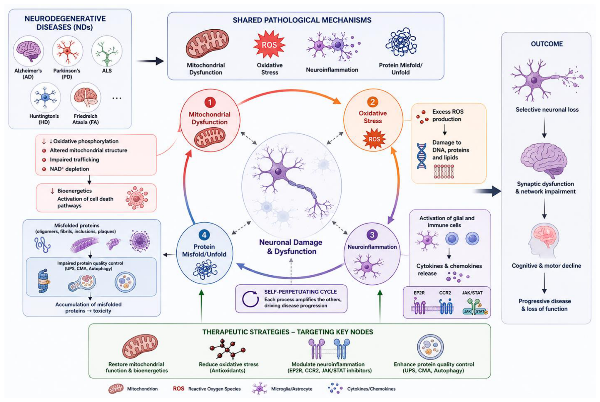
Figure 2.
Synergistic GLP-1R activation and melatonin action for enhanced neuroprotection. GLP-1 receptor (GLP-1R) agonists activate cAMP/PKA/CREB and PI3K/Akt signaling, enhancing neurotrophic support, mitochondrial function, glucose utilization, autophagy, and reducing neuroinflammation and oxidative stress. Melatonin complements these effects by stabilizing mitochondrial redox balance, preserving membrane potential, enhancing SIRT3/Nrf2 antioxidant defenses, regulating DRP1/OPA1-dependent mitochondrial dynamics, and promoting PINK1/Parkin-mediated mitophagy. Together, these complementary mechanisms may overcome the limitations of GLP-1R activation under sustained mitochondrial stress, resulting in enhanced neuronal survival, synaptic function, mitochondrial health, and long-term neuroprotection. BBB, blood–brain barrier; BDNF, brain-derived neurotrophic factor; cAMP, cyclic adenosine monophosphate; CREB, cAMP response element-binding protein; DRP1, dynamin-related protein 1; GLP-1R, glucagon-like peptide-1 receptor; GSK-3β, glycogen synthase kinase-3 beta; NLRP3, NOD-like receptor family pyrin domain containing 3; Nrf2, nuclear factor erythroid 2–related factor 2; OPA1, optic atrophy 1; PINK1, PTEN-induced kinase 1; PI3K, phosphoinositide 3-kinase; PKA, protein kinase A; ROS, reactive oxygen species; RNS, reactive nitrogen species; SIRT3, sirtuin 3.
Figure 2.
Synergistic GLP-1R activation and melatonin action for enhanced neuroprotection. GLP-1 receptor (GLP-1R) agonists activate cAMP/PKA/CREB and PI3K/Akt signaling, enhancing neurotrophic support, mitochondrial function, glucose utilization, autophagy, and reducing neuroinflammation and oxidative stress. Melatonin complements these effects by stabilizing mitochondrial redox balance, preserving membrane potential, enhancing SIRT3/Nrf2 antioxidant defenses, regulating DRP1/OPA1-dependent mitochondrial dynamics, and promoting PINK1/Parkin-mediated mitophagy. Together, these complementary mechanisms may overcome the limitations of GLP-1R activation under sustained mitochondrial stress, resulting in enhanced neuronal survival, synaptic function, mitochondrial health, and long-term neuroprotection. BBB, blood–brain barrier; BDNF, brain-derived neurotrophic factor; cAMP, cyclic adenosine monophosphate; CREB, cAMP response element-binding protein; DRP1, dynamin-related protein 1; GLP-1R, glucagon-like peptide-1 receptor; GSK-3β, glycogen synthase kinase-3 beta; NLRP3, NOD-like receptor family pyrin domain containing 3; Nrf2, nuclear factor erythroid 2–related factor 2; OPA1, optic atrophy 1; PINK1, PTEN-induced kinase 1; PI3K, phosphoinositide 3-kinase; PKA, protein kinase A; ROS, reactive oxygen species; RNS, reactive nitrogen species; SIRT3, sirtuin 3.
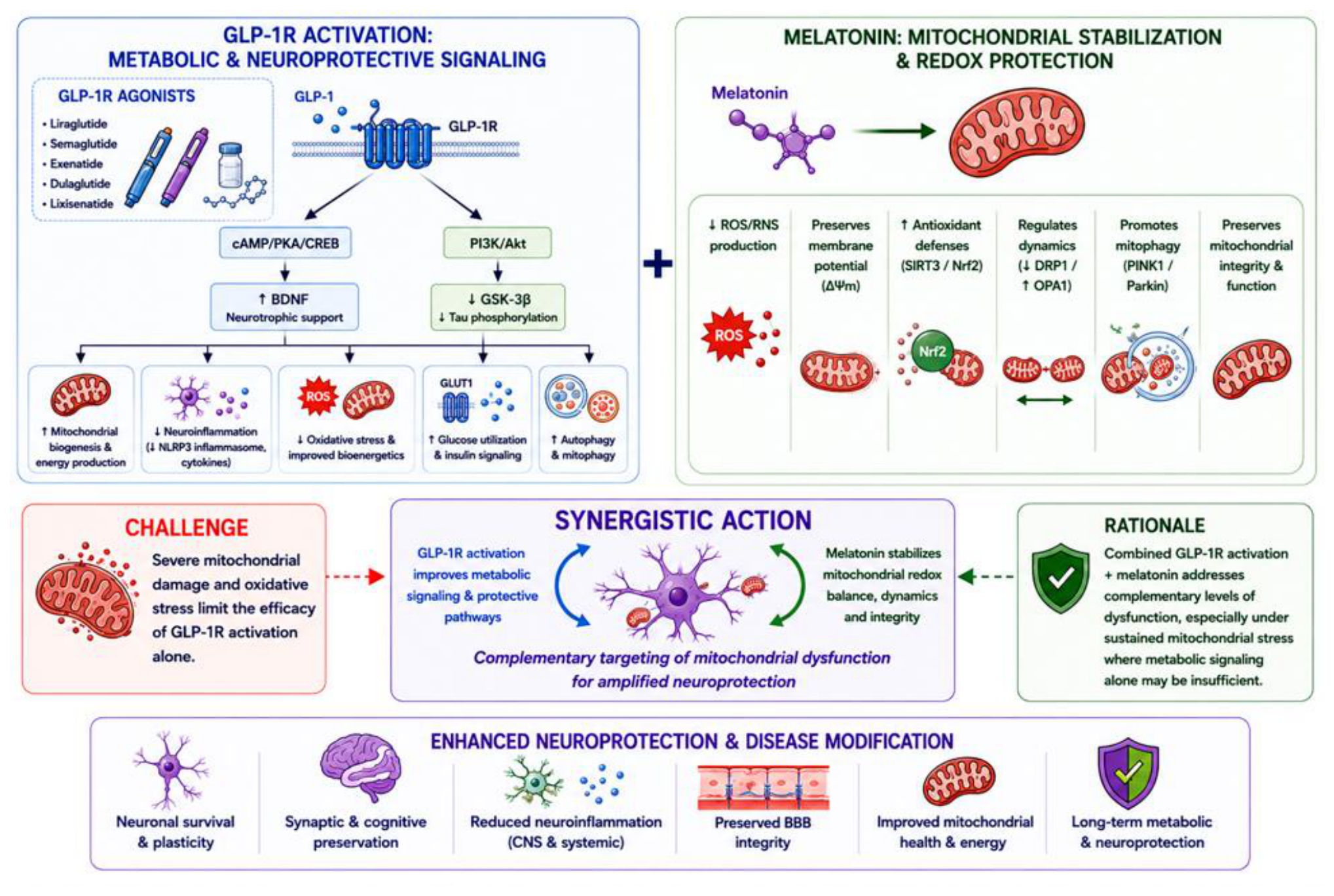
Figure 3.
Synergistic mitochondrial and neuroprotective mechanisms mediated by GLP 1 receptor agonists and melatonin, converging on the AMPK/SIRT1/PGC 1α axis. This schematic illustrates how GLP-1 receptor agonists (GLP-1RAs) and melatonin engage complementary signaling pathways that converge to enhance mitochondrial function and promote neuroprotection. On the left, GLP 1RA activation of the neuronal GLP 1 receptor stimulates the cAMP/PKA/CREB pathway and the PI3K/Akt/mTOR cascade, increasing metabolic flexibility and mitochondrial biogenesis, and suppressing neuroinflammatory signaling, including inhibition of the NLRP3 inflammasome. On the right, melatonin acts through MT1/MT2 receptors and direct antioxidant activity to increase ROS scavenging, reduce oxidative stress, and improve electron transport chain efficiency and ATP production. Both signaling arms converge on the AMPK/SIRT1/PGC 1α axis, a master regulatory pathway governing mitochondrial biogenesis, mitophagy, dynamics, and oxidative metabolism. Activation of this axis results in reduced ROS generation, enhanced ATP synthesis, improved mitochondrial quality control, and strengthened cellular resilience. Together, these synergistic effects reduce protein misfolding (Aβ, tau, α-synuclein), support neuronal survival and synaptic plasticity, and mitigate CNS injury and apoptosis. The combined actions of GLP 1RA and melatonin reinforce mitochondrial homeostasis and provide a multifaceted neuroprotective strategy.
Figure 3.
Synergistic mitochondrial and neuroprotective mechanisms mediated by GLP 1 receptor agonists and melatonin, converging on the AMPK/SIRT1/PGC 1α axis. This schematic illustrates how GLP-1 receptor agonists (GLP-1RAs) and melatonin engage complementary signaling pathways that converge to enhance mitochondrial function and promote neuroprotection. On the left, GLP 1RA activation of the neuronal GLP 1 receptor stimulates the cAMP/PKA/CREB pathway and the PI3K/Akt/mTOR cascade, increasing metabolic flexibility and mitochondrial biogenesis, and suppressing neuroinflammatory signaling, including inhibition of the NLRP3 inflammasome. On the right, melatonin acts through MT1/MT2 receptors and direct antioxidant activity to increase ROS scavenging, reduce oxidative stress, and improve electron transport chain efficiency and ATP production. Both signaling arms converge on the AMPK/SIRT1/PGC 1α axis, a master regulatory pathway governing mitochondrial biogenesis, mitophagy, dynamics, and oxidative metabolism. Activation of this axis results in reduced ROS generation, enhanced ATP synthesis, improved mitochondrial quality control, and strengthened cellular resilience. Together, these synergistic effects reduce protein misfolding (Aβ, tau, α-synuclein), support neuronal survival and synaptic plasticity, and mitigate CNS injury and apoptosis. The combined actions of GLP 1RA and melatonin reinforce mitochondrial homeostasis and provide a multifaceted neuroprotective strategy.
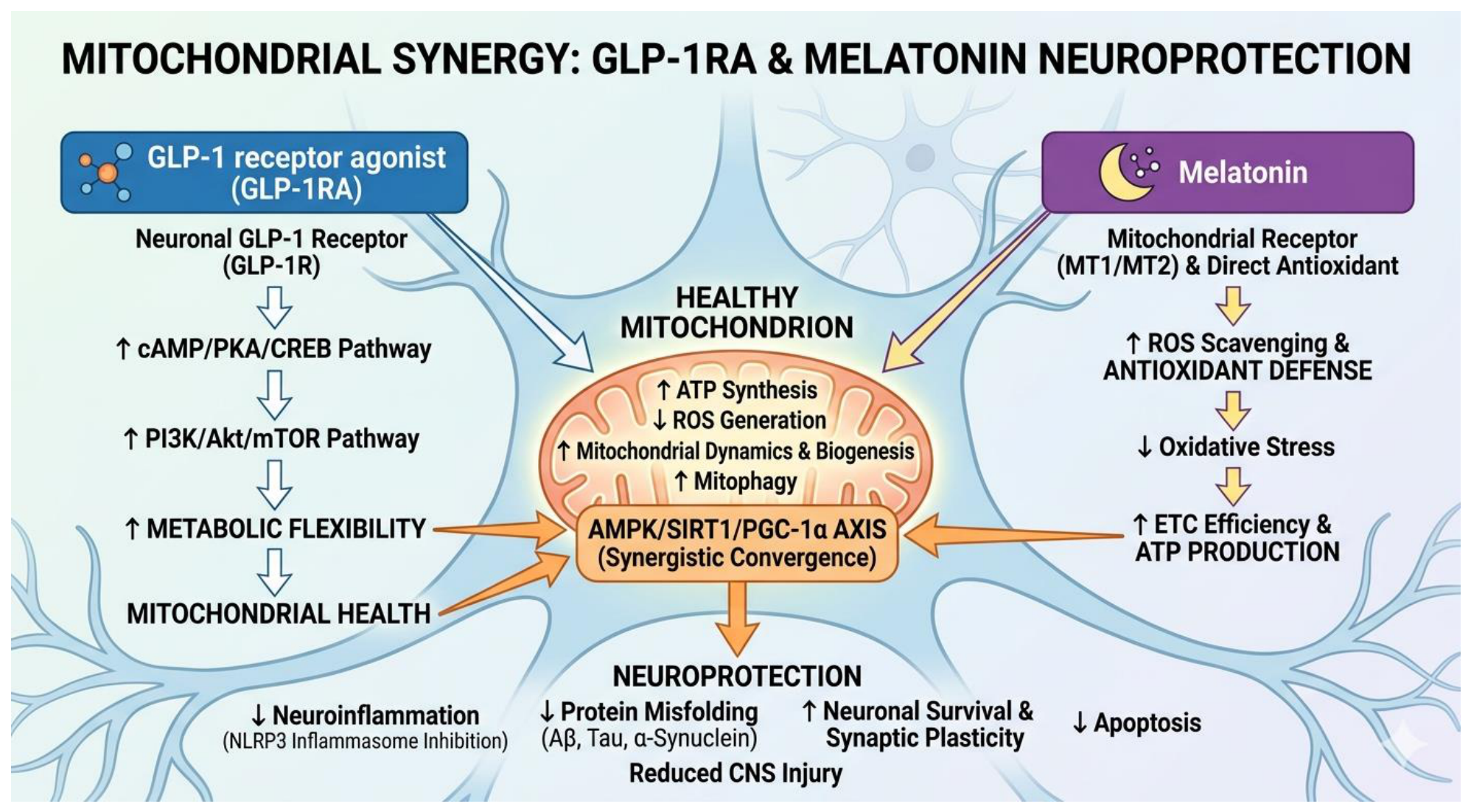
Disclaimer/Publisher’s Note: The statements, opinions and data contained in all publications are solely those of the individual author(s) and contributor(s) and not of MDPI and/or the editor(s). MDPI and/or the editor(s) disclaim responsibility for any injury to people or property resulting from any ideas, methods, instructions or products referred to in the content. |
© 2026 by the authors. Licensee MDPI, Basel, Switzerland. This article is an open access article distributed under the terms and conditions of the Creative Commons Attribution (CC BY) license (http://creativecommons.org/licenses/by/4.0/).
Copyright: This open access article is published under a Creative Commons CC BY 4.0 license, which permit the free download, distribution, and reuse, provided that the author and preprint are cited in any reuse.