Preprint
Article

This version is not peer-reviewed.

Percentage of Gutta-Percha-Filled-Areas in Canals Obturated by Two Different Core Techniques with Endodontic Bioceramics Sealer

Submitted:

10 November 2025

Posted:

12 November 2025

You are already at the latest version

Abstract

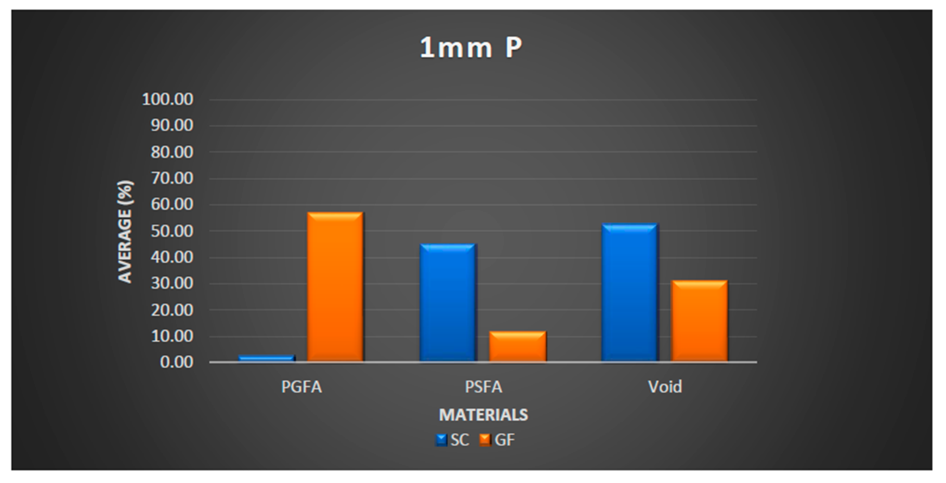
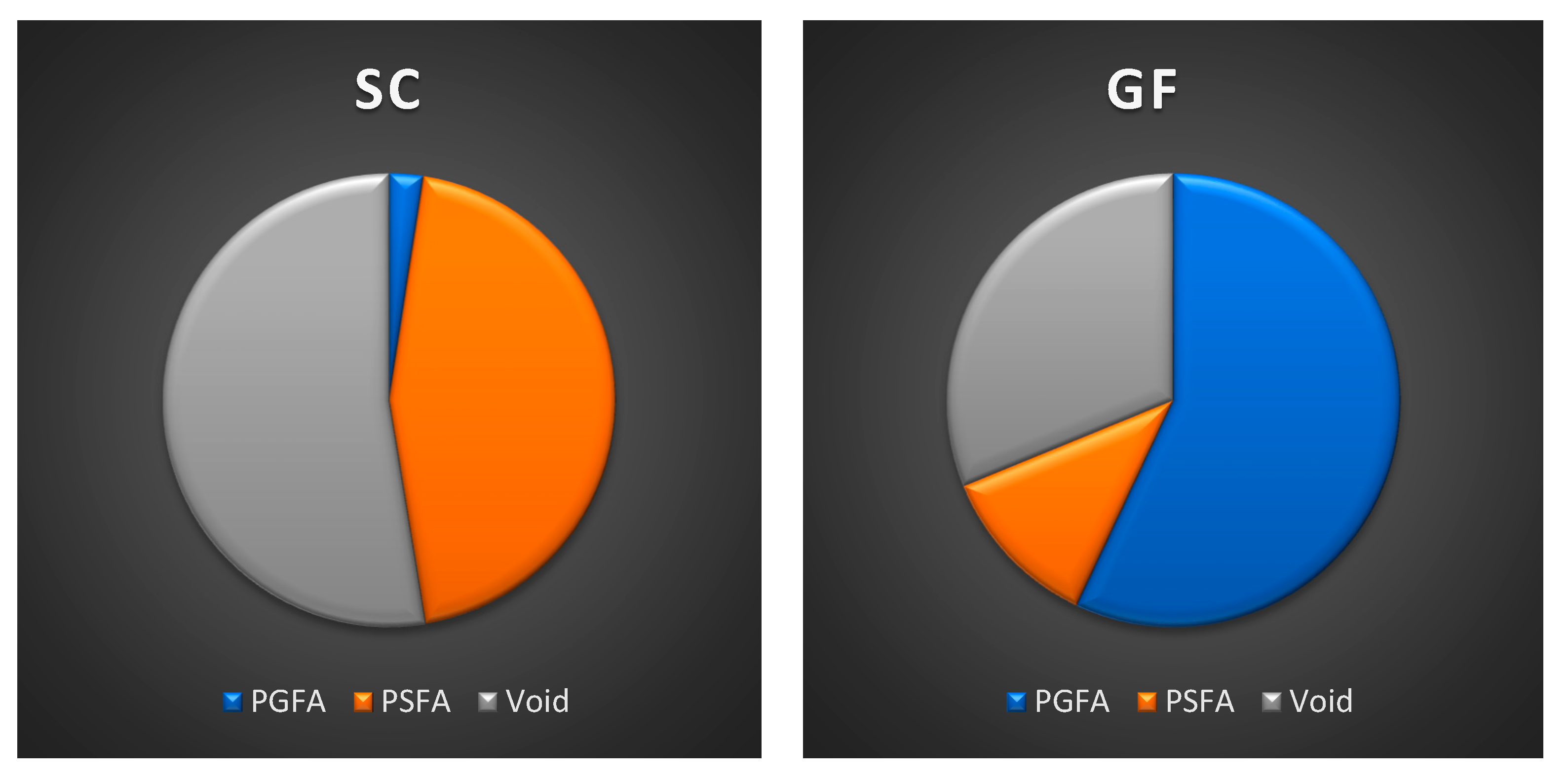
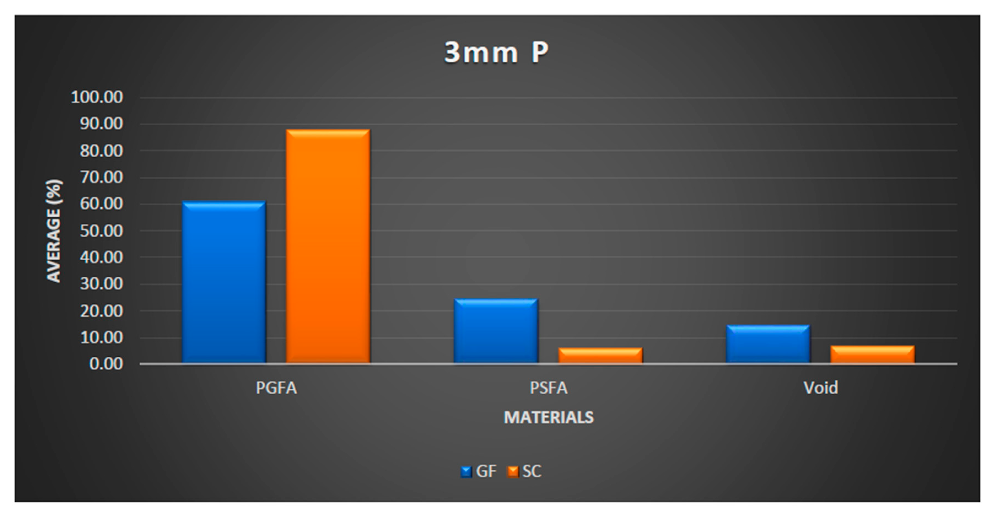
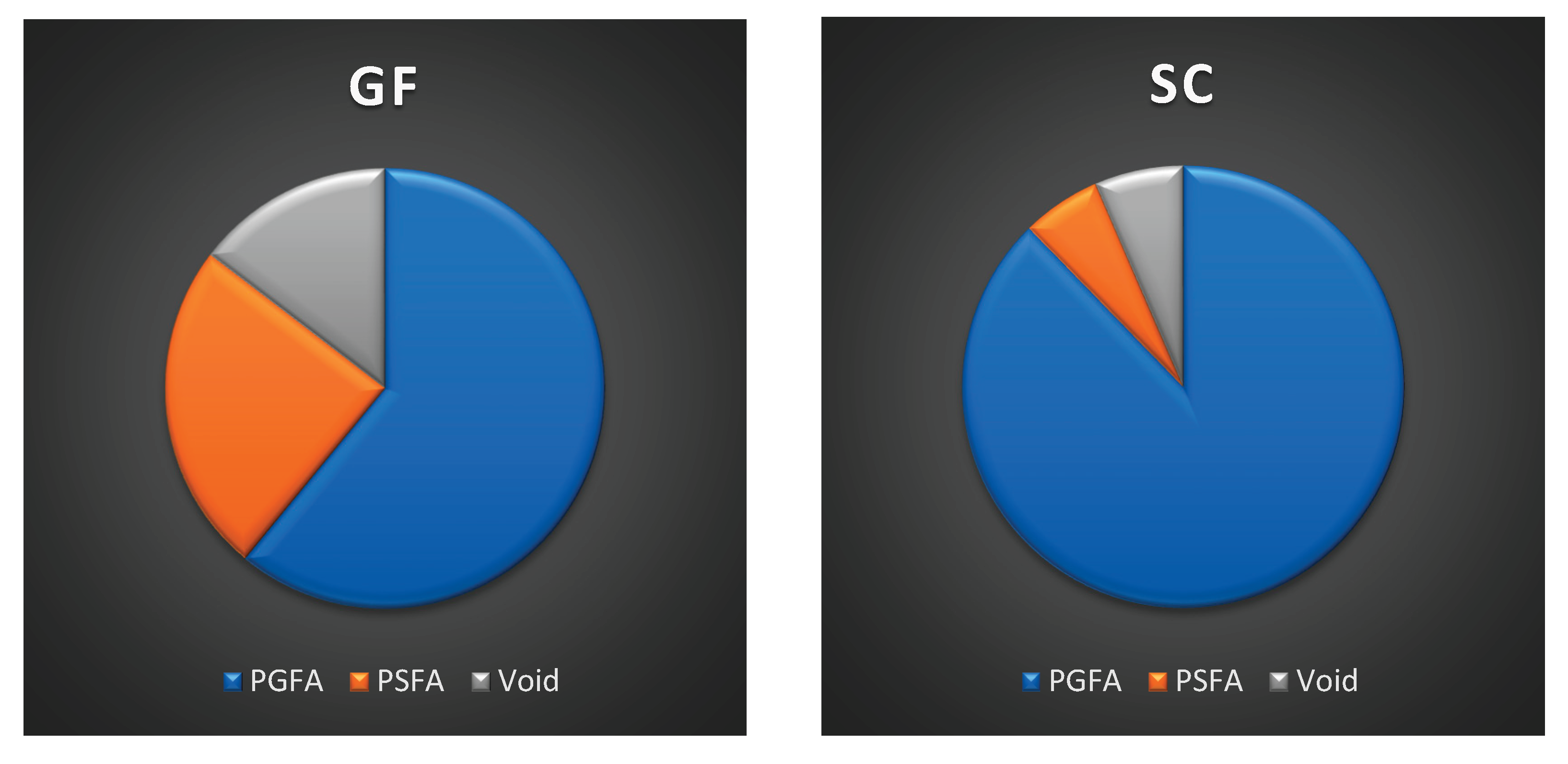
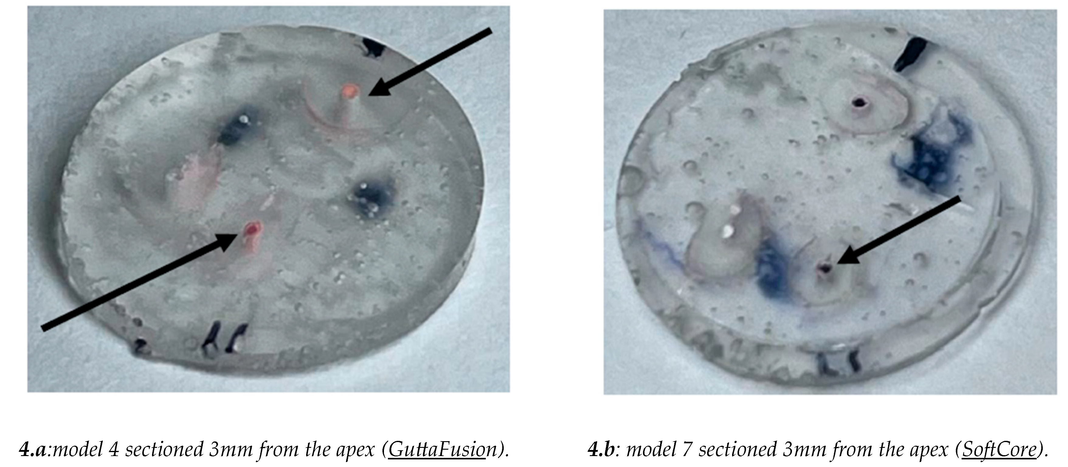
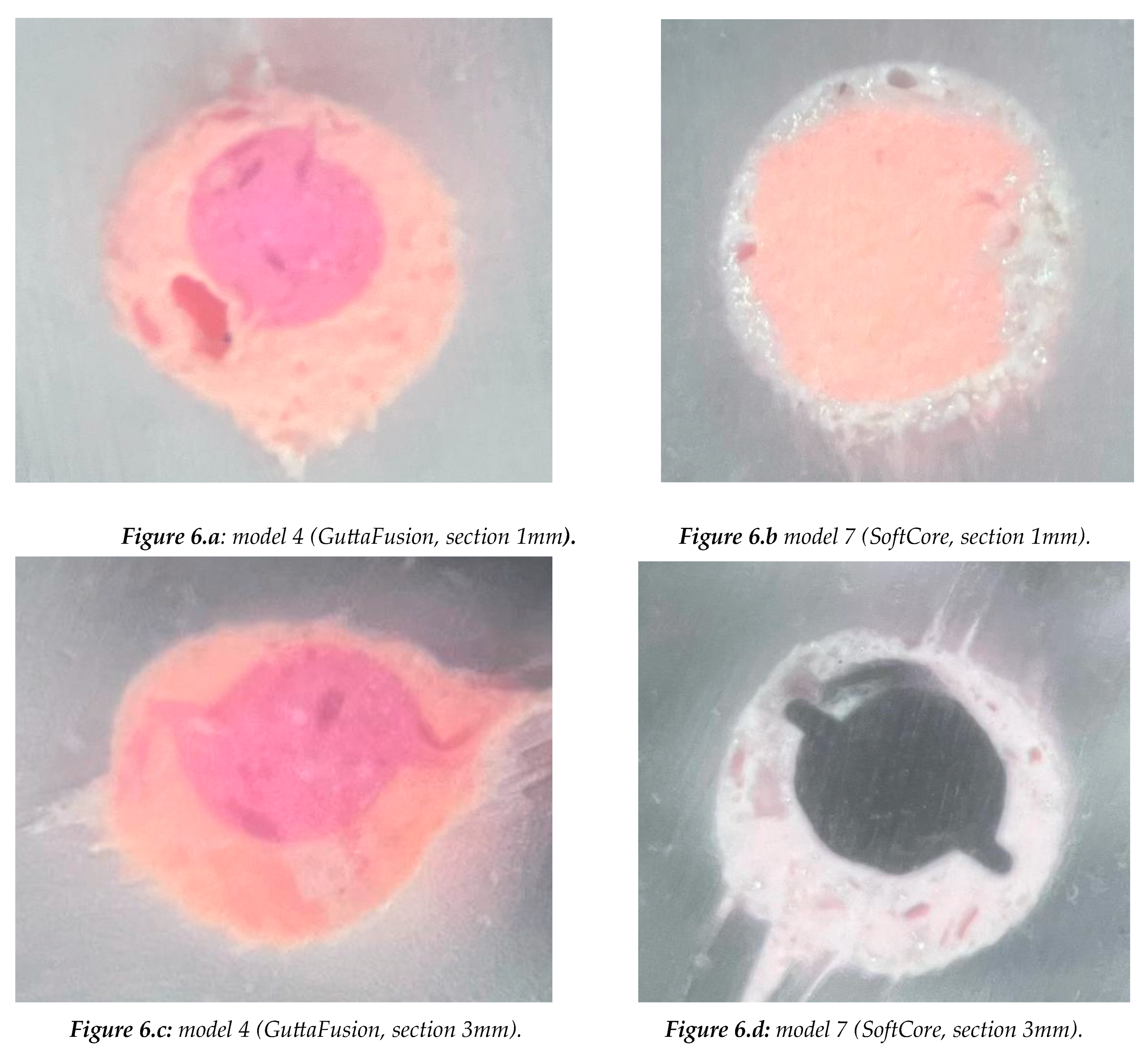
The aim of the study was to compare the sealing of two different core techniques (SoftCore and Guttafusion) associated to a bioceramic sealer (NeoSealer Flo). Eight tooth models (upper first molar) with simulated canals were used in this study. The models were divided into two groups: Group 1 (samples 1 to 4): Root canals were obturated with GuttaFusion. Group 2 (samples 5 to 8): Root canals were obturated with SoftCore. Both groups were filled with NeoSealer Flo sealer. Only the P and DV canals were obturated for each model. The models were sectioned at 1mm and 3mm from the apex. The total area of each canal segment was measured, and the areas were converted into the percentage of “Gutta-percha-filled areas”, ” Cement-filled areas”, “Void areas”. At 1mm from the apex GuttaFusion (GF) produced a higher PGFA and a lower PSFA value compared to SoftCore (SC). Additionally, SC produces a higher PGVA compared to GF. The values of PGFA, PSFA, and PVA are also comparable in the sections taken at 3mm from the apex. Comparing the two different techinques, a higher PGFA in GuttaFusion (GF) and a higher PSFA in SoftCore (SC) were found, while the VA was greater but comparable in both techniques, with a slight increase in percentage in the SC technique. At 3 mm, a higher PGFA was found compared to the PGFA at 1 mm.

Keywords: 
;  ;  ;  ;  ;  ;  

1. Introduction

The primary objectives of endodontic treatment are achieving a three-dimensional filling of the root canals with an atoxic, stable material, and creating an hermetically sealed apical area [1,2]. The filling of the canals, prevention of communication between the periodontal and endodontic spaces, and the mechanical preparation and irrigation are all essential steps to counteract the defense mechanisms of bacteria [1,2,3,4,5]. Gutta-percha, along with cement, has been successfully used, like core materials, for filling the root canal space. However, a hermetic seal cannot be achieved without the use of cement. This is because gutta-percha is unable to adhere to the dentinal walls of the canals, and the cement plays a critical role in filling imperfections and enhancing adhesion [6,7]. It has been reported that some cements shrink during setting, while others are subject to dissolution when in contact with tissue fluids [8,9]. Nevertheless, the amount of cement used should be kept to a minimum, while the amount of gutta-percha should be maximized to the greatest extent possible in order to optimize the filling of the canal [10,11]. Many techniques have achieved good adaptability for three-dimensional root canal obturation. One of the first described is the hot vertical condensation technique. Among the more modern techniques, SoftCore aims to fill the canal with heated “alpha guttapercha”, which contains a plastic carrier made of polysulfone [12,13]. In contrast, the Guttafusion technique, also a hot vertical condensation core technique, uses a carrier made from crosslinked gutta-percha (hardened gutta-percha with a modified lattice structure) [14,15]. To assess the quality of the root canal obturation, techniques such as cross-sectional analysis of obturated canals, infiltration tests, and the combination of both have been widely used. The methodology followed in our study is based on approaches used in previous studies by significant authors [16,17,18,19]. Some previous studies [20,21] have evaluated the possibility of avoiding the combination of endodontic cement with carrier-based obturation techniques. However, in our view, there has been a continuous improvement over the years in root canal obturation techniques, particularly in apical preparation, as many studies report good filling percentages and effective minimization of void spaces. Our goal is to confirm the effectiveness of these techniques in root canal obturation by minimizing excess cement and optimizing the space occupied by cement, Gutta-percha and Void spaces. The two core techniques we compare, SoftCore and Guttafusion, will be examined to evaluate their efficiency in cement usage and the quality of the canal filling. This preliminary study aims to analyze two "core" root canal obturation systems: SoftCore and GuttaFusion, using the NeoSealer Flo sealer, and to compare the results between them. The primary objective of this study is to compare the percentage of the Gutta-Percha-filled areas in the canals obturated with SoftCore and GuttaFusion, both with the same bioceramic sealer.

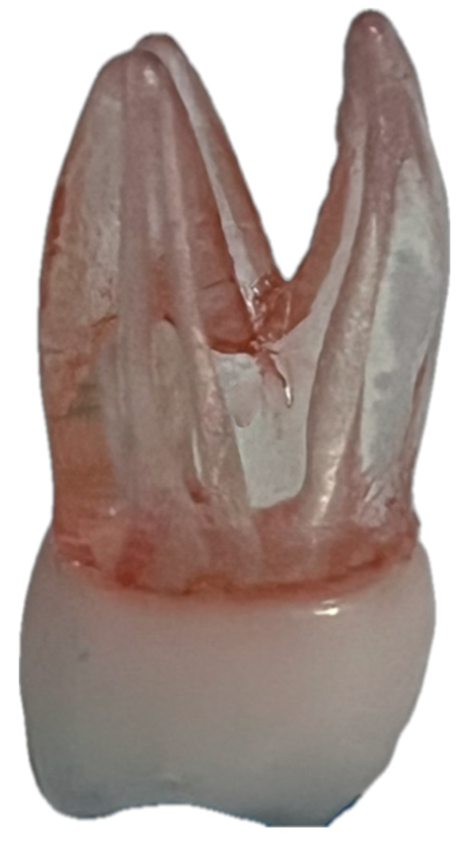
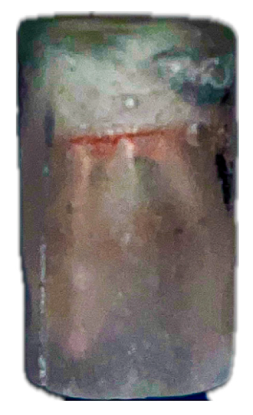
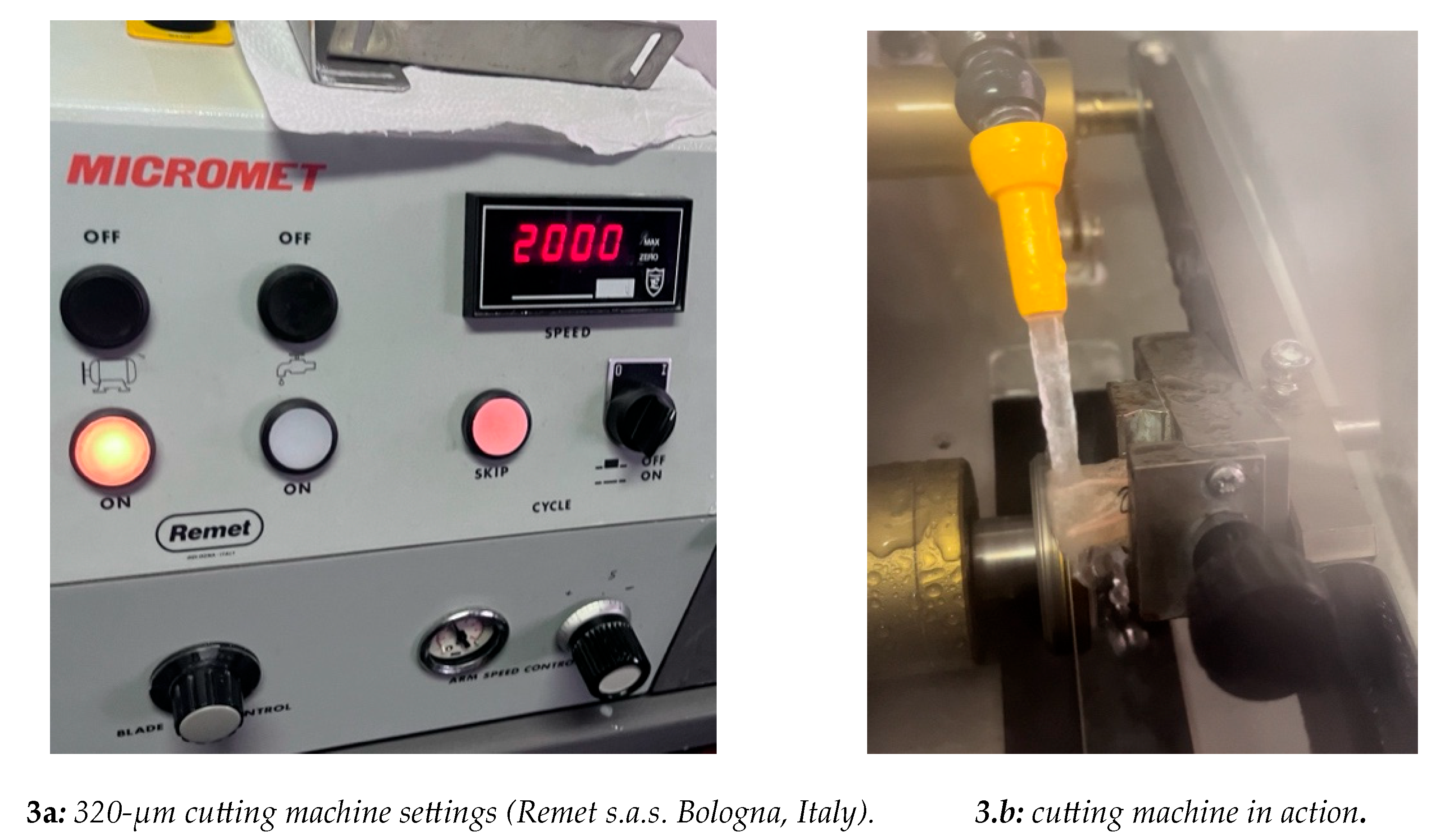
2. Materials and Methods

Eight identical models of upper first molars, made of resin (Figure 1), were examined. All the models had simulated canals (canal P; canal DB; canal MB1 and canal MB2). For our study, canal P and canal DB were chosen for each sample. The canals were instrumented to obtain the following working lengths (WL) for each sample (Table 1).
The working length (WL) was determined using a #10 K-file (Dentsply Maillefer, Bellaigues, Switzerland) until the tip of the instrument was clearly visible from the apical foramen. The mechanical preparation was performed using only a reciprocating file from the “Zarc4Endo” series, the Excalibur 25/05. The file was mounted on a Morita endodontic micromotor set to reciprocation (150°/30°; 450 rpm). During the preparation, the canals were irrigated using 30-G side-vented syringes, with 2 mL of 5.25% sodium hypochlorite and 10 mL of 17% EDTA. Apical gouging was verified using a #30 K-file. All the simulated canal samples were dried with paper points and then grouped into two groups:
  • GuttaFusion (samples 1 to 4)
  • SoftCore (samples 5 to 8).
A total of 16 CBO 25 obturators (ISO Red color) were selected, 8 for the first 4 samples and the remaining 8 for the last 4 samples. The obturators were thermoplasticized using the SoftCore Oven [22]. The NeoSealer Flosealer was used as the endodontic cement [23,24,25]. The samples were kept for 14 days at 37°C and 100% humidity to allow complete setting of the cement. The teeth were then embedded in epoxy resin blocks (Buehler Ltd; Evanston, IL) (Figure 2).
The teeth were sectioned at 1 mm and 3 mm from the apex, orthogonally to their long axis, using a 320-µm disc cutter (Remet s.a.s., Bologna, Italy) (Figure 3a) with water cooling (Figure 3b). The coronal face of the sections was polished with abrasive paper of decreasing grit (320, 1200, and 2500 grit) to obtain a smooth surface without obvious deformations [26]. The sections were analyzed using a Nikon TS100-F microscope, with a modified light plane, at 40x magnification. Images were captured with an iPhone 15 Pro Max camera (Apple Inc.). The images were analyzed in a nano compressed TIFF format, and measurements were taken using Adobe Photoshop CS3 (Adobe, San Jose, CA). The following areas were evaluated:
  • Gutta-percha area
  • Cement area
  • Void area
The values obtained were expressed as percentages of the total area (PGFA, PSFA, and VA). The data were recorded in Excel tables (Microsoft), with corresponding pie charts and histograms.
Additionally, the percentage of error due to human error during the processing with Adobe Photoshop was calculated, and this error was included in the total percentage.
Images of two samples sectioned 3mm from the apex: the arrows indicate the sections of the DV and P canals (Figure 4.a; Figure 4.b)
Images of the same two samples from Figure 6 (Figure 6.a; Figure 6.b; Figure 6.c; Figure 6.d), with 40x magnification on the palatal canal.

2. Results

Comparing the two techniques with the endodontic cement, we found the following results at 1mm from the apex. GuttaFusion (GF) produced a higher PGFA and a lower PSFA value compared to SoftCore (SC) (Graph 1). Additionally, SC produces a higher PGVA compared to GF. The values of PGFA, PSFA, and PVA are also comparable in the sections taken at 3mm from the apex (Graph 2-2.1-2.2). The values shown are clinically relevant but do not have a statistically significant value. Pie charts are also provided for simplicity of evaluation (Graph 1.1-1.2). The PFSA values increase in the SC technique compared to GF. Noting that the PSFA in SC occupies a good portion of the total % area. In the SC technique, it was also observed that the VA is increased), while the VA in the GF samples is decreased
Graph 1. Percentage area of material at 1 mm from the apex palatal root. 
Preprints 184509 gr001
Graph 1.1-1.2. Percentage area of material at 1 mm from the apex palatal root Illustrated in pie charts.
Preprints 184509 gr002
Graph 2. Percentage area of material at 3 mm from the apex palatal root. 
Preprints 184509 gr003
Graph 2.1-2.2. Percentage area of material at 3 mm from the apex palatal root Illustrated in pie charts. 
Preprints 184509 gr004

4. Discussion

The quality of root canal obturation, calculated through the percentage of gutta-percha, cement, and void, was used to evaluate the differences in quality between the two core materials [27,28,29]. The apical seal should create a tight seal, especially since only 3-4 mm of length remains in the canal after post-space preparation. However, studies have shown that numerous lateral canals are present in the apical third [30]. For this reason, sections at 1 mm and 3 mm from the apex—critical points for evaluating the apical seal—were analyzed in this and other studies. In our study, comparing two obturators with different core materials and endodontic cement, we found a higher PGFA in GuttaFusion (GF) and a higher PSFA in SoftCore (SC), while the VA was greater but comparable in both techniques, with a slight increase in percentage in the SC technique. However, as previously mentioned, these results lack statistical significance but do have clinical relevance. Our study, based on the measurements obtained, is consistent with the findings reported in reference studies. Previous studies on gutta-percha core techniques, including SoftCore and GuttaFusion, report a higher gutta-percha-to-sealer ratio compared to techniques like Microseal or System B. At 3 mm, our results show a higher PGFA compared to the PGFA found at 1 mm (for the palatal canal). Some earlier studies emphasized that Thermafil, like SoftCore, exhibited lower microleakage values compared to lateral condensation techniques or System B. In our study, the two core techniques, when compared to other studies conducted with different techniques, show that the PGFA is still acceptable at both 1 mm and 3 mm, albeit lower than that found with techniques like Microseal or System B, as reported in the reference study for this preliminary study. On the other hand, SC and GF present a lower percentage of VA and a higher PSFA at the second cutting level, indicating better cement adaptability. Nevertheless, the obtained values align with the statistical evidence of the cases.

5. Conclusions

Based on the PSFA, we conclude that for core endodontic techniques, the use of cement is important, if not essential, to achieve proper filling, particularly at 1 mm and 3 mm from the apex.

Author Contributions

A.L.: Supervision, Conceptualization, Investigation, Methodology, Resources. D.M.: Investigation., Resources. G.B.: Data curation, Writing, Formal Analysis, Methodology. G.C.: Investigation. V.C.: Reviewing, Editing, and Conceptualization. All authors have read and agreed to the published version of the manuscript.

Funding

This research received no external funding.

Institutional Review Board Statement

Not applicable.

Informed Consent Statement

Not applicable.

Data Availability Statement

The raw data supporting the conclusions of this article will be made available by the authors on request.

Conflicts of Interest

The authors declare no conflicts of interest.

References

  1. Schilder, H. Filling root canals in three dimensions. J. Endod. 1967, 23, 732–740. [Google Scholar] [CrossRef]
  2. Tomson, R.M.; Polycarpou, N.; Tomson, P.I. Contemporary obturation of the root canal system. Br. Dent. J. 2014, 216, 315–322. [Google Scholar] [CrossRef] [PubMed]
  3. Whithworth, J. Methods of filling root canals: principles and practice. Endod. Topics 2005, 12, 2–24. [Google Scholar] [CrossRef]
  4. Gallusi, G.; Campanella, V.; Montemurro, E.; Di Taranto, V.; Libonati, A. Antibacterial activity of first and latest generation bioceramic sealers on the elimination of Enterococcus faecalis: an in vitro study. J. Biol. Regul. Homeost. Agents 2020, 34 (3 Suppl. 1), 73–79. [Google Scholar]
  5. Ciani, L.; Libonati, A.; Dri, M.; Pomella, S.; Campanella, V.; Barillari, G. About a possible impact of endodontic infections by Fusobacterium nucleatum or Porphyromonas gingivalis on oral carcinogenesis: a literature overview. Int. J. Mol. Sci. 2024, 25, 5083. [Google Scholar] [CrossRef]
  6. Skinner, R.; Van Himel, T. The sealing ability of injection-molded thermoplasticized gutta-percha with and without the use of sealers. J. Endod. 1987, 13, 315–317. [Google Scholar] [CrossRef]
  7. Wu, M.K.; Ozok, A.R.; Wesselink, P.R. Sealer distribution in root canals obturated by three techniques. Int. Endod. J. 2000, 33, 340–345. [Google Scholar] [CrossRef]
  8. Kontakiotis, E.G.; Wu, M.K.; Wesselink, P.R. Effect of sealer thickness on long-term sealing ability: a two-year follow-up study. Int. Endod. J. 1997, 30, 307–312. [Google Scholar]
  9. Peters, D.D. Two-year in vivo solubility evaluation of four gutta-percha sealer obturation techniques. J. Endod. 1986, 12, 139–145. [Google Scholar] [CrossRef]
  10. Souza, E.M.; Wu, M.K.; Van Der Sluis, L.W. Effect of filling techniques and root canal area on the percentage of gutta-percha in laterally compacted root fillings. Int. Endod. J. 2009, 42, 719–726. [Google Scholar] [CrossRef]
  11. Jarrett IS, Marx D, Covey D, Karmazin M, Lavin M, Gound T. Percentage of canals filled in apical cross sections - an in vitro study of seven obturation techniques. Int Endod J. 2004 Jun;37(6):392-8.
  12. Johnson, W.B. A new gutta-percha technique. J. Endod. 1978, 4, 184–188. [Google Scholar] [CrossRef]
  13. Soft Core Manual. Soft Core Dental Production, Copenhagen, Denmark.
  14. Mark, J.E. Physical Properties of Polymers Handbook. Springer, 2007.
  15. Eguchi, D.E.; Peters, D.D.; Hollinger, J.O.; Lorton, L.A. Comparison of the area of the canal space occupied by gutta-percha following four gutta-percha obturation techniques using ProConsol sealer. J. Endod. 1985, 11, 166–175. [Google Scholar] [CrossRef] [PubMed]
  16. Silver, G.K.; Love, R.M.; Purton, D.G. Comparison of two vertical condensation obturation techniques: Touch and Heat modified and System B. Int. Endod. J. 1999, 32, 287–295. [Google Scholar] [CrossRef] [PubMed]
  17. Gencoglu, N.; Garip, Y.; Bas, M.; Samani, S. Comparison of different gutta-percha root filling techniques: Thermafil, Quick-Fill, System B, and lateral condensation. Oral Surg. Oral Med. Oral Pathol. Oral Radiol. Endod. 2002, 93, 333–336. [Google Scholar] [CrossRef] [PubMed]
  18. De Deus, G.; Gurgel-Filho, E.D.; Magalhães, K.M.; Coutinho-Filho, T. A laboratory analysis of gutta-percha-filled area obtained using Thermafil, System B and lateral condensation. Int. Endod. J. 2006, 39, 378–383. [Google Scholar] [CrossRef]
  19. Ishley, D.J.; ElDeeb, M.E. In vitro assessment of the quality of apical seal of thermomechanically obturated canals with and without sealer. J. Endod. 1986, 9, 242–246. [Google Scholar] [CrossRef]
  20. ElDeeb, M.E. The sealing ability of injection-molded thermoplasticized gutta-percha. J. Endod. 1985, 11, 84–86. [Google Scholar] [CrossRef]
  21. Natural GP Soft-Core: A 3rd Generation Endodontic Obturator Manual. Dental Production Aps, Copenhagen, 2001.
  22. Zamparini, F.; Prati, C.; Taddei, P.; Spinelli, A.; Di Foggia, M.; Gandolfi, M.G. Chemical-physical properties and bioactivity of new premixed calcium silicate–bioceramic root canal sealers. Int. J. Mol. Sci. 2022, 23, 13914. [Google Scholar] [CrossRef]
  23. Sanz, J.L.; López-García, S.; Rodríguez-Lozano, F.J.; Melo, M.; Lozano, A.; Llena, C.; Forner, L. Cytocompatibility and bioactive potential of AH Plus Bioceramic sealer: an in vitro study. Int. Endod. J. 2022, 55, 1066–1080. [Google Scholar] [CrossRef]
  24. Libonati, A.; Gallusi, G.; Angotti, V.; Di Taranto, V. The effect of different polishing systems on the surface roughness of two restorative dental materials. Oral Implantol. 2018, 11, 185–190. [Google Scholar]
  25. Gençoğlu, N. Comparison of six different gutta-percha techniques (Part II): Thermafil, JS Quick-Fill, Soft-Core, Microseal, System B and lateral condensation. [Journal not specified].
  26. Wu, M.K.; Van Der Sluis, L.W.; Wesselink, P.R. A preliminary study of the percentage of gutta-percha–filled areas in the apical canal filled with vertically compacted warm gutta-percha. Int. Endod. J. 2002, 35, 527–535. [Google Scholar] [CrossRef]
  27. Rubach, W.C.; Mitchell, D.F. Periodontal disease, accessory canals and pulp pathosis. J. Periodontol. 1965, 36, 34–38. [Google Scholar] [CrossRef] [PubMed]
  28. De Deus, Q.D. Frequency, location, and direction of lateral, secondary and accessory canals. J. Endod. 1975, 20, 361–366. [Google Scholar] [CrossRef] [PubMed]
  29. Nica, L.M.; Didilescu, A.; Rusu, D.; et al. Photomicrographic evaluation of the apical sealing capacity of three types of gutta-percha master cones: an in vitro study. Odontology 2012, 100, 54–60. [Google Scholar] [CrossRef] [PubMed]
  30. Libonati, A.; Montemurro, E.; Nardi, R.; Campanella, V. Percentage of gutta-percha–filled areas in canals obturated by three different techniques with and without the use of endodontic sealer. J. Endod. 2017, 1–4. [Google Scholar]
Figure 1. element model used.
Preprints 184509 g001
Figure 2. element model embedded in epoxy resin.
Preprints 184509 g002
Figure 3.
Figure 3.
Preprints 184509 g003
Figure 4.
Figure 4.
Preprints 184509 g004
Figure 6.
Figure 6.
Preprints 184509 g005
Table 1. WL of instrumented canals. 
SAMPLES P
ROOT CANAL
DB
ROOT CANAL
MB2
ROOT CANAL
MB1
ROOT CANAL
1-8 WL: 22.5mm
Ref: MP cuspid
Apex: K-file (30)
WL: 20.5 mm
Ref: DV cuspid
Apex: K-file (30)
WL: 22 mm
Ref: MP cuspid
Apex: K-file (30)
WL: 22 mm
Ref: MV cuspid
Apex: K-file (30)
Disclaimer/Publisher’s Note: The statements, opinions and data contained in all publications are solely those of the individual author(s) and contributor(s) and not of MDPI and/or the editor(s). MDPI and/or the editor(s) disclaim responsibility for any injury to people or property resulting from any ideas, methods, instructions or products referred to in the content.
Copyright: This open access article is published under a Creative Commons CC BY 4.0 license, which permit the free download, distribution, and reuse, provided that the author and preprint are cited in any reuse.
Prerpints.org logo

Preprints.org is a free preprint server supported by MDPI in Basel, Switzerland.

Subscribe

Disclaimer

Terms of Use

Privacy Policy

Privacy Settings

© 2025 MDPI (Basel, Switzerland) unless otherwise stated