
Submitted:
11 February 2025
Posted:
11 February 2025
You are already at the latest version
Abstract
NDV-based vector has been used as a veterinary vaccine and, recently also for human COVID-19 vaccine; however, data about the potential immune response against vector in humans is scarce, therefore, it is important evaluate levels of antibodies produced. The HI assay is the gold standard for assessing the humoral response against NDV in poultry serum. Here the objective of was validate the HI assay against the NDV-vectored vaccine to analyze antibodies in human serum. First, we standardized conditions in human sera before validation. The results from analytical performance: selectivity, sensitivity, specificity, positive and negative predictive value, as well as positive and negative diagnostic reliability, indicate that the assay is highly selective, as it allows clear discrimination between positive and negative samples. Regarding repeatability and inter-mediate precision, we demonstrated that the assay has precision to obtain consistent results and guarantees their reliability and truthfulness. Finally, the results of accuracy, linearity and robustness, indicate that the assay is accurate at the concentration intervals evaluated with a linear correlation between the low and high level and demonstrated that it is robust and consistent when the serum-antigen interaction times changed. We conclude that the suitability of the analytical method for its intended use is confirmed and guarantees the reliability of the results obtained under the established operating conditions.
Keywords:
NDV-vectored vaccine
; HI assay
; validation of analytical methods
1. Introduction
The Newcastle disease virus (NDV) is a pleomorphic virus measuring between 150 and 350 nm in diameter, which can be found as spherical or filamentous particles. It is an enveloped, non-segmented virus from the Paramyxoviridae family and subfamily Avulavirinae [1]. Its genome is a single-stranded, negative-sense RNA molecule measuring between 13 and 19 Kb that encodes six structural proteins: nucleocapsid protein (NP), phosphoprotein (P), matrix protein (M), fusion protein (F), hemagglutinin-neuraminidase (HN), and polymerase (L), along with two non-structural proteins (V and W). This virus is responsible for Newcastle disease, which is highly contagious and primarily affects birds. It can manifest in three clinical forms in chickens: mild, caused by lentogenic strains; moderate, caused by mesogenic strains; and severe, caused by velogenic strains, representing an economic problem in the poultry sector [2,3].
NDV is categorized as a hemagglutinating virus due to the interaction of the HN protein on its surface with sialic acid residues present in the glycoproteins of red blood cells (RBC) from chickens, pigs, guinea pigs, and humans, among others. This interaction leads to a network called hemagglutination, which can be evaluated in vitro and is dependent on the affinity of the viral proteins for the receptors on the RBC, as well as on temperature, interaction time, concentration, and the origin of the RBC [4,5,6].
Lentogenic strains such as LaSota and B1 have low virulence and propensity for recombination, in addition to eliciting a strong immune response, making them suitable as a biotechnological tool, for example, as an oncolytic therapy due to their immunomodulatory properties and high replication in tumor cells. They are also used as virus-based vaccine vectors or as vaccine strains in the poultry sector [7,8,9,10]. In the case of poultry, it is necessary to ensure the efficacy of vaccination by evaluating antibodies against NDV in vitro, as this prevents the spread of the virus.
The Hemagglutination Inhibition (HI) technique is the gold standard, which involves serial dilutions of a serum sample and contact with the NDV antigen, preventing the binding of the HN protein in the presence of RBCs, causing the RBCs to sediment and form a button with defined edges that slides in a drop shape when the plate is tilted at 45°. The concentration of antibodies is defined by the maximum dilution at which the button movement is observed [4].
There is evidence of the use of the HI assay to evaluate the immune response against NDV in humans, as this virus can potentially cause zoonosis with symptoms in the eyes, such as tearing, irritation, and swelling, although these symptoms are generally self-limiting and are not considered a risk [11]. Therefore, this technique can be used to evaluate the immune response to an NDV-vectored vaccine, which is of great importance, as it has been reported that the use of Adenovirus (Ad)-based vaccines, such as Ad5 in repeated immunizations, affects the organism’s effectiveness in producing antibodies against the antigen of interest due to high antibody production against the vector, which can limit its utility [8,9,12,13]. In contrast, the use of NDV as a vector has been shown to be safe and produces low antibody titers against the vector, as seen in the case of the AVX/COVID-12 "Patria" vaccine approved by the Comisión Federal para la Protección contra Riesgos Sanitarios (COFEPRIS) in Mexico [12].
To carry out the technique with human sera, it is necessary to standardize the conditions of each parameter involved, such as the minimum amount of antigen capable of causing hemagglutination, expressed in Hemagglutinating Units (HU) [14,15]. Additionally, the nature of serum samples is an important factor to consider, as mammalian serum samples, unlike avian serum, contain nonspecific inhibitors that can adhere to the surface of RBCs, potentially causing false positives [15,16,17]. Various research groups have proposed strategies to reduce the presence of nonspecific inhibitors: heating sera to high temperatures (56-65 °C), pretreatments with a kaolin solution, pre-absorption of serum samples in an RBC suspension, or the use of receptor-destroying enzymes (RDE) such as filtrates derived from Vibrio cholerae [16,18,19]. Although the HI assay has been an established test, there are still no reports in the context of the human NDV vaccine vector within an analytical validation process.
In this work, we present the standardization and validation of an HI assay that allowed the evaluation of the immune response against the NDV vector in human sera from volunteers vaccinated against SARS-CoV-2 with the AVX/COVID-12 "Patria" vaccine, demonstrating the suitability of the analytical method for its intended use. We also confirm that it guarantees the reliability of the results obtained under the established operating conditions.
2. Materials and Methods
2.1. Reagents
RDE Vibrio cholerae Filtrate Solution (Sigma-Aldrich C8772-IVC; MA, USA), 25 % kaolin solution in 1X PBS, pH 7.4 (Gibco REF 10010-031, MA, USA). The conditions of use of each reagent are described in subsequent sections.
2.2. Newcastle Disease Virus (NDV) Antigen
The NDV antigen LaSota strain was propagated in pathogen-free bird embryos and inactivated with 3.7% formaldehyde (Batch 23920102, Diagnósticos Clínicos Veterinarios, S.A. de C.V., Mexico). The concentration is represented in a titer of 1:256 hemagglutinating units (HU). This batch was used for all tests. Before starting each series of tests, the HU titer of the antigen was confirmed by titrating the NDV antigen, in addition to performing the test for HU control following the instructions of the protocol established by the supplier (Figure 1a,b).
2.3. Red Blood Cell (RBC) Suspension
A 5 % suspension of chicken RBC in 1X PBS v/v with a certificate of analysis issued by the DCV company (Batch 23-08, Mexico City, Mexico) were used. Pig RBC were donated by the “Instituto Nacional de Enfermedades Respiratorias, Ismael Cosío Villegas (INER)” Biotherium (through its research protocol code: B18-20), and human RBC were obtained from volunteers with O- blood type (Protocol code: C49-22), in both cases, a 5 % suspension in 1X PBS v/v was prepared from whole blood following the guidelines of good laboratory and biological safety practices.
2.4. Serum Samples
For assay standardization: one serum sample from a human volunteer vaccinated against SARS-CoV-2 (AVX/COVID-12), obtained 21 days post-vaccination, was used as exposure control for NDV (Protocol code: C49-22). One serum sample from a pig vaccinated against SARS-CoV-2 (AVX/COVID-12) was used as a positive control for NDV, with a known HI titer of 1:512 (Protocol code: C24_42), and human type AB serum (Batch 21C0457, Valley Biomedical, VA, USA) was used as a negative control for NDV.
For assay validation: Human type AB serum (Batch 21C0457, Valley Biomedical, VA, USA), which was negative for pathogens, was used as a serum matrix during the experiments; 10 serum samples from volunteers recovered from influenza and 10 serum samples from volunteers recovered from COVID-19, one and two months after diagnosis confirmation, were used for the selectivity test (pre-pandemic control and unrelated viral respiratory disease). Four serum samples from pigs were also included: three positive samples against SARS-CoV-2 (vaccinated with AVX/COVID-12), with known HI titers of 1:64, 1:32, and 1:256, and one sample without vaccination as a negative control (Protocol code: C24_42).
2.5. Test Controls
Positive and negative controls used during the tests were produced by the DCV company (with a certificate of analysis). A chicken serum immunized with the NDV vaccine (LaSota) from batch 2301, which has a known HI titer of 1:256, and a chicken serum without an HI titer from batch 2301, were produced using pathogen-free farm chickens (Figure 1c).
2.6. Standardization of Experimental Conditions from HI
Three concentrations of RBC suspension from chicken, pig, and human were prepared (1%, 1.3%, and 2%) to evaluate the best conditions for plate reading (sedimentation time and temperature). The plate reading is obtained when the RBC forms a circular button with defined edges at the bottom of the plate and slides in the form of a "teardrop" or "droplet" when tilting the plate at 45°. The pig and human serum samples were inactivated at 56 ± 1 °C for 30 ± 5 min in a water bath and then subjected to four treatments: a) PBS 1X 1:1 v/v was added and incubated at 4 °C for 16 – 18 h; b) RDE and 5% chicken RBC in a 1:5:4 v/v/v ratio were added and incubated at 4 °C for 16 – 18 h; in the end, samples were inactivated again; c) kaolin and 5% chicken RBC in a 1:2:2 v/v/v ratio were added and incubated at 4 °C for 16 – 18 h; d) 5% chicken RBC at a 1:1 v/v ratio was added and incubated at 4 °C for 16 – 18 h (Figure 2).
2.7. Experimental Strategy for the Validation of the HI Assay
We used a modification of the assay developed by the Food and Agriculture Organization of the United Nations (FAO) [19], which was obtained by our laboratory through a technology transfer agreement with DCV Company. This assay is considered a serological assay that is semi-quantitative, not normalized, and a non-pharmacopoeial method. The protocol is briefly described below based on the use of serum samples from farm chickens vaccinated against NDV. In 96-well "U" bottom plates, 1X PBS is added; the problem samples and test controls are placed in the assigned column of the plate following a sequential order. Mix and perform serial dilutions by transferring an equal volume to the next column. This step is repeated until reaching the last column. Add the NDV antigen, previously adjusted to 8 HU, to all the wells of the plate and incubate at room temperature (19-25 °C) for 30 minutes (for antigen-antibody interaction). Finally, add the 1% chicken RBC suspension, incubate for 40 minutes at room temperature, and proceed to plate reading. The results are expressed as the highest serum dilution that presents inhibition of hemagglutination.
2.8. Parameters and Validation Criteria
To support the validation process of the hemagglutination inhibition assay, we aligned ourselves with national and international guidelines for the adequacy and validation of analytical methods (Eurachem, Eurolab, 2016; ICH-Q2 (R2) Guidelines, 2018) [4,6,20,21,22,23]. To evaluate the analytical performance of the technique, we calculated qualitative parameters: selectivity, sensitivity, specificity, positive predictive value (PPV), negative predictive value (NPV), positive diagnostic reliability (PDR), and negative diagnostic reliability (NDR) using the contingency tables. These parameters represent the analytical behavior concerning the rate of true positive and negative data, as well as false positive and negative data, and are essential to understanding the errors associated with the method and their effect on the results. The established acceptance criteria were: Selectivity (χ² calculated greater than χ² from tables, degrees of freedom = 1 and α = 0.05); Sensitivity (a/(a+c) ≥ 0.95); Specificity (d/(d+b) ≥ 0.95); PPV (a/(a+b) ≥ 0.95); NPV (d/(d+c) ≥ 0.95); PDR (a/(a+b) ≥ 0.95); and NDR (c/(c+d) < 0.05). Repeatability and intermediate precision (coefficient of variation (CV) ≤ 20% and t-test with a P value < 0.05); accuracy (recovery percentage = 80 – 120% and CV ≤ 20%); linearity (linear regression with R² ≥ 0.90); robustness (different interaction time (serum – antigen): recovery percentage = 80 – 120% and CV ≤ 20%).
2.9. Analysts
Based on the validation design, two analysts were considered for the development of the experimental procedures. Both were trained and demonstrated that they had the skills required to develop the test under good laboratory practices and international standards such as ISO 9001:2015 and ISO/IEC 17025:2017.
To evaluate the qualitative parameters of the assay, an analyst used 20 samples of unrelated viral respiratory disease enriched with pig serum from both negative and positive samples for NDV at different levels of HI titers. Repeatability, intermediate precision, and accuracy were evaluated by two analysts on different days analyzing the same three samples. The samples analyzed were human serum type AB enriched with positive pig serum for NDV at three different levels of HI titers: 1:4 (low), 1:32 (middle), and 1:128 (high). The linearity parameter was evaluated by one analyst with three replicates of six human serum samples, type AB enriched with positive pig serum, obtaining HI titers calculated at three levels of quantification: 1:4, 1:8 (low), 1:32, 1:64 (middle), and 1:128, 1:256 (high). Robustness was evaluated by one analyst with 30 replicates of a sample of human serum type AB enriched with positive pig serum, obtaining HI titer values calculated at 1:128, subjecting them to three different antigen-sample incubation times (15, 30, and 45 min). Each result was obtained from samples evaluated in triplicate.
2.10. Statistical Treatment
The results obtained from each assay were expressed in HI titer and logarithm base 2 (Log2). The statistical treatment involved obtaining the average, standard deviation (SD), and the coefficient of variation (CV). The t-test and linear regression analysis were conducted with the help of the statistical program GraphPad Prism Ver. 10.1, according to the requirements of each aspect of the validation.
3. Results
3.1. Standardization of Test Conditions
The HI serological test is mainly used to evaluate antibody-mediated immunity against NDV or influenza in birds. In humans, it has only been reported with influenza [6,8,9] and only one report exists for NDV [11]. This is why we standardized and adapted several parameters of the original technique. The origin of the RBCs is a critical factor that can influence the results, so we evaluated different types of RBCs (chicken, pig, and human), their concentrations (%), ambient temperatures (two temperature intervals), and sedimentation times during the standardization of the assay. The results obtained showed that the sedimentation time of the pig RBCs was significantly more than 1 hour, regardless of the concentration percentage and ambient temperature. In contrast, the chicken and human RBCs showed sedimentation times lower than 1 hour at percentages between 1-1.5%. However, at 17-19 °C, the time for bottom slides (tear drop) was higher than at 20-25 °C. Therefore, the 20-25 °C range was established for the validation of the test, and the percentages of the RBC suspension were between 1.3-1.5%, since the button was better defined and ran properly at 20-35 seconds (Table 1).
Another important element to consider is the presence of nonspecific inhibitors in mammalian serum samples; thus, we evaluated the effect of pretreatment [14,18]. When the HI was performed using samples without pretreatment (human and pig serum), the samples did not agglutinate, forming a homogeneous network. On the contrary, partial precipitations without displacement were observed. This phenomenon is known as "spongy buttons or partial agglutination." However, once the serum was diluted, the spongy button became more discreet until it disappeared in positive and negative human samples and formed a hemagglutination network. In contrast, in the positive sample from pig, sedimentation but not teardrop formation was observed (Fig 2a). When comparing the pretreatments between kaolin and RDE with PBS 1X (without pretreatment), no differences were observed in terms of the formation of spongy buttons in human samples (Fig 2b and 2c); however, in the sample from pig, teardrop formation was observed (HI titer 1:320). Meanwhile, with the pretreatment using 5% chicken RBCs, we observed teardrop formation in both positive samples (HI titers 1:8 and 1:512); and, partial agglutination was still observed (Fig 2d). Therefore, we conclude that the best pretreatment was the adsorption with 5% chicken RBCs for 16-18 h at a temperature of 2-8 °C, starting with a first concentrated dilution of 1:2 and a late dilution of 1:4 (Figure 2d and S1).
3.2. Validation
To demonstrate that the assay is capable of detecting or measure a particular analyte or biological activity, parameters of analytical performance such as selectivity, sensitivity, specificity, PPV, NPV, PDR, and NDR were evaluated. The results obtained by one analyst from 20 samples of unrelated viral respiratory diseases enriched with pig serum (negative for NDV and positive for NDV at different levels of HI titers) show an inhibitory activity of hemagglutination in 100% of those enriched with positive pig serum with HI titers between 4 and 8 (Log2), while the negative samples did not exhibit inhibitory activity. These results indicate that the assay is highly selective under the experimental conditions used, as it allows for clear discrimination between positive and negative samples (Figure 3, Table 2 and Table 3)
The results obtained for the parameters of sensitivity, specificity, PPV, NPV, PDR, and NDR showed that the assay meets the established performance criteria. The absence of false positives and negatives, as well as values greater than the established criterion of 0.95, confirms the assay (Table 2 and Table 3).
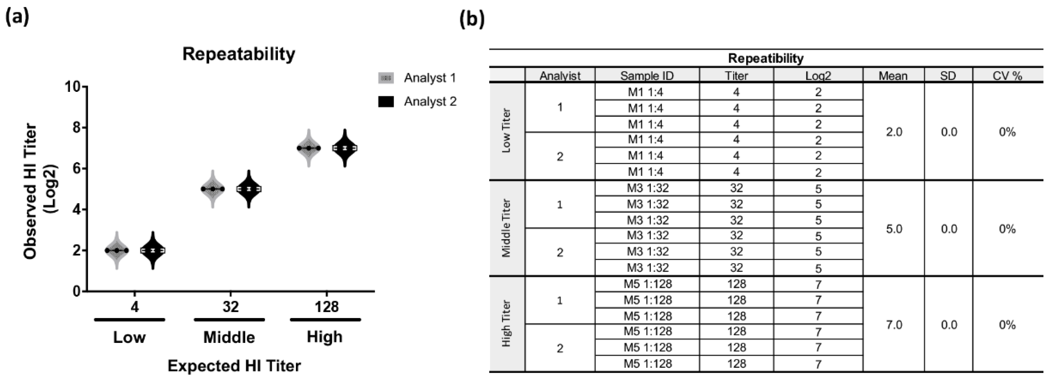
Based on the guidelines for the validation of analytical methods using statistical parameters, repeatability represents the distribution of results from repeated independent determinations of a sample under specific conditions. In this regard, the results obtained by the analysts, analyzed independently on different days, yielded a standard deviation (SD) and a coefficient of variation (CV) equal to 0 at the three levels of quantification evaluated. The student’s t-test showed that there are no statistically significant differences between the results obtained by each analyst, indicating that the test has the necessary precision to obtain consistent results from the same sample with an absence of inter-analyst variability (Figure 4).
The next parameter was to evaluate the intermediate precision; this represents the relative agreement obtained when evaluating independent results under variable conditions in the same laboratory. The results obtained by Analyst 1 maintained a CV of 0 % at the three levels of quantification. Regarding the analysis of the data obtained by Analyst 2, an increase of one logarithm was observed at the low titer level on the second day of analysis. Despite the fact that the % CV observed was higher at the three evaluated levels (low: 20 %, medium: 9 %, and high: 6 %), these values remained below the established acceptance criteria of the test. This was complemented by applying the t-test, where it was demonstrated that there are no significant differences between the results of both analysts, obtaining a p-value of 0.712. This confirms that the behavior of the results obtained under the established experimental conditions is sufficiently consistent, which guarantees their reliability and truthfulness (Figure 5).
To assess the agreement between the observed value and the expected value of the method, the accuracy of the assay was calculated by determining the recovery percentages at three concentration levels. The results obtained showed individual recovery percentages of 113%, 105%, and 104%, and CV values of 20%, 9%, and 6% for the low, medium, and high levels, respectively. In none of these cases were the established acceptance criteria for the test exceeded. These results indicate that the assay maintains its accuracy and precision at the different concentration intervals evaluated.
Figure 5.
Results of intermediate precision of HI assay. The results were obtained by two analysts under the same conditions on different days from three human serum type AB enriched with pig serum positive for NDV at three different levels of HI titer: 1:4 (low), 1:32 (middle) and 1:128 (high). Each result was obtained from samples evaluated from triplicate. (a) Representative graph of the average values obtained by each of the analysts in the different concentrations. (b) Summary table of results obtained at the levels of titers expected and obtained by each analyst, as well as the values obtained from the average, SD and CV%.
To ensure that the results obtained directly or by a mathematical transformation are proportional to the concentration or inhibition titer present in the sample, the linearity of the assay was determined. The results obtained corresponded to those expected when observing a linear correlation between the low and high levels; however, at the middle level, a logarithmic increase in the observed values of the expected HI titer of 1:64 was observed. Despite this, the general linear trend was maintained with a coefficient of determination (R²) of 0.9725 and a CV of 0 at each of the points. These results demonstrate that the established assay complies with this condition and presents linearity in the concentration interval evaluated, which guarantees the reliability for the issuance of accurate results. (Figure 6).
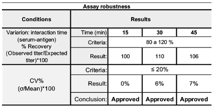
Finally, to assess whether the hemagglutination inhibition assay has the ability to maintain its precision and accuracy in the face of variations in the established operating conditions, the robustness test was evaluated by modifying the interaction time between the serum samples and the NDV antigen. The results obtained demonstrate that the assay is robust and maintains its performance, as neither the CV% nor the % recovery exceeded the acceptance criteria when the serum-antigen interaction times were changed (Table 4). Based on the results obtained in each of the tests, we conclude that the assay designed to evaluate the inhibitory capacity of human sera with the HI assay against the NDV antigen possesses the analytical attributes of consistency, reliability, and truthfulness of the results obtained, making it a useful tool for evaluating the immune response in serological studies.
4. Discussion
In this work, we report the different technical and analytical performance elements needed to develop and validate a serological assay that allows us to evaluate the level of antibodies against NDV vaccine vector in human sera using the HI assay. Previous reports have described that it is possible to use RBC obtained from the same species as the sera to be analyzed by HI or in combination with those of other species as a testing reading system [6]. In this way, to obtain the optimal conditions to establish the assay robustly and reproducibly, we made combinations between RBC from three different species and sera belonging to these same species, intending to implement and validate the assay in an easy, fast, and reliable way aligned with the international norms of adequacy and validation of analytical methods, under the criteria of ISO17025:2017. Based on the results obtained and considering factors such as availability, traceability, and stability of the biological material, we used chicken RBC, coming from animals specifically destined for this purpose. In the standardization tests, we observed that chicken RBC settle in an adequate time and present an easily observable shift pattern concerning the tested mammalian RBC, which ensures that we can assess their integrity and functionality during the test, guaranteeing that we can define samples with subtle changes in inhibitory activity more precisely compared to those without such changes. We also evaluated during standardization was the use of pre-treatments to eliminate potential non-specific inhibitors present in human serum samples. Since, sera pre-treatments such as heating the sample at temperatures above 65 °C, pre-adsorption with RBCs, and mixing the sample with kaolin or RDE solutions have been reported [17]; Therefore, we evaluated some of these treatments both individually and in combination, aiming to optimize and appreciate a complete agglutination pattern or eliminate possible interferences in the reading. However, we did not observe substantial differences between the use of kaolin and RDE with the pretreatment of 5% chicken RBC, as reported by another working group [16], with the advantage that the 5% chicken RBC has minor dilution of the sample. Similarly, treatments with kaolin or RDE dilute the sample ten-fold in the first dilution, making it impossible to detect low antibody titers, which is a disadvantage for the assay if it aims to identify the smallest potential response induced by the NDV vaccine vector. It is known, at least in one report, that the titers of infected people are not higher than 1:32 [11]. In another report made by Boliar et al, 2006, it was indicated that they do not have an effect for determining titers correctly in the HI assay [16]. Meanwhile, the pre-adsorption treatment with chicken RBC suspension results in a two-fold dilution of the sample and does not interfere with the assay reading. We found several discrepancies concerning the dilution values of the reported samples; they do not agree with the experimental design described, suggesting a discordance with the concept between proportion and dilution. Therefore, in our opinion, it is not trivial to omit the working dilution [16,24]. For the above, we considered the use of RBC to carry out the HI assay to adsorb the samples and reduce nonspecific agglutination; these conditions were used to perform the method validation. According to the guidelines of the Eurachem guide, analytical methods adopted from standards or a commercial system require verification to ensure correct implementation in the laboratory [21]. However, when modifications are made to an existing method or the methodology is developed internally, a validation process is necessary. For this reason, we submitted our modified method to the validation process, aiming to ensure that the results provided were accurate, reliable, and truthful for the analysis of human sera [22]. The tests of selectivity, sensitivity, specificity, PPV, NPV, PDR, and NDR are qualitative parameters that determine the degree of interference of the matrix in the assay and evaluate the probability of obtaining positive or negative results when there is or is not the presence of anti-NDV antibodies [21]. The parameters considered in the validation showed that the assay is specific and sensitive for the detection of positive or negative samples under the tested conditions. The accuracy was evaluated by analyzing repeatability and intermediate precision. Repeatability allows the evaluation of the variability of results obtained under practically identical conditions in short periods or with the same analyst, while intermediate precision allows the evaluation of variability between different days, analysts, or equipment, respectively [22]. The results indicate that the method is accurate, as no significant variation was observed in the conditions evaluated in our laboratory; thus, the method meets the proposed use objective. However, it is essential to recognize that the validation of the test is limited by the absence of reproducibility studies. While the publication of the International Conference on Harmonization of Technical Requirements for Registration of Pharmaceuticals for Human Use (ICH) establishes reproducibility as an essential parameter to evaluate the test in two different laboratories, the results obtained suggest that the method is suitable for use in our specific environment [22]. The results gained in terms of accuracy and linearity of the method were consistent with those reported by Morokutti and collaborators [4], in a modified HI test to evaluate antibodies against the influenza virus. Finally, our results demonstrated that the interaction time between antigen and sample, within a range of 15 to 45 minutes of incubation of serum samples and the NDV antigen, does not significantly influence the test results. This provides flexibility for the method, making it an easy, fast, and effective tool for detecting antibodies against NDV in human serum from individuals vaccinated with vaccines based on the NDV virus or received a prophylactic and therapeutic treatment based in NDV virus [25]. Previous studies evaluating anti-vector antibodies in adenoviral platforms demonstrated the utility for elucidating the effects of homologous or heterologous immunization regimens, concluding that homologous regimens decrease the production of antibodies against the antigen of interest while heterologous regimens favor the decrease of anti-vector antibodies and promote the production of the antigen of interest [26,27]. Therefore, the use of the new vaccines that include the NDV viral vector and other vectors has great potential and makes it necessary to have sensitive and validated assays that allow the identification the production of neutralizing antibodies against NDV in clinical studies where this vector is utilized. Additionally, experimental validation of the HI assay is essential to guarantee the reliability of the results developed in research laboratories. This supports the request for sanitary registration before the competent authority, requested through a Common Technical Document (CTD) developed by the ICH [28].
5. Conclusions
The tests carried out for the validation of the serological assay, "Analysis Method to Evaluate the Inhibitory Capacity of Human Sera with the Hemagglutination Inhibition (HI) Technique for Newcastle Disease Antigen," demonstrated that the method is statistically selective, sensitive, and specific, indicating high diagnostic reliability, as well as satisfactory results in the acceptance criteria of positive or negative predictive value. The method is accurate and precise, and it is observed that there is a linear correspondence between the expected titer value and the observed titer value, as well as being robust against the evaluated variations. These variations did not affect the results in the analysis to evaluate the inhibitory capacity of human sera with HI technique for the NDV antigen. Therefore, we conclude that the suitability of the analytical method for its intended use is confirmed and guarantees the reliability of the results obtained under the established operating conditions.
Author Contributions
Conceptualization, M.N., E.R, M.T. and H.Z.; Methodology, M.N. E.R, G.A., M.R. and H.Z; assay development and validation, A.P, C.B., M.N., E.R., and H.Z.; formal analysis, A.P, C.B., M.N., E.R., and H.Z.; investigation, M.N., E.R., and H.Z.; resources, C.C., M.T., and B.L.; data curation, A.P., CB, and H.Z; writing—original draft preparation, M.N., E.R., M.T., and H.Z.; writing—review and editing, M.N., E.R., C.C., M.T and H.Z.; visualization, G.A., M.R, and L.R.; project administration, B.L., and M.T.; funding acquisition, C.C., and M.T. All authors have read and agreed to the published version of the manuscript. Authorship must be limited to those who have contributed substantially to the work reported.
Funding
This study was funded by Instituto Nacional de Enfermedades Respiratorias “Ismael Cosío Villegas”, INER and Laboratorio Avi-Mex, S.A. de C.V. (Avimex®).
Institutional Review Board Statement
This study is a part of a research protocol conducted in accordance with the Declaration of Helsinki; reviewed and approved by the Research Ethic and Biosecurity Committees of the Instituto Nacional de Enfermedades Respiratorias “Ismael Cosío Villegas” (INER), with approval code C49-22 and approved date on November 09, 2022 for studies involving analysis with human samples. DCV, S.A. de C.V., is authorized by the General Directorate of SENASICA Mexico (Health, Safety and Agri-Food Quality Service) as a Zoosanitary Clinical Diagnostic Laboratory with number 116. Laboratorio Avi-Mex, S.A. de C.V. (Avimex®), provided the pig sera used as positive controls and its protocol was approved by the Research Committee for the Care and Use of Animals in Experiments (CICUAE-FESC), National Autonomous University of Mexico. Approval Code: C24_42; Approval Date: December 09, 2024, for studies involving animals. The Pig red blood cells provided by the INER´s Biotherium is a part of a research protocol reviewed and approved by The Research Ethic Committee from the Instituto Nacional de Enfermedades Respiratorias “Ismael Cosío Villegas” (INER), with approval code B18-20 and approved date on June 21st, 2021 for studies involving animals.
Informed Consent Statement
Informed consent is not required for de HI assay validation process.
Data Availability Statement
The data presented in this study are available upon request from the corresponding author.
Acknowledgments
We would like to extend our thanks to MBA Monica Pérez for her technical support in the validation data analysis; to the sample bank managers Dulce Cinthia Soriano Hernández and Montserrat A. Garcia Ramos, and to Francisco Enrique Camacho, for his invaluable support with the edition of the English language.
Conflicts of Interest
The authors declare that the research was conducted in the absence of any commercial or financial relationships that could be construed as a potential conflict of interest.
References
- International Committee on Taxonomy of Viruses (ICTV). Paramyxoviridae: Orthoavulavirus. [Online]. https://ictv.global/report/chapter/paramyxoviridae/paramyxoviridae/orthoavulavirus (accessed Jan 29, 2025).
- Rasekhi Kazeruni, A.; Babaei, N.; Esmaeili Gouvarchin Ghaleh, H.; Doosti, A.; Farzanehpour, M. Newcastle Disease Virus Enhances the Antitumor Efficacy of Doxorubicin in a Cervical Cancer Mouse Model. BMC Cancer 2024, 24 (1), 1253. [CrossRef]
- Suarez, D. L.; Miller, P. J.; Koch, G.; Mundt, E.; Rautenschlein, S. Newcastle Disease, Other Avian Paramyxoviruses, and Avian Metapneumovirus Infections. In Diseases of Poultry, 14th ed.; Swayne, D. E., Boulianne, M., Logue, C. M., McDougald, L. R., Nair, V., Suarez, D. L., Wit, S., Grimes, T., Johnson, D., Kromm, M., Prajitno, T. Y., Rubinoff, I., Zavala, G., Eds.; John Wiley & Sons: Hoboken, NJ, 2020; Chapter 3. [CrossRef]
- Yusoff, K.; Tan, W. S. Newcastle disease virus: macromolecules and opportunities. Avian Pathol. 2001, 30 (5), 439-455. ). [CrossRef]
- Morokutti, A., Redlberger-Fritz, M., Nakowitsch, S., Krenn, B. M., Wressnigg, N., Jungbauer, A., Romanova, J., Muster, T., Popow-Kraupp, T., & Ferko, B. Validation of the modified hemagglutination inhibition assay (mHAI), a robust and sensitive serological test for analysis of influenza virus-specific immune response. Journal of clinical virology: the official publication of the Pan American Society for Clinical Virology, 2013. 56(4), 323–330. [CrossRef]
- Makkoch, J., Prachayangprecha, S., Payungporn, S., Chieochansin, T., Songserm, T., Amonsin, A., & Poovorawan, Y. Erythrocyte binding preference of human pandemic influenza virus a and its effect on antibody response detection. Annals of laboratory medicine. 2012. 32(4), 276–282. [CrossRef]
- Kaufmann, L., Syedbasha, M., Vogt, D., Hollenstein, Y., Hartmann, J., Linnik, J. E., & Egli, A. An Optimized Hemagglutination Inhibition (HI) Assay to Quantify Influenza-specific Antibody Titers. Journal of visualized experiments : JoVE. 2017. (130), 55833. [CrossRef]
- Fulber JPC, Kamen AA. Development and Scalable Production of Newcastle Disease Virus-Vectored Vaccines for Human and Veterinary Use. Viruses. 2022. May 6;14(5):975. [CrossRef]
- Duan, Z., Xu, H., Ji, X., & Zhao, J. Recombinant Newcastle disease virus-vectored vaccines against human and animal infectious diseases. 2015. Future Microbiology, 10(8), 1307–1323. [CrossRef]
- Kim, S.-H.; Samal, S.K. Newcastle Disease Virus as a Vaccine Vector for Development of Human and Veterinary Vaccines. Viruses 2016, 8, 183. [CrossRef]
- Nelson, C. B., Pomeroy, B. S., Schrall, K., Park, W. E., & Linderman, R. J. An outbreak of conjunctivitis due to Newcastle disease virus (NDV) occurring in poultry workers. American journal of public health and the nation’s health. 1952. 42(6), 672–678. [CrossRef]
- López-Macías, C.; Torres, M.; Armenta-Copca, B.; Wacher, N. H.; Castro-Castrezana, L.; Colli-Domínguez, A. A.; Rivera-Hernández, T.; Torres-Flores, A.; Damián-Hernández, M.; Ramírez-Martínez, L.; la Rosa, G. P.; Rojas-Martínez, O.; Suárez-Martínez, A.; Peralta-Sánchez, G.; Carranza, C.; Juárez, E.; Zamudio-Meza, H.; Carreto-Binaghi, L. E.; Viettri, M.; Romero-Rodríguez, D.; ... Lozano-Dubernard, B. Phase II Study on the Safety and Immunogenicity of Single-Dose Intramuscular or Intranasal Administration of the AVX/COVID-12 "Patria" Recombinant Newcastle Disease Virus Vaccine as a Heterologous Booster Against COVID-19 in Mexico. Vaccine 2025, 43 (Pt 2), 126511. [CrossRef]
- Santra S, Seaman MS, Xu L, Barouch DH, Lord CI, Lifton MA, Gorgone DA, Beaudry KR, Svehla K, Welcher B, Chakrabarti BK, Huang Y, Yang ZY, Mascola JR, Nabel GJ, Letvin NL. Replication-defective adenovirus serotype 5 vectors elicit durable cellular and humoral immune responses in nonhuman primates. J Virol. 2005. May;79(10):6516-22. [CrossRef]
- Bautista-Juárez J. Factores que intervienen en la reacción antígeno-anticuerpo y clasificación antigénica eritrocitaria. Revista Médica del IMSS. 2005. (43) 9-12. https://www.medigraphic.com/pdfs/imss/im-2005/ims051c.pdfBautista-Juárez J. Factores que intervienen en la reacción antígeno-anticuerpo y clasificación antigénica eritrocitaria. Revista Médica del IMSS. 2005. (43) 9-12. https://www.medigraphic.com/pdfs/imss/im-2005/ims051c.pdf.
- Food and Agriculture Organization of the United Nations. Manual of Procedures for the Implementation of the Mobile Desert Locust Information Surveillance System. [Online]; FAO: Rome, 2002; https://www.fao.org/4/AC802E/ac802e00.htm (accessed Apr 01, 2024).
- Boliar, S., Stanislawek, W., & Chambers, T. M. Inability of kaolin treatment to remove nonspecific inhibitors from equine serum for the hemagglutination inhibition test against equine H7N7 influenza virus. Journal of veterinary diagnostic investigation: official publication of the American Association of Veterinary Laboratory Diagnosticians, Inc. 2006. 18(3), 264–267. [CrossRef]
- Kim, H. R., Lee, K. K., Kwon, Y. K., Kang, M. S., Moon, O. K., & Park, C. K. Comparison of serum treatments to remove nonspecific inhibitors from chicken sera for the hemagglutination inhibition test with inactivated H5N1 and H9N2 avian Influenza A virus subtypes. Journal of veterinary diagnostic investigation : official publication of the American Association of Veterinary Laboratory Diagnosticians, Inc. 2012. 24(5), 954–958. [CrossRef]
- World Health Organization. Manual for the Laboratory Diagnosis and Virological Surveillance of Influenza [Online]; WHO, 2011. https://www.who.int/publications/i/item/manual-for-the-laboratory-diagnosis-and-virological-surveillance-of-influenza (accessed Apr 01, 2024).
- Hidalgo-Lara, D. R., De la Luz-Armendáriz, J., Rivera-Benítez, J. F., Gomez-Nuñez, L., Salazar-Jiménez, E. N., Madrigal-Valencia, T. L., & Ramírez-Mendoza, H. Comparison of hemagglutination inhibition tests, immunoperoxidase monolayer assays, and serum neutralizing tests in detecting antibodies against blue eye disease in pigs. Journal of immunological methods. 2021. 496, 113088. [CrossRef]
- Rabenau, H. F., Kessler, H. H., Kortenbusch, M., Steinhorst, A., Raggam, R. B., & Berger, A. Verification and validation of diagnostic laboratory tests in clinical virology. Journal of clinical virology : the official publication of the Pan American Society for Clinical Virology. 2007. 40(2), 93–98. [CrossRef]
- EURACHEM. Quantifying Uncertainty in Analytical Measurement, 3rd ed.; EURACHEM: London, 2012; https://www.eurachem.org/index.php/publications/guides/mv (accessed Apr 01, 2024).
- European Medicines Agency. ICH Q2(R2) Validation of Analytical Procedures - Scientific Guideline [Online]. https://www.ema.europa.eu/en/ich-q2r2-validation-analytical-procedures-scientific-guideline(accessed Apr 01, 2024).
- Sánchez, R. Validación de Métodos Analíticos no Cuantitativos. Rev. Mex. Cienc. Farm. 2010, 4 (12), 15-24.
- Trombetta, C. M., Remarque, E. J., Mortier, D., & Montomoli, E. Comparison of hemagglutination inhibition, single radial hemolysis, virus neutralization assays, and ELISA to detect antibody levels against seasonal influenza viruses. Influenza and other respiratory viruses, 2018. 12(6), 675–686. [CrossRef]
- Yang, H., Tian, J., Zhao, J., Zhao, Y., & Zhang, G. The Application of Newcastle Disease Virus (NDV): Vaccine Vectors and Tumor Therapy. Viruses. 2024. 16(6), 886. [CrossRef]
- Cervantes-Torres, J., Cabello-Gutiérrez, C., Ayón-Núñez, D. A., Soldevila, G., Olguin-Alor, R., Diaz, G., Acero, G., Segura-Velázquez, R., Huerta, L., Gracia-Mora, I., Cobos, L., Pérez-Tapia, M., Almagro, J. C., Suárez-Güemes, F., Bobes, R. J., Fragoso, G., Sciutto, E., & Laclette, J. P. Caveats of chimpanzee ChAdOx1 adenovirus-vectored vaccines to boost anti-SARS-CoV-2 protective immunity in mice. Applied microbiology and biotechnology. 2024. 108(1), 179. [CrossRef]
- Byazrova, M. G., Astakhova, E. A., Minnegalieva, A. R., Sukhova, M. M., Mikhailov, A. A., Prilipov, A. G., Gorchakov, A. A., & Filatov, A. V. Anti-Ad26 humoral immunity does not compromise SARS-COV-2 neutralizing antibody responses following Gam-COVID-Vac booster vaccination. NPJ vaccines. 2022. 7(1), 145. [CrossRef]
- European Medicines Agency. ICH M4Q - Common technical document for the registration of pharmaceuticals for human use - Quality (scientific guideline). EMA [Online]. https://www.ema.europa.eu/en/ich-m4q-common-technical-document-registration-pharmaceuticals-human-use-quality-scientific-guideline. (accessed Apr 01, 2024).
Figure 1.
Representative images of NDV antigen titration, control of HU for HI assay and controls of HI assay. (a) NDV antigen was diluted 1:2 and 1:5 in triplicate (rows 1, 2 and 3) and the titration was assessed by serial dilutions. The titer is the last dilution where hemagglutination of the RBC is observed (green box). The orange triangle box, represents the concentration of NDV antigen. (b) The control test HU for IH assay. The NDV antigen suspension at 8 HU is loaded in position A1 to A3 and serial two-fold dilutions were performed (8, 4, 2, 1 and 0.5 HU) at 0.5 HU indicates that the agglutination is partial (light blue box), in column A4 to H4 is the RBC integrity control. (c) Controls used in HI Assay; Positive control (C+) hyperimmune serum from pathogen-free chicken immunized with the attenuated NDV vaccine (A1-H1), with an established titer of 1:256 (purple box); Negative control (C-) non immunized serum from pathogen-free chicken (A2-H2), Integrity control of the suspension RBC from pathogen-free chicken obtained from peripheral blood, used as a read-out system (A3-H3). The integrity of the RBC was determined with the test of the displacement of the precipitate forming a "teardrop" when the plate was tilted at an angle of 45°, used as part of the HI trial acceptance criteria.
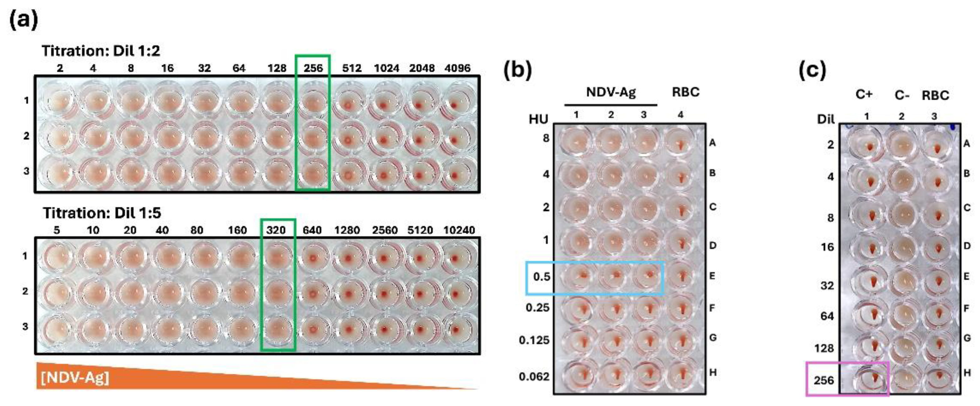
Figure 2.
The effect of pretreatments on serum samples in the HI assay. (a) 1X PBS with a 1:1 v/v ratio at 4 °C for 16-18 h, (initial dilution 1:4). (b) 5 % RDE and 5 % RBC, 1:5:4 v/v/v ratio at 4 °C for 16-18 h, (initial dilution 1:20). (c) 25 % kaolin solution, and 5 % RBC 1:2:2 v/v/v ratio at 4 °C for 16-18 h (Initial dilution 1:10). (d) absorption with 5 % RBC 1:1 v/v ratio at 4 °C for 16-18 h (initial dilution 1:4). (S1) Human serum sample vaccinated against SARS-CoV-2 with AVX/COVID-12, (S2) pig serum sample vaccinated against SARS-CoV-2 with AVX/COVID-12 and (S3) human serum sample negative to NDV.
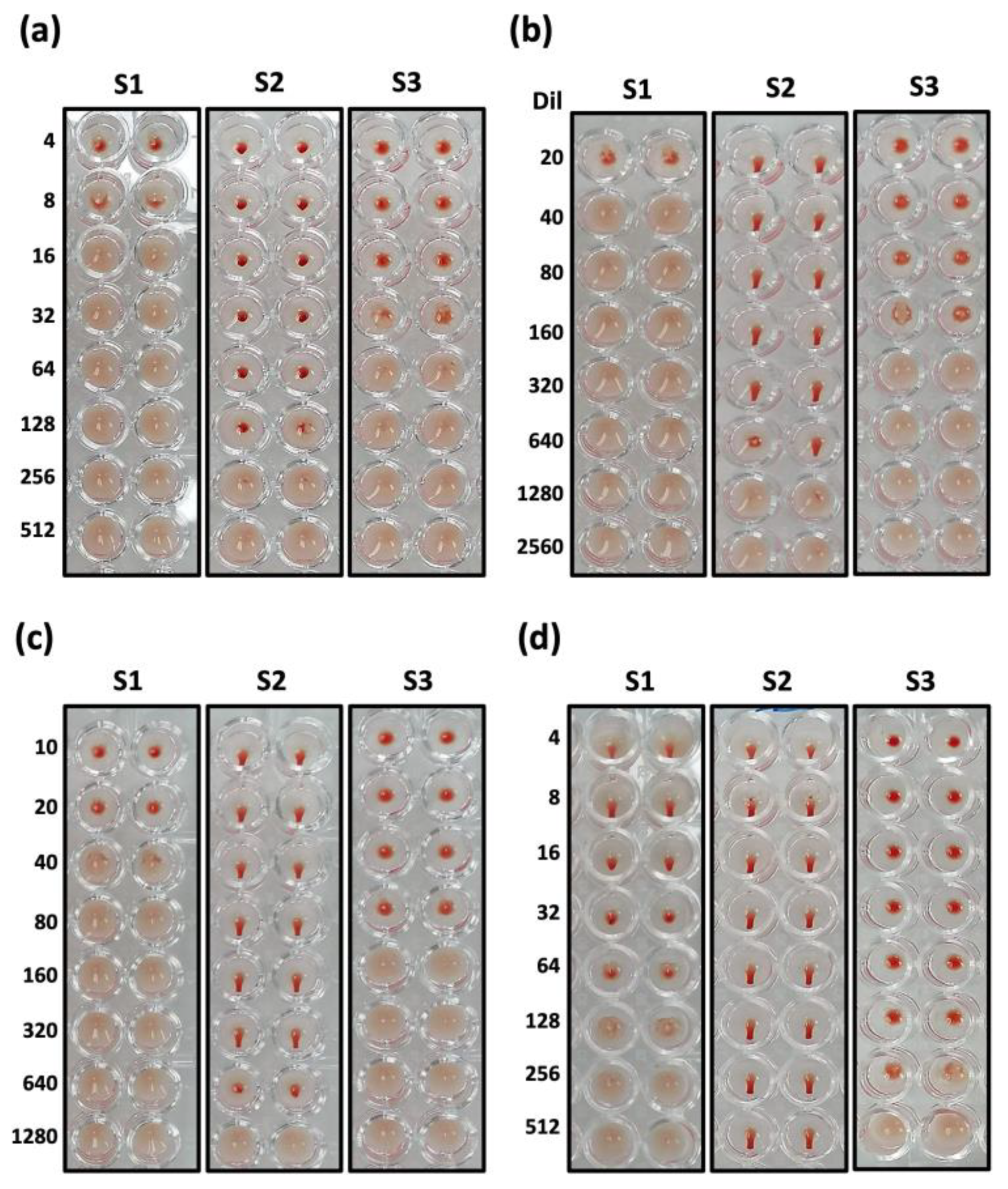
Figure 3.
Analytical performance of HI assay. The results to experimentally evaluate the qualitative selectivity parameter were obtained by one analyst from 20 samples of unrelated viral respiratory disease enriched with pig serum from negative for NDV and positive for NDV at different levels of HI titer. Each result was obtained from samples evaluated from triplicate. (a) Representative graph of the titers obtained in Log2 of the samples analyzed, indicating the cut-off point 2 established with the positive samples. (b) Summary of the calculations for the evaluation of analytical selectivity using the Xi2 statistical test.
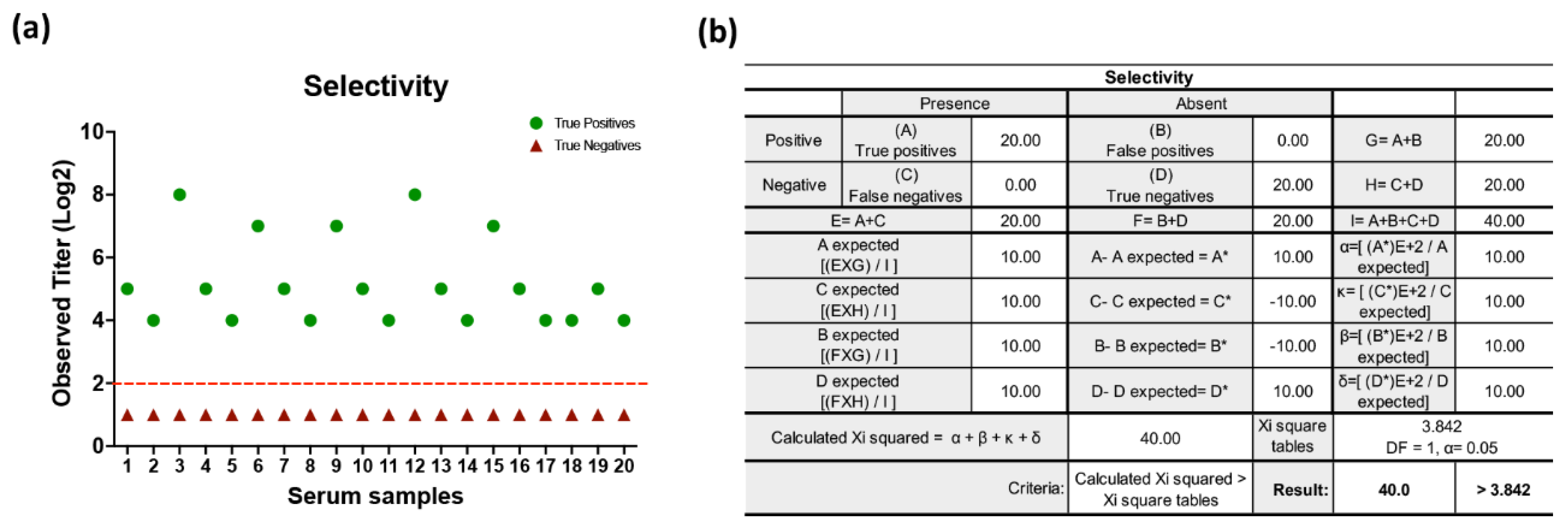
Figure 4.
Results of repeatability for HI assay. The results were obtained by two analysts under the same analysis conditions on different days from three human serum type AB enriched with pig serum positive for NDV at three different levels of HI titers: 1:4 (low), 1:32 (middle) and 1:128 (high). Each result was obtained from samples evaluated from triplicate. (a) Representative graph of the average values obtained by each of the analysts in the different concentrations analyzed. (b) Summary table of results obtained at the levels of titers expected and obtained by each analyst, as well as the values obtained from the average, SD and CV.
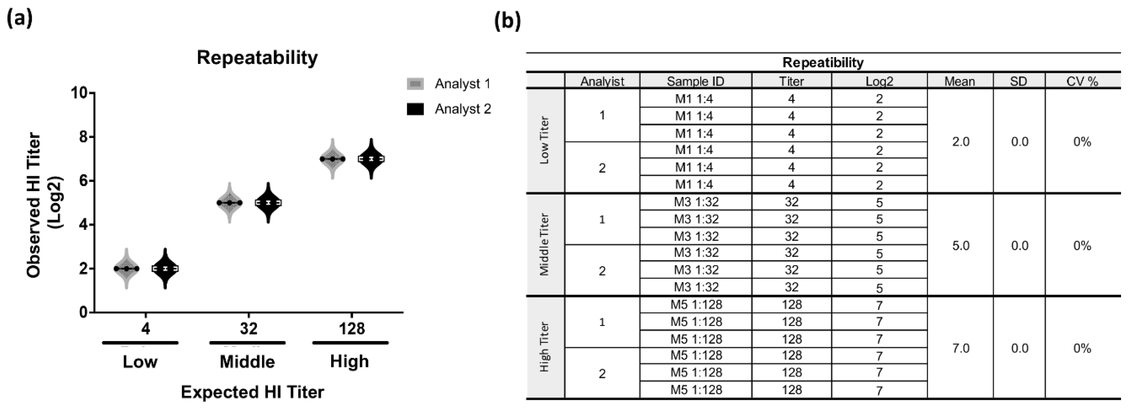
Figure 6.
Results of linearity parameter of HI. The results were obtained by one analyst from six human serum samples type AB enriched with pig serum positive obtaining HI titers value calculated at three levels of quantification: 1:4, 1:8 (low), 1:32, 1:64 (middle) and 1:128, 1:256 (high). Each result was obtained from samples evaluated from triplicate. (a) Representative graph of the linear regression test between the expected versus observed values of the HI titers and obtaining the coefficient of determination (R2) and the graph formula. (b) Table of results obtained at the titers levels expected and obtained from the assay and statistical values obtained.
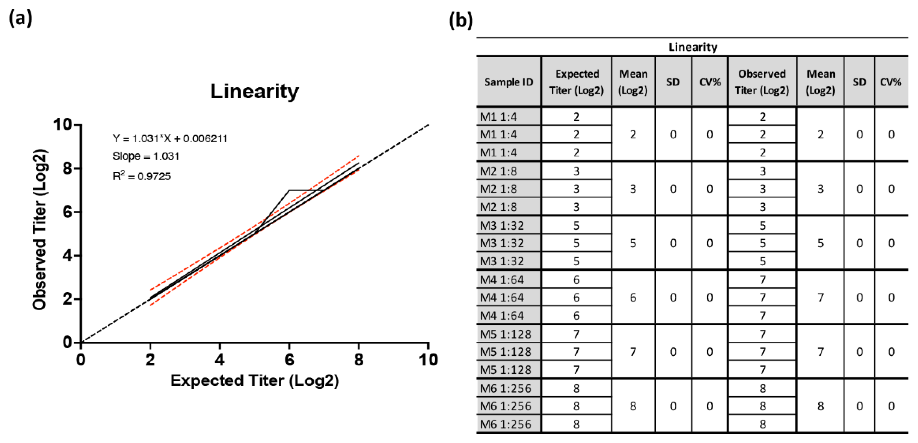

Table 1.
Summary tested conditions for standardization of RBC sedimentation and “teardrop” formation .
Table 1.
Summary tested conditions for standardization of RBC sedimentation and “teardrop” formation .
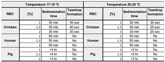
Table 2.
Statistical tests to evaluate the analytical performance of HI assay: Sensitivity, Specificity, PPV, NPV and acceptance criteria.
Table 2.
Statistical tests to evaluate the analytical performance of HI assay: Sensitivity, Specificity, PPV, NPV and acceptance criteria.
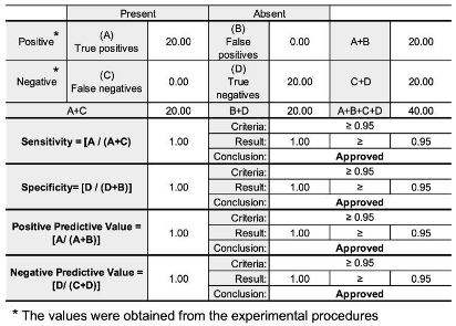
Table 3.
Statistical tests to evaluate the analytical performance of HI assay: PDR, NDR and acceptance criteria.
Table 3.
Statistical tests to evaluate the analytical performance of HI assay: PDR, NDR and acceptance criteria.
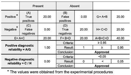
Table 4.
Summary of results obtained from HI assay to determine robustness. Robustness is a parameter that evaluates the influence of noise variations such as the interaction time between serum samples and the NDV antigen, 30 replicates of a sample of human serum type AB enriched with positive pig serum obtaining HI titer value calculated 1:128, subjecting to three different antigen-sample incubation times (15, 30 and 45 min). Each result was obtained from samples evaluated from triplicate.
Disclaimer/Publisher’s Note: The statements, opinions and data contained in all publications are solely those of the individual author(s) and contributor(s) and not of MDPI and/or the editor(s). MDPI and/or the editor(s) disclaim responsibility for any injury to people or property resulting from any ideas, methods, instructions or products referred to in the content. |
© 2025 by the authors. Licensee MDPI, Basel, Switzerland. This article is an open access article distributed under the terms and conditions of the Creative Commons Attribution (CC BY) license (http://creativecommons.org/licenses/by/4.0/).
Copyright: This open access article is published under a Creative Commons CC BY 4.0 license, which permit the free download, distribution, and reuse, provided that the author and preprint are cited in any reuse.


