Preprint
Article

This version is not peer-reviewed.

Breast Cancer in Young Women in Michoacan—A Retrospective Comparative Cohort Study

Submitted:

01 February 2025

Posted:

03 February 2025

You are already at the latest version

Abstract

Background: Invasive breast cancer (IBC) is the most prevalent neoplasm and the leading cause of death in women worldwide. Young women under 40 years of age, despite their low incidence, have different clinical-pathological characteristics than older women, giving them a worse prognostic profile. Objective: To determine the association between risk factors associated with MIC in young women in Michoacan. Methods: descriptive, observational, retrospective and cross-sectional study that included 1178 patients < 40 years of age with MIC diagnosed in the period 2015-2022 in a second-level hospital of the IMSS in Michoacán. Sample size at convenience, with database registration, with the use of means, standard deviation, chi-square and Student's t-test, p 0.05 as an indicator of significance. Registration Number: R-2022-1602-025. Results: of 1178 cases, 12.1% were in young women, who had an increased risk of tumors > 2 cm (p = 0.02, 1.062–2.058), ER–/RP– (p = 0.0067, 1.158–2.501) and triple negative (p = 0.0006; 1.377–5.563). Obesity III was associated with larger tumors (P = 0.0028, 1.051–3.518), advanced TNM stages (P = 0.0445), and greater tissue involvement (T3-T4, P = 0.0301). Conclusions: Breast cancer in young women in Michoacan showed worse prognostic clinical profiles compared to older women.

Keywords: 
;  ;  ;  ;  

1. Introduction

Invasive breast cancer (IBC) is the most prevalent neoplasm in women, with an estimated global incidence in 2020 of over 2.2 million cases [1]. In Mexico, an incidence of 19.3% of new cases in young women under 44 years of age was reported in 2022, which were mainly reported in the Mexican Institute of Social Security (IMSS) [2]. In that same year, in the State of Michoacan, 564 new cases were registered, more than double compared to 2020, with the municipalities of Morelia, Zamora, and Uruapan being the most affected [3].
In Mexico, more than 65% of cases are diagnosed in advanced stages [4], which translates to higher mortality, where more than 13% corresponds to young women [5]. This underscores the great challenge that early identification of risk factors associated with the patient's clinical context represents in our environment, such as: sex, age, age of menarche and menopause, age at first birth, parity index, and breastfeeding [4,6]. For example, the age of diagnosis in Mexico occurs in younger women compared to what is reported in developed countries [7], and they have a higher prevalence of poor prognosis tumors compared to older women [8], such findings have been a subject of our investigative efforts as previously submitted by our team [9].
Another important risk factor is elevated body mass index (BMI) which, by itself, constitutes a serious public health problem, with a higher incidence of IBC in Mexican women with overweight and obesity (41% and 30%, respectively) [10], and a higher risk for poor prognosis tumors [8]. Additionally, endogenous exposure to estrogens, such as the age of menarche and menopause at extreme ages, and its association with poor prognosis IBC [11,12]. These factors support the great diversity of clinical behaviors and treatment responses that characterize this complex disease [13].
Other associated factors are those that derive from tumor characteristics, the number of affected nodes, and distant metastases, which are classic prognostic factors integrated into systems for classification to define prognosis, predictive value for treatments, and survival estimation. The above recovers the relevance of early detection programs in the State of Michoacan, where understanding their relationship is essential to develop effective risk assessment and detection strategies for different age groups as has been implemented in other countries [14]; Therefore, this study sought to compare clinical, pathological, and molecular findings between women younger and older than 40 years of age in Michoacan.

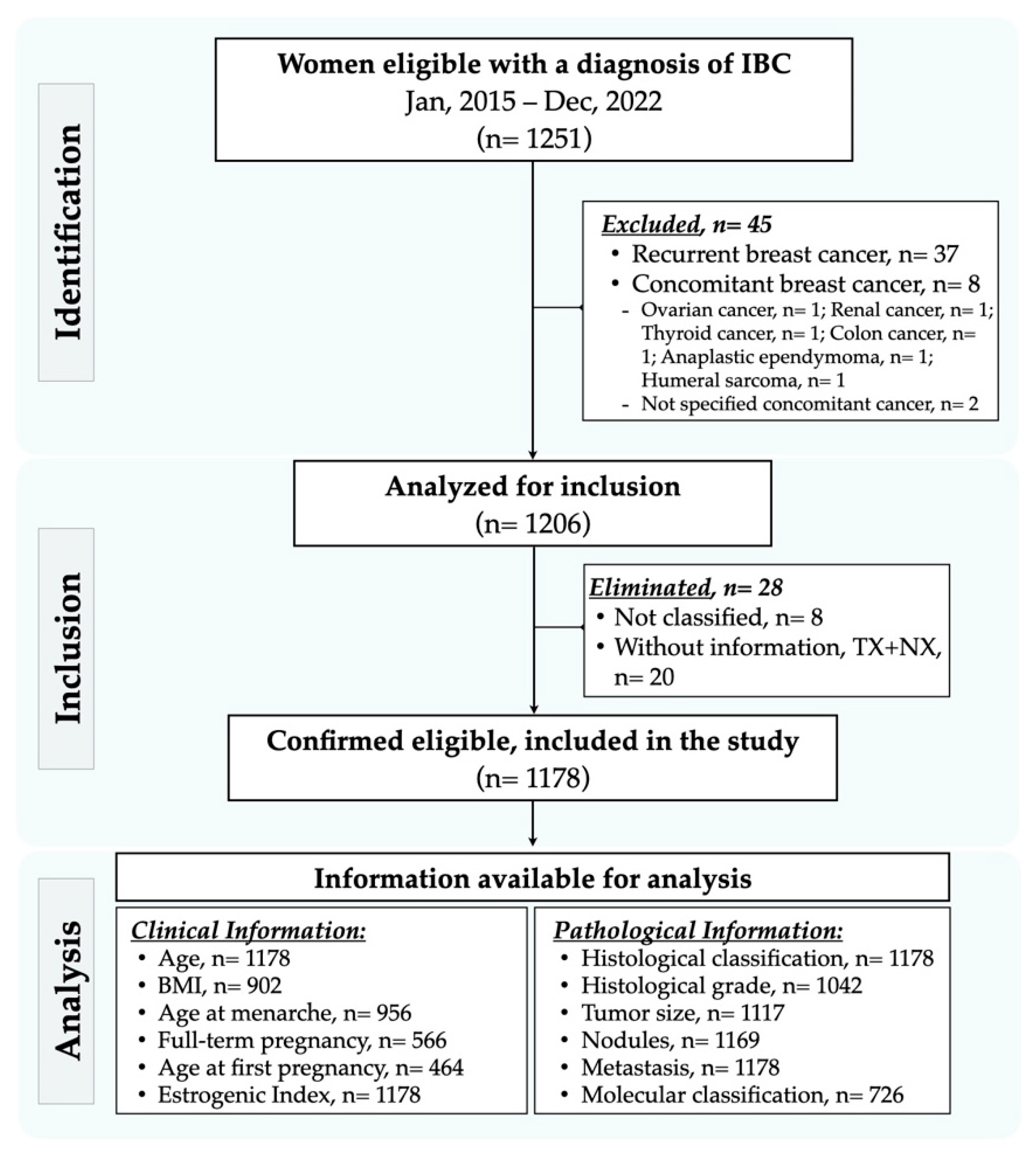
2. Materials and Methods
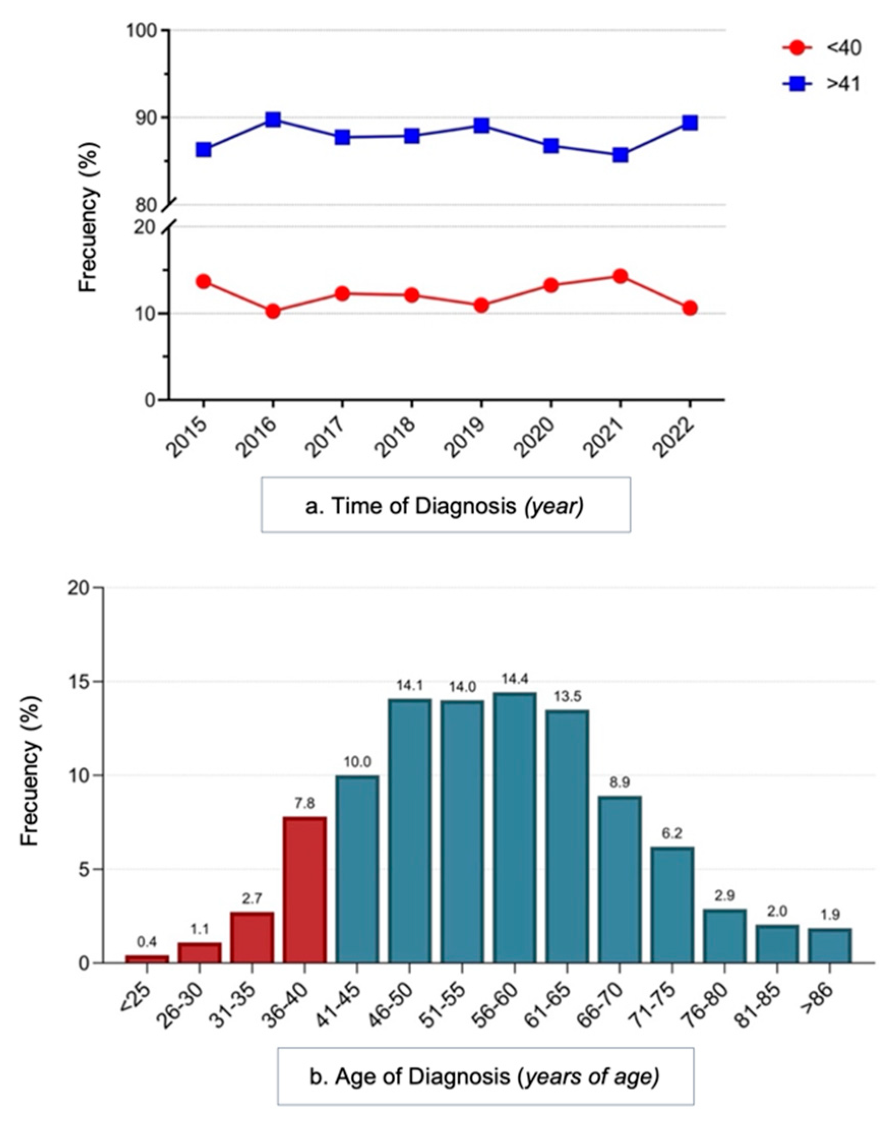
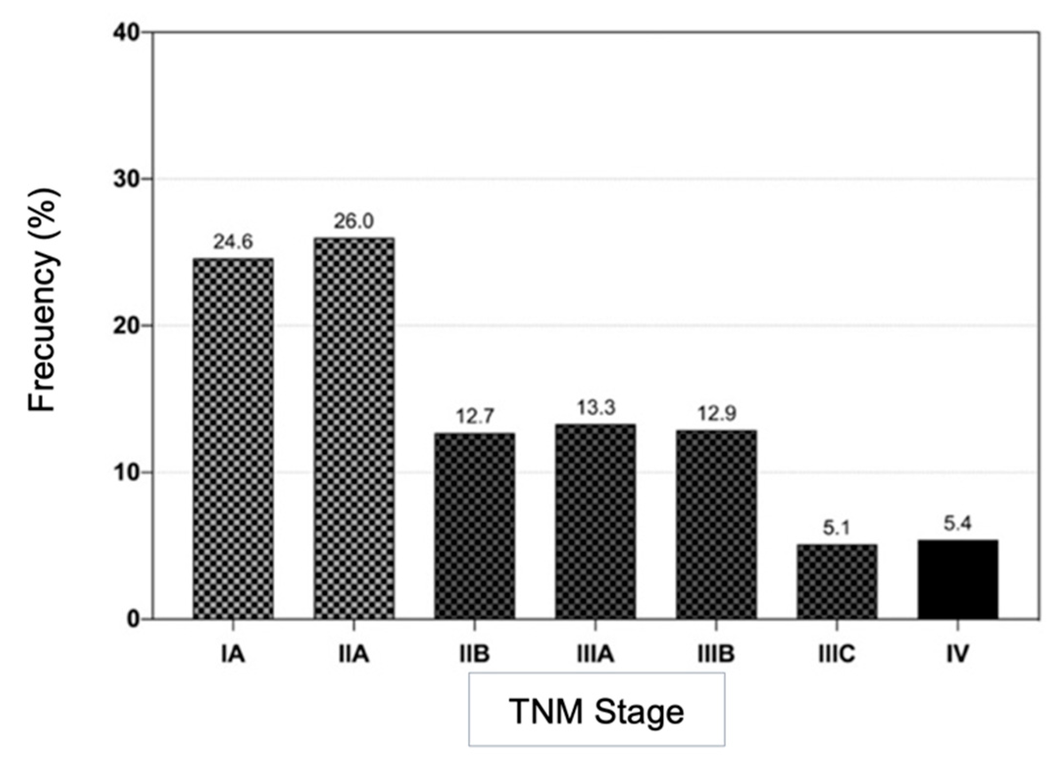
This was a descriptive, observational, retrospective, and cross-sectional cohort study conducted from January 2015 to December 2022 among beneficiaries of the IMSS at the Regional General Hospital No. 1 (HGR1) in Morelia, Michoacán, Mexico. The study included 1,178 women over 18 years of age selected through a systematic convenience sampling approach, wherein all eligible patients who met the inclusion criteria during the study period were consecutively enrolled. The study followed STROBE (Strengthening the Reporting of Observational Studies in Epidemiology) guidelines (Figure 1).
Patients were excluded if their clinical records were incomplete or if they had received initial treatment at another institution. Variables of interest were obtained through clinical record review: age at diagnosis, BMI (weight and height), and gynecological-obstetric history. Young women were defined as those aged 40 years or younger.
Variables of interest from the histopathological study included histological classification, lymphovascular permeation, and tumor size in centimeters. For cases where surgical specimens were obtained through mastectomy, quadrantectomy, tumorectomy, or excisional biopsy, these were identified as pT. In cases diagnosed through fine-needle aspiration biopsy (FNAB), core needle biopsy, sentinel lymph node biopsy, or core needle aspiration (CNA), tumor size was recorded based on clinical and/or ultrasound findings, identified as cT. Metastatic nodes were determined in cases with histopathological studies that included axillary extension of the breast. Distant metastasis was determined through extension studies such as nuclear magnetic resonance and axial tomography in selected cases as indicated by the oncologist.
Molecular studies for hormone receptor determination (estrogen and progesterone receptors), HER2, and Ki-67 were performed on tissue samples analyzed for initial diagnosis. These were conducted as subrogated studies. Cases with missing molecular data were included in the general analysis but excluded from molecular subtype analyses. Molecular subtypes were classified as luminal A (ER+ and/or PR+, HER2−), luminal B (ER+ and/or PR+, HER2+), HER2 (ER−, PR−, HER2+), and TN (ER−, PR−, HER2−).
Data analysis included absolute values (n), simple frequencies (percentages), measures of central tendency (arithmetic mean and median), lower and upper limits of values (LL-UL), measures of dispersion (standard deviation), and position measures (quartiles and percentiles). Normality testing was performed using the Shapiro-Wilk test. Based on normality results, differences between means were determined using Student's t-test for normally distributed data and Mann-Whitney test for non-normal distributions, while proportion comparisons used Chi-square test with odds ratio calculation and 95% confidence intervals (95% CI). Sample size was calculated using a power analysis with α = 0.05 and β = 0.20, based on previously reported prevalence rates. A p-value ≤ 0.05 was considered statistically significant. Analysis was performed using GraphPad Prism statistical package version 10.1.1 (GraphPad Software, San Diego, CA, USA, www.graphpad.com).
This study was evaluated and approved by the IMSS Local Ethics and Health Research Committee (R-2022-1602-025). All participants were informed of the research objectives and participated freely.

3. Results

3.1. Clinical Characteristics of the Study Population

A total of 1178 cases with a mean age (SD) of 55.8 ± 12.8 years (IQR 46-64, LL-UL of 24-94 years) were included in the period 2015-2022 (Figure 2a), of which 142 (12.1%) were young women <40 years, with a mean age of 35.8 ± 4.0 years (IQR 33–39) (Figure 2b). The most affected age group was 31-40 years old with 87.3% (124/142), with a higher concentration between 36 and 40 years old; while in older women, a higher proportion of cases was observed in those aged between 51-60 years (335 cases, 28.4%).
The mean BMI of the population was 28.9 ±5.4 kg/m2 (IQR 25.2-32, LL-UL of 14.4-49.4), significantly higher in women >41 years (Table 1).
Both young women and those over 40 years of age had normal age at menarche, however, young women with early menarche (<10 years of age) had a 4-fold increased risk of IBC (p=0.0202; RR 4.0; 95%CI: 1.211-13.37). Of those with one or more gestations, their mean age of the population was 22.2 ±5.19 years, with no differences between groups, with a greater number of nulls in those cases younger than 40 years, and a protective effect was observed after the 5th child (p < 0.0001, RR 2.560).

3.2. Pathological and Molecular Characteristics

The mean tumor size was 3.52 ±2.96 cm (range: 0.1–30 cm), most of which were infiltrating ductal carcinoma without specific pattern (CDI-SPE) with 79.4% of cases, followed by infiltrating lobular carcinoma (ILC) with 8.7% (Table 2).
Young women showed an increased risk of TN tumors (p=0.0110; RR 1,799; 95% CI 1.142-2.823), while higher hormone receptor-positive (HR+) receptors were significantly associated with older women (RR 1.2446; 95% CI 1,028-1,544), especially ER+ tumors (RR 1,555; 95% CI 1.168-2.051) and Luminal B (p=0.0290; RR 1,390; 95% CI 1.033-1.908) (Table 3).
The most frequent histological grade was moderately differentiated with 58.9% of cases, followed by high-grade (22.9%). Lymph node dissection was performed in 931 women, of whom 54.5% had nodules positive for neoplastic infiltration (mean of 4.75 ±4.8 nodes), while 48.8% had lymph node hyperplasia. Of the cases with lymph node metastases, 59.8% (116/463) had capsular rupture and 40.2% had adipose infiltration.
A total of 66 women were found to have metastases at the time of diagnosis, with distribution mainly to the lung and pleura with 29.6%, followed by bone with 19.4%. At diagnosis, most cases in young women were identified in early stages, followed by locally advanced stages (Figure 3).

4. Discussion

Our study provides important insights into breast cancer characteristics among Mexican women, with particular attention to age-related differences. The key findings demonstrate a mean age of 55.8 years at diagnosis, with a notable prevalence of cases in young women (<40 years) that is threefold higher than international reports [15]. In Mexico, worse prognosis has been reported in young women [16,17], likely due to different screening strategies in these age groups, leading to delayed diagnosis and poorer outcomes. En nuestro estudio, la edad promedio fue de 55.8 años, con predominio de casos en mujeres >41 años, lo cual coincide con otros estudios en México [18].
Age-related patterns in our population reveal significant clinical and molecular distinctions. Young women presented with larger tumors and more aggressive features, including higher rates of undifferentiated histology and triple-negative molecular subtypes. This finding aligns with previous studies but highlights a particularly concerning pattern in our Mexican population, where the prevalence of aggressive subtypes exceeds that reported in developed nations [19,20].
The relationship between BMI and breast cancer characteristics emerged as a significant factor in our analysis. Women with BMI >30 kg/m2 showed a 3.5-fold increased risk for larger tumors and greater involvement of surrounding tissues (T3-T4), compared to those with normal weight. This association with poor prognostic factors underscores the importance of weight management as a modifiable risk factor in breast cancer outcomes [21,22].
Regarding reproductive factors, our findings challenge some previously reported associations. While earlier studies suggested early menarche as a risk factor [11], our population showed age at menarche within normal ranges. Furthermore, we found that multiple full-term pregnancies conferred a protective effect in young women, supporting recent findings from our research group and others [9,23]. This highlights the complex interplay between reproductive history and breast cancer risk in different populations.
Most cases were diagnosed as infiltrating carcinomas, with IDC-NOS being the most frequent, followed by ILC. The most common histological grade was moderately differentiated, followed by high grade, where young women were more likely to present undifferentiated tumors. Based on this, histological grade has been found to be a significant predictor of overall survival and cancer-specific survival in breast cancer patients [24]. During our investigation, it was possible to investigate its correlation with age and size, mostly because some studies report a statistically significant correlation between histological grade, age, and tumor size when associated with ER, PR, and HER-2 positivity [25], which confirms age-related heterogeneity [6].
The molecular profile analysis revealed distinct patterns across age groups. Older women showed higher prevalence of hormone receptor-positive tumors, particularly estrogen receptor-positive cases. In contrast, younger women exhibited higher rates of triple-negative tumors and, unexpectedly, a protective effect against Luminal B subtypes compared to postmenopausal women. This molecular heterogeneity by age supports the growing evidence for age-specific approaches to breast cancer screening and treatment [26].
Disease staging and progression patterns in our cohort showed some distinctive features. Unlike national reports indicating late-stage predominance [4], our population showed a higher proportion of early-stage disease (IA-IIA). However, among young women who presented with metastatic disease (n=66), we observed a different pattern of metastatic spread compared to previous reports, with lung and pleural involvement predominating over bone metastases [27].
Limitations and Future Directions Several limitations should be considered when interpreting our results. First, the retrospective nature of the study and reliance on medical records led to incomplete clinical information for some parameters. Second, molecular typing was not available for all confirmed cases, potentially introducing selection bias in our molecular subtype analysis. Third, the single-center design may limit generalizability to other Mexican populations. Future prospective studies with comprehensive molecular profiling and longer follow-up periods would help validate these findings and explore their implications for clinical outcomes.

5. Conclusiones

Our research demonstrates significant heterogeneity between women under 40 years of age and older women with breast cancer. Non-modifiable risk factors, particularly age, play a crucial role in determining pathological and molecular characteristics associated with poor outcomes. Specifically, our findings reveal that younger women face substantially higher risks related to larger tumor sizes, lymph node involvement, metastatic spread, and hormone receptor status. These correlations highlight important age-dependent variations in disease presentation and progression.
The distinct pattern of disease characteristics in young women carries important implications for clinical practice and public health strategies. Our observations suggest that current screening and diagnostic approaches may need refinement to better serve this vulnerable population. Furthermore, the higher prevalence of aggressive disease features in younger patients underscores the necessity for age-specific risk assessment and management protocols.
These findings emphasize three critical areas for future development: (1) enhanced research into age-specific risk factors and disease mechanisms, (2) optimization of early detection strategies for young women, and (3) development of targeted interventions that account for age-related biological differences. Understanding these age-dependent variations is essential for improving prognostic accuracy, developing preventive strategies, and enhancing screening programs to address the unique challenges faced by young women with breast cancer.

Author Contributions

The research and manuscript preparation involved distinct contributions from each team member: A.G.R.-L. and S.G.-C. led the primary research efforts, including study conceptualization and design, A.G.R.-L. conducted the comprehensive clinical data analysis, development of visual representations through tables and figures, initial manuscript preparation. A.G.-G. provided expertise in statistical methodology, conducting thorough statistical analyses and subsequent validation of findings. J.A.D-P. ensured data integrity through rigorous clinical data validation processes. S.G.S-C. contributed to manuscript development and conducted validation procedures to ensure research quality. S.G.-C., as primary research advisor, provided comprehensive oversight of the project, including study conceptualization and design, clinical data analysis supervision, guidance on data visualization, and final manuscript preparation. All authors have reviewed and endorsed the published version of the manuscript, ensuring its academic integrity and scientific accuracy.

Funding

This research was conducted independently of external funding sources. The work represents a component of the principal author's thesis to obtain the grade as Family Medicine Especialist, completed under the academic endorsement of UNAM/CONACyT (CVU: 1030352). No financial support was received from public or private entities.

Acknowledgments

We extend our sincere appreciation to the following institutions and departments for their invaluable support throughout this research: The Teaching and Medical Education Coordination Department at UMF 80; The Department of Pathological Anatomy and its dedicated team at HGR1; The ARIMAC Department at HGR1 and the Michoacán Biomedical Research Center, specifically the Research Department of IMSS. Their collaborative support and expertise were instrumental in the successful completion of this study.

Conflicts of Interest

The authors have thoroughly reviewed potential conflicts of interest and formally declare that no competing interests exist that could have influenced the research process, findings, or presentation of results in this study.

References

  1. Ferlay J, Colombet M, Soerjomataram I, Parkin DM, Pineros M, Znaor A, et al. Cancer statistics for the year 2020: An overview. Int J Cancer. 2021;149(4):778–89. Epub 20210405. PubMed PMID: 33818764. [CrossRef]
  2. Secretaría de Salud, DGE. Anuario de Morbilidad 1984 - 2022 México: Secretaría de Salud; 2023 [cited 01/2025 01/2025]. Available from: https://www.gob.mx/salud/acciones-y-programas/anuarios-de-morbilidad-1984-a-2022.
  3. Rivera-Cruz F. Este año, en Michoacán 238 casos de cáncer de mama y 157 muertes. Gente del Balsas. 2021.
  4. Cárdenas-Sánchez J, Valle-Solís AA, Arce-Salinas C, Bargalló-Rocha JE, Bautista-Piña V, Cervantes-Sánchez MG, et al. Consenso Mexicano sobre diagnóstico y tratamiento del cáncer mamario. 10 ed. Colima, México. 2023. 266 p.
  5. INEGI. Estadísticas a propósito del día internacional de la lucha contra el cáncer de mama (19 de octubre). Comunicado de Prensa Núm. 595/23 [Internet]. 2023 [01/2025]. 10/2023:[1–7]. Available from: https://www.inegi.org.mx/contenidos/saladeprensa/aproposito/2023/EAP_CMAMA23.pdf.
  6. Moncada-Madrazo M A-GA, Isojo-Gutiérrez R, Issa-Villarreal ME, Elizondo-Granillo C, Ramos-Reyes A. et al Factores de riesgo modificables del cáncer de mama: una comparación entre mujeres menores y mayores de 40 años. Ginecología y Obstetricia de México. 2020;88(3):131-8. [CrossRef]
  7. Maffuz-Aziz A, Labastida-Almendaro S, Espejo-Fonseca A, Rodriguez-Cuevas S. [Clinical and pathological features of breast cancer in a population of Mexico]. Cir Cir. 2017;85(3):201-7. Epub 20160919. PubMed PMID: 27658545. [CrossRef]
  8. Alarcon Rojas CA, Alvarez-Banuelos MT, Morales-Romero J, Suarez-Diaz H, Hernandez-Fonseca JC, Contreras-Alarcon G. Breast Cancer: Metastasis, Molecular Subtypes, and Overweight and Obesity in Veracruz, Mexico. Clin Breast Cancer. 2019;19(1):e166-e71. Epub 20180822. PubMed PMID: 30236925. [CrossRef]
  9. Rocha López AG, Gómez-García A, Montecillo-Aguado M, Sánchez-Ceja SG, Huerta-Yepez S, Gutiérrez-Castellanos S. Risk Factors Associated to Poor Survival in Primary Breast Cancer of Premenopausal and Postmenopausal Patients: A Mexican-Based Population Study. Preprints: Preprints; 2025.
  10. Reynoso-Noveron N, Villarreal-Garza C, Soto-Perez-de-Celis E, Arce-Salinas C, Matus-Santos J, Ramirez-Ugalde MT, et al. Clinical and Epidemiological Profile of Breast Cancer in Mexico: Results of the Seguro Popular. J Glob Oncol. 2017;3(6):757-64. Epub 20170208. PubMed PMID: 29244990; PubMed Central PMCID: PMC5735969. [CrossRef]
  11. Lozano-Esparza S, Jansen EC, Hernandez-Avila JE, Zamora-Munoz S, Stern D, Lajous M. Menarche characteristics in association with total and cause-specific mortality: a prospective cohort study of Mexican teachers. Ann Epidemiol. 2021;62:59-65. Epub 20210622. PubMed PMID: 34166807. [CrossRef]
  12. Rojas-Lima E, Gamboa-Loira B, Cebrian ME, Rothenberg SJ, Lopez-Carrillo L. A cumulative index of exposure to endogenous estrogens and breast cancer by molecular subtypes in northern Mexican women. Breast Cancer Res Treat. 2020;180(3):791-800. Epub 20200222. PubMed PMID: 32086656. [CrossRef]
  13. Tan J, Le A. The Heterogeneity of Breast Cancer Metabolism. Adv Exp Med Biol. 2021;1311(1311):89-101. PubMed PMID: 34014536; PubMed Central PMCID: PMC9703239. [CrossRef]
  14. Tran TXM, Moon SG, Kim S, Park B. Association of the Interaction Between Mammographic Breast Density, Body Mass Index, and Menopausal Status With Breast Cancer Risk Among Korean Women. JAMA Netw Open. 2021;4(12):e2139161. Epub 20211201. PubMed PMID: 34940866; PubMed Central PMCID: PMC8703253. [CrossRef]
  15. Liu R, Xiao Z, Hu D, Luo H, Yin G, Feng Y, et al. Cancer-Specific Survival Outcome in Early-Stage Young Breast Cancer: Evidence From the SEER Database Analysis. Front Endocrinol (Lausanne). 2021;12:811878. Epub 20220118. PubMed PMID: 35116010; PubMed Central PMCID: PMC8805172. [CrossRef]
  16. Perez-Montemayor DF, Orozco-Licona CA. Automated breast ultrasound (ABUS) in asymptomatic young women: an exploratory study. Journal of the Mexican Federation of Radiology and Imaging. 2022;1(4). [CrossRef]
  17. Sánchez-Hernández AG, Ortiz-Mendoza CM. Frequency of mammography use in women insured by the ISSSTE in an urban general hospital. Gaceta Mexicana de Oncolog�a. 2022;18(3). [CrossRef]
  18. Flores-Diaz D, Arce C, Flores-Luna L, Reynoso-Noveron N, Lara-Medina F, Matus JA, et al. Impact of invasive lobular carcinoma on long-term outcomes in Mexican breast cancer patients. Breast Cancer Res Treat. 2019;176(1):243-9. Epub 20190417. PubMed PMID: 30997623. [CrossRef]
  19. Chen JQ, Russo J. ERalpha-negative and triple negative breast cancer: molecular features and potential therapeutic approaches. Biochim Biophys Acta. 2009;1796(2):162-75. Epub 20090613. PubMed PMID: 19527773; PubMed Central PMCID: PMC2937358. [CrossRef]
  20. Jung AY, Ahearn TU, Behrens S, Middha P, Bolla MK, Wang Q, et al. Distinct Reproductive Risk Profiles for Intrinsic-Like Breast Cancer Subtypes: Pooled Analysis of Population-Based Studies. J Natl Cancer Inst. 2022;114(12):1706-19. PubMed PMID: 35723569; PubMed Central PMCID: PMC9949579. [CrossRef]
  21. Lee K, Kruper L, Dieli-Conwright CM, Mortimer JE. The Impact of Obesity on Breast Cancer Diagnosis and Treatment. Curr Oncol Rep. 2019;21(5):41. Epub 20190327. PubMed PMID: 30919143; PubMed Central PMCID: PMC6437123. [CrossRef]
  22. Herr D, Wischnewsky M, Joukhadar R, Chow O, Janni W, Leinert E, et al. Does chemotherapy improve survival in patients with nodal positive luminal A breast cancer? A retrospective Multicenter Study. PLoS One. 2019;14(7):e0218434. Epub 20190708. PubMed PMID: 31283775; PubMed Central PMCID: PMC6613686. [CrossRef]
  23. Chie WC, Hsieh C, Newcomb PA, Longnecker MP, Mittendorf R, Greenberg ER, et al. Age at any full-term pregnancy and breast cancer risk. Am J Epidemiol. 2000;151(7):715-22. PubMed PMID: 10752799. [CrossRef]
  24. Hu T, Chen Z, Hou M, Lin K. Overall and cancer-specific survival in patients with breast Paget disease: A population-based study. Exp Biol Med (Maywood). 2022;247(3):187-99. Epub 20211129. PubMed PMID: 34842487; PubMed Central PMCID: PMC8851527. [CrossRef]
  25. Yadav P, Rathore SS, Verma V, Yadav MK, Sangwan Y. Immunohistochemical profile of carcinoma breast and its association with clinicopathological features: a single institutional experience. International Surgery Journal. 2022;9(4). [CrossRef]
  26. Anderson WF, Rosenberg PS, Prat A, Perou CM, Sherman ME. How many etiological subtypes of breast cancer: two, three, four, or more? J Natl Cancer Inst. 2014;106(8). Epub 20140812. PubMed PMID: 25118203; PubMed Central PMCID: PMC4148600. [CrossRef]
  27. Kurniawan BN, Ferianto D, Pieter J, Jr. Evaluation of breast cancer metastasis and mortality rates based on molecular subtype: A description study. Breast Dis. 2022;41(1):427-32. PubMed PMID: 36591651. [CrossRef]
Figure 1. STROBE flowchart of the study population.
Preprints 147984 g001
Figure 2. Distribution by year of diagnosis and age groups. Source: data collection sheet. Figure (a) represents the frequency of cases (in percentage) according to the year of diagnosis, and Figure (b) indicates the distribution of cases according to age group.
Preprints 147984 g002
Figure 3. Distribution of cases according to TNM stage. Source: data collection sheet. The figure represents the frequency of cases (in percentage) according to the TNM stage.
Preprints 147984 g003
Table 1. General characteristics of the study population.
Clinical variables ≤40 years (n= 142)
n/%
>40 years (n= 780)
n/%
RR (95%CI) p-value
Age—years, mean (SD) 35.8 ± 4.0 58.6 ± 11.0 - <0.0001*
BMI—kg/m2, mean (SD) 26.7 ± 4.9 29.3 ± 5.4 - <0.0001*
  <18.4 4 / 3.3 4 / 0.5 3.79 (1.61 – 6.23) 0.0024*
  18.5 – 24.9 36 / 29.5 173 / 22.2 1.39 (0.97 – 1.97) 0.0744
  25.0 – 29.9 61 / 50.0 296 / 37.9 1.53 (1.10 – 2.12) 0.0114*
  30.0 – 34.9 14 / 11.5 204 / 26.2 0.41 (0.24 – 0.69) 0.0004*
  35.0 – 39.9 4 / 3.3 69 / 8.8 0.39 (0.15 – 0.95) 0.0360*
  >40 3 / 2.5 34 / 4.4 0.59 (0.20 – 1.57) 0.3251
Age at Menarche—years, mean (SD) 12.3 ± 1.2 12.3 ± 1.2 - 0.4454
  < 10 4 / 2.8 24 / 2.9 0.96 (0.38 – 2.16) 0.9317
120 / 84.5 671 / 82.4 1.14 (0.76 – 1.75) 0.5461
  > 13 18 / 12.7 119 / 14.6 0.87 (0.55 – 1.35) 0.5420
Full-term gestations—no., mean (SD) 2.4 ± 1.0 3.7 ± 2.4 - <0.0001*
Nulligest 21 / 22.3 58 / 12.3 1.77 (1.15 – 2.65) 0.0102*
  1 – 2 39 / 41.5 138 / 29.2 1.56 (1.08 – 2.24) 0.0193*
  3 – 4 32 / 34.0 167 / 35.4 0.95 (0.64 – 1.40) 0.8039
  > 5 2 / 2.1 109 / 23.1 0.09 (0.02 – 0.32) <0.0001*
Age at 1st gestation—years, mean (SD) 21.9 ± 5.4 22.3 ± 4.8 - 0.1440
  < 19 29 / 42.0 116 / 29.4 1.60 (1.03 – 2.45) 0.0363*
  20-34 38 / 55.1 269 / 68.1 0.63 (0.41 – 0.97) 0.0348*
  > 35 2 / 2.9 10 / 2.5 1.12 (0.31 – 3.13) 0.8594
Breastfeeding —months, mean (SD) 8.6 ± 8.9 9.0 ± 10.7 - 0.9187
  < 6 13 / 34.2 66 / 27.4 1.32 (0.71 – 2.39) 0.3855
  ≥ 6 25 / 65.8 175 / 72.6 0.76 (0.42 – 1.41)
⊥ Chi-square test unless otherwise specified. † Values presented as means and standard deviation, p-value obtained with the unpaired Student's t-test, Mann-Whitney. * Statistical significance p-value ≤ 0.05, in bold. Abbreviations. BMI: body mass index; RR: Relative risk; SD: standard deviation; 95%CI: 95% confidence interval. Note: The percentages correspond to the total of the columns.
Table 2. Pathological factors associated with invasive breast cancer in young women.
Pathological variables ≤40 years (n= 142)
n/%
>40 years (n= 780)
n/%
RR (95%CI) p-value Total (N= 1178)
n/%
Tumor size — cm, mean (SD) 3.95 ± 3.04 3.53 ± 2.96 0.0285* 3.58 ± 2.97
  < 2 45 / 32.8 418 / 42.2 0.68 (0.48 – 0.96) 0.0374* 463 / 41.0
  ≥ 2 92 / 67.2 573 / 57.8 1.30 (0.95 – 1.79) 665 / 59.0
HG
  HG1 23 / 18.4 164 / 17.9 1.03 (0.67 – 1.56) 0.8879 187 / 17.9
  HG 2 67 / 53.6 564 / 61.5 0.75 (0.54 – 1.05) 0.0898 631 / 60.6
  HG 3 35 / 28 189 / 20.6 1.42 (0.99 – 2.02) 0.0592 224 / 21.5
Tumor TNM
  T1–T2 98 / 71.5 742 / 74.1 0.89 (0.63 – 1.26) 0.5173 840 / 73.8
  T3–T4 39 / 28.5 259 / 25.9 1.12 (0.79 – 1.58) 298 / 26.2
Nodules TNM — no. , mean (SD) 3.1 ± 4.5 2.4 ± 4.0 0.0142* 4.75 ± 4.53
  N0 55 / 41.4 441 / 48.7 1.77 (1.15 – 2.65) 0.0102* 496 / 47.8
  NI 42 / 31.6 241 / 26.6 1.56 (1.08 – 2.24) 0.0193* 283 / 27.3
  NII 28 / 21.1 161 / 17.8 0.95 (0.64 – 1.40) 0.8039 189 / 18.2
  NIII 8 / 6.0 62 / 6.9 0.09 (0.02 – 0.32) <0.0001* 70 / 6.7
Metástasis TNM
  M1 11 / 7.8 51 / 4.9 1.51 (0.85 – 2.54) 0.1576 62 / 5.3
  M0 131 / 92.3 985 / 95.1 0.66 (0.39 – 1.18) 1116 / 94.7
TNM Stage
  IA– IIA 63 / 48.1 517 / 52.5 0.86 (0.62 – 1.18) 0.3441 580 / 52.0
  IIB–IIIC 68 / 51.9 468 / 47.5 1.17 (0.85 – 1.61) 536 / 48.0
⊥ Chi-square test unless otherwise specified. † Values presented as means and standard deviation, p-value obtained with the unpaired Student's t-test, Mann-Whitney. * Statistical significance p-value ≤ 0.05, in bold. Note: The percentages correspond to the total of the columns. Abbreviations. HG: histological grade; RR: Relative risk; SD: standard deviation; 95%CI: 95% confidence interval.
Table 3. Molecular factors associated with invasive breast cancer in young women.
Molecular Variables ≤40 years (n= 142)
n/%
>40 years (n= 780)
n/%
RR (95%CI) p-value Total (N= 1178)
n/%
ER — % 32. 5 ± 40.3 40.7 ± 27.5 0.0009* 72.95 ± 1.53*
  ER– 40 / 43.5 173 / 27.3 1.85 (1.27 – 2.70) 0.0014* 228 / 30
  ER+ 52 / 56.5 461 / 72.7 0.54 (0.37 – 0.79) 533 / 70
  PR — % 27.8 ± 38.1 32.5 ± 38.9 0.2895 57.82 ± 1.87*
  PR– 41 / 44.6 238 / 37.5 1.29 (0.88 – 1.88) 0.1954 466 / 61.2
  PR+ 51 / 55.4 396 / 62.5 0.78 (0.53 – 1.14) 295 / 38.8
HR 0.0388*
  ER+ / PR+ 49 / 58.3 376 / 59.9 0.95 (0.63 – 1.42) 0.7871 446 / 58.6
  ER– / PR– 30 / 35.7 158 / 25.2 1.55 (1.02 – 2.33) 0.0393* 208 / 27.3
  ER+ / PR– 3 / 3.6 79 / 12.6 0.28 (0.10 – 0.01) 0.0151* 87 / 11.4
  ER– / PR+ 2 / 2.4 15 / 2.4 0.99 (0.28 – 2.98) 0.9966 20 / 2.6
HER2
  Positive 23 / 25.0 138 / 21.8 1.17 (0.75 – 1.80) 0.4854 166 / 22
  Negative 69 / 75.0 496 / 78.2 0.86 (0.56 – 1.33) 588 / 78
Ki-67 — % 40.7 ± 27.5 33.8 ± 24.9 0.0588 34.44 ± 1.04*
  Positive (>20%) 51 / 12.7 340 / 87.3 1.44 (0.85 – 2.49) 0.1807 410 / 70.2
  Negative (<20%) 15 / 8.6 151 / 91.4 0.69 (0.40 – 1.18) 174 / 29.8
Molecular Subtype
  Luminal A 43 / 46.7 398 / 62.8 0.57 (0.39 – 0.83) 0.0032* 441 / 59.4
  Luminal B 11 / 12.0 78 / 12.3 0.97 (0.54 – 1.70) 0.9246 89 / 12.0
  HER2 12 / 13.0 60 / 9.5 1.36 (0.77 – 2.30) 0.2830 81 / 10.9
  Triple negative 26 / 28.3 98 / 15.5 1.91 (1.26 – 2.85) 0.0023* 131 / 17.7
⊥ Chi-square test unless otherwise specified. † Values presented as means and standard deviation, p-value obtained with the unpaired Student's t-test, Mann-Whitney. * Statistical significance p-value ≤ 0.05, in bold. Note: The percentages correspond to the total of the columns, Ki-67 stands for Nuclear marker of cell proliferation. Abbreviations. ER: Estrogen receptor; HER2: Human epidermal receptor 2; HR: Hormone receptors; PR: Progesterone receptor; RR: Relative risk; SD: standard deviation; 95%CI: 95% confidence interval.
Disclaimer/Publisher’s Note: The statements, opinions and data contained in all publications are solely those of the individual author(s) and contributor(s) and not of MDPI and/or the editor(s). MDPI and/or the editor(s) disclaim responsibility for any injury to people or property resulting from any ideas, methods, instructions or products referred to in the content.
Copyright: This open access article is published under a Creative Commons CC BY 4.0 license, which permit the free download, distribution, and reuse, provided that the author and preprint are cited in any reuse.
Prerpints.org logo

Preprints.org is a free preprint server supported by MDPI in Basel, Switzerland.

Subscribe

Disclaimer

Terms of Use

Privacy Policy

Privacy Settings

© 2025 MDPI (Basel, Switzerland) unless otherwise stated