Preprint
Article

This version is not peer-reviewed.

Enhancing Niacinamide Skin Penetration by Other Skin Brightening Agents: a Molecular Dynamics Simulation Study

Submitted:

21 January 2025

Posted:

22 January 2025

You are already at the latest version

Abstract

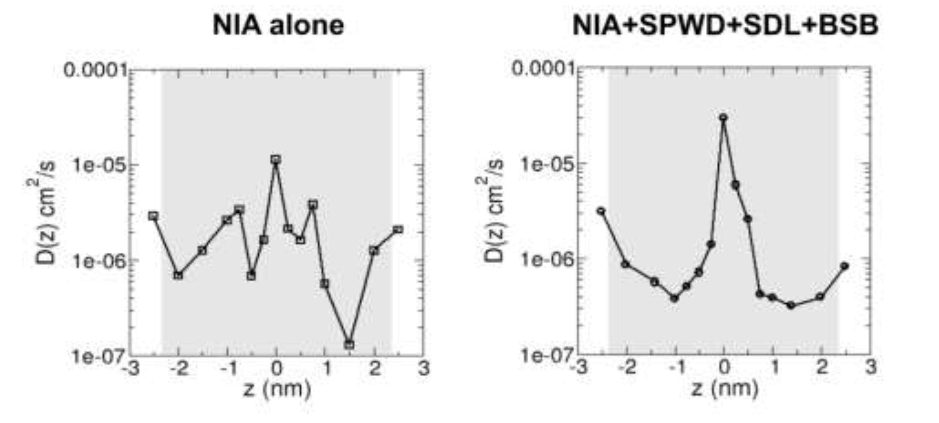
Niacinamide, a derivative of vitamin B3, has been shown to reduce skin pigmentation (i.e. acting as a brightening agent) and inflammatory responses such as dermatitis and acne vulgaris. However, niacinamide is a hydrophilic compound and poor partitioning to the lipid matrix in the uppermost layer of the skin (the stratum corneum or SC) limits its delivery to the skin. This necessitates the use of penetration enhancers to increase its bio-availability. In this study, we used computer simulations to investigate the skin penetration of niacinamide alone and in combination with other brightening agents that are also shown to be skin penetration enhancers, namely Sepiwhite®, bisabolol or sucrose dilaurate. Molecular dynamics simulations were performed to reveal molecular interactions of these brightening agents with a lipid bilayer model that mimics the SC lipid matrix. We observed minimal penetration of niacinamide into the SC lipid bilayer when applied alone or in combination with any one of the three compounds. However, when all three compounds were combined, a notable increase in penetration was observed. We showed 32% increase in the niacinamide diffusivity in the presence of other three brightening agents, which also work as penetration enhancer for niacinamide. These findings suggest that formulations containing multiple brightening agents, which works as penetration enhancers, may improve skin penetration of niacinamide and enhance the effectiveness of the treatment.

Keywords: 
;  ;  ;  ;  ;  ;  
Subject: 
Physical Sciences  -   Biophysics

1. Introduction

Niacinamide (nicotinamide) is the heterocyclic aromatic amide form of water-soluble vitamin B3 (niacin). Vitamin B3 deficiency can lead to a disease known as Pellagra [1]. Niacinamide has been used for anti-hyperpigmentation/anti-aging applications [2,3], as an anti-oxidant [4], improving skin barrier function [5], and for alleviating some skin conditions such as acne vulgaris [6] and dermatitis [7]. Niacinamide reduces hyperpigmentation and improves skin brightness by inhibiting the transfer of melanosomes from melanocytes to keratinocytes as found from clinical studies [8,9]. By inhibiting poly(ADP-ribose) polymerase to down-regulate the expression of inflammatory mediators, niacinamide helps to reduce inflammatory responses such as dermatitis and acne vulgaris [10,11,12,13].
As niacinamide is highly soluble in water (Alog P of −0.32, Table 1), it is challenging to be delivered into the skin, whereby the uppermost layer known as the stratum corneum (SC) consists of dead keratin cells embedded in a lipid matrix [14]. The hydrophobic lipid matrix represents the primary permeation pathway for any skin active compound [15]. To overcome this barrier to skin permeation, various strategies have been explored and are either active or passive in nature. Active strategies to disrupt the SC barrier include electrical or mechanical means, or use of ultrasound or laser radiation [16]. Passive strategies include use of simple solvent systems as skin penetration enhancers [17,18], and surfactants as emulsifying agents and as skin penetration enhancers [19]. Propylene glycol (PG) is a commonly used solvent, and it was shown that adding propylene glycol monolaurate to PG at 1:1 ratio increased the skin flux and permeability coefficient of niacinamide by > 400-fold and adding Miglyol 812N® to the binary solvent further doubled the skin flux and permeability coefficient [17]. In another study, PG:linolenic and PG:oleic acid showed the highest cumulative permeation of niacinamide through human skin at 24-hrs, with a 100-fold enhancement compared to PG alone [18]. The fatty acids might have increased lipid fluidity prior to PG-promoted niacinamide penetration. Surfactants consist of a polar head and a non-polar tail which is either a straight or branched hydrocarbon or fluorocarbon chain with 8-18 carbon atoms [19]. Surfactants can be divided into natural, anionic, cationic, nonionic and zwitterionic ones. An nonionic surfactant, Tween®80, in dipropylene glycol solvent was found to enhance the skin permeability of niacinamide by about 13-fold compared to niacinamide in distilled water [20].
Molecular dynamics (MD) simulations are a useful tool to gain valuable molecular-level insights into drug permeation across the skin and how various penetration enhancers can facilitate the permeation. In MD simulations, the motions of various molecular species and their interactions are monitored in time. MD simulations have been used to study permeation of various small molecules through skin lipid bilayer model composed of equal molar ratio of Ceramide24 NS, C24 free fatty acid and cholesterol [21]. Permeability of each molecule was computed and compared to experimental values, showing similar trends. The effect of chemical enhancers in modulating the permeability of various reference compounds have also been investigated by MD simulations [22,23]. Calculated permeability coefficients showed that the effect of various penetration enhancers depends on the compounds and their concentration [22]. The mechanism of the drug penetration enhancer propylene glycol (a simple solvent mentioned above) has also been studied via MD simulations, whereby a similar SC lipid bilayer composed of equal molar ratios of Ceramide24 NS, C24 free fatty acid and cholesterol was used [24]. PG was found to localize to hydrophilic lipid head-groups and result in an increase in the area per lipid, with a slight increase in the lipid tail disorder in a concentration-dependent manner [24]. However, bilayer penetration by PG molecules were not observed even on microsecond simulation time-scales despite evidence from experiments, likely due to energetic barriers of multiple kJ/mol that makes spontaneous PG permeation a low probability event on the time-scales of atomistic MD simulations.
Brightening molecules which are more amphiphilic (namely Sepiwhite®(SPWD), bisabolol (BSB) and sucrose dilaurate (SDL), see Table 1) may enhance the permeation of niacinamide through the skin. SDL is a natural surfactant derived from sugars and fatty acid. As a sucrose ester, SDL is non-toxic and readily biodegradable. Sucrose laurate/dilaurate was shown to significantly reduce bilirubin levels in the skin and hence skin yellowness [25], and also enhances skin permeation of ibuprofen by over 2-fold [26]. Sucrose esters interact with SC lipids and fluidize the intercellular lipids thereby reducing the SC barrier function [27,28]. Sepiwhite® (undecylenoyl phenylalanine) is a commercial lipophilic derivative of phenylalanine that inhibits activation of tyrosinase, an enzyme that converts L-tyrosine amino acid to melanin during melanogenesis [29]. Hyperpigmentation is a common skin problem with darkened patches or spots on the skin due to increased melanin production because of UV radiation, disease or medications. Undecylenoyl phenylalanine was shown to be helpful in the treatment of melasma [30], a common skin pigmentary problem. Combined formulation of undecylenoyl phenylalanine and niacinamide was found to be significantly more effective than either the control vehicle or niacinamide alone in reducing hyperpigmentation over 8 weeks [31]. The same study also showed that undecylenoyl phenylalanine enhances the skin penetration of niacinamide, with over 50% of the applied niacinamide penetrated into the human skin over a 24-hrs period [31]. Lastly, bisabolol is an unsaturated, optically active sesquiterpene alcohol from essential oil of chamomile. It has anti-microbial and anti-skin inflammation properties [32,33]. Bisabolol is effective against skin inflammation by inhibiting the production of pro-inflammatory cytokines in macrophage cells and skin inflammation in mice [34], and shown to significantly lighten pigmented skin of Asian women in a clinical study [35]. Bisabolol was also demonstrated to enhance trans-epidermal drug penetration through the skin by possibly increasing lipid fluidity [36].
In this work, we aim to explore whether amphiphilic skin brightening agents, namely Sepiwhite®, bisabolol and sucrose dilaurate, enhances the skin permeability of niacinamide by using computer simulations. These three brightening agents are hydrophobic as their 1-octanol/water partition coefficient values (Alog) are greater than 4 (Table 1) which suggest stronger preference of these three skin brightening agents in the lipid matrix than the hydrophilic niacinamide (Table 1). We performed MD simulations of niacinamide alone or in combination with the other compounds on a model SC lipid bilayer composed of equimolar ratios of Ceramide24 NS, free fatty acids and cholesterol as used in previous simulation studies [21,24] but with three free fatty acids (C16:0, C18:0, C20:0) in equimolar ratios to better represent the heterogeneity of free fatty acids in the SC. Previously we have reported the effect of palmitoylation (enhancing hydrophobicity) on the adsorption and diffusion of a short polar(hydrophilic) peptide across a model SC lipid bilayer using molecular dynamics simulations [37]. No significant penetration of niacinamide into the SC lipid bilayer was observed for niacinamide alone or together with either Sepiwhite®, bisabolol and sucrose dilaurate on the simulation time-scales. However, some degree of penetration into the SC lipid bilayer was observed for niacinamide in combination with all three compounds. Calculated niacinamide diffusivity across the SC lipid bilayer also showed higher average diffusivity in the presence of the enhancers. Our simulations thus suggest that these three compounds may act synergistically as penetration enhancers for niacinamide.

2. Results

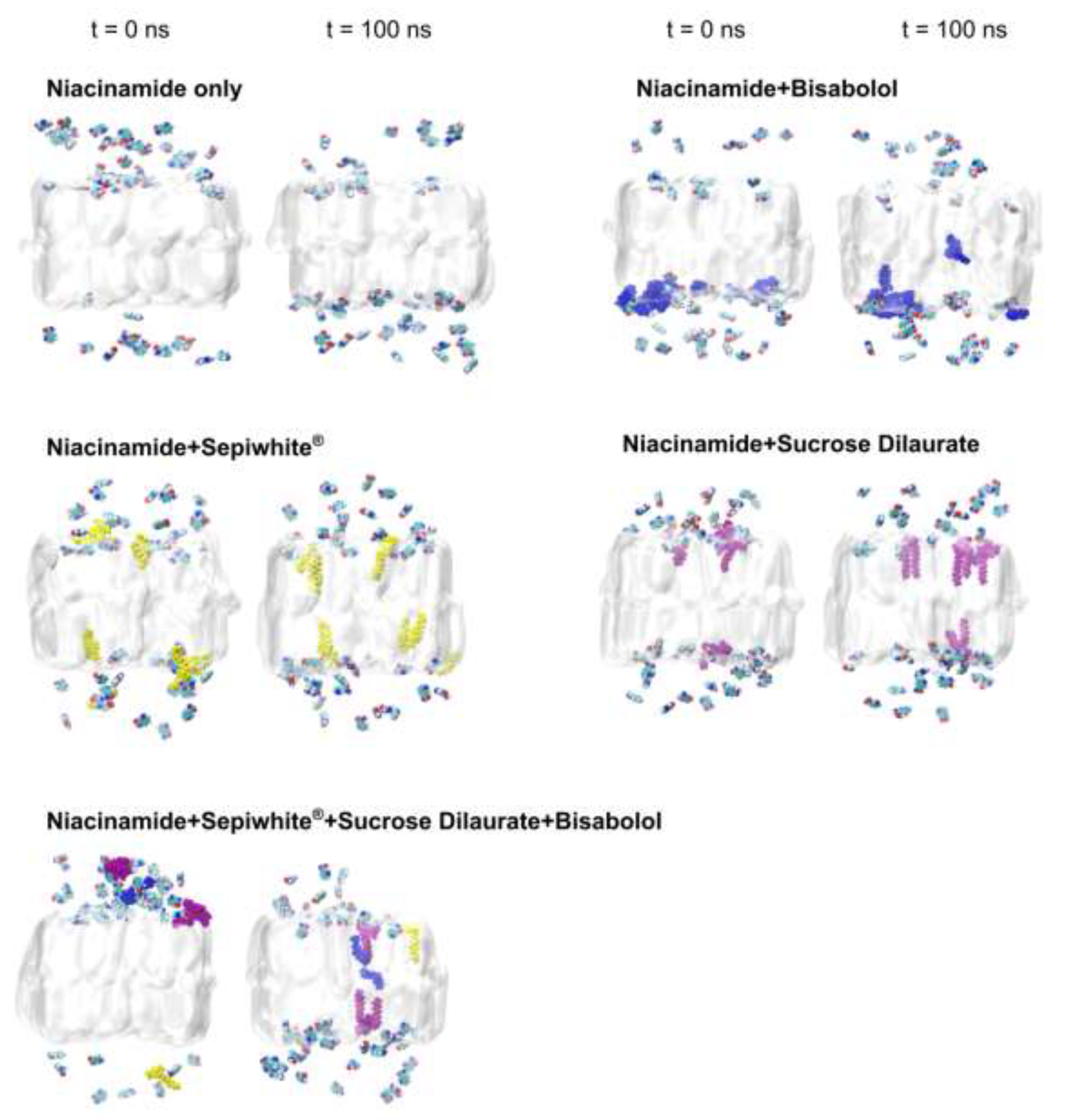
2.1. Brightening Agents Adsorb onto the SC Lipid Bilayer

To evaluate how hydrophobic brightening agents interact with the SC lipid bilayer and influence niacinamide permeation, we carried out six separate MD simulations (Table 2). The simulations include: a control featuring solely the SC lipid bilayer (None), a system with 5% niacinamide alone (NIA), and four systems combining 5% niacinamide with 2% bisabolol (NIA+BSB), 2% Sepiwhite® (NIA+SPWD), 2% sucrose dilaurate (NIA+SDL), and a complex mixture of 5% niacinamide, 0.2% Sepiwhite®, 1% sucrose dilaurate, and 0.4% bisabolol (NIA+SPWD+SDL+BSB). The chemical structures of the SC lipid bilayer components, as well as the brightening agents are illustrated in Figure 1. Atomistic model of the equilibrated SC lipid bilayer is also shown in Figure 1. Each simulation was run for 100 nanoseconds (ns) and performed in duplicate to ensure reproducibility.
To elucidate how brightening agents affect SC lipid bilayer integrity and packing, we monitored bilayer thickness, area per lipid, and hydrogen bond formation between the between the lipid headgroups of SC lipid bilayer across different simulation systems (Table 2). Compared to the control system (None), the presence of brightening agents induced subtle alterations in the SC lipid bilayer structure. Specifically, the bilayer thickness showed fluctuations between 4.51 and 4.55 nm, indicative of minor perturbations in SC lipid bilayer packing. In contrast, the area per lipid remained largely unchanged across all systems, with values ranging from 0.98 ± 0.007 nm² in NIA to 1.01 ± 0.021 nm² in NIA+SPWD+SDL+BSB. This minimal variation suggests that brightening agents do not significantly disrupt the lateral packing of lipid tails, thereby maintaining the bilayer's fundamental structural integrity. Additionally, analysis of hydrogen-bond formation within the SC lipid bilayer revealed that the incorporation of brightening agents led to a slight increase in hydrogen bonds (Table 2). Specifically, the control system exhibited an average of 33.67 ± 0.21 hydrogen bonds, whereas systems containing niacinamide alone (NIA) and in combination with various brightening agents showed elevated hydrogen bond counts, ranging from 34.74 ± 1.32 to 37.48 ± 1.35 hydrogen bonds. These increases indicate a trend of enhanced hydrogen bond formation with the addition of brightening agents, suggesting that these agents facilitate additional molecular interactions within the SC lipid bilayer. Collectively, these findings demonstrate that brightening agents subtly modulate the structural and interactive properties of the SC lipid bilayer, contributing to an environment that supports niacinamide permeation without compromising SC lipid bilayer integrity.
In our simulations, visual inspections revealed that niacinamide did not fully traverse the SC bilayer, whereas other brightening agents partially embedded themselves within the bilayer (Figure 2). This partial embedding suggests the formation of transient interactions with lipid headgroups that can modify SC lipid bilayer properties. To quantify how deeply niacinamide could infiltrated the SC lipid bilayer over time without and without the other brightening agents, we monitored the minimum distance between niacinamide atoms and the center of the bilayer over time for each of the systems (Figure 3a). The shaded regions in the distance profiles represent the standard deviation at each time point, illustrating fluctuations in penetration depth. Notably, in systems containing multiple brightening agents (i.e. the mixture of niacinamide, bisabolol, Sepiwhite®, and sucrose dilaurate), niacinamide molecules showed the largest penetration (Figure 3a), with some of the other agent molecules penetrated more extensively into the bilayer core (Figure 3b). Niacinamide was observed to accompany SDL as it moved further into the bilayer interior at around 70 ns (Figure 3b), suggesting a cooperative effect wherein SDL alters local lipid packing, thereby facilitating niacinamide’s entry. Although niacinamide did not completely cross the bilayer within 100 ns, its capacity to reach deeper bilayer regions in the presence of brightening agents underscores its potential role in modulating permeability pathways.

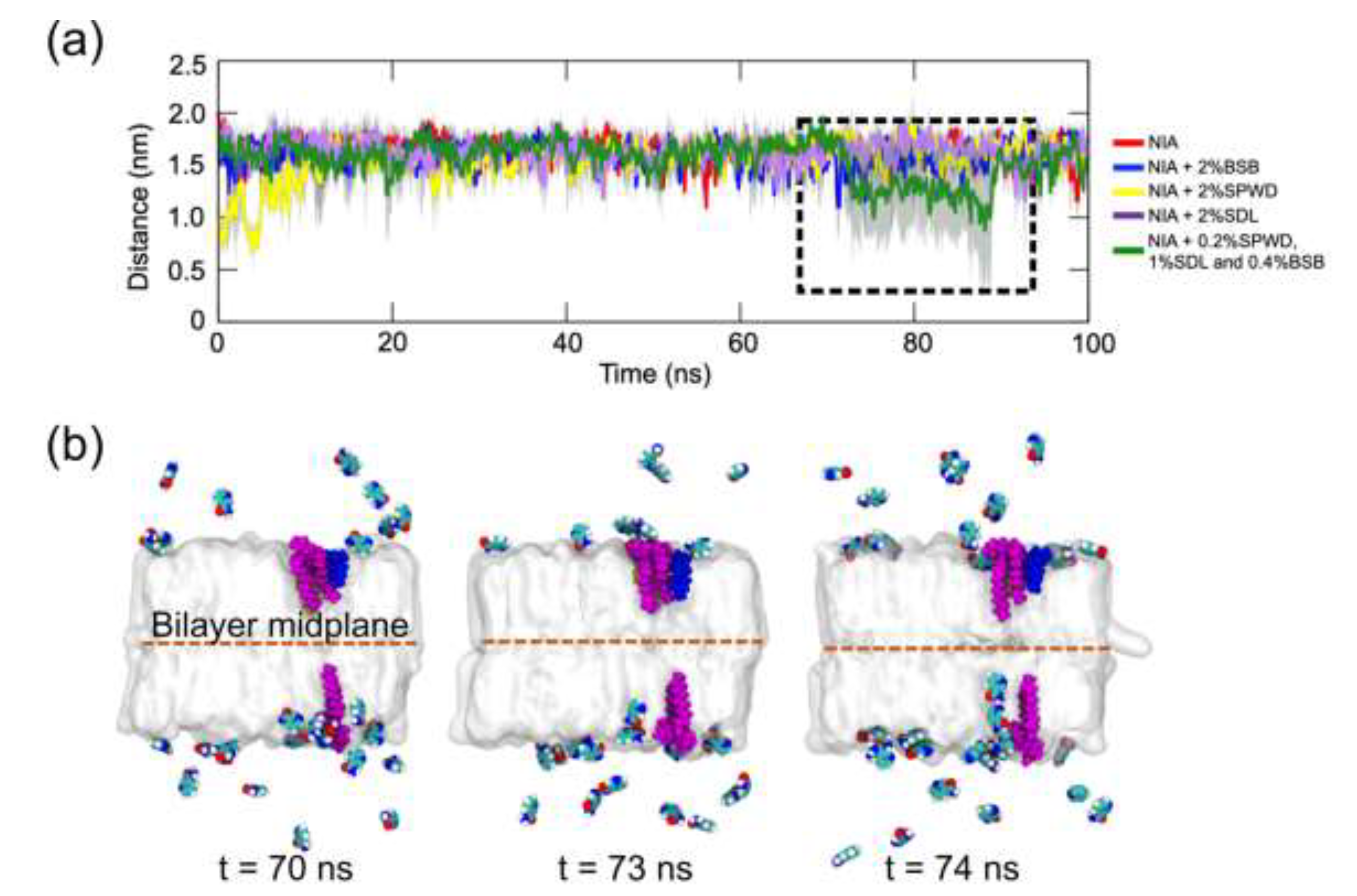
2.2. Brightening Agents Absorbed onto SC Lipid Bilayer Enhance Diffusivity of Niacinamide Across the Bilayer

In our MD simulations, we investigated the influence of brightening agents on niacinamide diffusivity across the SC lipid bilayer under two primary conditions: a control system without any enhancers and a system containing these penetration-promoting agents. As depicted in Figure 4, the analysis focused on the central hydrophobic region of the bilayer from −2.3 nm to 2.3 nm (highlighted by the grey overlay), which represents the primary barrier region for molecular transport [21]. By averaging the values of the diffusivity profile D(z) over this region, we have derived an average diffusivity value that captures the overall mobility of niacinamide within the bilayer.
The control system without enhancers exhibited an average diffusivity of 2.41×10⁻⁶ cm2/s (Figure 4, left). In contrast, the enhancer-containing system showed a notable increase to 3.18×10⁻⁶ cm2/s (Figure 4, right), corresponding to a 32% enhancement. While this difference might seem modest in absolute terms, it underscores how small perturbations—such as slight disruptions in lipid packing or localized boosts in SC lipid bilayer fluidity—can facilitate both lateral and transverse motion of niacinamide.

3. Discussion

Niacinamide acts as a skin brightening agent for anti-aging/anti-hyperpigmentation applications and also helpful to reduce skin inflammatory responses such as dermatitis and acne vulagris. Being a hydrophilic molecule, niacinamide do not partition easily into the hydrophobic lipid matrix of the skin stratum corneum (uppermost layer of the skin). Hence, skin penetration enhancing strategies such as the use of skin penetration enhancing molecules are required for the effective delivery of niacinamide into the skin.
We have used computer simulations to explore how niacinamide alone and in the presence of other brightening agents such as Sepiwhite®, bisabolol and sucrose dilaurate may partition into a model of the skin SC lipid matrix. Brightening agent molecules were placed in the water phase above a lipid bilayer representing a unit of the SC lipid matrix in the stratum corneum. We found that niacinamide alone is not able to partition into the skin SC lipid bilayer and mostly stay in the water phase during the simulation. Niacinamide molecules were also observed to interact with the hydrophilic head-group of SC lipids but could not penetrate into the hydrophobic lipid bilayer interior. In the presence of any one of the other brightening agents, there was also only minimal penetration of niacinamide into the lipid interior. However, in the presence of all three other brightening agents (SPWD, BSB, and SDL), a notable increase in lipid bilayer penetration of niacinamide was observed. Being amphiphilic in nature, SPWD, BSB and SDL all have both hydrophilic head group and hydrophobic tail moieties which facilitates their partitioning into the SC lipid bilayer. These molecules were able to penetrate the SC lipid bilayer within the 100 ns time scale of our simulations, with SPWD and SDL inserting their hydrophobic chains into the SC lipid bilayer whereas the smaller bisabolol molecules were able to completely insert into the hydrophobic interior of the lipid bilayer as BSB has only one polar hydroxyl group (-OH). SDL has been suggested to interact with SC lipids and increase their fluidity thereby compromising the SC barrier function [27,28], with bisabolol also acting to increase lipid fluidity as it enhances drug penetration into the skin [36]. However, our simulations suggest that only when acting together do these three brightening agents significantly enhance the penetration of niacinamide that can be observed on our 100 ns time scale. Although niacinamide did not completely cross the SC lipid bilayer within 100 ns, its ability to reach deeper bilayer regions in the presence of brightening agents underscores the potential role that these agents collectively play in modulating skin permeability pathways.
From our simulations, we also computed the average diffusivity of niacinamide across the SC lipid bilayer interior using enhanced sampling techniques. We found that the average diffusivity increased by about 32% in the presence of the other three brightening agents which potentially acted as penetration enhancers for niacinamide. Though seemingly modest, this finding underscores the importance of small perturbations to lipid packing or localized increases of lipid fluidity via insertion of amphiphilic molecules that can facilitate motion of niacinamide into the lipid bilayer. These enhancements to niacinamide bilayer penetration are likely driven by transient interactions between the brightening agents and various regions of the bilayer, including both lipid hydrophilic headgroups and hydrophobic tail domains. By slightly altering the bilayer’s microstructure (1-2 percent changes to bilayer thickness and area per lipid were observed in our simulations), these agents might reduce the SC lipid bilayer’s resistance to solute diffusion, thereby supporting deeper penetration. Additionally, the hydrogen-bonding propensity of the brightening agents (via carbonyl and hydroxyl groups) may help form more “fluid-like” or less rigid zones within the SC lipid bilayer, promoting niacinamide’s passage through otherwise tightly packed lipid regions.
In summary, incorporating the other brightening agents may act to enhance the bio-availability of each other and of niacinamide in the skin. Having multiple brightening agents in the same formulation further facilitates skin brightening via various biological pathways.

4. Materials and Methods

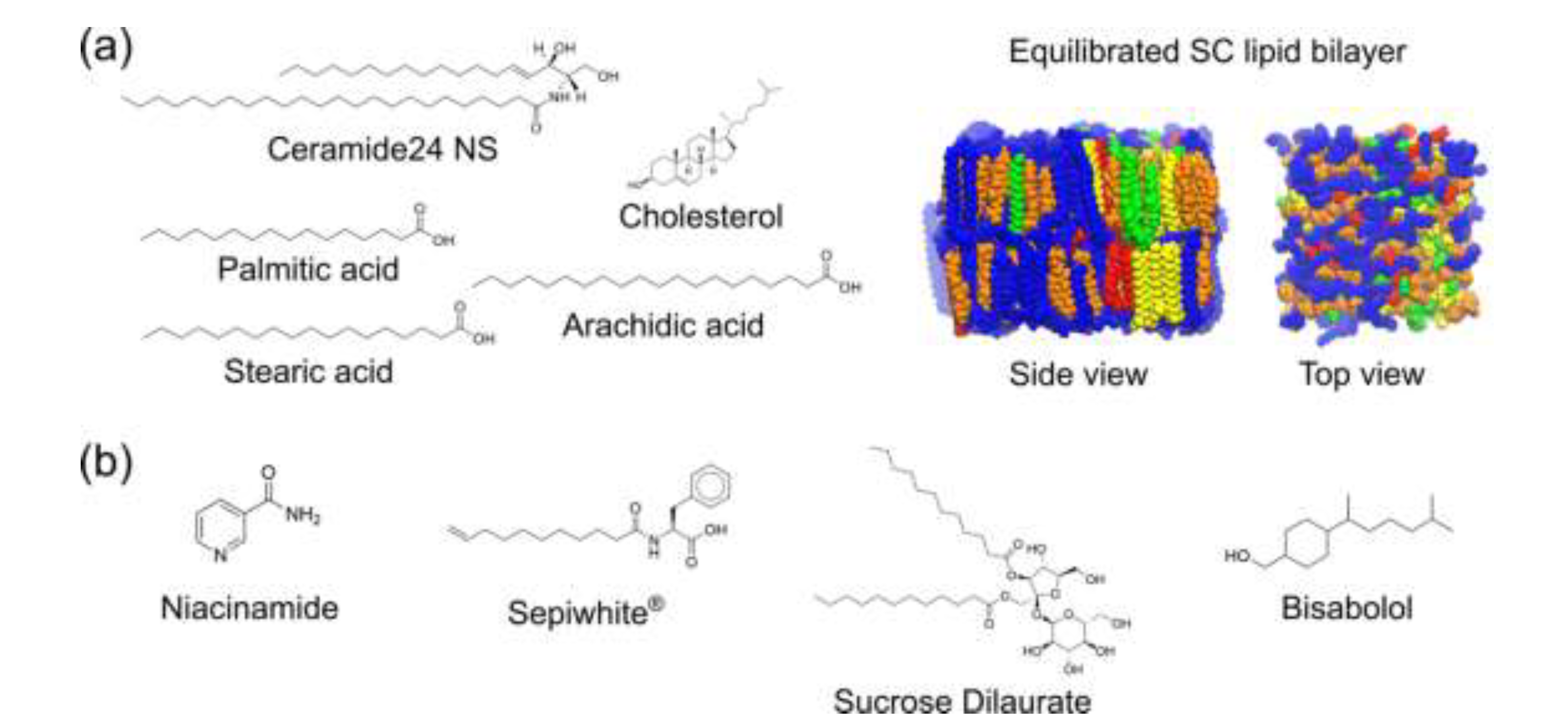
4.1. Generation and Equilibration of SC Lipid Bilayer Model

A model of SC lipid bilayer composed of 100 molecules of ceramide Cer24 NS (C24:0 fatty acid tail and C18:1 sphingosine tail), 100 molecules of cholesterol and 100 molecules of protonated FFA (equal numbers of C16:0 or palmitic acid, C18:0 or stearic acid and C20:0 or arachidic acid) was generated using PackMol software [38]. In terms of composition by weight, ceramides, cholesterols and FFAs make up 49.2%, 29.3% and 21.5% of the SC lipid bilayer, respectively. Thus, the percentages by weight of ceramides and fatty acids fall in the range reported for SC intercellular lipid domains, with that of cholesterol being slightly higher. TIP3P waters were then added above and below the bilayer in a rectangular box with periodic boundaries. The Solution Builder module of CHARMM-GUI webserver established since 2006 was used to generate parameters based on the well-established CHARMM36 additive force-field for each of the lipid species as well as files for optimal GROMACS simulation protocol including energy minimization and equilibration MD simulation runs [39,40]. The SC lipid bilayer model was then subjected to steepest descent energy minimization for 1000 steps followed by a series of short equilibration MD runs (three steps of 125 ps each at 1 fs time-step, followed by three steps of 500 ps each at 2 fs time-step), with restraints on the lipid head-group atoms progressively decreased from 1000 kJ/mol/nm2 to 0 kJ/mol/nm2. GROMACS MD simulation package version 2020.3 was used for energy minimization and MD simulations [41,42,43]. System temperature was maintained at 310 K using the Velocity-rescale method with time constant of 1.0 ps, whereas system pressure was maintained at 1 bar using the Berendsen method with semi-isotropic coupling (i.e. X-Y directions are coupled) with time constant of 5 ps and compressibility of 4.5 × 10⁻⁵ bar−1. Lastly, unrestrained (production) simulation run was carried out for 100 ns at 310 K and 1 bar with a 2 fs simulation time-step to generate an equilibrated SC lipid bilayer model for use in compound interaction study below. Here, the system temperature was maintained at 310 K using Nosé-Hoover method with the same time constant, whereas system pressure was maintained using Parrinello-Rahman method with semi-isotropic coupling and with the same time constant and compressibility. Electrostatic interactions were computed using Particle Mesh Ewald method with cut-off distance of 1.2 nm. Van der Waals (vdW) interactions were computed using cut-off method with forces smoothly switched to zero between 1.0 to 1.2 nm.

4.2. Calculation of Biophysical Properties of the SC Bilayer

Biophysical properties of the equilibrated SC lipid bilayer were computed based on the simulation data obtained over 100 ns of production run. The bilayer thickness was calculated based on the difference in the average Z-locations of Cer24 NS and arachidic acid head-group atoms using an in-house TCL script running in Visual Molecular Dynamics (VMD) software v1.9.4 to extract the Z coordinates [44]. An in-house Python script was then used to compute the average Z coordinates of head-group atoms in the top and bottom monolayers and take the difference. The bilayer thickness calculated in this way is an approximation as the calculated average Z coordinates of head-group atoms do not represent the actual interface between the bilayer surface and aqueous solvent.
The area/lipid (APL) was computed using the FATSlim program (Fast and Accurate Toolkit for lipid bilayer Simulations) [45]. The APL was calculated as the average area of these Voronoi polygons over the equilibrated simulation frames. For this study, frames from the final 20 ns of the simulation (80–100 ns) were used to ensure analysis of equilibrated properties. The resulting APL values were averaged across all lipid molecules in the bilayer.
In addition to the bilayer thickness and APL, hydrogen bond interactions within the SC lipid bilayer were analysed to understand the molecular interactions influencing lipid bilayer stability and permeability. Utilizing VMD's built-in hydrogen bond analysis tool, hydrogen bonds formed between lipid molecules and brightening agents were identified based on a geometric cut-off of 3.0 Å for donor-acceptor distances. The hydrogen bond data generated were subsequently processed using an in-house Python script to calculate the total number of hydrogen bonds formed during each simulation. To ensure that the analysis reflected equilibrated interactions, only hydrogen bonds occurring within the final 20 ns (80-100 ns) of each simulation were averaged.
To evaluate niacinamide penetration into the SC lipid bilayer, we employed the GROMACS tool gmx mindist to calculate the minimum distance between niacinamide molecules and the center of the SC bilayer throughout the simulation. This analysis was conducted for each replicate simulation, and the resulting minimum distance data from both replicates were averaged using a custom Python script. Specifically, the minimum distance between the niacinamide atoms and the midpoint of the bilayer was monitored over the 100 ns simulation period to assess the depth of penetration.

4.3. Generation of brightening agent models and simulation of brightening agent-SC lipid bilayer interactions

The three-dimensional model of each compound (niacinamide, bisabolol, Sepiwhite®, and sucrose dilaurate) was generated using Accelrys Discovery Studio and manually edited to match the required format for input into CHARMM-GUI webserver which automatically generated the all-atom CHARMM36 force-field parameters for the compounds [46,47,48]. MD simulation of the compounds in aqueous solution (TIP3P water models) was first carried out to assess the stability of each model. After energy minimization to relax any possible steric clashes and 125 ps of simulation with compound heavy atoms restrained, unrestrained simulation was then carried out with a 2 fs simulation time-step for 10 ns under a temperature of 310 K and pressure of 1 bar following the same protocol using the same associated parameters as for the SC lipid bilayer. Electrostatic and vdW interactions were also calculated using the same methods as for the SC lipid bilayer. For each system containing either niacinamide only or in presence with the other compounds (see Table 2), the molecules were initially placed atop the SC lipid bilayer and solvated with TIP3P water molecules. For instance, the system containing all four compounds consists of 54 niacinamide, two bisabolol, one Sepiwhite®, and two sucrose dilaurate molecules. Energy minimization followed by short equilibration runs with positional restraints on compound heavy atoms and lipid head-group atoms were carried out before unrestrained (production) runs were performed for 100 ns at 310 K and 1 bar as for simulations of just the SC lipid bilayer. For post-processing, VMD software was used for visualization of the simulation trajectories and GROMACS tools were used to calculate the temporal evolution of the minimum distance between compound and midplane of the bilayer.

4.4. Calculation of Position-Dependent Diffusivity of Niacinamide Across the SC Lipid Bilayer

For the calculation of position-dependent diffusivity, a niacinamide that was bound on the SC lipid bilayer was selected from the final simulation configuration and pulled both towards the bilayer center as well as away from the surface to generate configurations at various positions along the bilayer normal. Next, the niacinamide was then restrained at each of these positions with a force constant of 1000-3000 kJ/mol/nm2 and simulated for 40 ns (umbrella sampling method). Constrain forces were stored at every 0.1 ps during the last 10 ns of each sampling simulation and then used to calculate the position-dependent diffusivity in terms of a time correlation function involving constrain forces [21],
D z = R T 2 0 F z , t F z , 0 d t
where R is the gas constant, T is temperature in Kelvin, and F z , t = F z , t F z , t   is the instantaneous deviation of the constrain force from the average value over time F ( z , t ) . The time correlation function in the denominator of D(z) was written as a time integral over different starting times t’,
F z , t F z , 0 = lim T 1 T 0 T F z , t ' F z , t ' + t d t '
The algorithm to compute D(z) was implemented in Python using NumPy library.

5. Conclusions

The brightening efficacy of brightening agents not only depends on their bio-activity but also bio-availability of all these brightening actives in the skin. We showed through our simulations that niacinamide being hydrophilic skin brightening agent interact only with the hydrophilic head-group of SC lipids but could not penetrate the SC lipid bilayer interior and majorly staying in water. The other three brightening agents (SPWD, BSB, and SDL) partially embedded themselves within the SC lipid bilayer during the simulation. Our simulation suggests that only when these three brightening agents acting together, significantly enhance the penetration of niacinamide. We showed the 32% increase in niacinamide average diffusivity in the presence of all three brightening agents which potentially acted as penetration enhancers and helps niacinamide to partition into the skin SC lipid matrix and being more bio-available in the skin for better efficacy. While these brightening agents providing the brightening efficacy via different biological pathways but incorporating these brightening agents together may act to enhance the bio-availability of each other and of niacinamide in the skin. Having multiple brightening agents in the same formulation further facilitates better skin brightening efficacy.
Our simulation study provides the importance of small perturbations to lipid packing or localized increases of lipid fluidity via insertion of amphiphilic skin brightening molecules, which modulate skin permeability pathways and facilitate partitioning of niacinamide into the lipid bilayer.
Collectively, these findings demonstrate that amphiphilic brightening agents subtly modulate the structural and interactive properties of the SC lipid bilayer, contributing to an environment that supports niacinamide permeation without compromising membrane integrity, which is very crucial to enhancing the brightening efficacy without causing any adverse and harmful impact on the skin.

Author Contributions

Conceptualization, C.H. and S.G.; Methodology, C-P.C.; Formal Analysis, K.S. and C-P.C.; Investigation, K.S.; Resources, C.H. and S.G.; Writing – Original Draft Preparation, K.S. and C-P.C.; Writing – Review & Editing, C.H. and S.G.; Visualization, K.S.; Supervision, C.H. and S.G.; Funding Acquisition, S.G. All authors have read and agreed to the published version of the manuscript.

Funding

The work was financially supported by the A*STAR BMRC Strategic. Positioning Fund (SPF)-A*STAR-P&G Collaboration Grant (H23HW10006).

Data Availability Statement

Data may be made available upon reasonable written request to the corresponding author.

Conflicts of Interest

The authors declare no conflicts of interest.

References

  1. Matts, P.J.; Oblong, J.E.; Bissett, D.L. Int Fed Soc Cosmet Chem Mag. 2002, pp. 285–289.
  2. Boo, Y.C. Mechanistic Basis and Clinical Evidence for the Applications of Nicotinamide (Niacinamide) to Control Skin Aging and Pigmentation. Antioxidants 2021, 10. [Google Scholar] [CrossRef] [PubMed]
  3. Castanedo-Cazares, J.P.; Lárraga-Piñones, G.; Ehnis-Pérez, A.; Fuentes-Ahumada, C.; Oros-Ovalle, C.; Smoller, B.R.; Torres-Álvarez, B. Topical Niacinamide 4% and Desonide 0.05% for Treatment of Axillary Hyperpigmentation: A Randomized, Double-Blind, Placebo-Controlled Study. Clin Cosmet Investig Dermatol 2013, 6, 29–36. [Google Scholar] [CrossRef] [PubMed]
  4. Bowes, J.; Piper, J.; Thiemermann, C. Inhibitors of the Activity of Poly (ADP-Ribose) Synthetase Reduce the Cell Death Caused by Hydrogen Peroxide in Human Cardiac Myoblasts. Br J Pharmacol 1998, 124, 1760–1766. [Google Scholar] [CrossRef] [PubMed]
  5. Tanno, O.; Ota, Y.; Kitamura, N.; Inoue, S. Nicotinamide Increases Biosynthesis of Ceramides as Well as Other Stratum Corneum Lipids to Improve the Epidermal Permeability Barrier. British Journal of Dermatology 2000, 143, 524–531. [Google Scholar] [CrossRef]
  6. Khodaeiani, E.; Fouladi, R.F.; Amirnia, M.; Saeidi, M.; Karimi, E.R. Topical 4% Nicotinamide vs. 1% Clindamycin in Moderate Inflammatory Acne Vulgaris. Int J Dermatol 2013, 52, 999–1004. [Google Scholar] [CrossRef]
  7. Zhu, J.R.; Wang, J.; Wang, S.S. A Single-Center, Randomized, Controlled Study on the Efficacy of Niacinamide-Containing Body Emollients Combined with Cleansing Gel in the Treatment of Mild Atopic Dermatitis. Skin Research and Technology 2023, 29. [Google Scholar] [CrossRef]
  8. Hakozaki, T.; Minwalla, L.; Zhuang, J.; Chhoa, M.; Matsubara, A.; Miyamoto, K.; Greatens, A.; Hillebrand, G.G.; Bissett, D.L.; Boissy, R.E. The Effect of Niacinamide on Reducing Cutaneous Pigmentation and Suppression of Melanosome Transfer. British Journal of Dermatology 2002, 147, 20–31. [Google Scholar] [CrossRef]
  9. Greatens, A.; Hakozaki, T.; Koshoffer, A.; Epstein, H.; Schwemberger, S.; Babcock, G.; Bissett, D.; Takiwaki, H.; Arase, S.; Wickett, R.R.; et al. Effective Inhibition of Melanosome Transfer to Keratinocytes by Lectins and Niacinamide Is Reversible. Exp Dermatol 2005, 14, 498–508. [Google Scholar] [CrossRef]
  10. Khodaeiani, E.; Fouladi, R.F.; Amirnia, M.; Saeidi, M.; Karimi, E.R. Topical 4% Nicotinamide vs. 1% Clindamycin in Moderate Inflammatory Acne Vulgaris. Int J Dermatol 2013, 52, 999–1004. [Google Scholar] [CrossRef]
  11. Zhu, J.R.; Wang, J.; Wang, S.S. A Single-Center, Randomized, Controlled Study on the Efficacy of Niacinamide-Containing Body Emollients Combined with Cleansing Gel in the Treatment of Mild Atopic Dermatitis. Skin Research and Technology 2023, 29. [Google Scholar] [CrossRef]
  12. Fabbrocini, G.; Cantelli, M.; Monfrecola, G. Topical Nicotinamide for Seborrheic Dermatitis: An Open Randomized Study. Journal of Dermatological Treatment 2014, 25, 241–245. [Google Scholar] [CrossRef] [PubMed]
  13. Ungerstedt, J.S.; Blombäck, M.; Söderström, T. 2003.
  14. Elias, P.M. Epidermal Lipids, Barrier Function, and Desquamation. Journal of Investigative Dermatology 1983, 80, S44–S49. [Google Scholar] [CrossRef]
  15. Elias, P.M.; Goerke, J.; Friend, D.S. Mammalian Epidermal Barrier Layer Lipids: Composition and Influence on Structure. Journal of Investigative Dermatology 1977, 69, 535–546. [Google Scholar] [CrossRef]
  16. Brown, M.B.; Martin, G.P.; Jones, S.A.; Akomeah, F.K. Dermal and Transdermal Drug Delivery Systems: Current and Future Prospects. Drug Delivery: Journal of Delivery and Targeting of Therapeutic Agents 2006, 13, 175–187. [Google Scholar] [CrossRef] [PubMed]
  17. Mohammed, D.; Matts, P.J.; Hadgraft, J.; Lane, M.E. In Vitro-in Vivo Correlation in Skin Permeation. Pharm Res 2014, 31, 394–400. [Google Scholar] [CrossRef] [PubMed]
  18. Zhang, Y.; Kung, C.P.; Sil, B.C.; Lane, M.E.; Hadgraft, J.; Heinrich, M.; Sinko, B. Topical Delivery of Niacinamide: Influence of Binary and Ternary Solvent Systems. Pharmaceutics 2019, 11. [Google Scholar] [CrossRef] [PubMed]
  19. Ghafourian, T.; Nokhodchi, A.; Kaialy, W. Surfactants as Penetration Enhancers for Dermal and Transdermal Drug Delivery. In Percutaneous Penetration Enhancers Chemical Methods in Penetration Enhancement: Modification of the Stratum Corneum; Dragicevic, N., Maibach, H.I., Eds.; Springer Berlin Heidelberg, 2015; pp. 207–230 ISBN 9783662470398.
  20. Sohn, J.S.; Choi, J.S. Development and Evaluation of Niacinamide Transdermal Formulation by Artificial Membrane Permeability. Saudi Pharmaceutical Journal 2023, 31, 1229–1236. [Google Scholar] [CrossRef]
  21. Gupta, R.; Sridhar, D.B.; Rai, B. Molecular Dynamics Simulation Study of Permeation of Molecules through Skin Lipid Bilayer. Journal of Physical Chemistry B 2016, 120, 8987–8996. [Google Scholar] [CrossRef]
  22. Lundborg, M.; Wennberg, C.L.; Narangifard, A.; Lindahl, E.; Norlén, L. Predicting Drug Permeability through Skin Using Molecular Dynamics Simulation. Journal of Controlled Release 2018, 283, 269–279. [Google Scholar] [CrossRef]
  23. Wennberg, C.; Lundborg, M.; Lindahl, E.; Norlén, L. Understanding Drug Skin Permeation Enhancers Using Molecular Dynamics Simulations. J Chem Inf Model 2023, 63, 4900–4911. [Google Scholar] [CrossRef]
  24. Mistry, J.; Notman, R. Mechanisms of the Drug Penetration Enhancer Propylene Glycol Interacting with Skin Lipid Membranes. Journal of Physical Chemistry B 2024, 128, 3885–3897. [Google Scholar] [CrossRef] [PubMed]
  25. Fang, B.; Card, P.D.; Chen, J.; Li, L.; Laughlin, T.; Jarrold, B.; Zhao, W.; Benham, A.M.; Määttä, A.T.; Hawkins, T.J.; et al. A Potential Role of Keratinocyte-Derived Bilirubin in Human Skin Yellowness and Its Amelioration by Sucrose Laurate/Dilaurate. Int J Mol Sci 2022, 23. [Google Scholar] [CrossRef] [PubMed]
  26. Csizmazia, E.; Erős, G.; Berkesi, O.; Berkó, S.; Szabó-Révész, P.; Csányi, E. Penetration Enhancer Effect of Sucrose Laurate and Transcutol on Ibuprofen. J. Drug Del. Sci. Tech. 2011, 21, 411–415. [Google Scholar] [CrossRef]
  27. Bolzinger, M.A.; Carduner, C.; Poelman, M.C. Bicontinuous Sucrose Ester Microemulsion: A New Vehicle for Topical Delivery of Niflumic Acid. Int J Pharm 1998, 176, 39–45. [Google Scholar] [CrossRef]
  28. Walters, K.A.; Florence, A.T.; Dugard, P.H. Interaction of Polyoxyethylene Alkyl Ethers with Cholesterol Monolayers 1. J Colloid Interface Sci 1982, 89. [Google Scholar] [CrossRef]
  29. Kabiri, H.; Tayarani-Najaran, Z.; Rahmanian-devin, P.; Vaziri, M.S.; Nasirizadeh, S.; Golmohammadzadeh, S.; Kamali, H. Preparation, Characterization, and Evaluation of Anti-Tyrosinase Activity of Solid Lipid Nanoparticles Containing Undecylenoyl Phenylalanine (Sepiwhite®). J Cosmet Dermatol 2022, 21, 6061–6071. [Google Scholar] [CrossRef]
  30. Katoulis, A.; Alevizou, A.; Soura, E.; Mantas, N.; Bozi, E.; Gregoriou, S.; Makris, M.; Rigopoulos, D. A Double-Blind Vehicle-Controlled Study of a Preparation Containing Undecylenoyl Phenylalanine 2% in the Treatment of Melasma in Females. J Cosmet Dermatol 2014, 13, 86–90. [Google Scholar] [CrossRef]
  31. Bissett, D.L.; Robinson, L.R.; Raleigh, P.S.; Miyamoto, K.; Hakozaki, T.; Li, J.; Kelm, G.R.; Johnson, M. Reduction in the Appearance of Facial Hyperpigmentation by Topical N-Undecyl-10-Enoyl-L-Phenylalanine and Its Combination with Niacinamide. J Cosmet Dermatol 2009, 8, 260–266. [Google Scholar] [CrossRef]
  32. Forrer, M.; Kulik, E.M.; Filippi, A.; Waltimo, T. The Antimicrobial Activity of Alpha-Bisabolol and Tea Tree Oil against Solobacterium Moorei, a Gram-Positive Bacterium Associated with Halitosis. Arch Oral Biol 2013, 58, 10–16. [Google Scholar] [CrossRef]
  33. De, O. Leite, G.; Leite, L.H.I.; De S. Sampaio, R.; Araruna, M.K.A.; De Menezes, I.R.A.; Da Costa, J.G.M.; Campos, A.R. (-)-α-Bisabolol Attenuates Visceral Nociception and Inflammation in Mice. Fitoterapia 2011, 82, 208–211. [Google Scholar] [CrossRef]
  34. Maurya, A.K.; Singh, M.; Dubey, V.; Srivastava, S.; Luqman, S.; Bawankule, D.U. α-(-)-Bisabolol Reduces Pro-Inflammatory Cytokine Production and Ameliorates Skin Inflammation. Curr Pharm Biotechnol 2014, 15, 173–181. [Google Scholar] [CrossRef] [PubMed]
  35. Lee, J.; Jun, H.; Jung, E.; Ha, J.; Park, D. Whitening Effect of α-Bisabolol in Asian Women Subjects. Int J Cosmet Sci 2010, 32, 299–303. [Google Scholar] [CrossRef] [PubMed]
  36. Kadir, R.; Barry, B.W. Alpha-Bisabolol, a Possible Safe Penetration Enhancer for Dermal and Transdermal Therapeutics. Int J Pharm 1991, 70, 87–94. [Google Scholar] [CrossRef]
  37. Chng, C.P.; Zhang, L.; Gupta, S.; Huang, C. Palmitoylation Enhances Short Polar Peptide Permeation across Stratum Corneum Lipid Bilayer: A Molecular Dynamics Study. Extreme Mech Lett 2024, 71, 102213. [Google Scholar] [CrossRef]
  38. Martinez, L.; Andrade, R.; Birgin, E.G.; Martinez, J. Packmol: A Package for Building Initial Configurations for Molecular Dynamics Simulations. J Comput Chem 2009, 30, 2157–2164. [Google Scholar] [CrossRef]
  39. Jo, S.; Kim, T.; Iyer, V.G.; Im, W. CHARMM-GUI: A Web-Based Graphical User Interface for CHARMM. J Comput Chem 2008, 29, 1859–1865. [Google Scholar] [CrossRef]
  40. Lee, J.; Cheng, X.; Swails, J.M.; Yeom, M.S.; Eastman, P.K.; Lemkul, J.A.; Wei, S.; Buckner, J.; Jeong, J.C.; Qi, Y.; et al. CHARMM-GUI Input Generator for NAMD, GROMACS, AMBER, OpenMM, and CHARMM/OpenMM Simulations Using the CHARMM36 Additive Force Field. J Chem Theory Comput 2016, 12, 405–413. [Google Scholar] [CrossRef]
  41. Abraham, M.J.; Murtola, T.; Schulz, R.; Páll, S.; Smith, J.C.; Hess, B.; Lindahl, E. GROMACS: High Performance Molecular Simulations through Multi-Level Parallelism from Laptops to Supercomputers. SoftwareX 2015, 1, 19–25. [Google Scholar] [CrossRef]
  42. Páll, S.; Abraham, M.J.; Kutzner, C.; Hess, B.; Lindahl, E. Tackling Exascale Software Challenges in Molecular Dynamics Simulations with GROMACS. In Proceedings of the Solving software challenges for exascale; Markidis, S., Laure, E., Eds.; Springer, Cham; 2015; pp. 3–27. [Google Scholar]
  43. Pronk, S.; Páll, S.; Schulz, R.; Larsson, P.; Bjelkmar, P.; Apostolov, R.; Shirts, M.R.; Smith, J.C.; Kasson, P.M.; van der Spoel, D.; et al. GROMACS 4.5: A High-Throughput and Highly Parallel Open Source Molecular Simulation Toolkit. Bioinformatics 2013, 29, 845–854. [Google Scholar] [CrossRef]
  44. Humphrey, W.; Dalke, A.; Schulten, K. VMD: Visual Molecular Dynamics. J Mol Graph 1996, 14, 33–38. [Google Scholar] [CrossRef]
  45. Buchoux, S. FATSLiM: A Fast and Robust Software to Analyze MD Simulations of Membranes. Bioinformatics 2017, 33, 133–134. [Google Scholar] [CrossRef] [PubMed]
  46. Lee, J.; Cheng, X.; Swails, J.M.; Yeom, M.S.; Eastman, P.K.; Lemkul, J.A.; Wei, S.; Buckner, J.; Jeong, J.C.; Qi, Y.; et al. CHARMM-GUI Input Generator for NAMD, GROMACS, AMBER, OpenMM, and CHARMM/OpenMM Simulations Using the CHARMM36 Additive Force Field. J Chem Theory Comput 2016, 12, 405–413. [Google Scholar] [CrossRef] [PubMed]
  47. Kim, S.; Lee, J.; Jo, S.; Brooks, C.L.; Lee, H.S.; Im, W. CHARMM-GUI Ligand Reader and Modeler for CHARMM Force Field Generation of Small Molecules. J Comput Chem 2017, 38, 1879–1886. [Google Scholar] [CrossRef] [PubMed]
  48. Jo, S.; Kim, T.; Iyer, V.G.; Im, W. CHARMM-GUI: A Web-Based Graphical User Interface for CHARMM. J Comput Chem 2008, 29, 1859–1865. [Google Scholar] [CrossRef]
Figure 1. All-atom model of stratum corneum (SC) lipid bilayer interacting with brightening agents as penetration enhancers. (a) Chemical structures of the lipids present in our SC lipid bilayer model (left) and simulation snapshots showing the equilibrated SC lipid bilayer model as side and top views (middle and right). The number of Ceramide24 NS, cholesterol and free fatty acid (FFA) molecules are present at equimolar ratio. The three FFAs are also present in equimolar ratio. Ceramides and cholesterol are colored blue and orange, respectively, while FFAs are colored as follows: palmitic acid in yellow, stearic acid in red and arachidic acid in green. Water layers above and below the bilayer are omitted for clarity.(b) Chemical structures of brightening agents in this study.
Preprints 146763 g001
Figure 2. MD simulations of the interaction of brightening agents with the SC lipid bilayer. Simulation snapshots showing the initial (start of unrestrained production run) and final configurations of the brightening agents relative to the SC bilayer surface. Niacinamide is color-coded according to atom type, whereas bisabolol, Sepiwhite®, and sucrose dilaurate are represented in blue, yellow, and purple, respectively. The SC bilayer is shown in a transparent style for enhanced clarity.
Preprints 146763 g002
Figure 3. Penetration of the niacinamide molecules into the SC lipid bilayer in the presence of other brightening agents. (a) The temporal evolution of the minimum distance between niacinamide atoms and the middle of SC lipid bilayer showing depth of penetration of niacinamides in the different systems in Table 2. Shaded areas represent the standard deviation at each time point. (b) Zoomed-in area in (a) for system with all three enhancers showing niacinamide molecules penetrating into the bilayer in proximity to SDL molecules (in purple) that were absorbed into the bilayer.
Preprints 146763 g003
Figure 4. Diffusivity profiles of niacinamide across SC lipid bilayer in the absence or presence of other brightening agents as penetration enhancers. The average diffusivity taken from −2.3 nm to 2.3 nm (within the gray overlay) of the profile without and with enhancers are 2.41×10⁻⁶ cm2/s and 3.18×10⁻⁶ cm2/s respectively, with the latter value being 32% higher relative to the former.
Preprints 146763 g004
Table 1. Chemical properties of brightening agents considered in this study. ALogP is an estimation of log P (the logarithm of 1-octanol/water partition coefficient). Molar volume is the ratio of molar mass to mass density.
Brightening agent ALogP Molar volume (m3/mol)
Niacinamide –0.32 322
Sepiwhite® 5.19 852
Sucrose Dilaurate 5.99 1607
Bisabolol 4.31 593
Table 2. Biophysical properties of the SC lipid bilayer in the absence and presence of brightening agents as penetration enhancers. Niacinamide concentration is 5% by weight of the SC lipid bilayer. Average ± standard errors are reported based on two replicate simulations for each system.
Brightening agent(s) added Bilayer thickness (nm) Area/lipid (nm2) # of lipid head-group H-bonds
None 4.53 ± 0.02 0.99 ± 0.004 33.67 ± 0.21
Niacinamide (NIA) 4.55 ± 0.00 0.98 ± 0.007 35.84 ± 1.18
NIA + 2% BSB 4.54 ± 0.00 1.01 ± 0.000 37.34 ± 0.94
NIA + 2% SPWD 4.51 ± 0.01 1.01 ± 0.000 34.74 ± 1.32
NIA + 2% SDL 4.54 ± 0.01 0.99 ± 0.002 36.72 ± 0.40
NIA + 0.2% SPWD, 1% SDL and 0.4% BSB 4.51 ± 0.01 1.01 ± 0.021 37.48 ± 1.35
Disclaimer/Publisher’s Note: The statements, opinions and data contained in all publications are solely those of the individual author(s) and contributor(s) and not of MDPI and/or the editor(s). MDPI and/or the editor(s) disclaim responsibility for any injury to people or property resulting from any ideas, methods, instructions or products referred to in the content.
Copyright: This open access article is published under a Creative Commons CC BY 4.0 license, which permit the free download, distribution, and reuse, provided that the author and preprint are cited in any reuse.
Prerpints.org logo

Preprints.org is a free preprint server supported by MDPI in Basel, Switzerland.

Subscribe

Disclaimer

Terms of Use

Privacy Policy

Privacy Settings

© 2025 MDPI (Basel, Switzerland) unless otherwise stated